

Choroidal Macrovessel
<< Back to Cases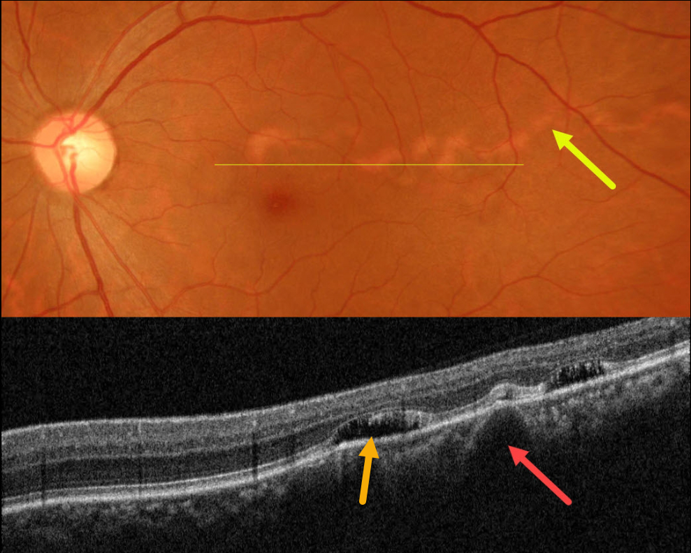
CHOROIDAL MACROVESSEL
Asma Samsudeen and Ashish Sharma
Originally posted on @retina.rocks June 20, 2023
This healthy 41YO female presented with an asymptomatic lesion in her left eye. Vision was 20/20 bilaterally. She is a -7.00 myope.
Color photography shows a serpiginoid-like choroidal atrophic tract extending from the superior macula into the temporal midperiphery.
OCT shows a large dilated choroidal vessel corresponding to this lesion with some pockets of overlying subretinal fluid.
Fortunately, her fovea is uninvolved, and we are following her without treatment.
Learning Points:
Originally reported by Lima et al (BJO 2011;95:1333-1334), choroidal macrovessels are benign dilated choroidal vessels that radiate from the temporal macula towards the midperiphery, somewhat resembling a parasitic tract. They are usually unilateral and found as an incidental finding in an asymptomatic eye.
They appear on OCT as enlarged hyporeflective choroidal structures that compress the surrounding choroid, and changes in the outer retinal bands and RPE are commonly found (Gallo et al, BJO 2020;106:568-575). Localized subretinal fluid overlying the macrovessel is seen in about one-quarter of eyes, which may be caused by overlying choriocapillaris compression with RPE dysfunction.
See Bowen et al (Survey Ophthalmology 2022;67:570-578) for a systematic review, suggesting that choroidal macrovessels are dilated posterior ciliary arteries.


