

Choroidal Melanocytosis
<< Back to Cases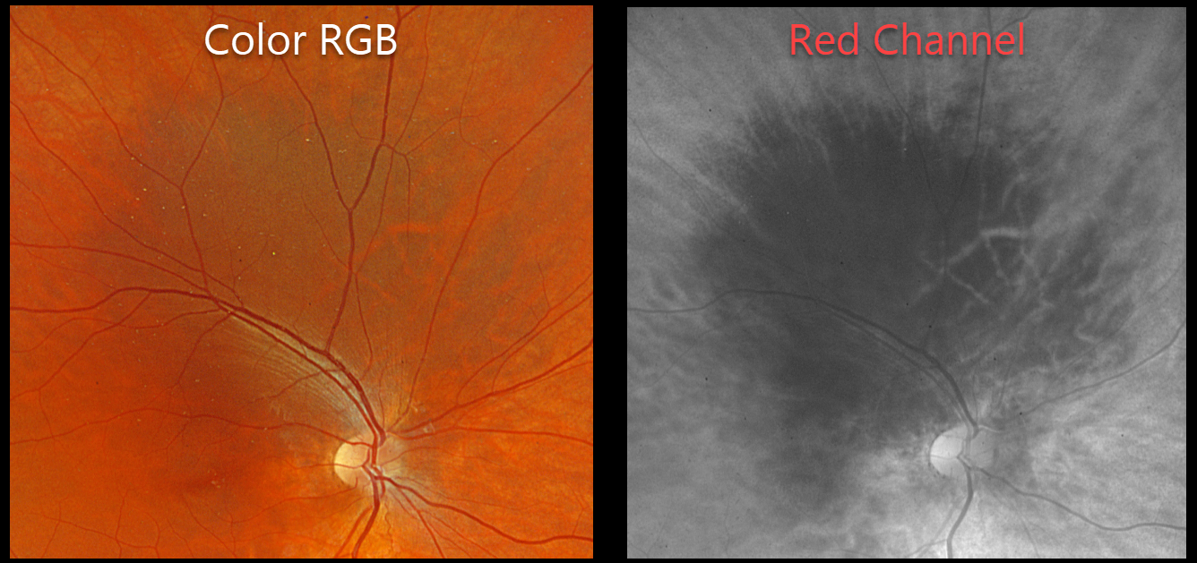
CHOROIDAL MELANOCYTOSIS
Originally posted on @retina.rocks January 22, 2026
This healthy 15YO male was referred for an asymptomatic pigmented lesion in his right eye. Family history was negative. Vision was 20/40 OD and 20/30 in his normal OS.
Optos color RGB imaging shows a region of increased choroidal pigmentation extending from the superonasal macula into the superior midperiphery. This lesion is best seen with the red channel.
Learning Points:
Choroidal melanocytosis is part of the oculodermal spectrum which occasionally includes pigmentation of the globe or periocular skin (Nevus of Ota, melanosis oculi). There are increased dendritic melanocytes in the affected tissues following the distribution of the first and second branches of the trigeminal nerve. Patients need ongoing monitoring since about 1 in 400 will develop uveal melanoma. Of those that develop melanoma, they are twice as likely to undergo metastasis when compared to patients with uveal melanoma and no pre-existing melanocytosis.
Although pretty color images that match what we see on biomicroscopy are most intuitive clinically, newer imaging modalities that distort the normal fundus colors often contain very clinically relevant information. Standard Optos color RG images are generated from a red (635nm) and green (532nm) laser. Although these images have a greenish tint, they essentially contain two images in one: the RPE/neurosensory retina is best captured with the green channel, and the choroid is best seen with the red channel. The newer Optos true color RGB unit maintains the ability to view the separate RG channels but also generates a true to life color image by adding a blue (488nm) laser. In our patient’s case, the red channel from the RG image best shows the melanocytosis.
WAARDENBURG SYNDROME
Gil Calvão-Santos and Keissy Sousa
Originally posted on @retina.rocks May 24, 2023
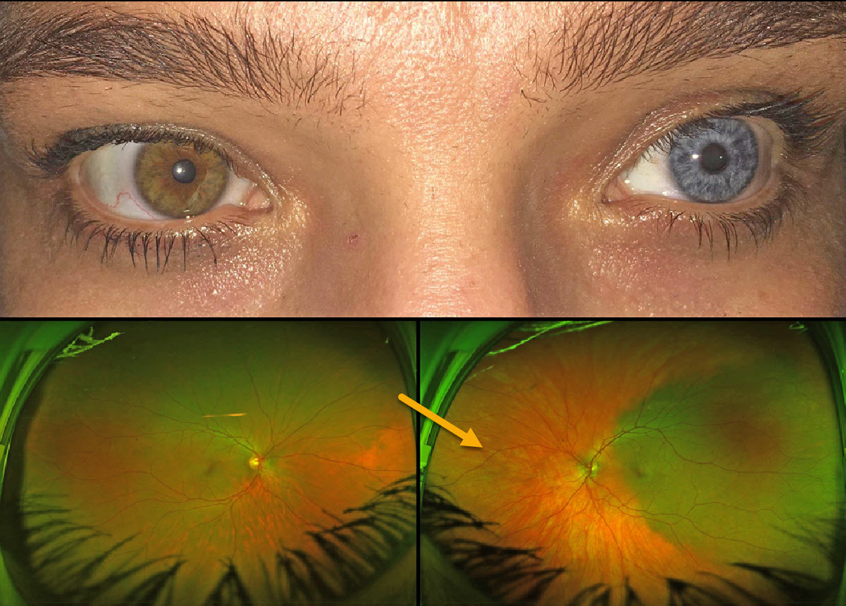
This 43YO female has a constellation of findings that are pathognomonic for Waardenburg syndrome type 1. Vision was 20/20 OU.
External examination shows telecanthus with iris heterochromia. When questioned, she mentioned synophrys (fusion of the eyebrows), which she removed with a laser, poliosis, which she dyed, and a family history of poliosis and heterochromia. She is also congenitally deaf.
Optos imaging shows apparent choroidal melanocytosis temporally in her left eye. But in actuality, this is the only normally pigmented choroid in each eye, with diffuse choroidal hypopigmentation elsewhere.
Learning Points:
Waardenburg syndrome is a group of usually autosomal dominant genetic disorders characterized by achromia of the hair and/or skin, congenital deafness, partial or total iris heterochromia, synophrys, broad and high nasal root, telecanthus, and choroidal hypopigmentation. Vision is usually normal, as was the case in our patient.
See Shields at al for a review of the iris and choroidal abnormalities found in Waardenburg syndrome (JAMA Ophthalmol 2013;131:1167-1173).
UVEAL MELANOMA
Giovanni Cuffaro
Originally posted on @retina.rocks February 6, 2023
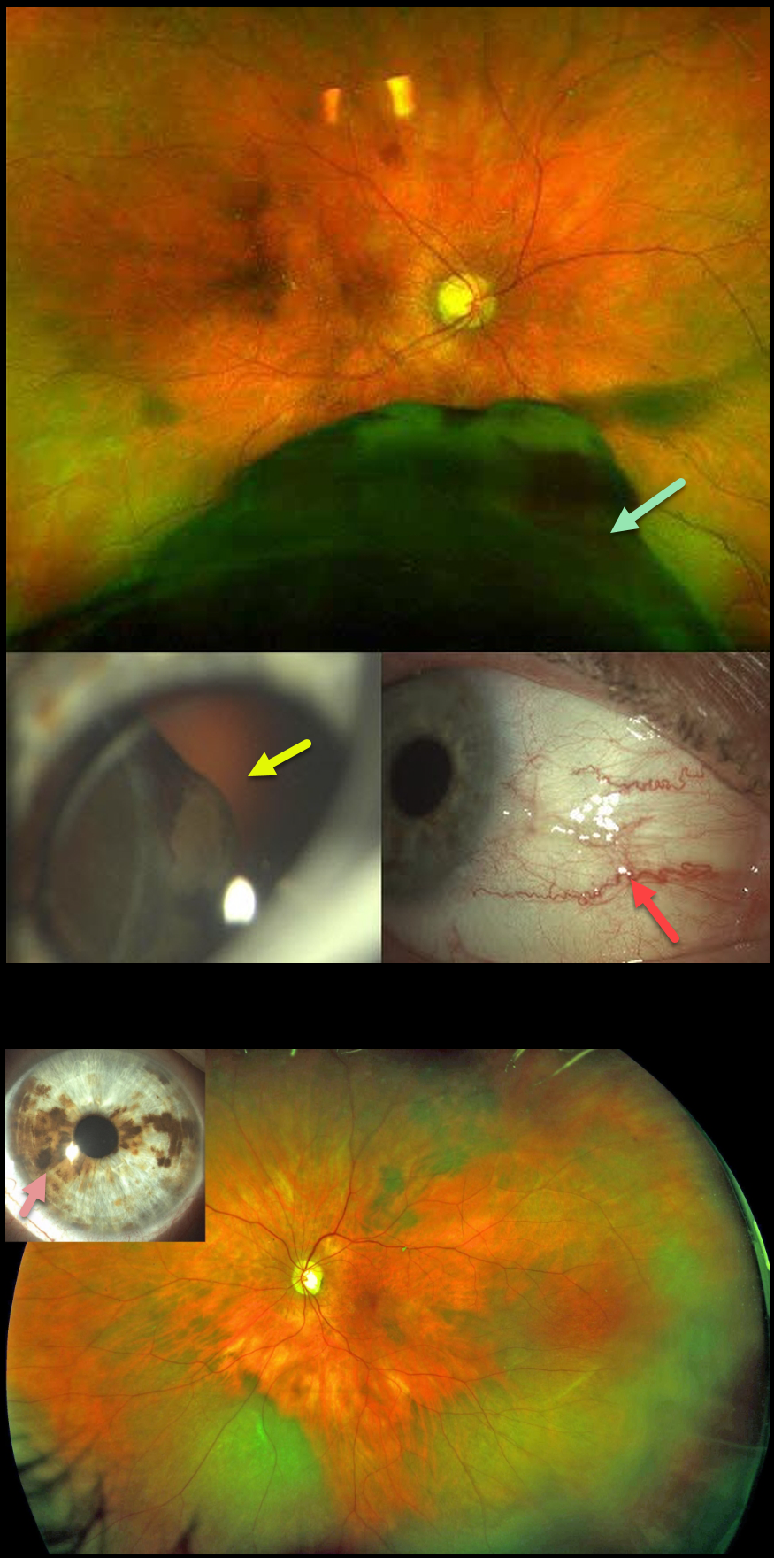
This 71YO female presented in January 2019 with one month of photopsias in her right eye. Optos imaging shows a large inferior malignant melanoma (MM) extending from the inferior retinal periphery into the posterior pole. The tumor was visible through the pupil at the slit lamp, and sentinel vessels were noted. Ocular melanocytosis was noted in the left eye.
She underwent primary enucleation of the right eye one month later. There was complete monosomy 3, and HLA-A*0201 testing was positive.
She subsequently developed a peripheral inferotemporal uveal malignant melanoma in her left eye (axial thickness 5mm, lateral dimensions 13.5 by 13 mm), which was treated with brachytherapy in March 2020. In October 2021, she developed liver metastases.
She was placed on systemic Nivolumab, in addition to external radiotherapy to the liver lesions. Vision is currently 20/20 OS with no signs of local recurrence.
Learning Points:
Our patient had a unique combination of unfortunate multiple risk factors for developing uveal malignant melanoma with metastatic disease (see Kaliki et al, Indian J Ophthalmol 2015;62:93-102).
Monosomy 3 is strongly associated with metastatic disease and melanoma-related mortality. The tumor suppressor gene BRCA1-associated protein 1 (BAP1) is mapped to chromosome 3p21.1, which is associated with autosomal dominant uveal MM and other primary cancers.
Choroidal melanocytosis is part of the oculodermal spectrum, which occasionally includes pigmentation of the globe or periocular skin (Nevus of Ota, melanosis oculi). There are increased dendritic melanocytes in the affected tissues following the distribution of the first and second branches of the trigeminal nerve. Patients need ongoing monitoring since about 1 in 400 will develop uveal melanoma. Those who develop melanoma are twice as likely to undergo metastasis when compared to patients with uveal melanoma and no pre-existing melanocytosis.
For a recent review of oculodermal melanocytosis, see Abdolrahimzadeh et al, Graefe’s 2023;261:291-301.
CHOROIDAL MELANOCYTOSIS
Originally posted on @retina.rocks May 6, 2022
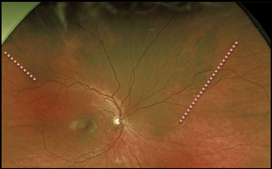
This 16YO male presented with asymptomatic, unilateral disseminated patches of choroidal melanocytosis. There were no anterior segment or skin abnormalities.
Learning Points:
Choroidal melanocytosis is part of the oculodermal spectrum, which occasionally includes pigmentation of the globe or periocular skin (Nevus of Ota, melanosis oculi).
There are increased dendritic melanocytes in the affected tissues following the distribution of the first and second branches of the trigeminal nerve.
Patients need ongoing monitoring since about 1 in 400 will develop uveal melanoma. Of those who develop melanoma, they are twice as likely to undergo metastasis when compared to patients with uveal melanoma and no pre-existing melanocytosis.
CHOROIDAL MELANOCYTOSIS
Originally posted on @retina.rocks August 27, 2021
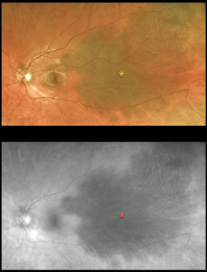
This 17YO female presented with asymptomatic, unilateral disseminated patches of choroidal melanocytosis on Optos color and red-channel imaging. There were no anterior segment or skin abnormalities.
Learning Points:
Choroidal melanocytosis is part of the oculodermal spectrum, which occasionally includes pigmentation of the globe or periocular skin (Nevus of Ota, melanosis oculi).
There are increased dendritic melanocytes in the affected tissues following the distribution of the first and second branches of the trigeminal nerve.
Patients need ongoing monitoring since about 1 in 400 will develop uveal melanoma. Of those who develop melanoma, they are twice as likely to undergo metastasis when compared to patients with uveal melanoma and no pre-existing melanocytosis.
CHOROIDAL MELANOCYTOSIS
Originally posted on @retina.rocks August 10, 2020
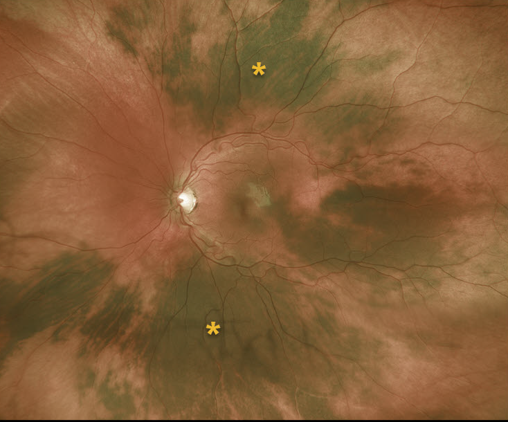
This 13YO female has unilateral disseminated patches of choroidal melanocytosis, but without anterior segment or skin abnormalities.
Learning Points:
Choroidal melanocytosis is part of the oculodermal spectrum, which occasionally includes pigmentation of the globe or periocular skin (Nevus of Ota, melanosis oculi).
There are increased dendritic melanocytes in the affected tissues following the distribution of the first and second branches of the trigeminal nerve.
Patients need ongoing monitoring since about 1 in 400 will develop uveal melanoma. Those who develop melanoma are twice as likely to undergo metastasis when compared to patients with uveal melanoma and no pre-existing melanocytosis.


