

Choroidal Metastases
<< Back to Cases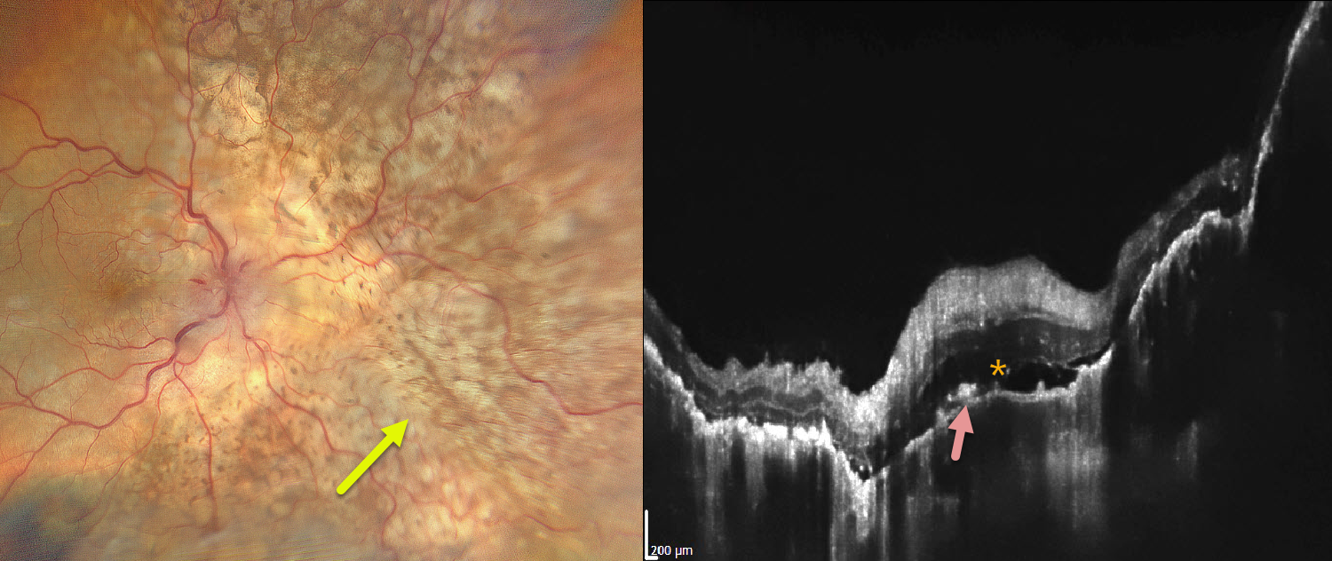
CHOROIDAL METASTASES
Ayushi Gupta and Vishal Agrawal
Originally posted on @retina.rocks July 13, 2026
This 68YO male presented with severe vision loss in his right eye for several weeks. There was a known history of metastatic lung adenocarcinoma. Vision was light perception OD and 20/20 in his normal OS.
Color photography shows variably confluent, creamy-yellow subretinal lesions scattered within the macula and extending into the midperiphery. OCT scanning shows an undulating thickened choroid with shallow overlying subretinal fluid and scattered hyperreflective material above the RPE, likely representing lipofuscin.
Learning Points:
Although uveal melanoma is the most common primary intraocular malignancy, metastatic choroidal tumors are the most common intraocular malignancies. Lung and breast cancer are the most common sources.
METASTASES
The European VitreoRetinal Society, Aysegul Mavi Yildiz and Hikmet Ozcetin
Originally posted on @retina.rocks March 6, 2026
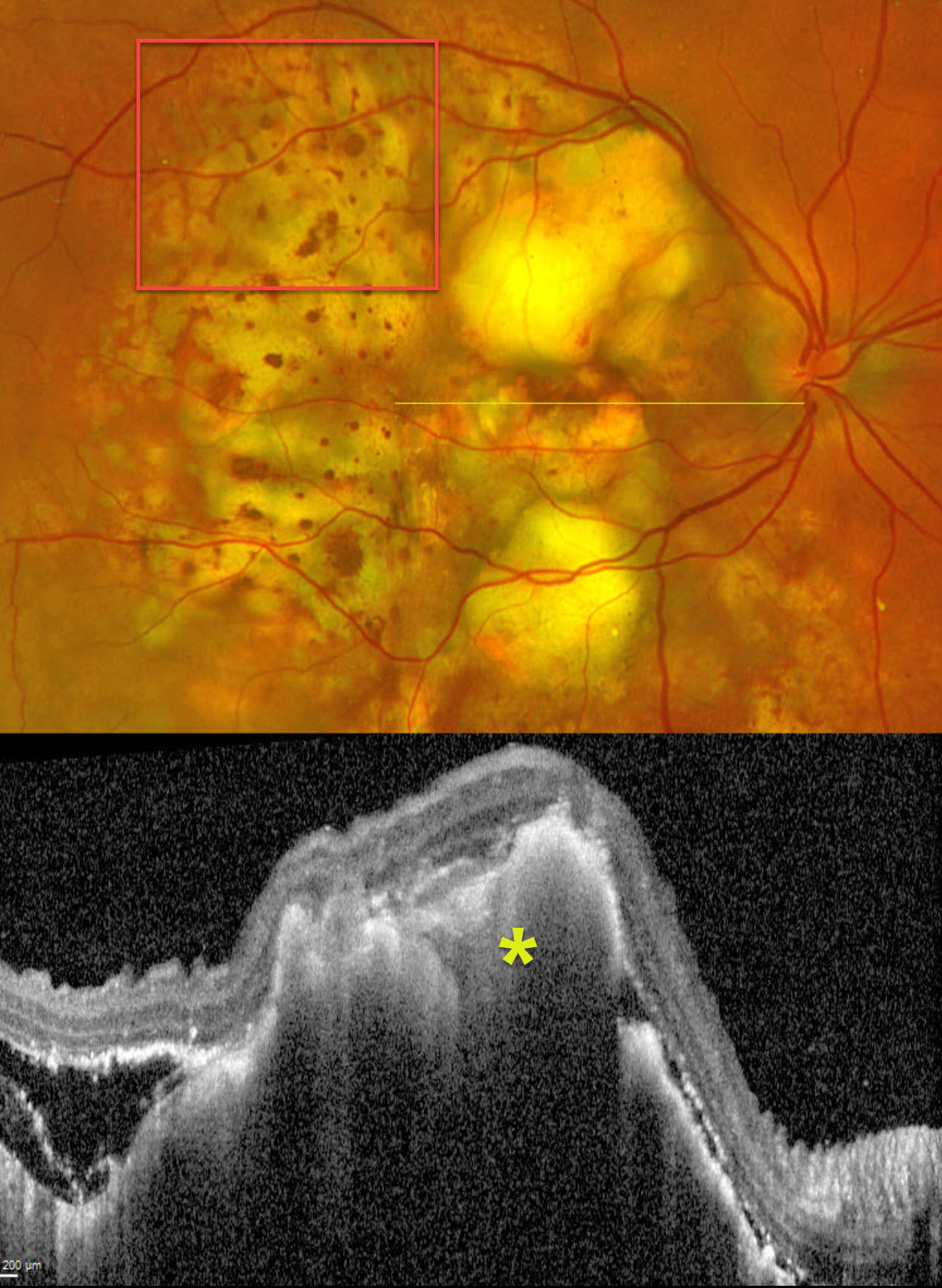
This 67YO male presented with progressive decreased vision in his right eye. He had a known history of metastatic lung cancer. Vision was hand motion OD.
Optos color RG imaging shows multifocal, variably confluent, creamy-yellow subretinal lesions scattered within the macula and extending into the midperiphery. A leopard spot pattern of pigmentary changes is noted temporally. OCT scanning shows marked hyperreflective lumpy choroidal thickening with posterior shadowing. There is temporal subretinal fluid containing variable hyperreflective material.
Learning Points:
Although uveal melanoma is the most common primary intraocular malignancy, metastatic choroidal tumors are the most common intraocular malignancies. Lung and breast cancer are the most common sources.
METASTATIC MELANOMA OF UNKNOWN ORIGIN, SUSPECT CHOROIDAL NEVUS OD AND PROBABLE MELANOMA OS
Mattie Adams
Originally posted on @retina.rocks January 19, 2026
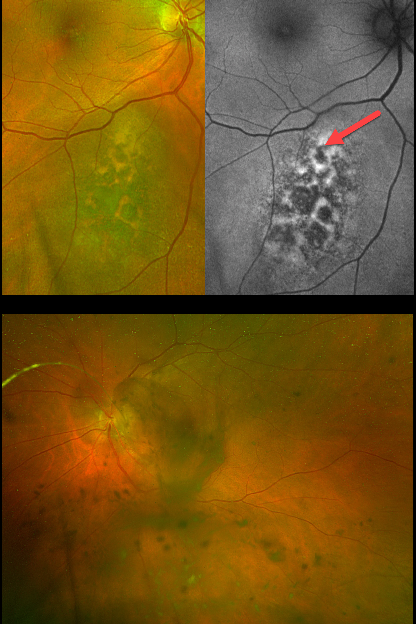
This 67YO female presented with a 3-week history of floaters in her left eye. She has a known history of metastatic melanoma of unknown origin, including numerous brain lesions. There was no family history of ocular or cutaneous melanoma. Vision was 20/20 OD and 20/40 OS.
Optos color RG imaging OD shows a suspect choroidal nevus just inferonasal to the macula. There is overlying orange lipofuscin pigment, which is hyperautofluorescent. Optos imaging OS shows extensive pigmented vitreous debris variably obscuring the posterior pole.
Learning Points:
Uveal malignant melanoma (MM) and cutaneous melanoma are rarely associated with each other. The exception is the presence of a mutated tumor suppressor gene, BRCA1-associated protein 1 (BAP1), which is associated with autosomal-dominant uveal MM and other primary cancers, including cutaneous melanoma.
Our patient did not have a primary for her metastatic disease. The presence of a suspect nevus in her right eye and possible metastatic melanoma to her left eye argues for a pathogenic BAP1 mutation. We discussed this possibility with her oncologist, who felt this was highly unlikely given the negative family history. Given her relatively good vision and poor systemic prognosis, observation was recommended as the initial management for her ocular findings. Unfortunately, she died 3 months later.
CHOROIDAL METASTASTES
Keissy Sousa
Originally posted on @retina.rocks August 13, 2025
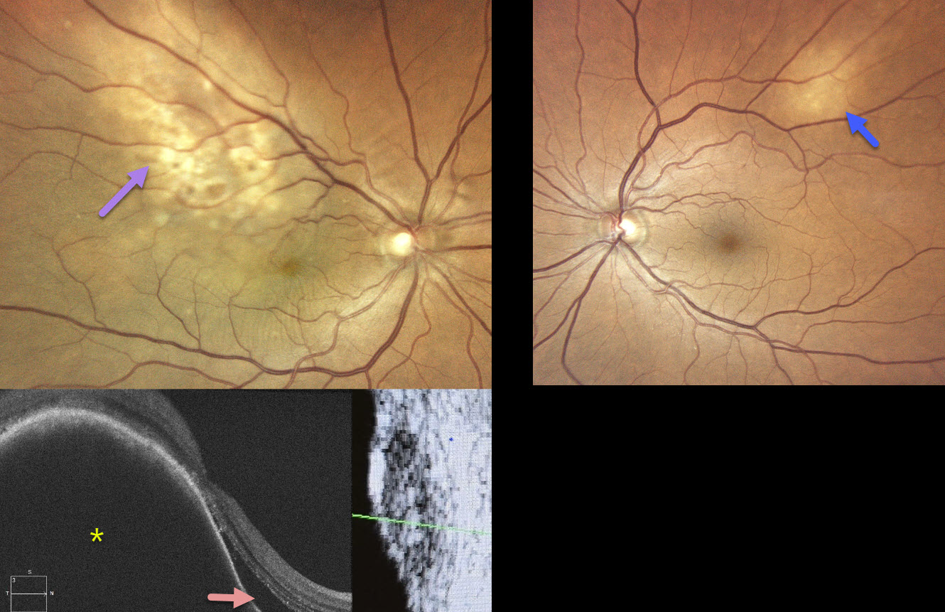
This healthy 70YO female recently underwent cataract surgery elsewhere for vision loss in her right eye. Vision failed to improve postoperatively. Vision was 20/40 OD and 20/20 OS.
Color photography OD shows an ill-defined amelanotic macular choroidal lesion extending into the superotemporal midperiphery. There are some macular chorioretinal folds. OCT scanning shows a markedly thickened choroidal lesion with overlying subretinal fluid. B-scan ultrasonography shows moderate homogenous internal reflectivity. The left eye shows a much smaller amelanotic choroidal lesion superotemporally.
MRI scanning showed a right front paramedial metastatic lesion, and she was subsequently diagnosed with a right-sided primary pulmonary neoplasia. Systemic chemotherapy with pulmonary radiotherapy began without local treatment for the choroidal metastases. When examined 5 months later, vision was no light perception OD and 20/20 OS. The right optic nerve was pale, and the metastatic lesions were fairly stable bilaterally (not shown).
Learning Points:
Although uveal melanoma is the most common primary intraocular malignancy, metastatic choroidal tumors are the most common intraocular malignancies. Lung and breast cancer are the most common sources. Although we can’t be certain, we believe that our patient’s ‘cataract’ symptoms were most likely caused by a missed choroidal metastasis. This underscores the importance of a thorough preoperative dilated funduscopic exam before cataract surgery. In our patient’s case, this not only led to an unnecessary cataract procedure but also delayed the diagnosis and treatment of her metastatic disease.
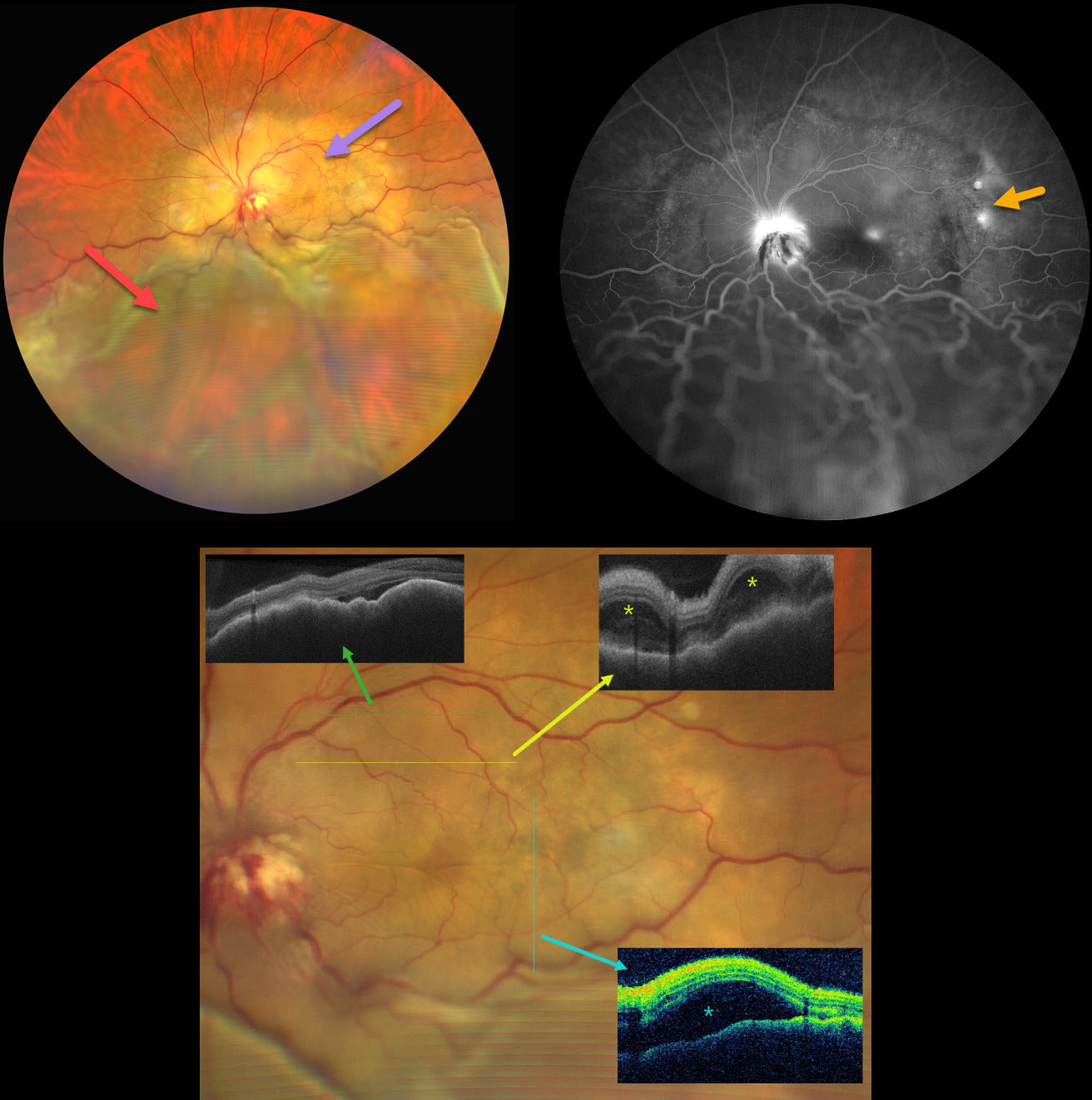
CHOROIDAL METASTASES WITH EXUDATIVE RETINAL DETACHMENT
Ayushi Gupta and Vishal Agrawal
Originally posted on @retina.rocks June 18, 2025
This 47YO female with a known history of metastatic breast carcinoma presented with several weeks of decreased vision in her left eye. She was on active chemotherapy and hormonal therapy. Vision was no light perception OD following trauma 30 years earlier, and hand motion OS.
Ultrawidefield color photography shows a large, amelanotic yellow choroidal lesion involving the entire macula and peripapillary choroid, which extends into the midperiphery. A large secondary inferior exudative retinal detachment involves the entire inferior hemisphere. This fluid shifted when laid supine. The optic disc is hyperemic with indistinct margins and associated blood.
OCT scanning shows a thickened, undulating choroid with variable overlying subretinal fluid and bacillary layer detachments. Fluorescein angiography shows multifocal subretinal leaks, choroidal lesion staining with possible overlying pooling, and intense nerve leakage.
Learning Points:
Although uveal melanoma is the most common primary intraocular malignancy, metastatic choroidal tumors are the most common intraocular malignancies. Lung and breast cancer are the most common sources.
Our patient had a known history of metastatic disease when she presented with this large lesion occupying her entire posterior pole. The extent of the metastasis and the overlying exudation are atypical in our experience. She was immediately referred to her oncologist for external-beam radiation, but was lost to follow-up.
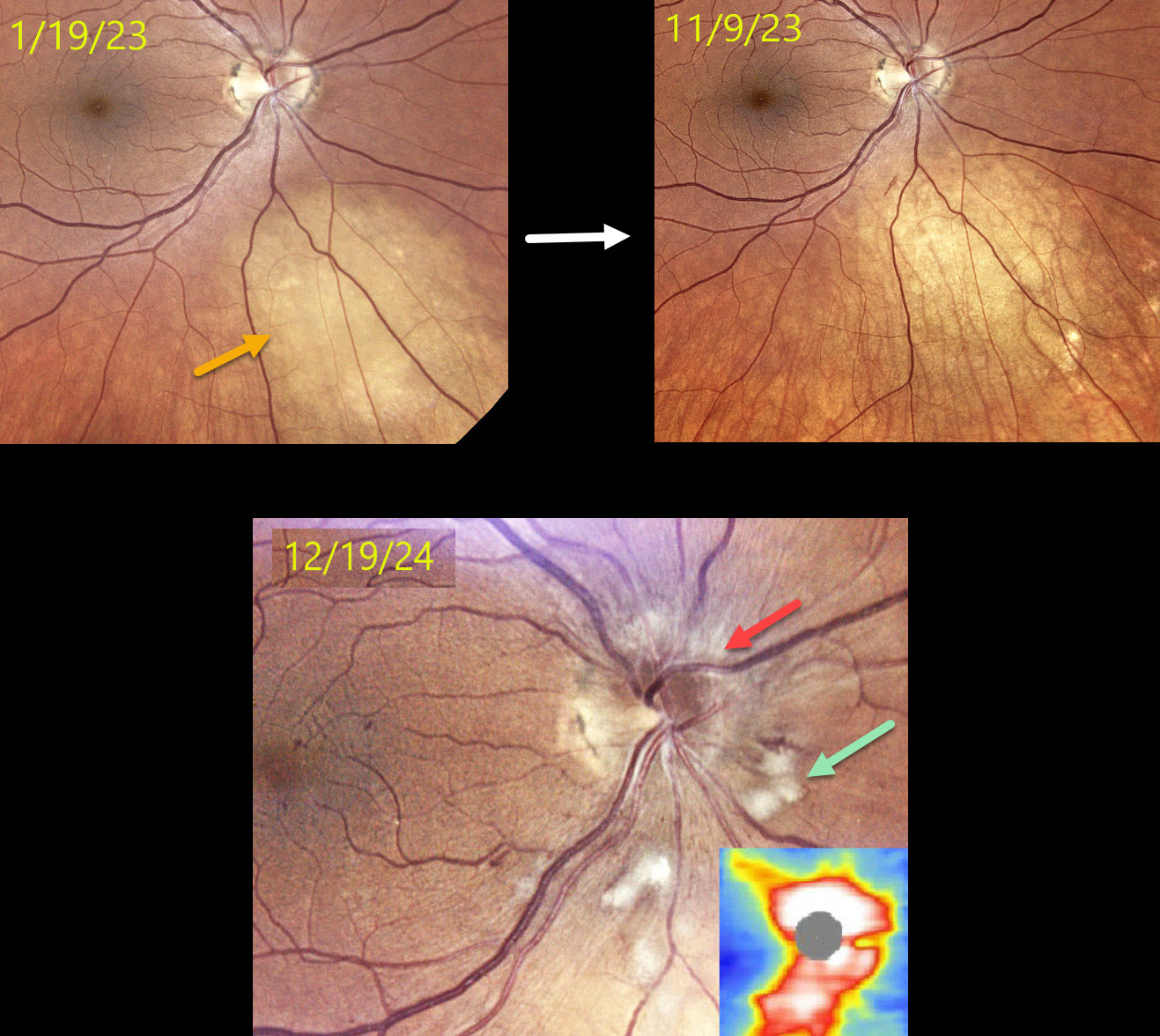
RADIATION RETINOPATHY AND PAPILLOPATHY
Mattie Adams
Originally posted on @retina.rocks May 27, 2025
This 52YO female presented on 1/19/23 with a history of metastatic breast cancer and an asymptomatic uveal metastasis in her right eye. The lesion completely regressed following external beam radiation.
When examined on 12/19/24, she remained visually asymptomatic with 20/60 vision. Color photography shows a new, mildly swollen nerve, which is confirmed on OCT. Scattered nerve fiber layer infarcts (cotton-wool spots, CWS) and some nasal macular telangiectasia are also noted.
Learning Points:
Radiation retinopathy usually occurs 3 months to 3 years after external-beam radiation or plaque radiotherapy. The ocular findings closely resemble those seen with diabetic retinopathy. Radiation optic neuropathy (Carey et al, Br J Ophthalmol 2023;107:743-749) can also develop.
Since our patient was completely asymptomatic without macular edema, we recommended close observation. We will begin intravitreal anti-VEGF therapy if she develops macular edema or worsening nerve findings.
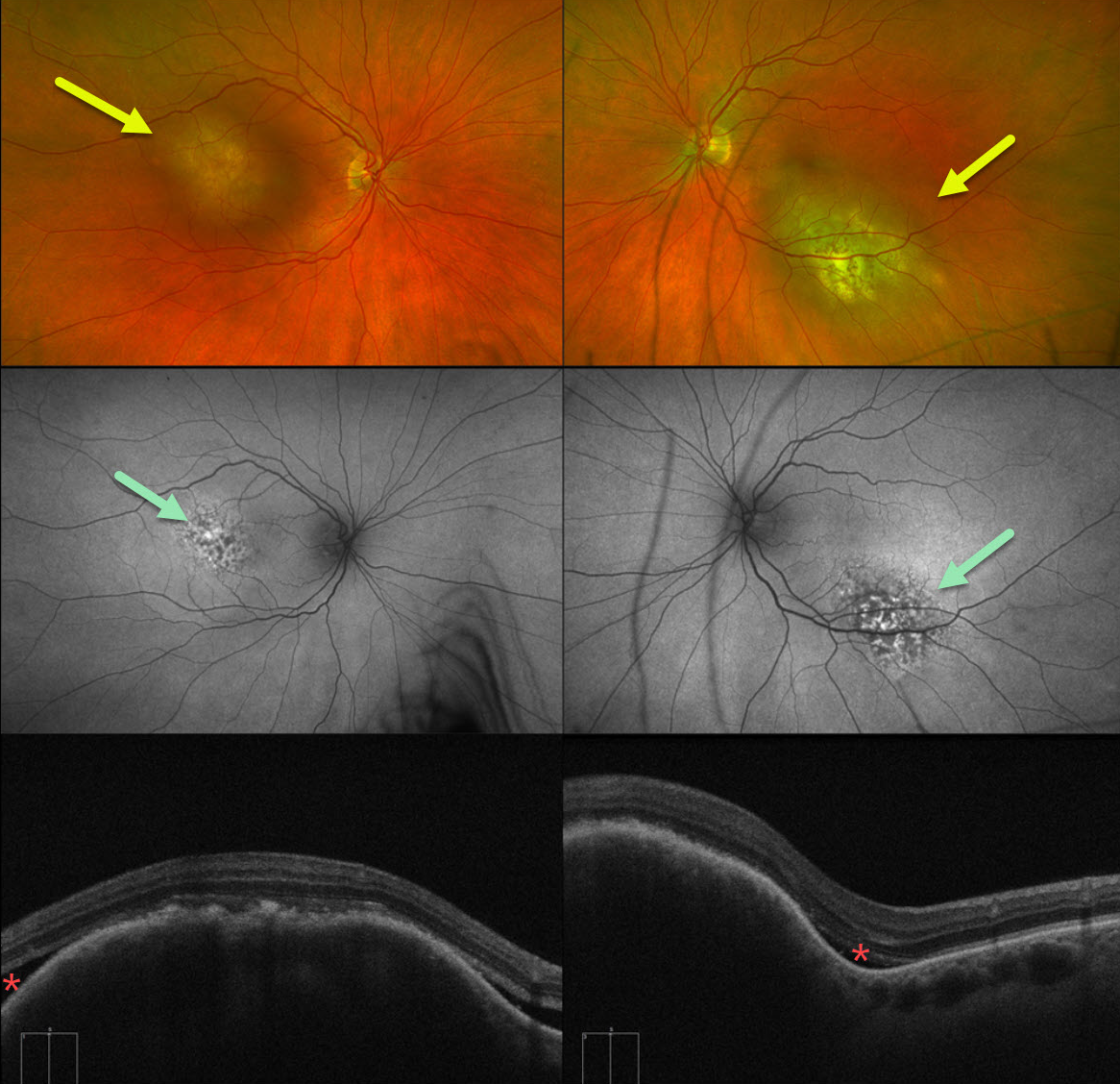
CHOROIDAL METASTASES
Mattie Adams
Originally posted on @retina.rocks May 20, 2025
This 62YO female presented with 1 week of bilateral vision loss. She had a known history of metastatic urothelial bladder carcinoma. Vision was counting fingers OD and 20/100 OS.
Optos color RG imaging shows bilateral elevated submacular choroidal lesions. These lesions were clinically creamy in coloration, although they appeared variably amelanotic and pigmented on Optos pseudocolor imaging. Fundus autofluorescence (FAF) shows speckled hyper- and hypo-FAF overlying the superonasal portion of the lesion OD and the inferonasal portion of the lesion OS. The remainder of each lesion is mildly hyper-FAF. Some localized overlying subretinal fluid is present along the edges of the choroidal tumors.
Although uveal melanoma is the most common primary intraocular malignancy, metastatic choroidal tumors are the most common intraocular malignancies. Lung and breast cancer are the most common sources.
We immediately spoke with the patient’s oncologist and suggested referral to a radiation oncologist for external beam radiation to the choroidal metastases. Unfortunately, she died within several months of seeing us.
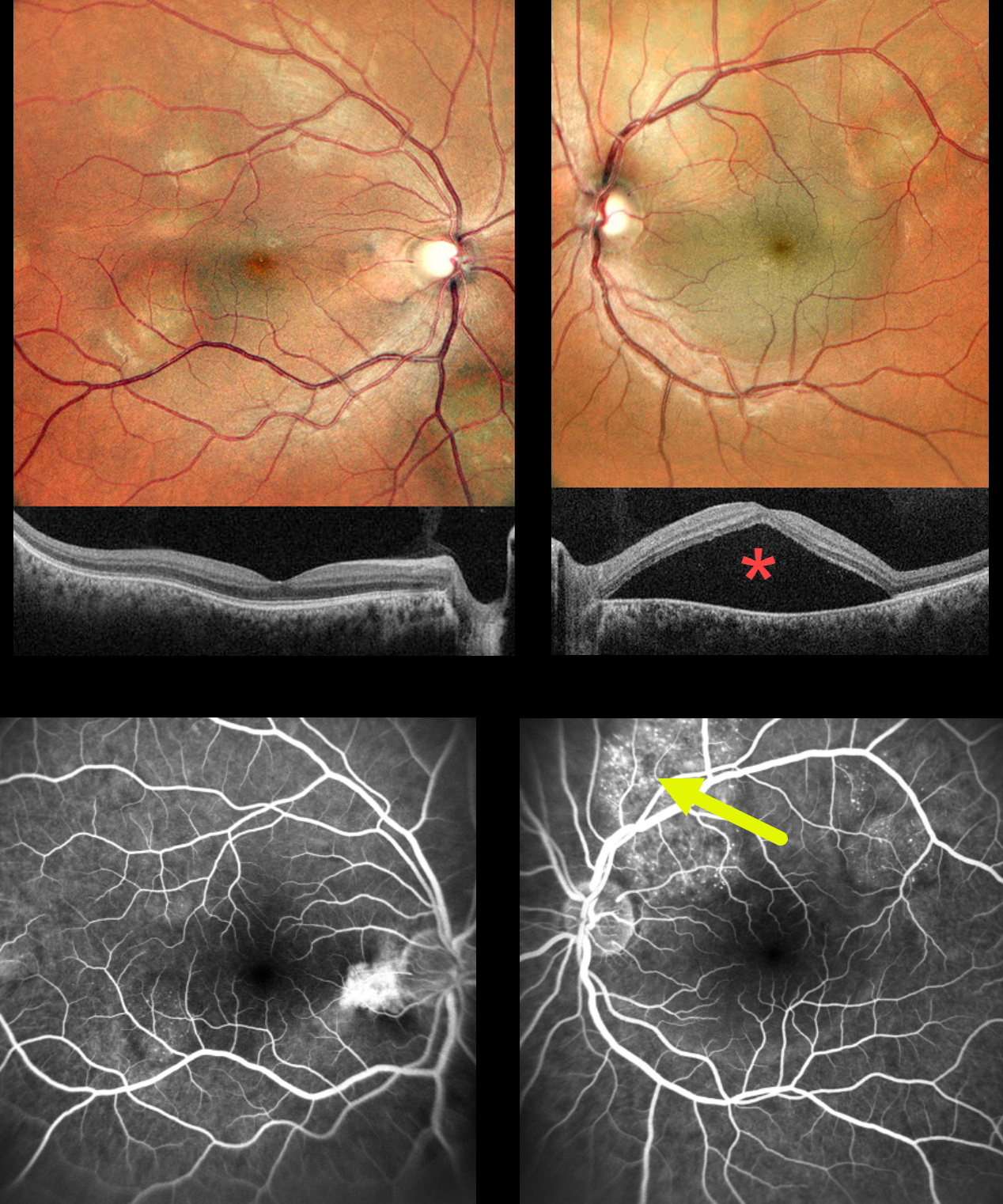
CHOROIDAL METASTASES
Ankit Jain, Navneet Mehrotra and Manish Nagpal
Originally posted on @retina.rocks November 14, 2024
This previously healthy 30YO female presented with several weeks of decreased vision in her left eye. Vision was 20/20 OD and 20/60 OS.
MultiColor imaging shows bilateral multifocal variably sized yellow-white choroidal lesions throughout each posterior pole. A large serous detachment occupies the entire left macula, which is confirmed on SD-OCT. Fluorescein angiography of the right eye shows some speckled subretinal hyperfluorescence overlying a choroidal lesion in the distal inferotemporal macula. Angiography of the left eye shows similar but more extensive hyperfluorescence overlying the superior and superotemporal choroidal lesions, likely responsible for the serous macular fluid.
An extensive medical workup revealed widely metastatic pulmonary adenocarcinoma, and she was subsequently referred to oncology. She was unfortunately lost to follow-up immediately.
Learning Points:
Although uveal melanoma is the most common primary intraocular malignancy, metastatic choroidal tumors are the most common intraocular malignancies. Lung and breast cancer are the most common sources. The extensive multifocal lesions in each of our patient’s eyes are unusual, since most patients have 1-2 choroidal lesions (Shields et al Retina 2020;40:204-213).
CHOROIDAL CARCINOID METASTASIS
Originally posted on @retina.rocks April 23, 2024
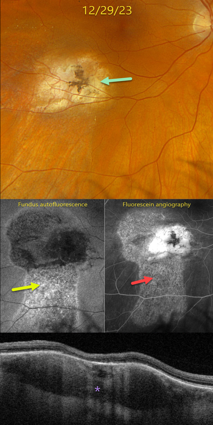
This 43YO female presented to our office with recent vague symptoms of vision loss OD. She was followed elsewhere since 2006 for a carcinoid lesion metastatic to the right choroid. Her right bronchial carcinoid tumor was treated earlier that year with a right pneumonectomy without systemic treatment. Initial observation of the choroidal lesion was recommended, but she was almost immediately lost to follow-up for 13 years.
She returned to the outside practice in 2019, and the choroidal lesion showed slight growth with mild new fluid. She was again lost to follow-up until June 2021, with continued growth and increased fluid. Photodynamic therapy was applied on 6/8/21 and 9/21/21 with subsequent tumor shrinkage and resolution of the fluid.
On 12/29/23, vision was 20/25. Optos color RGB imaging shows an atrophic, variably pigmented, lumpy choroidal lesion in the inferotemporal macula.
Fundus autofluorescence (FAF) shows a hyper-FAF gutter extending inferiorly from the lesion. This gutter is well-visualized on fluorescein angiography, with staining of the inactive tumor.
Triton swept-source OCT shows a variably thickened and mostly hyporeflective choroidal lesion with overlying outer retinal atrophy. These findings were all stable compared to images seen 1 year earlier at the outside practice. We plan to follow her twice a year.
Learning Points:
Although uveal melanoma is the most common primary intraocular malignancy, metastatic choroidal tumors are the most common intraocular malignancies. Lung and breast cancer are the most common sources. These lesions can be treated with systemic chemotherapy and are also usually very responsive to external beam radiation.
Neuroendocrine (carcinoid) tumors are usually low-grade malignancies that arise in the bronchopulmonary system or the gastrointestinal or urogenital tracts. In a recent review of over 1100 patients referred to the Wills ocular oncology service over a 43-year period, pulmonary carcinoid accounted for 2% of cases (Shields et al, Retina 2020;40:204-213). These lesions have a characteristic orange appearance, in contrast to most other choroidal metastases, which are usually creamy yellow (Shields et al, BJO 1996;80:852-853).
CHOROIDAL METASTASES
Originally posted on @retina.rocks March 2, 2023
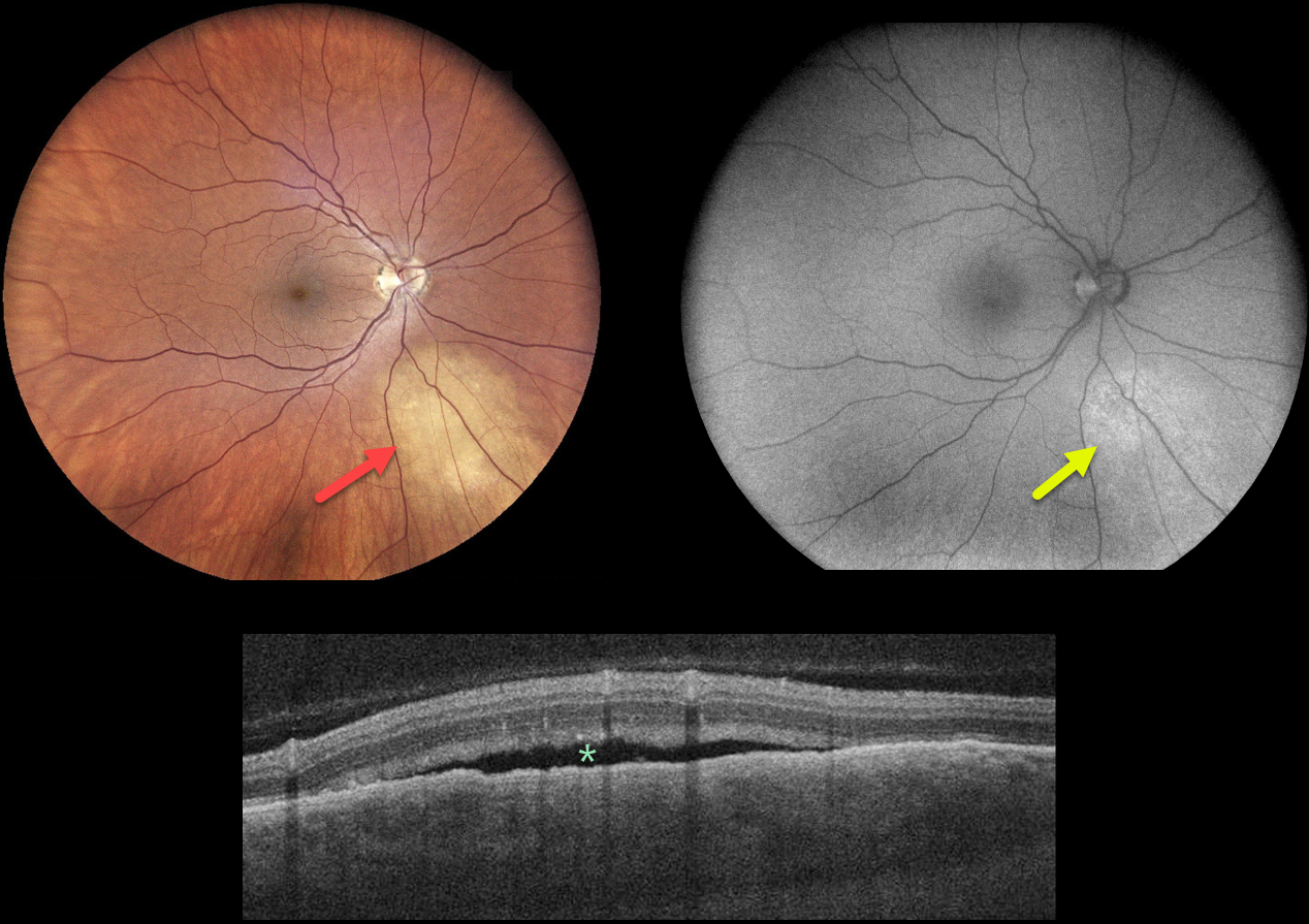
This 52YO female has a 10-year history of breast cancer and a 3-year history of metastatic pulmonary disease, which is controlled with systemic chemotherapy. She was referred for an asymptomatic lesion in her right eye.
Fundus photography shows an amelanotic subretinal lesion inferonasal to the nerve. This lesion faintly hyper-autofluoresces.
OCT scanning through this lesion shows a thickened choroid with a somewhat undulating overlying RPE and shallow subretinal fluid.
Learning Points:
Although uveal melanoma is the most common primary intraocular malignancy, metastatic choroidal tumors are the most common intraocular malignancies. Lung and breast cancer are the most common sources. These lesions can be treated with systemic chemotherapy and are also usually very responsive to external beam radiation.
After discussing the findings with her oncologist, the lesion will be treated with external beam radiation.
CHOROIDAL METASTASES
Originally posted on @retina.rocks October 17, 2022
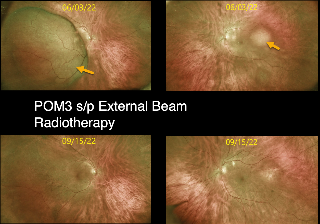
This is a follow-up post from 6/6/22 of a 74YO female who initially presented with a several-week history of progressive vision loss in her right eye. Vision was counting fingers OD and 20/25 OS.
There was a known history of metastatic breast cancer when we diagnosed her with bilateral uveal metastases.
After three months of external beam radiotherapy, the metastatic choroidal lesions dramatically resolved. Vision improved to 20/50 OD and 20/25 OS.
Learning Points:
Although uveal melanoma is the most common primary intraocular malignancy, metastatic choroidal tumors are the most common intraocular malignancies. Lung and breast cancer are the most common sources.
CHOROIDAL METASTASIS
Originally posted on @retina.rocks September 23, 2022
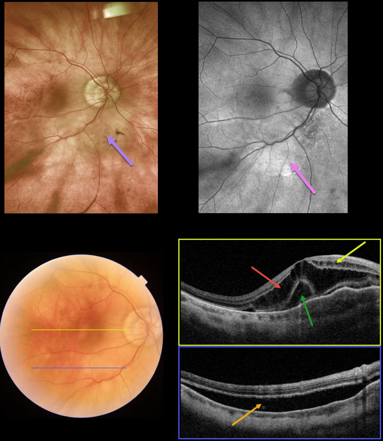
This 87YO male presented with recent blurred vision in his right eye. Vision was 20/200 OD. He has a medical history of hypertension and prostate cancer that was successfully treated with radiation 15 years earlier.
Optos color imaging shows a creamy white subretinal discoloration in the nasal macula extending inferiorly.
Fundus autofluorescence (FAF) shows mostly hyper-FAF within this region. Swept-source OCT through the macular center shows severe cystic fluid in the outer nuclear layers and milder fluid in the inner nuclear layers. An unusual collection of outer vs subfoveal fluid is also seen. A B-scan through the inferior macula shows subretinal fluid.
The patient was referred to his oncologist for a metastatic workup, which disclosed previously undiagnosed colon cancer.
Learning Points:
Although uveal melanoma is the most common primary intraocular malignancy, metastatic choroidal tumors are the most common intraocular malignancies.
Lung and breast cancer are the most common sources. Prostate cancer rarely metastasizes to the choroid, found in only 2% of uveal metastases (Shields et al, Retina 2020;40:204-213).
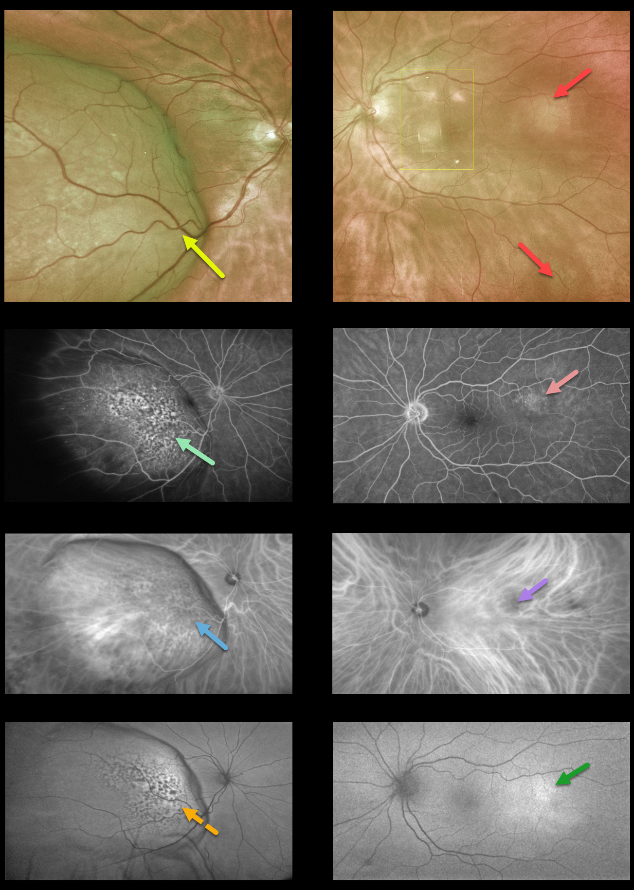
CHOROIDAL METASTASES
Originally posted on @retina.rocks June 6, 2022
This 74YO female presented with a 2-week history of progressive vision loss in her right eye. Vision was 20/200 OD and 20/20 OS.
Optos imaging shows a large, creamy-colored temporal choroidal lesion extending into the right temporal macula. The fovea is markedly elevated and tilted along the nasal edge of the tumor. Two smaller and fainter amelanotic choroidal lesions are noted in the left eye (the lesions in the left macula are artifacts).
Fundus fluorescein angiography (FFA) shows patchy window defects over the nasal dome of the lesion in her right eye and faint staining from the temporal lesion in the left eye.
Indocyanine green angiography shows mostly hypofluorescence corresponding to the FFA window defects with some increased fluorescence from the intrinsic vasculature more temporally in the right eye, and hypofluorescence from the temporal lesion in the left eye.
Fundus autofluorescence (FAF) shows hypo-FAF corresponding to the FFA window defects in the right eye and hyperfluorescence from the temporal lesion in the left eye.
There was a known history of metastatic breast cancer, and a recent PET scan showed possible new lung metastases.
We are currently working with the patient’s oncologist for the next step forward, which will likely include external beam radiotherapy for a presumed choroidal metastasis in her right eye. Photodynamic therapy or transpupillary thermotherapy will be considered for the left eye.
Learning Points:
Although uveal melanoma is the most common primary intraocular malignancy, metastatic choroidal tumors are the most common intraocular malignancies. Lung and breast cancer are the most common sources.
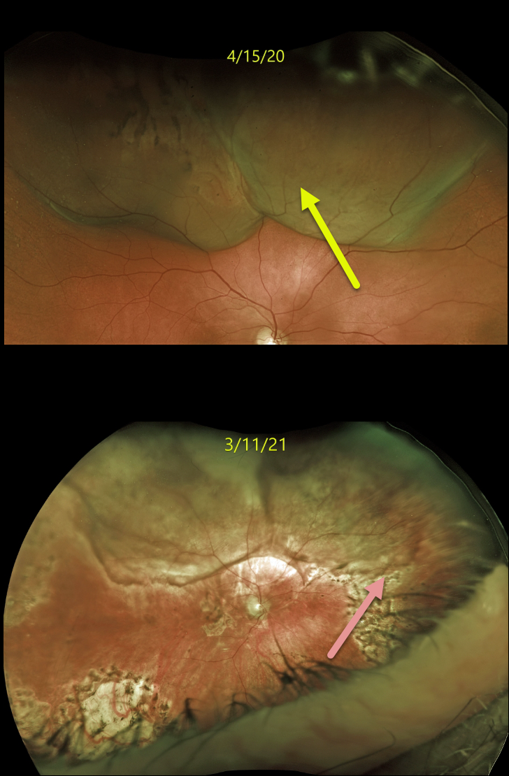
CHOROIDAL METASTASES
Originally posted on @retina.rocks August 2, 2021
This 82YO male with a history of retinal detachment treated with a segmental buckle and cryotherapy presented on 4/15/20 with some pain and new floaters in his right eye. Vision was 20/40.
Optos imaging shows an exudative retinal detachment secondary to a large, elevated, irregular, and amelanotic choroidal mass in the superior hemisphere. B-scan ultrasonography (not shown) revealed the tumor to be 6mm in height and 23 by 13 mm in basal diameter.
There was a prior history of metastatic clear cell renal carcinoma that was treated with radical left nephrectomy and chemotherapy from 2012 to 2014. He remained in remission until 12/2019, when a thoracic lymph node was treated with external beam radiation. This new large choroidal metastatic lesion heralded more widespread metastatic disease.
A fine needle aspiration biopsy was performed, and metastatic renal cell carcinoma was confirmed. The right globe was treated with external beam radiation, and he was started on systemic immunotherapy.
After the lesion showed regression and stability, vitrectomy with silicone oil was performed for retinal detachment. When last examined on 3/11/21, the tumor had regressed. Vision was counting fingers.
Unfortunately, he died shortly after this last exam.
Learning Points:
Although uveal melanoma is the most common primary intraocular malignancy, metastatic choroidal tumors are the most common intraocular malignancy. Lung and breast cancer are the most common sources. Renal cell cancer is an uncommon cause for choroidal metastasis.
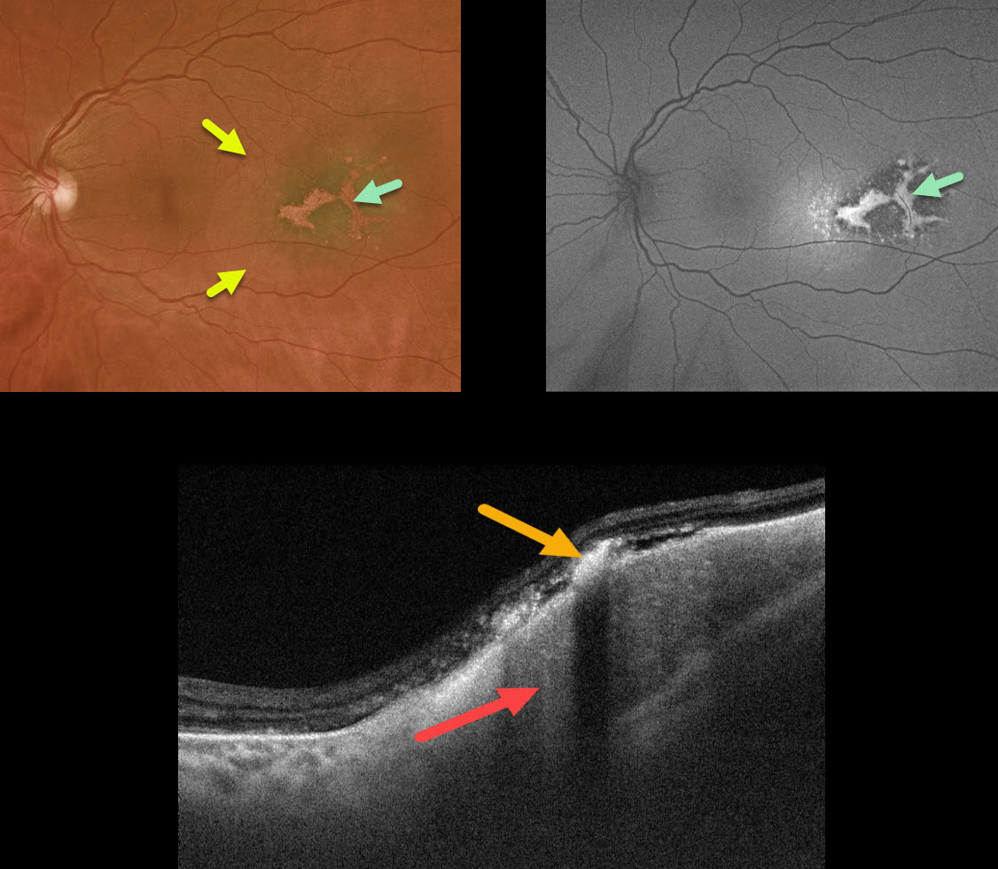
CHOROIDAL METASTASES
Originally posted on @retina.rocks March 31, 2021
This 50YO female with a known history of cutaneous melanoma and renal cell carcinoma presented with a variably pigmented temporal macular lesion.
The lesion had an associated shallow serous detachment and overlying lipofuscin, which was hyperautofluorescent. OCT B-scan showed the choroidal mass with overlying hyperreflective lipofuscin.
Transpupillary thermotherapy was recommended, but external beam radiation will be needed if the lesion further enlarges.
We assume this lesion is a choroidal metastasis, but we are unsure of the primary cancer. We can also not rule out that this represents a primary uveal malignant melanoma (MM). Her oncologist is aware of this choroidal lesion, and she continues to receive systemic treatment for her renal cell carcinoma.
Learning Points:
Although uveal MM is the most common primary intraocular malignancy, metastatic choroidal tumors are the most common intraocular malignancies. Lung and breast cancer are the most common sources.
Uveal MM is usually not familial nor associated with systemic cancers. The BAP1 mutation is a recently recognized autosomal dominant syndrome with predisposition to uveal melanoma and other primary cancers, including cutaneous melanoma, renal cell carcinoma, and mesothelioma (see Singh et al, AJO 2021;224:172-177).
We spoke at length with the patient’s oncologist, and at least for now, he did not want to check for BAP1.
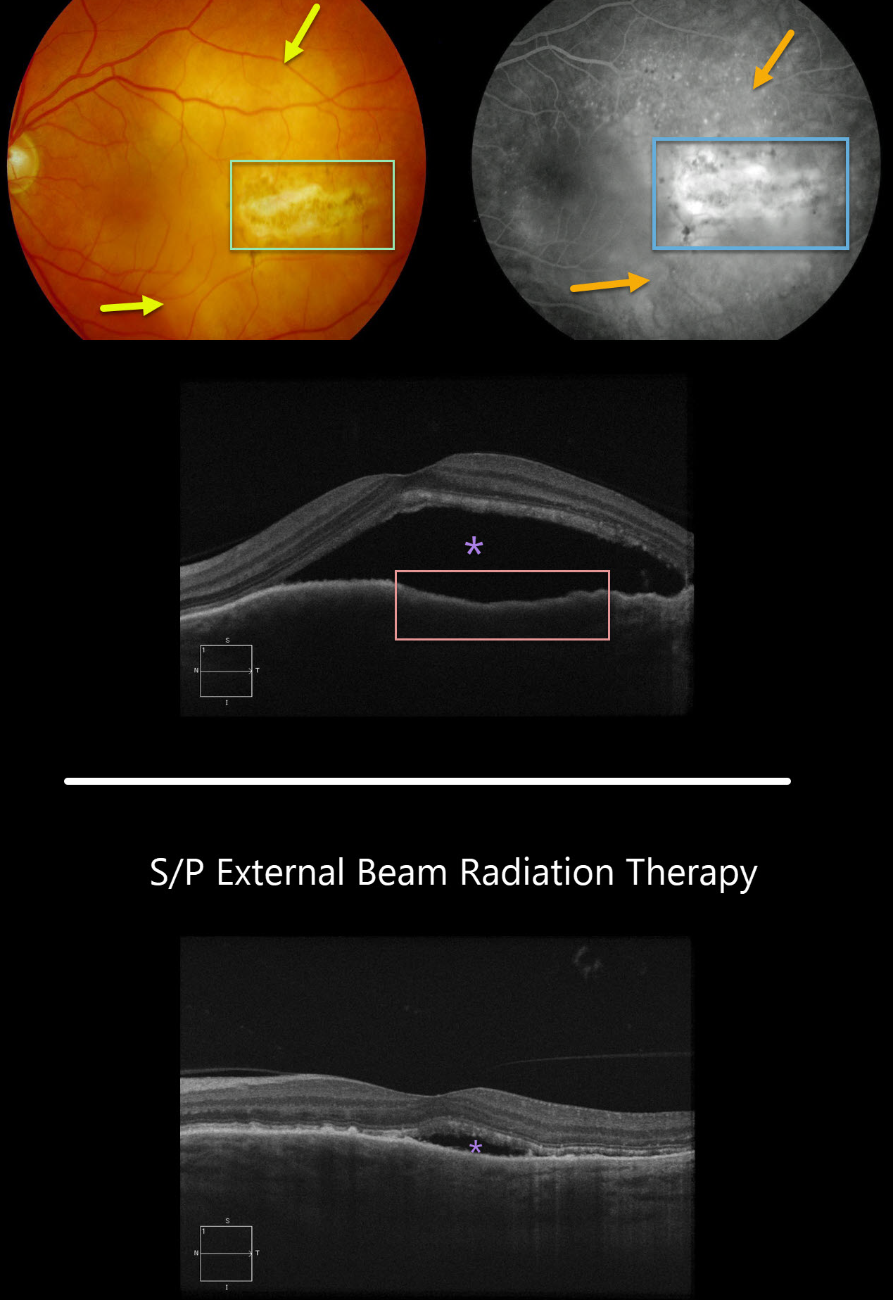
CHOROIDAL METASTASES
Originally posted on @retina.rocks February 9, 2021
This patient with a known history of breast cancer presented with a creamy colored choroidal lesion with central overlying pigmentary changes and scarring in the temporal macula.
Fluorescein angiography showed diffuse subretinal leakage with staining of the overlying scar. OCT scanning showed choroidal thickening with an irregular retinal pigment epithelial contour and overlying subretinal fluid.
The metastatic lesion was treated with localized external beam irradiation, with near immediate tumor regression and decreasing fluid 5 weeks later.
Learning Points:
Although uveal melanoma is the most common primary intraocular malignancy, metastatic choroidal tumors are the most common intraocular malignancies.
Lung and breast cancer are the most common sources.
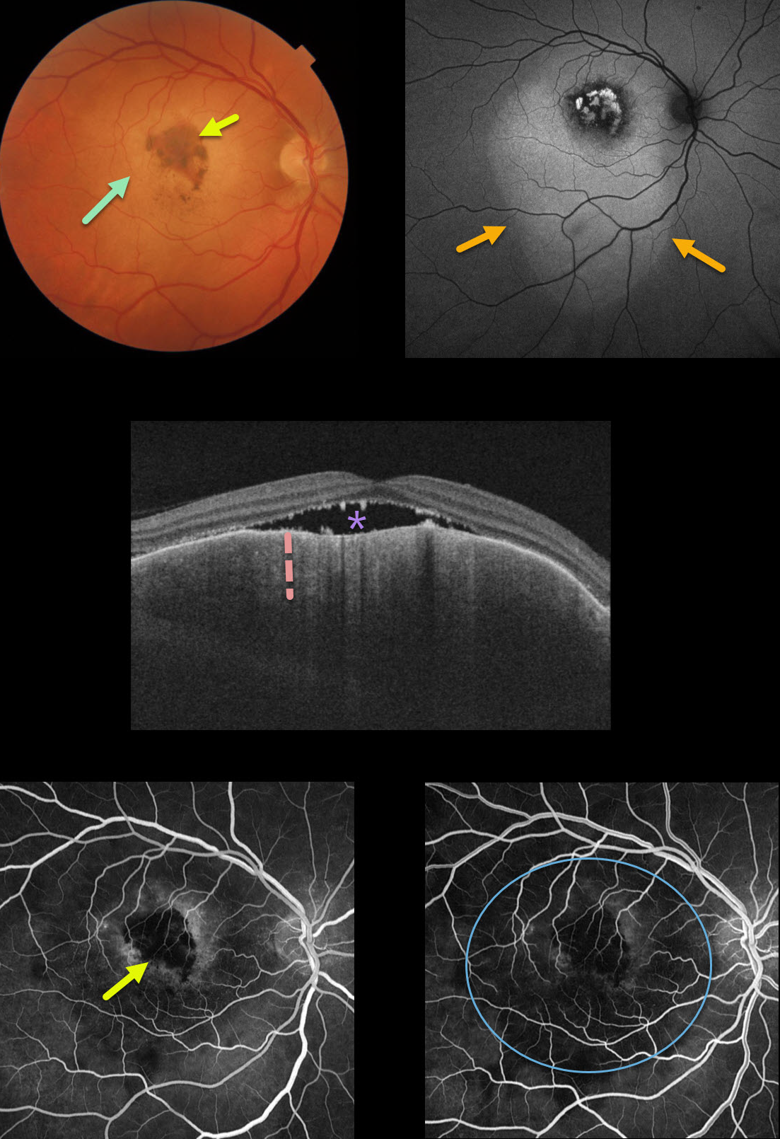
CHOROIDAL METASTASES
Originally posted on @retina.rocks February 10, 2020
This patient has an uncommon pancreatic choroidal metastasis. The lesion is clinically amelanotic with a more central area of increased pigmentation but is much
larger on fundus autofluorescence.
The OCT shows marked choroidal thickening with some subretinal fluid. Fluoroscein angiography (FA) shows no leakage with blockage from the central hyperpigmentation and fainter surrounding blockage from the tumor itself.
Learning Points:
Although uveal melanoma is the most common primary intraocular malignancy, metastatic choroidal tumors are the most common intraocular malignancies.
Lung and breast cancer are the most common sources.


