

Choroidal Nevus and Freckle
<< Back to Cases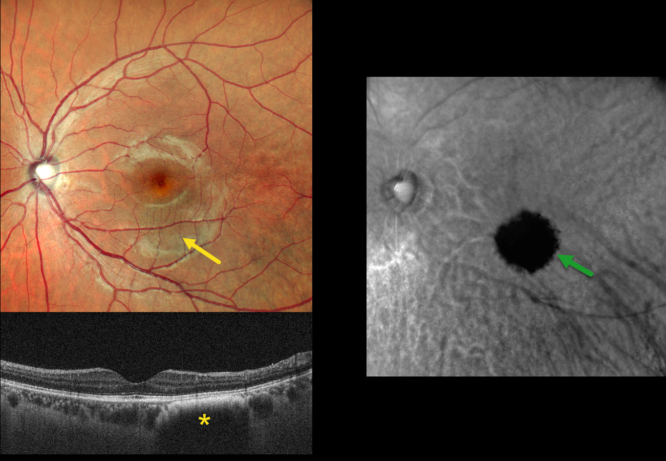
SUBCLINICAL CHOROIDAL NEVUS VS HEMANGIOMA
Shraddha Raj Shrivastava, Steven Bloom, and Manish Nagpal
Originally posted on @retina.rocks May 13, 2026
This asymptomatic 26YO male presented for a routine eye examination. Vision was 20/20.
Clinical biomicroscopy and MultiColor scanning laser ophthalmoscope (SLO) imaging are normal except for faint hyperpigmentation in the inferior macula. OCT in this region shows a dome-shaped hyporeflective lesion with an anterior hyperreflective border. Retromode imaging shows a well-defined hyporetroreflective lesion.
Learning Points:
Traditional color photography uses white light to image the posterior segment. Newer imaging devices utilize LED or SLO technology to illuminate the fundus with blue, green, and infrared light, which best image the vitreoretinal interface/inner retina, mid-retina to RPE, and choroid, respectively. Many of these technologies create a ‘color’ fundus image that is not true to life yet contains clinically useful depth information in a flat image. In our patient’s case, Nidek Mirante Retromode imaging best detected this subclinical lesion using a 790 nm SLO. For a recent review of Retromode imaging, see Sukkarieh et al, Surv Ophthalmology 2023;68:1027-1037.
So does our patient have a choroidal nevus or hemangioma? Internally, the authors are split between a nevus (SRS and MN) and a hemangioma (SB). Findings supporting a choroidal hemangioma include its orange color blending with the surrounding RPE/choroid and round shape on Retromode imaging. However, we have not seen a hemangioma with an anterior hyperreflective margin on OCT. Jonna et al described 5 OCT patterns of flat choroidal nevi (Ophthalmology Retina 2019;3:270-277), including a subtype with “anteriorly bowed hyperreflectivity with discrete borders and cascading edges,” which appears identical to our patient’s OCT.
SUSPECT CHOROIDAL NEVUS
Originally posted on @retina.rocks January 29, 2026
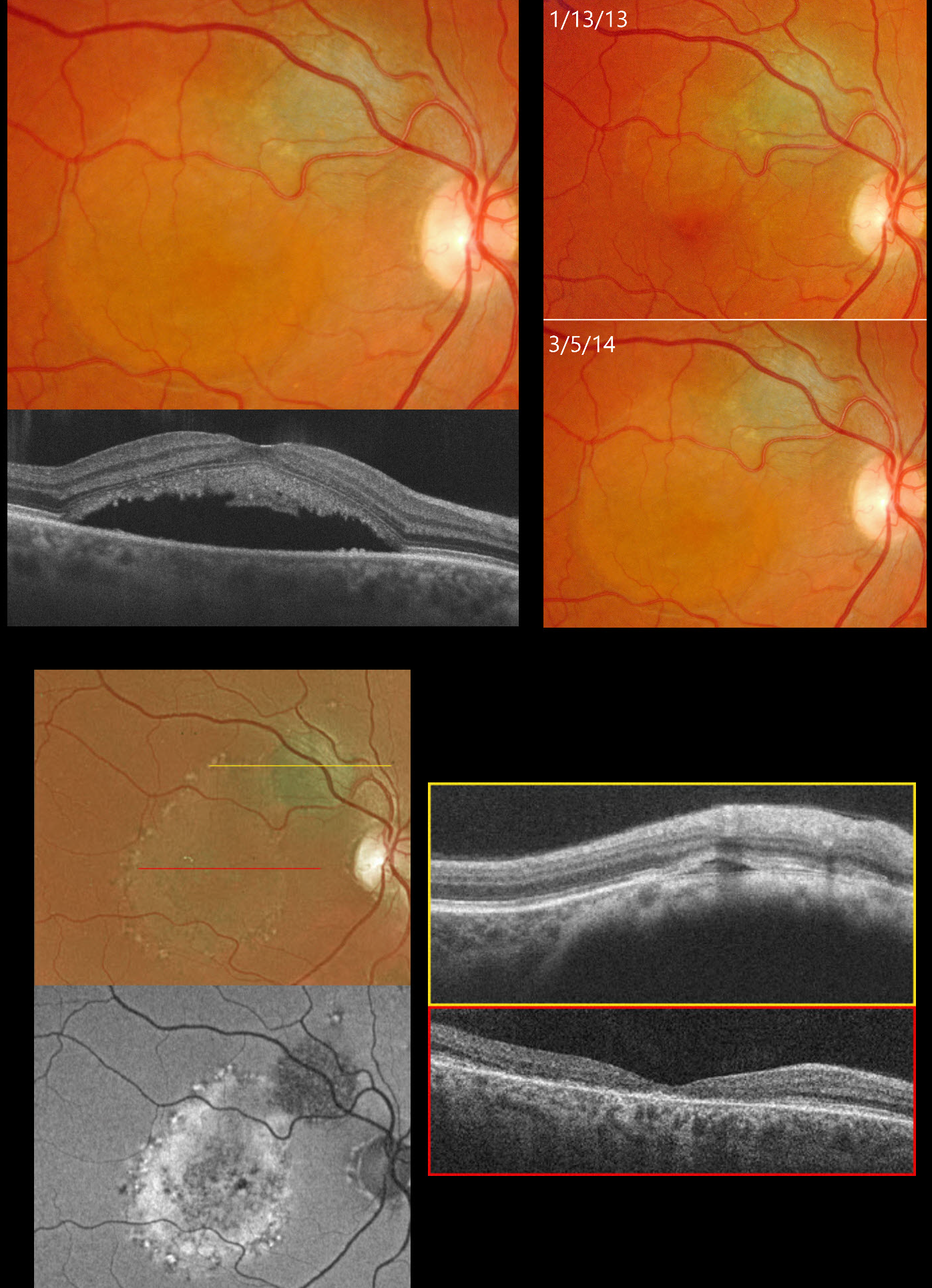
This 35YO female was examined for a suspect choroidal nevus on 1/13/13. Color photography shows about a 2x2mm minimally thickened choroidal nevus superotemporal to the right optic nerve (image 2). Subretinal fluid extends into the superior macular and some orange pigment overlies the nevus.
She returned on 3/15/14, now with fluid extending through the fovea (image 1). OCT scanning confirms the foveal fluid and hyperreflective material, likely representing shed outer segments, lines the outer surface of the detached macula.
She was referred to an ocular oncologist and immediately lost to follow-up until she presented on 4/2/25 without new complaints. She told us she had received two photodynamic therapies elsewhere shortly after we last saw her over 10 years earlier. Vision was 20/200. Optos color RG imaging shows the nevus to be stable in size but with new pigmentary changes throughout the macula within the area of prior fluid (image 3). On fundus autofluorescence (FAF), the nevus is hypo-FAF, and the region of prior fluid shows variable hyper-FAF. OCT scanning shows no fluid overlying the nevus with foveal atrophy. Continued observation was recommended.
Learning Points:
Clinical characteristics of choroidal nevi at risk for progressing to a uveal MM were initially described by Shields et al (Ophthalmology 1995;102:1351-1361). The most recent version of this famous acronym (Shields et al, Retina 2019;39:1840-1851) is To Find Small Ocular Melanoma Doing Imaging (TFSOM-DIM), which stands for To (Thickness >2mm) Find (subretinal Fluid) Small (Symptoms, vision <= 20/50) Ocular (Orange pigment) Melanoma (Melanoma hollow on ultrasonography) DIM (DIaMeter > 5mm).
METASTATIC MELANOMA OF UNKNOWN ORIGIN, SUSPECT CHOROIDAL NEVUS OD AND PROBABLE MELANOMA OS
Mattie Adams
Originally posted on @retina.rocks January 19, 2026
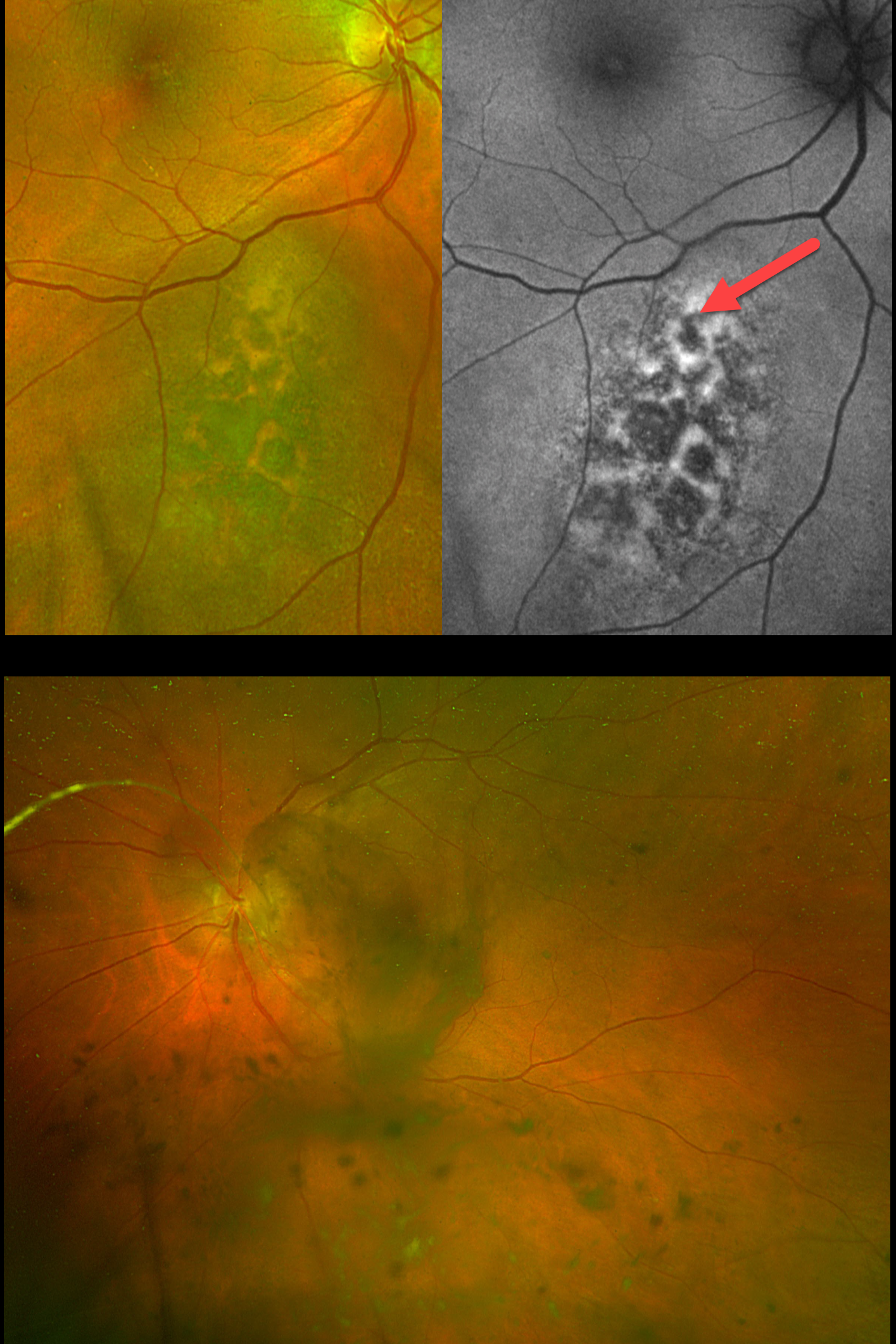
This 67YO female presented with a 3-week history of floaters in her left eye. She has a known history of metastatic melanoma of unknown origin, including numerous brain lesions. There was no family history of ocular or cutaneous melanoma. Vision was 20/20 OD and 20/40 OS.
Optos color RG imaging OD shows a suspect choroidal nevus just inferonasal to the macula. There is overlying orange lipofuscin pigment, which is hyperautofluorescent. Optos imaging OS shows extensive pigmented vitreous debris variably obscuring the posterior pole.
Learning Points:
Uveal malignant melanoma (MM) and cutaneous melanoma are rarely associated with each other. The exception is the presence of a mutated tumor suppressor gene, BRCA1-associated protein 1 (BAP1), which is associated with autosomal-dominant uveal MM and other primary cancers, including cutaneous melanoma.
Our patient did not have a primary for her metastatic disease. The presence of a suspect nevus in her right eye and possible metastatic melanoma to her left eye argues for a pathogenic BAP1 mutation. We discussed this possibility with her oncologist, who felt this was highly unlikely given the negative family history. Given her relatively good vision and poor systemic prognosis, observation was recommended as the initial management for her ocular findings. Unfortunately, she died 3 months later.
CHOROIDAL NEVUS WITH CNV
Originally posted on @retina.rocks October 15, 2025
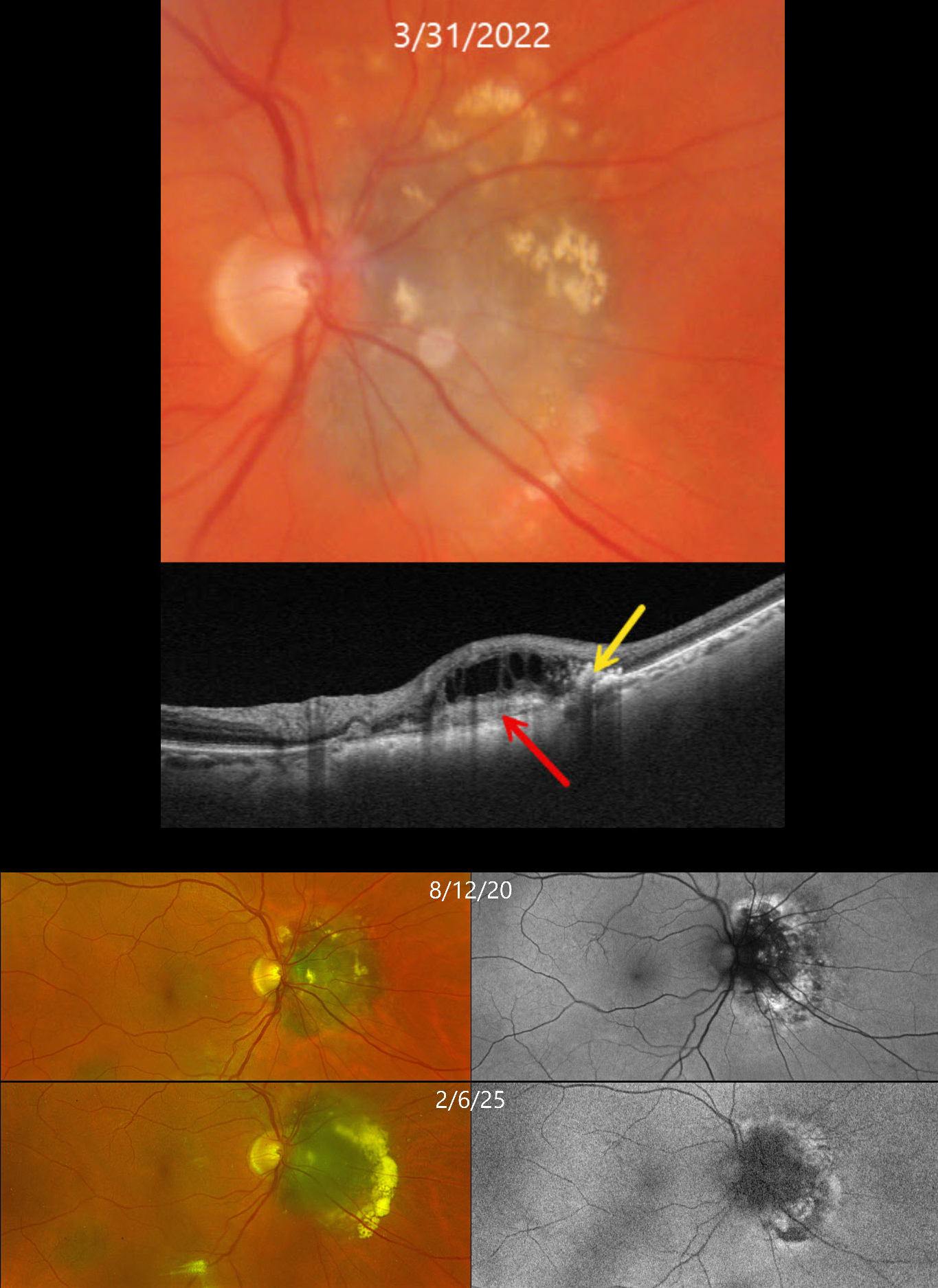
This 71YO female has been followed for several years with a choroidal nevus and chronic overlying choroidal neovascularization (CNV).
On March 31, 2022, Triton color photography shows a nasal peripapillary choroidal nevus that touches the optic nerve. Lipid is noted mostly along its superior and temporal edges. Swept-source OCT shows a hyperreflective type 2 (above the RPE) CNV, hyperreflective lipid, and overlying cystic retinal thickening.
The CNV has been observed due to her complete lack of symptoms and extramacular location of the lesion. When last examined on February 6, 2025, the nevus looks fairly stable on Optos RG imaging, although there is increased subretinal lipid along its temporal edge. On fundus autofluorescence (FAF), the CN is hypo-FAF with hyper-FAF along its periphery.
Learning Points:
Choroidal neovascularization is a rare finding in choroidal nevi, and associated exudation can be misinterpreted as exudation from possible malignant transformation. OCT angiography is helpful in diagnosing these lesions, which respond well to either intravitreal anti-VEGF monotherapy or combination treatment with photodynamic therapy (Munie and Demirci, Ophthalmology Retina 2018;2:53-58).
CHOROIDAL NEVUS WITH MNV
Originally posted on @retina.rocks April 24, 2025
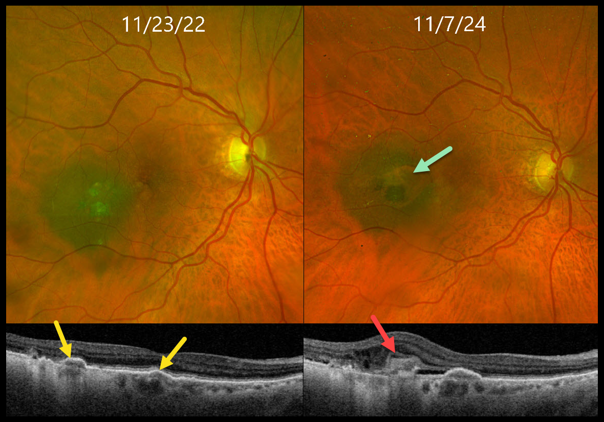
This 85YO female presented for her annual choroidal nevus examination, noticing worsening reading vision. Vision had decreased to 20/200 from 20/40 one year earlier.
When seen 2 years earlier, Triton swept-source OCT showed a few areas of drusen-like changes overlying a temporal macular nevus. At the current visit, although the margins of the nevus were unchanged, Optos color RGB imaging shows a new area of relative depigmentation with a nodular area of pigmented subretinal thickening. OCT scanning shows a hyperreflective type 2 (above the RPE) macular neovascularization (MNV) with mild associated fluid.
Monthly intravitreal Avastin therapy was started. The lesion regressed after several monthly injections, vision improved to 20/60, and a treat-and-extend regimen was started.
Learning Points:
Macular neovascularization is a rare complication of choroidal nevi. Intravitreal Avastin monotherapy is usually successful, although photodynamic therapy may be needed in some cases (Munie and Demirci, Ophthalmology Retina 2018;2:53-58).
We were initially somewhat concerned that the OCT appearance of the type 2 MNV might represent early tumor proliferation through Bruch’s membrane, but the absence of underlying choroidal thickening and the prompt response to anti-VEGF treatment argued against this.
CHOROIDAL NEVUS MISDIAGNOSED AS CENTRAL SEROUS
Emma Oreskovic and Natasa Draca
Originally posted on @retina.rocks September 7, 2023
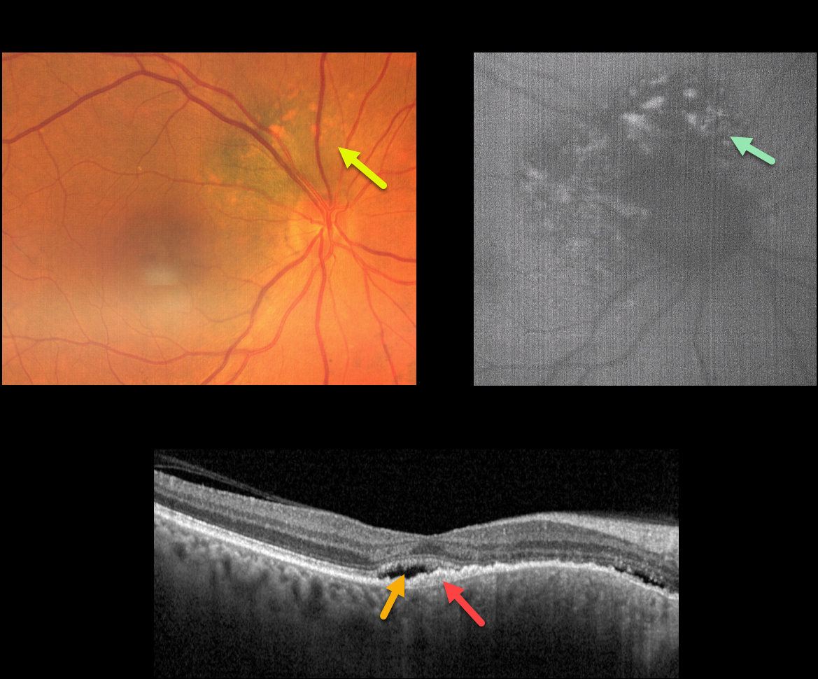
This 71YO female was referred for further treatment of chronic idiopathic central serous chorioretinopathy (ICSC) of her right eye, for which she had previously received unsuccessful anti-VEGF therapy at another institution. Vision was 20/100 OD and 20/20 in her normal left eye.
Fundus photography shows a variably pigmented, minimally thickened peripapillary choroidal nevus that extends from about 7-1 o’clock around the optic nerve margin. Overlying orange pigmentation is noted, which variably hyper-autofluoresces.
OCT scanning shows shallow foveal subretinal fluid with a subretinal layer of thickened hyperreflective material extending nasally overlying the thickened nevus. Beneath this is a very shallow, irregular elevation of the RPE.
Learning Points:
Clinical characteristics of choroidal nevi at risk of progressing to uveal malignant melanoma (MM) were initially described by Shields et al. (Ophthalmology 1995;102:1351-1361).
The most recent version of this famous acronym (Shields et al, Retina 2019;39:1840-1851) is To Find Small Ocular Melanoma Doing Imaging (TFSOM-DIM), which stands for To (Thickness >2mm) Find (subretinal Fluid) Small (Symptoms, vision <= 20/50) Ocular (Orange pigment) Melanoma (Melanoma hollow on ultrasonography) DIM (DIaMeter > 5mm).
ICSC and pachychoroid disorders, similar to syphilis, are one of the ‘great imitators,’ including uveal MM masquerading as ICSC (Negretti et al, Ophthalmology Retina 2023;7:171-177).
In our patient, on the central macular OCT B-scan, one sees a thickened choroid, subretinal fluid, and hyperreflective shed outer segments/lipofuscin, all of which are suggestive of ICSC.
This case emphasizes the importance of looking beyond the OCT, using both the clinical exam and multimodal imaging, for every patient, including those with chronic ICSC, to arrive at the correct diagnosis and treatment.
Since our patient has several high-risk characteristics (fluid, symptoms, and orange pigment), she was referred to an ocular oncologist for further management.
HALO CHOROIDAL NEVUS
Natasa Draca and Emma Oreskovic
Originally posted on @retina.rocks April 26, 2023
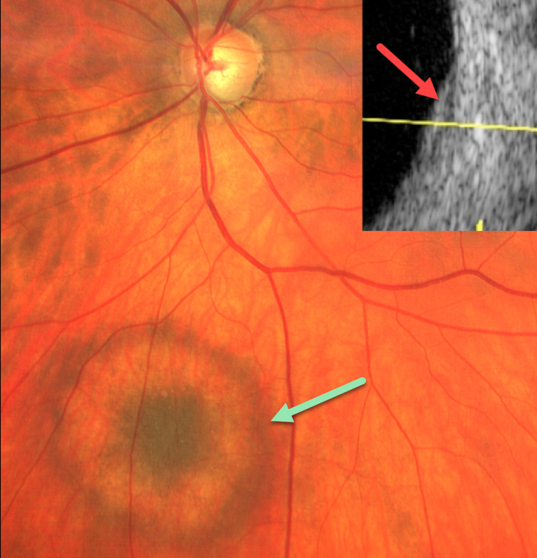
During a routine glaucoma examination, this 61YO male was found to have a halo choroidal nevus in his right eye. The nevus was hyperpigmented centrally, then encircled by relative depigmentation, and finally surrounded by a hyperpigmented rim. The lesion was clinically minimally thickened and measured about 2-3 disc diameters. The nevus was barely visible with B-scan ultrasonography.
Learning Points:
Halo choroidal nevi are a relatively rare variant of the more typical diffusely hyperpigmented lesions. A large retrospective series by Shields et al found that these lesions may be associated with cutaneous melanoma (Arch Ophthalmology 2010;128:859-864).
Malignant transformation to uveal melanoma is reduced when a halo is present (Shields et al, Arch Ophthalmol 2009;127:981-987), although this does not preclude malignant transformation.
CHOROIDAL NEVUS
Originally posted on @retina.rocks March 30, 2023
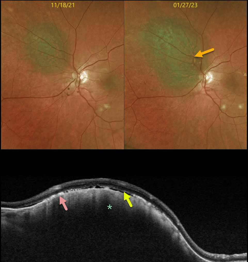
This 71YO female presented on 1/27/23 for follow-up for an asymptomatic, less than 1mm thick choroidal nevus measuring 6.3 x 5.5 mm.
Optos color imaging shows that the nevus has significantly increased in size (3.0 x 10.5 x 7.5 mm) compared with the prior visit on 11/18/21. The nevus enlarged laterally in all dimensions and now touched the nerve margin. There was significantly more overlying orange pigment.
Swept-source OCT shows a thickened choroidal lesion. The clinically noted orange pigment is seen on OCT as hyperreflective outer retinal deposits. Trace variable subretinal fluid vs loss of outer retinal tissue is also seen.
Radioactive plaque placement was performed. Fine needle biopsy revealed the tumor to be Class 1A, PRAME negative.
Learning Points:
Clinical characteristics of choroidal nevi at risk for progressing to a uveal MM were initially described by Shields et al (Ophthalmology 1995;102:1351-1361). The most recent version of this famous acronym (Shields et al, Retina 2019;39:1840-1851) is To Find Small Ocular Melanoma Doing Imaging (TFSOM-DIM), which stands for:
To (Thickness >2mm)
Find (subretinal Fluid)
Small (Symptoms, vision <= 20/50)
Ocular (Orange pigment)
Melanoma (Melanoma hollow on ultrasonography)
DIM (DIaMeter > 5mm).
Although our patient had several of these baseline risk factors (orange pigment, 6.3mm diameter), the growth in just 6 months is unusual.
Patients with Class 1A lesions have a lower 5-year metastatic risk (2%) than those with Class 1B or 2 lesions. PReferentially expressed Antigen in MElanoma (PRAME) is a gene that can be aberrantly expressed in some malignancies. Its positivity can indicate an increased risk for metastatic disease in Class 1 tumors, but its negativity does not tend to alter prognosis.
CHOROIDAL NEVUS
Originally posted on @retina.rocks August 30, 2022
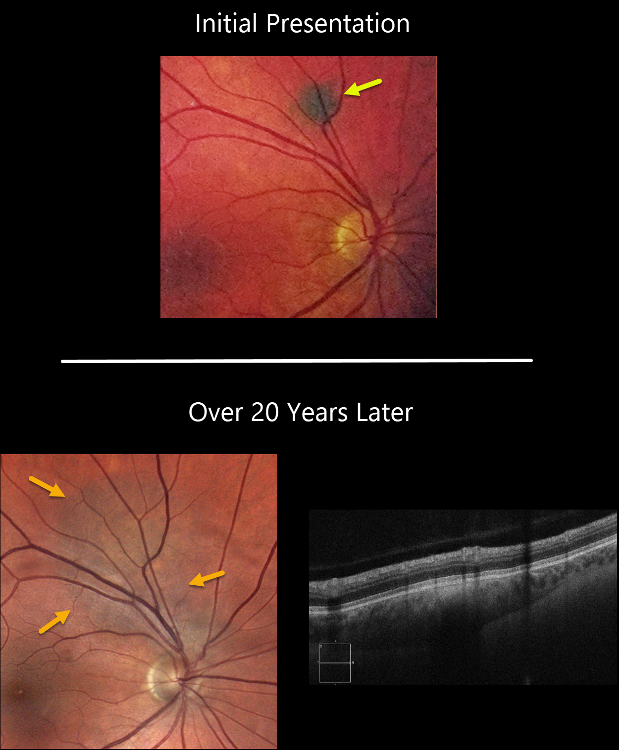
This 58YO male was initially seen in 2001 with a small asymptomatic choroidal nevus superior to the right optic nerve.
He presented to us in 2022 with a still asymptomatic but much enlarged nevus. The lesion now extends to the nerve. There is no overlying orange pigment or subretinal fluid clinically or on OCT. The lesion was 0.4mm thick.
Learning Points:
Clinical characteristics of choroidal nevi at risk for progressing to a uveal MM were initially described by Shields et al (Ophthalmology 1995;102:1351-1361). The most recent version of this famous acronym (Shields et al, Retina 2019;39:1840-1851) is To Find Small Ocular Melanoma Doing Imaging (TFSOM-DIM), which stands for:
To (Thickness >2mm)
Find (subretinal Fluid)
Small (Symptoms, vision <= 20/50)
Ocular (Orange pigment)
Melanoma (Melanoma hollow)
DIM (DIaMeter > 5mm)
Although our patient’s nevus has grown over time, this does not imply malignant transformation, as about one-third will enlarge with long-term follow-up (Shields et al., Ophthalmology 2011;118;382-388).
Shields et al found a marked increased risk for
malignant transformation for choroidal nevi over 2.0mm (Retina 2019;39:1840-1851).
Since our patient’s nevus is still well below this number and has no TFSOM-DIM risk factors other than diameter, we feel safe managing him conservatively with observation twice yearly.
CHRPE + CHOROIDAL NEVUS
Originally posted on @retina.rocks July 19, 2022
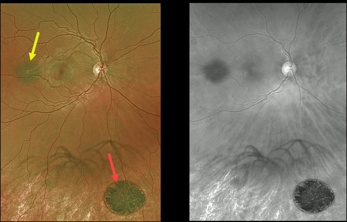
This 27YO male was referred for two distinct and asymptomatic pigmented fundus lesions.
Optos color imaging shows a small, flat choroidal nevus and a larger, flat patch of congenital hypertrophy of the RPE. The color image best shows the color difference between these lesions, with the CHRPE lesion appearing jet-black compared with the nevus.
The red channel best highlights the typical, somewhat indistinct margins of the nevus compared with the very sharp, round margins typical for the CHRPE. The patient is being followed yearly.
Learning Points:
Clinical characteristics of choroidal nevi at risk for progressing to a uveal malignant melanoma were initially described by Shields et al (Ophthalmology 1995;102:1351-1361).
The most recent version of this famous acronym (see Retina 2019;39:1840-1851) is To Find Small Ocular Melanoma Doing Imaging (TFSOM-DIM), which stands for:
To (Thickness >2mm)
Find (subretinal Fluid)
Small (Symptoms, vision <= 20/50)
Ocular (Orange pigment)
Melanoma (Melanoma hollow on ultrasonography)
DIM (DIaMeter > 5mm)
Although CHRPE lesions can enlarge with time (see Shields et al, Ophthalmology 2003;110:1968-1973), they carry virtually no malignant potential.
Histologic RPE hypertrophy presents in several ways. The typical isolated CHPRE lesions and bear tracks are not associated with intestinal polyps or cancer, and thus require no systemic evaluation. Only the multiple, small comet-shaped lesions are part of the familial polyposis spectrum.
CHOROIDAL NEVUS
Originally posted on @retina.rocks February 11, 2022
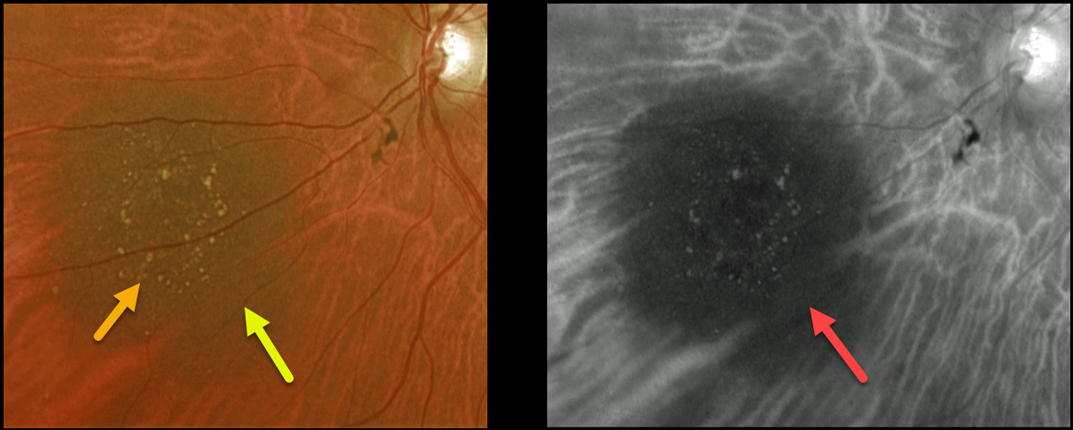
This 72YO female presented with a nasal choroidal nevus seen on Optos color and Optos red channel. The lesion is flat, has overlying drusen, and measures 4.5 by 5.1 mm. Is the nevus at risk for progressing to a uveal malignant melanoma (MM)?
Learning Points:
Clinical characteristics of choroidal nevi at risk for progressing to a uveal MM were initially described by Shields et al (Ophthalmology 1995;102:1351-1361).
The most recent version of this famous acronym is To Find Small Ocular Melanoma Doing Imaging (TFSOM-DIM), which stands for To (Thickness >2mm) Find (subretinal Fluid) Small (Symptoms, vision <= 20/50) Ocular (Orange pigment) Melanoma (Melanoma hollow on ultrasonography) DIM (DIaMeter > 5mm).
Other than measuring just over 5mm in diameter, our patient has none of these other risk factors. The presence of drusen also indicates chronicity and low risk for malignant transformation.
We will follow her yearly.
CHOROIDAL NEVUS
Originally posted on @retina.rocks December 16, 2020
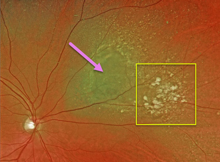
This 70YO male presented with this nasal choroidal nevus.
Other than being near the optic nerve, our patient has none of the risk factors for transformation to uveal melanoma. The presence of drusen also indicates chronicity and low risk for malignant transformation. We are following him yearly.
Learning Points:
Clinical characteristics of choroidal nevi at risk for progressing to a uveal MM were described by Shields et al. (Ophthalmology 1995;102:1351-1361). The most recent version of this famous acronym, To Find Small Ocular Melanoma Doing Imaging (TFSOM-DIM, Retina 2019;39:1840-1851), is:
To (Thickness >2mm)
Find (Subretinal Fluid)
Small (Symptoms, vision <= 20/50)
Ocular (Orange pigment)
Melanoma (Melanoma hollow)
DIM (DIIaMeter >5mm)


