

Choroidal Osteoma
<< Back to Cases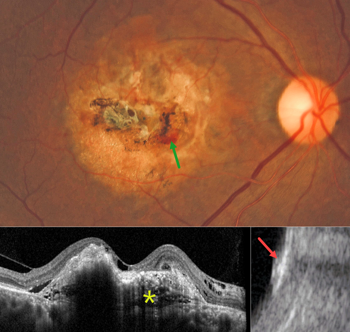
CHOROIDAL OSTEOMA
César Adrián Gómez Valdivia
Originally posted on @retina.rocks July 27, 2026
This 73YO male presented with 8 months of gradual vision loss in his right eye. Vision was 20/200 OD and 20/25 in his normal OS.
Color photography shows a yellowish, refractile amelanotic occupying nearly the entire macula. Some areas of increased pigment are noted centrally, along with some blood inferonasally. On OCT, the lesion has an internal lamellar, sponge-like architecture, characterized by horizontal hyporeflective lines and some intraretinal and subretinal fluid. The lesion is markedly hyperreflective on B-scan ultrasonography with posterior acoustic shadowing.
Learning Points:
Choroidal osteoma is a rare, benign ossifying choroidal tumor that typically affects young women, often in the peripapillary or macular region. Typical features include a clinical yellowish-orange lesion, a hyperreflective choroidal mass with a lamellar or trabecular internal architecture on OCT, and, most importantly, marked posterior acoustic shadowing on B-scan ultrasonography due to calcification. These imaging characteristics are distinct from those of other choroidal lesions, such as melanoma or hemangioma, which lack dense calcification and exhibit different reflectivity patterns.
Although benign, choroidal osteoma may cause vision loss due to macular neovascularization (MNV), subretinal fluid, and progressive decalcification, with atrophy of the retinal pigment epithelium and outer retinal layers. MNV can be successfully treated with anti-VEGF therapy (see Seong et al, Graefe’s 2022;250:1713-1721). Intravitreal Eylea was started for our patient.
CHOROIDAL OSTEOMA
Manuel Bande
Originally posted on @retina.rocks June 8, 2026
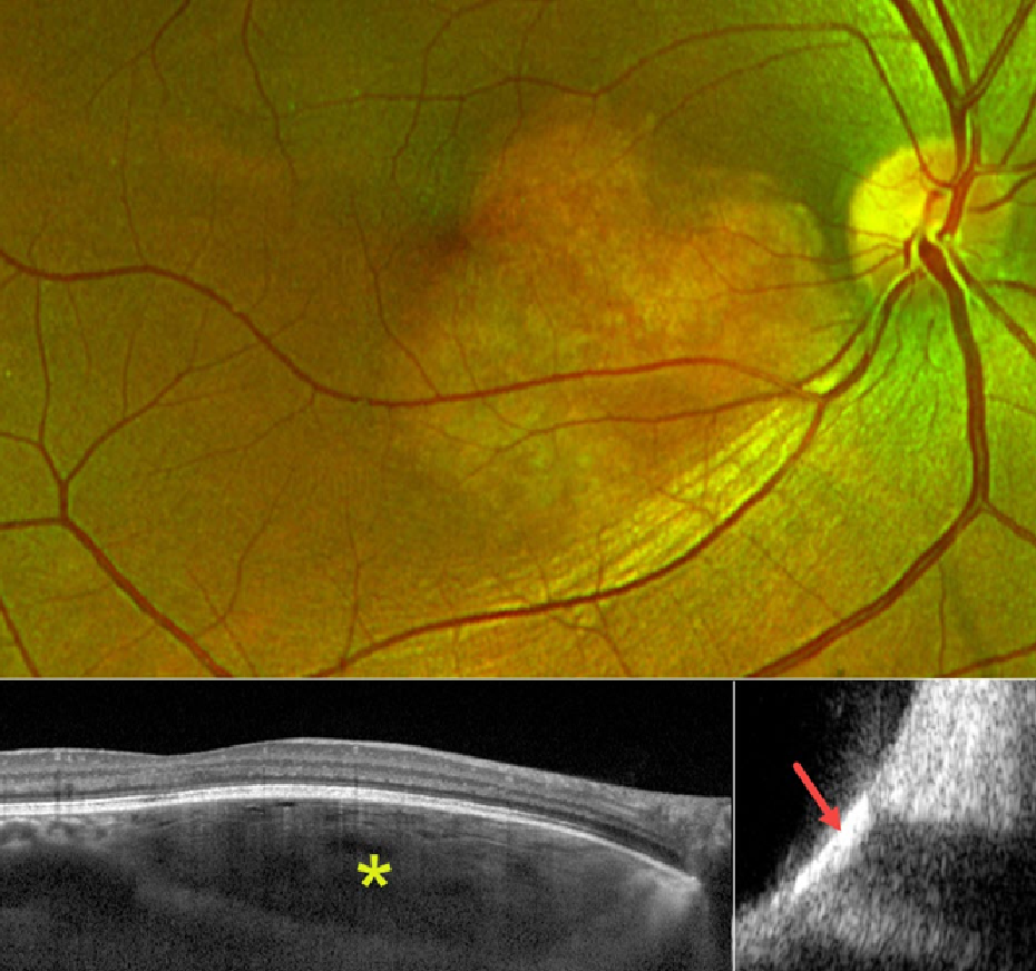
Optos color RG imaging shows a yellowish, well-circumscribed amelanotic lesion centered in the inferonasal macula. On OCT, the lesion has an internal lamellar, sponge-like architecture, characterized by horizontal hyporeflective lines corresponding to intralesional vascular channels and bony trabeculae. The overlying RPE and retina appear fairly normal. The lesion is markedly hyperreflective on B-scan ultrasonography with posterior acoustic shadowing.
Learning Points:
Choroidal osteoma is a rare, benign ossifying choroidal tumor that typically affects young women, often in the peripapillary or macular region. Typical features include a clinical yellowish-orange lesion, a hyperreflective choroidal mass with a lamellar or trabecular internal architecture on OCT, and, most importantly, marked posterior acoustic shadowing on B-scan ultrasonography due to calcification. These imaging characteristics are distinct from those of other choroidal lesions, such as melanoma or hemangioma, which lack dense calcification and exhibit different reflectivity patterns.
Although benign, choroidal osteoma may cause vision loss from macular neovascularization (MNV), subretinal fluid, and progressive decalcification with atrophy of the retinal pigment epithelium and outer retinal layers. MNV can be successfully treated with anti-VEGF therapy (see Seong et al, Graefe’s 2022;250:1713-1721).
CHOROIDAL OSTEOMA
Omar Mulki
Originally posted on @retina.rocks June 21, 2022
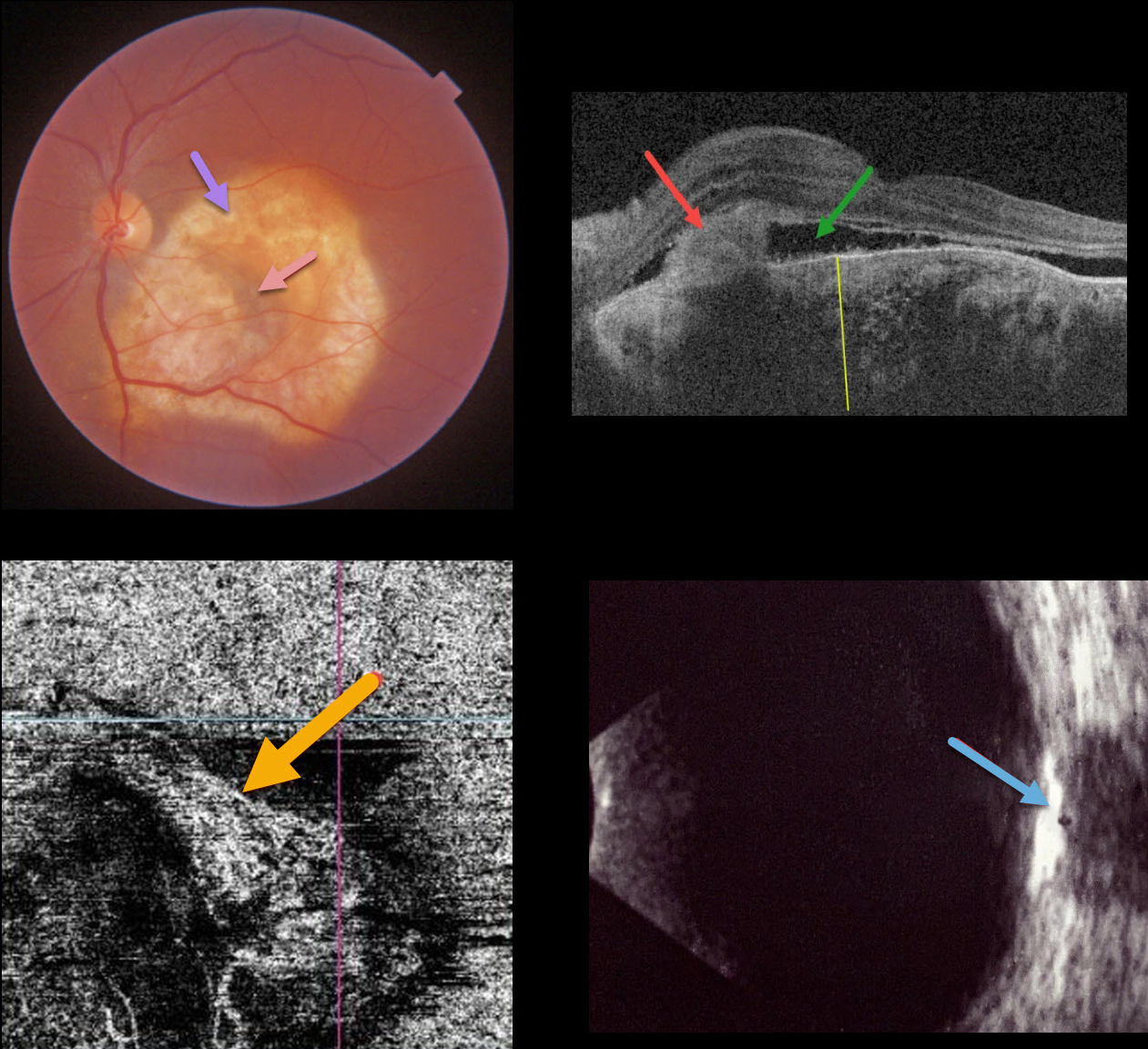
This 40YO female presented with recent vision loss in her left eye. Vision was 20/20 in her normal right eye and counting fingers in her left eye.
Triton color imaging shows an amelanotic choroidal lesion occupying virtually the entire macula. There is a small dot of central blood. Swept-source OCT shows a markedly thickened choroid with variable regions of hyper- and hyporeflectivity. A hyperreflective type 2 macular neovascularization (MNV) is seen along with exudative subretinal fluid. OCT angiography shows flow signals within the MNV. B-scan ultrasonography shows the lesion to be markedly hyperreflective.
An anti-VEGF injection was given, but she was unfortunately subsequently lost to follow-up.
Learning Points:
Choroidal osteomas are benign, calcified amelanotic choroidal tumors. They are more common in women, are almost always unilateral, and are usually located in the peripapillary or macular regions.
Choroidal lesions are best seen on the Optos red channel. Fluorescein angiography (FA) usually shows early patchy hyperfluorescence with late diffuse staining. Ultrasonography is often helpful in differentiating an osteoma (markedly hyperreflective) from other amelanotic choroidal tumors.
Macular neovascularization develops in a significant number of eyes and can be successfully treated with anti-VEGF therapy (see Seong et al, Graefe’s 2022;250:1713-1721).
CHOROIDAL OSTEOMA
Originally posted on @retina.rocks October 7, 2021
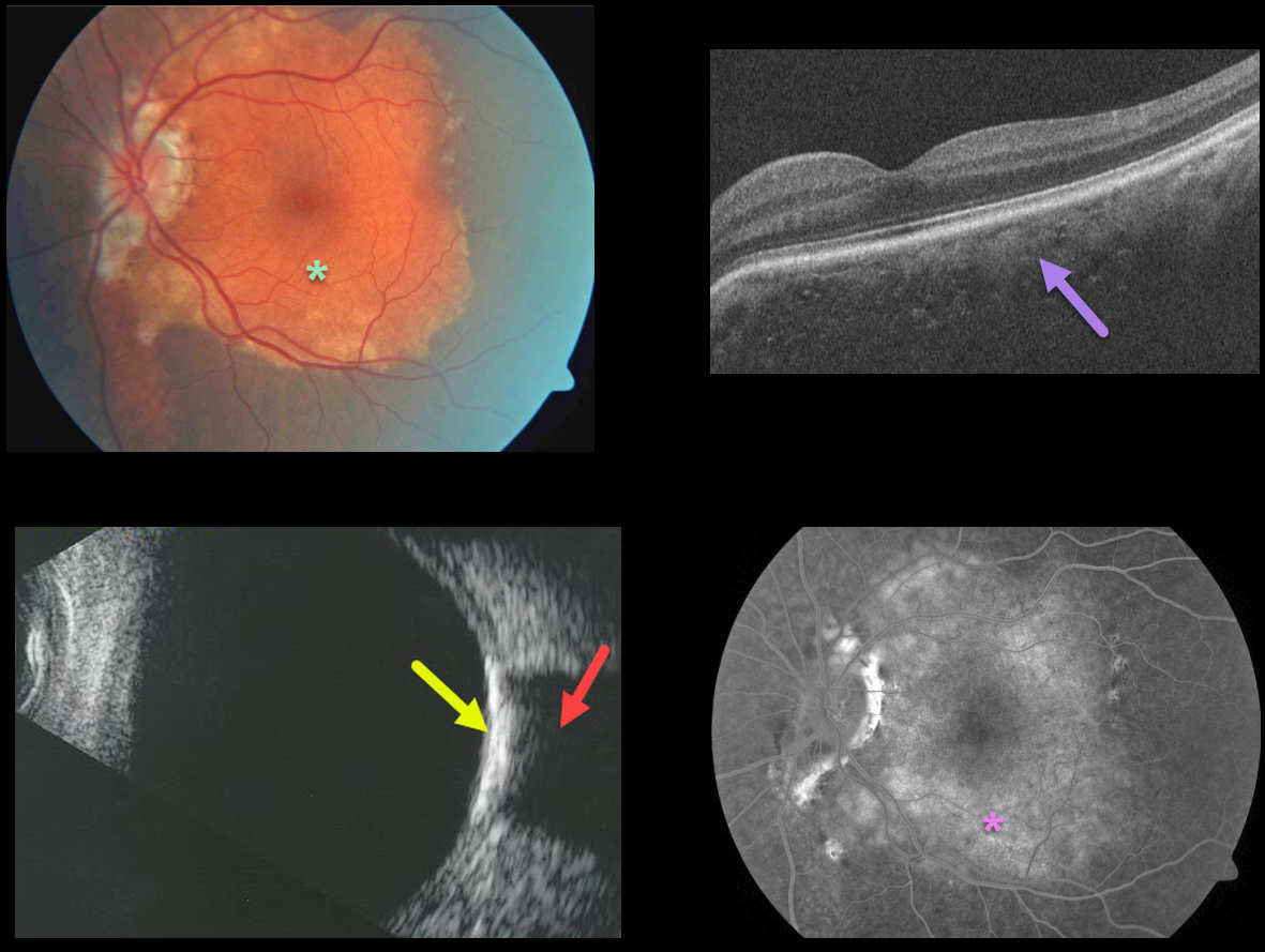
This patient presented with an orange-yellow choroidal lesion occupying the entire left macula.
OCT scanning shows diffuse choroidal thickening with small scattered specks of increased reflectivity.
B-scan ultrasonography shows marked increased reflectivity with marked posterior shadowing. The lesion diffusely stains on fluorescein angiography.
Learning Points:
Choroidal osteomas are benign, calcified amelanotic choroidal tumors. They are more common in women, are almost always unilateral, and are usually located in the peripapillary or macular regions.
Ultrasonography, as in this patient, is often helpful in differentiating an osteoma (markedly hyperreflective) from other amelanotic choroidal tumors. Fluorescein angiography usually shows early patchy hyperfluorescence with late diffuse staining.
Although the tumor itself does not require treatment, patients still need to be followed due to the risk of macular neovascularization.
CHOROIDAL OSTEOMA
Originally posted on @retina.rocks December 8, 2020
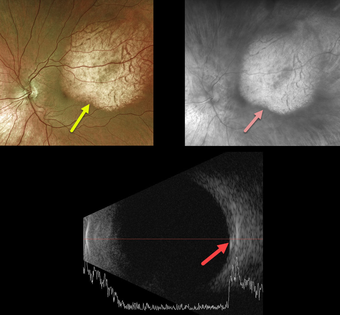
Our best guess is that this patient has a choroidal osteoma. As choroidal lesions, they are best seen on the Optos red channel.
We would, however, expect more reflectivity on the B-scan as osteomas are typically markedly hyperreflective.
Learning Points:
Choroidal osteomas are benign, calcified amelanotic choroidal tumors. They are more common in women, are almost always unilateral, and usually located in the peripapillary or macular regions.
Ultrasonography is useful in differentiating choroidal osteomas from other amelanotic choroidal tumors.
Fluorescein angiography (FA) usually shows early patchy hyperfluorescence with late diffuse staining.
CHOROIDAL OSTEOMA
Originally posted on @retina.rocks July 15, 2020
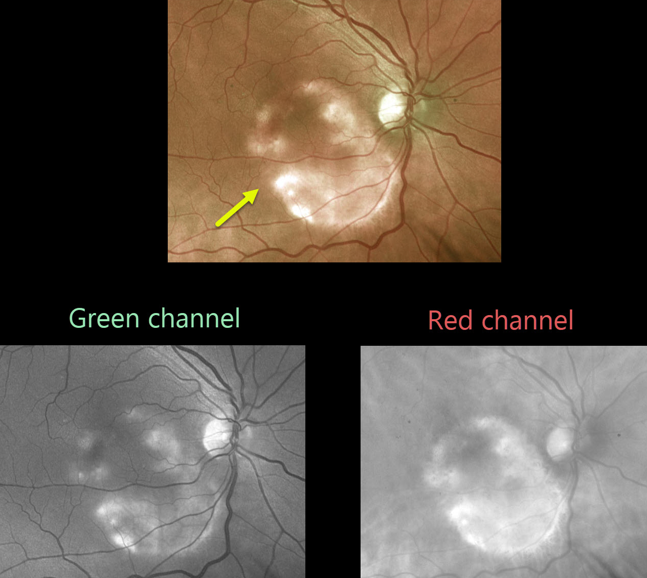
This asymptomatic patient was noted to have a choroidal osteoma.
As choroidal lesions, osteomas are best seen on the red channel compared with the green Optos channel. Ultrasonography is often helpful in differentiating an osteoma (markedly hyperreflective) from other amelanotic choroidal tumors.
Learning Points:
Choroidal osteomas are benign, calcified amelanotic choroidal tumors. They are more common in women, are almost always unilateral, and are usually located in the peripapillary or macular region.
Although many patients are asymptomatic, vision loss can occur from overlying subretinal fluid or macular neovascularization.


