

Choroidal Rupture
<< Back to Cases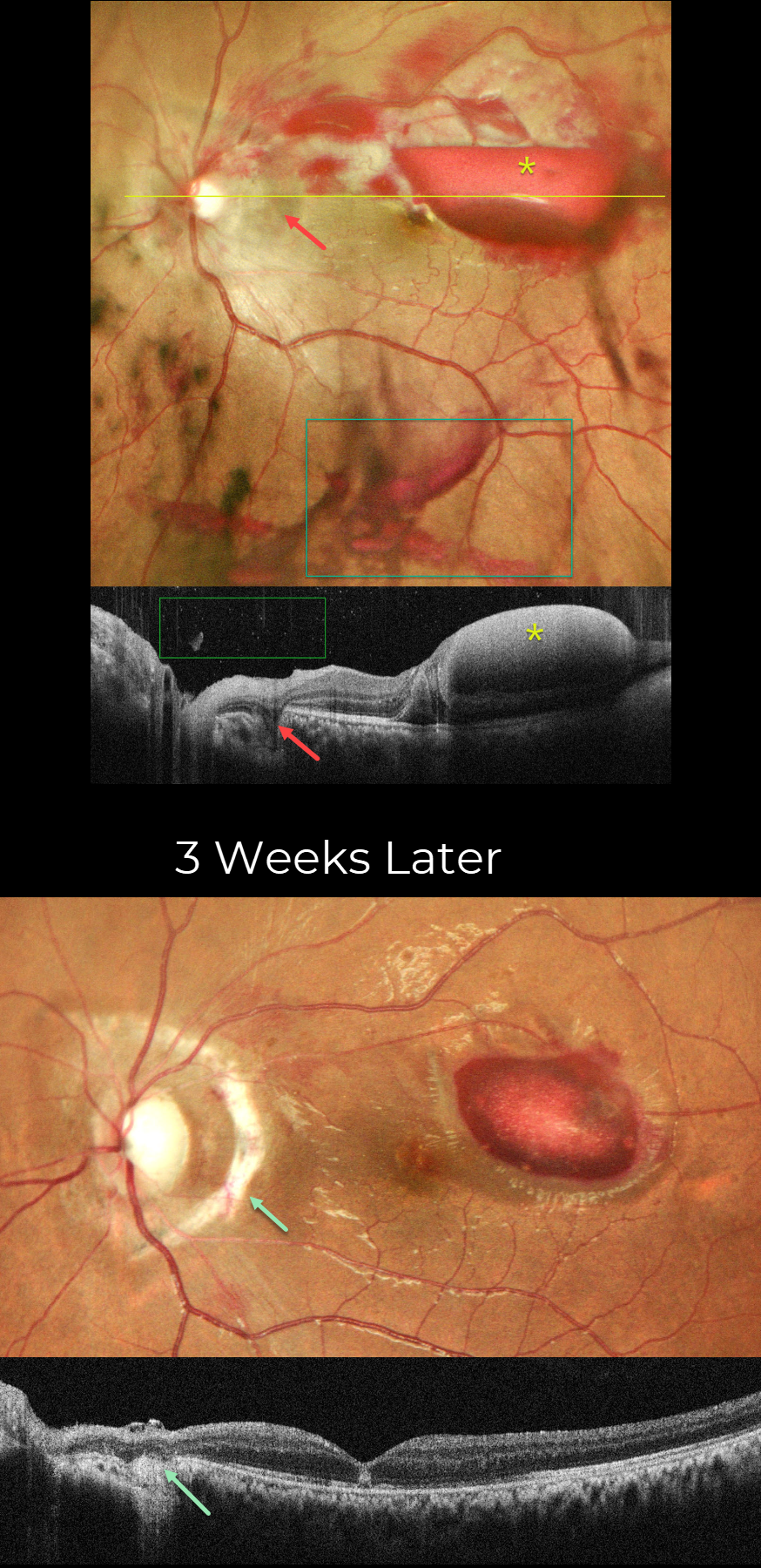
CHOROIDAL RUPTURE
Shraddha Raj Shrivastava and Manish Nagpal
Originally posted on @retina.rocks August 4, 2026
This 19YO male presented with vision loss in his left eye 2 days following blunt trauma from a tennis ball. Vision was counting fingers. Slit lamp examination revealed anterior chamber inflammation, traumatic mydriasis, and nasal subluxation of the crystalline lens.
Pseudocolor SLO imaging shows scattered retinal, sub-internal limiting membrane (ILM), and vitreous blood. The acute choroidal rupture appears as a crescent of increased subretinal pigmentation that arches around the nerve temporally and superiorly. Superior and superotemporal macular commotio retina is faintly visible. On OCT scanning, the vitreous and sub-ILM blood appear hyperreflective. The choroidal rupture appears as a hyporeflective defect extending from the outer retina to the sclera.
Topical steroids, atropine, and a short course of oral steroids were prescribed. Three weeks later, vision improved to 20/200. A prominent depigmented choroidal rupture with bare sclera surrounds the nerve nasally 180 degrees. The vitreous and retinal hemorrhages are virtually completely resolved, and the sub-ILM is decreasing. On OCT, the choroidal rupture shows loss of outer retinal and RPE band detail, with diffuse underlying choroidal hyperreflectivity. There is variable outer retinal band loss in the macula with a central hyperreflective lesion in the outer nuclear layer.
Learning Points:
An acute submacular hemorrhage following blunt trauma almost always indicates an underlying choroidal rupture. As the blood clears, the rupture appears as a curvilinear or crescent-shaped yellowish-white lesion. It occurs due to rapid compression-expansion stress on Bruch’s membrane, which is less elastic and has less tensile strength than the sclera, leading to its fracture. The final vision is usually good unless the rupture extends through the macular center. These patients need to be followed long-term due to the increased risk of macular neovascularization.

COMMOTIO RETINAE WITH CONTUSIVE RPE FLUID AND CHOROIDAL RUPTURE
Brahim Alfonso Khouri Lopez, Julian Villarreal, Alexis Ramirez, and Angie Maldonado
Originally posted on @retina.rocks July 22, 2026
This 13YO boy presented 2 days following a slingshot injury to his right eye. Vision was counting fingers.
Color photography shows an opaque white discoloration below the retina throughout the posterior pole. Diffuse faint commotio retinae is also noted in the superior retinal periphery. There is a large choroidal rupture inferior to the macula, along with an inner retinal hemorrhage inferonasal to the nerve. OCT scanning shows variably opaque central subretinal fluid and marked cystic edema and disorganization of the nasal outer retinal layers external to the outer plexiform layer. Unfortunately, he was immediately lost to follow-up.
Learning Points:
Commotio retinae is caused by direct, blunt eye trauma. The outer retinal whitening is due to shearing of the photoreceptor outer segments. The term “Berlin’s edema” is a misnomer and should not be used, as the retina is not edematous. Our patient’s fluid is likely due to a contusive RPE injury that disrupted the outer blood-retinal barrier, allowing fluid to accumulate in the subretinal space and outer retina (Mishra et al, Journal of Vitreoretinal Diseases 2021;5:165-169).
CHOROIDAL RUPTURE WITH LIKELY CONTUSIVE RPE FLUID
Akansha Sharma, Manish Nagpal and Navneet Mehrotra
Originally posted on @retina.rocks May 18, 2026
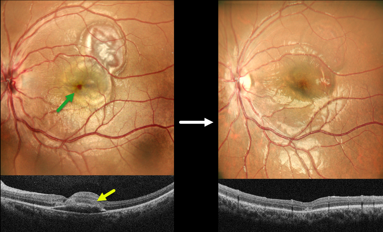
This 8YO boy presented with 2 days of vision loss in his left eye following a firecracker injury. Vision was 20/200 OS.
MultiColor SLO imaging shows a blister of foveal fluid with an overlying dot of foveal blood, and more superotemporal subretinal blood. OCT scanning shows foveal subretinal fluid with overlying hyperreflectivity from the blood. Intravenous methylprednisolone was given for 3 days, followed by a short course of oral steroids. One month later, vision improved to 20/60. The fluid and blood resolved with a choroidal rupture now visible temporally.
Learning Points:
An acute submacular hemorrhage following blunt trauma almost always indicates an underlying choroidal rupture as the source of the blood. The final vision is generally good unless the rupture extends through the macular center. These patients need long-term follow-up due to the increased risk of macular neovascularization.
Transient subretinal fluid is an uncommon finding with blunt trauma. Injury to the choroid and RPE allows fluid to accumulate in the subretinal space and outer retina (Mishra et al, Journal of Vitreoretinal Diseases 2021;5:165-169), as likely occurred in our patient.
CHOROIDAL RUPTURE
Malvika Singh and Manish Nagpal
Originally posted on @retina.rocks January 14, 2026
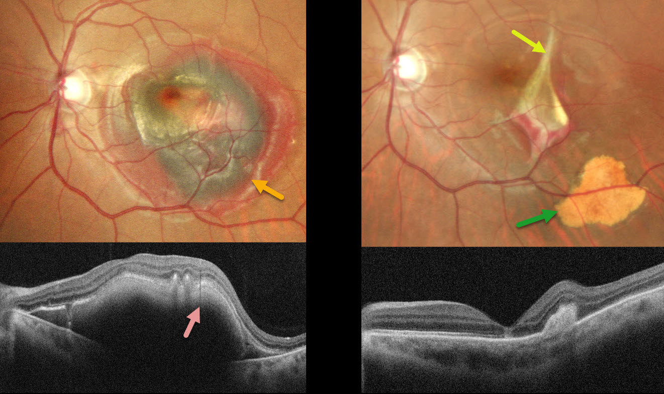
This 38YO male was hit by a stone in his left eye two days earlier. Vision was 20/20 in his normal OD and counting fingers OS.
Pseudocolor SLO imaging shows central macular greenish-brown subretinal blood surrounded by a rim of thinner red blood. OCT shows the hyperreflective blood with posterior shadowing. An in-office intravitreal C3F8 gas injection was performed, followed by face-down positioning. Six weeks later, the blood is virtually resolved except for some residual distal inferotemporal yellow devitalized subretinal blood. The causative choroidal rupture can be seen just temporal to the foveal center.
Learning Points:
An acute submacular hemorrhage following blunt trauma almost always indicates an underlying choroidal rupture as the source of the blood. The final vision is usually good unless the rupture extends through the macular center. These patients need long-term follow-up due to the increased risk of macular neovascularization.
CHOROIDAL RUPTURE
Malvika Singh, Akansha Sharma and Manish Nagpal
Originally posted on @retina.rocks December 10, 2025
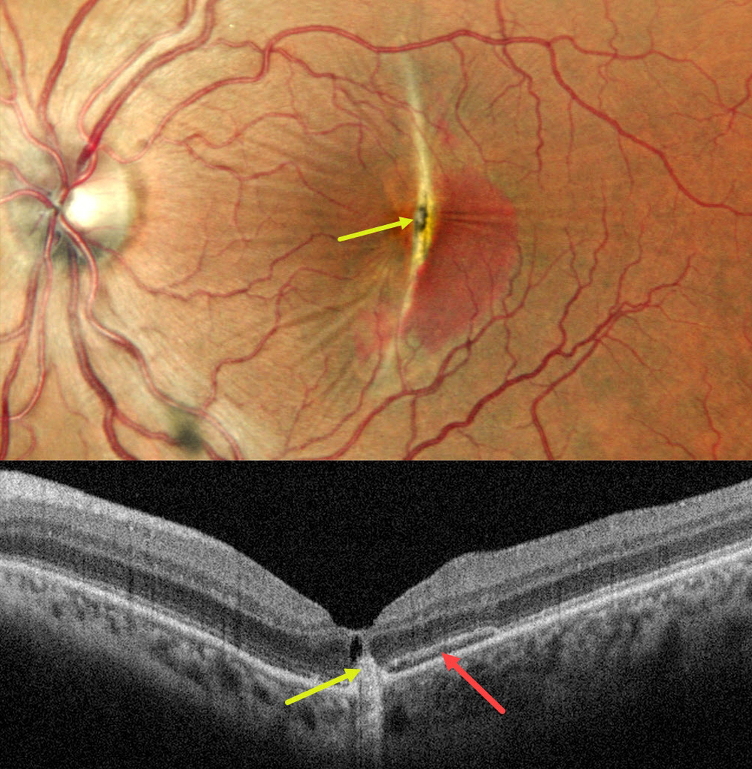
This 33YO female noted decreased vision in her left eye immediately following blunt trauma when a ceiling an fell on her face. Vision was 20/20 in her normal OD and 20/125 OS.
Pseudocolor SLO imaging shows a vertical choroidal rupture bisecting the central macula with surrounding subretinal blood. Radiating retinal striae extend outwards from the rupture. There is a central hyperpigmented lesion centrally, which appears hyperreflective in the outer retina on OCT. There is a hyporeflective cystic space anterior to this lesion. The subretinal blood appears as a hyper- and hyporeflective band on OCT.
This case was submitted by Malvika Singh, Akansha Sharma and Manish Nagpal.
Learning Points:
An acute submacular hemorrhage following blunt trauma almost always indicates an underlying choroidal rupture as the source for the blood. The final vision is usually good unless the rupture extends through the macular center. These patients need to be followed long-term due to the increased risk for macular neovascularization (MNV).
The central pigmented lesion with overlying fluid was suggestive for a MNV. An anti-VEGF injection was given, but she was unfortunately immediately lost to follow up.
CHOROIDAL RUPTURE WITH SECONDARY MACULAR NEOVASCULARIZATION
Nilesh Kumar
Originally posted on @retina.rocks November 24, 2025
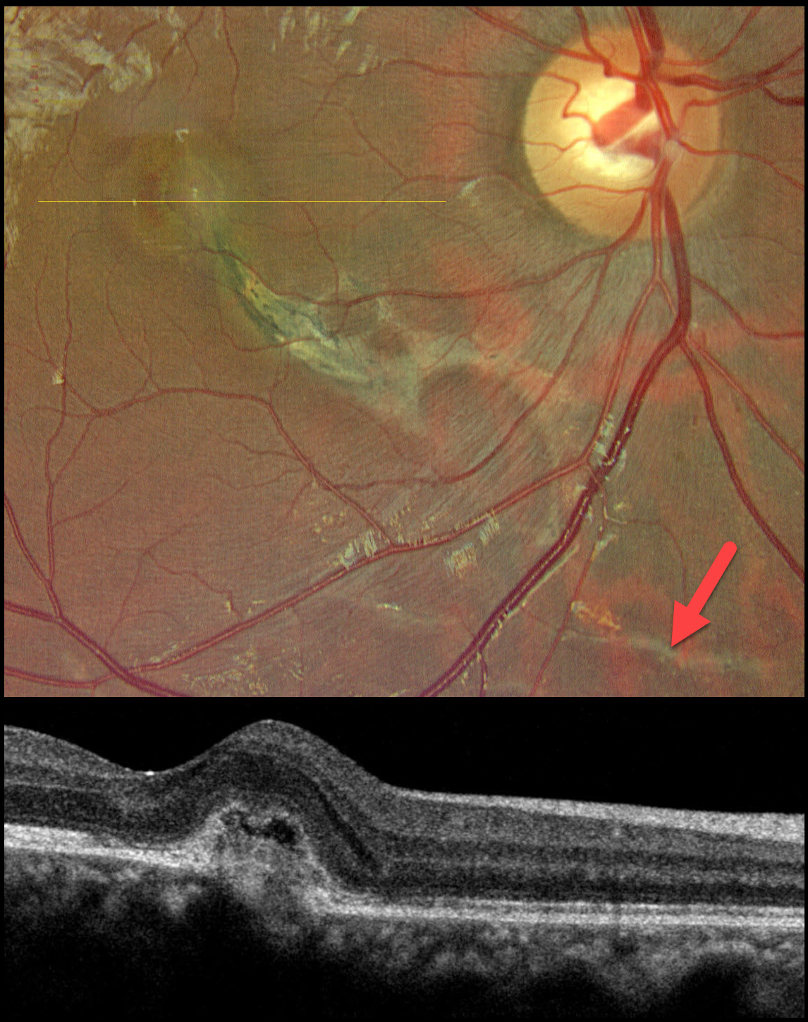
This 20YO male presented with 1 year of decreased vision in his right eye following blunt trauma from a small pebble that flew off a firecracker blast. Vision was 20/100 OD and 20/20 in his normal left eye.
Color photography shows an inferonasal macular choroidal rupture that extends through the fovea. A faint choroidal rupture is also noted inferior to the nerve. Subtle fluid and blood surround the foveal portion of the rupture. OCT scanning shows a thickened macular neovascularization (MNV) with mild overlying subretinal fluid.
Learning Points:
Choroidal ruptures are caused by blunt ocular trauma and are always circumferential with respect to the optic nerve. Subretinal blood following blunt trauma virtually always indicates the presence of an underlying choroidal rupture. The visual prognosis is generally good unless the rupture extends through the macular center. Patients need to be followed long-term due to the risk for MNV, as developed in our patient. We elected to closely follow him and will begin anti-VEGF therapy if increasing exudation develops.
ACUTE CHOROIDAL RUPTURES WITH SUBRETINAL BLOOD + TRAUMATIC MACULAR HOLE
Originally posted on @retina.rocks July 16, 2025
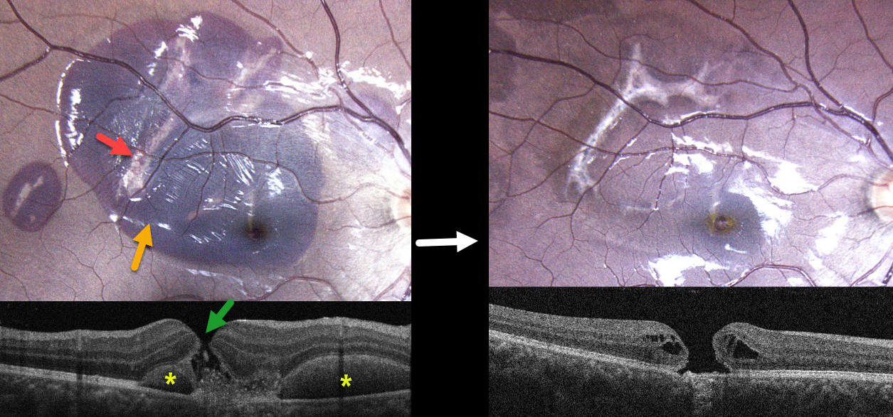
This healthy 17YO male was in a motor vehicle accident 2 weeks earlier. He was not wearing a seat belt and suffered a right upper lid laceration and closed head injury. He noticed immediate vision loss in his right eye. Vision was 20/200 OD and 20/20 in his normal OS.
Color photography shows a large macular and a smaller distal temporal submacular hemorrhage. Multiple choroidal ruptures are visible beneath this blood. OCT scanning shows a central full-thickness macular hole with speckled hyperreflective subretinal material. The submacular blood is hyperreflective with posterior shadowing on either side of the macular hole.
Learning Points:
An acute submacular hemorrhage following blunt trauma always indicates an underlying choroidal rupture as its source. The final vision is generally good unless the rupture extends through the macular center. These patients need long-term follow-up due to the increased risk of macular neovascularization.
Since fresh traumatic holes can spontaneously flatten, especially in pediatric cases (Liu et al, AJO 2020;210:174-183), an initial period of observation is recommended before considering vitrectomy. When examined about 6 months following his injury, all blood had resolved, and the temporal foveal choroidal rupture appears to spare the macular center. We will likely recommend vitrectomy if the hole fails to close with several more months of observation.
ANGOID STREAKS WITH TRAUMATIC CHOROIDAL RUPTURE AND SUBRETINAL BLOOD
Neelam Khatwani
Originally posted on @retina.rocks July 2, 2025
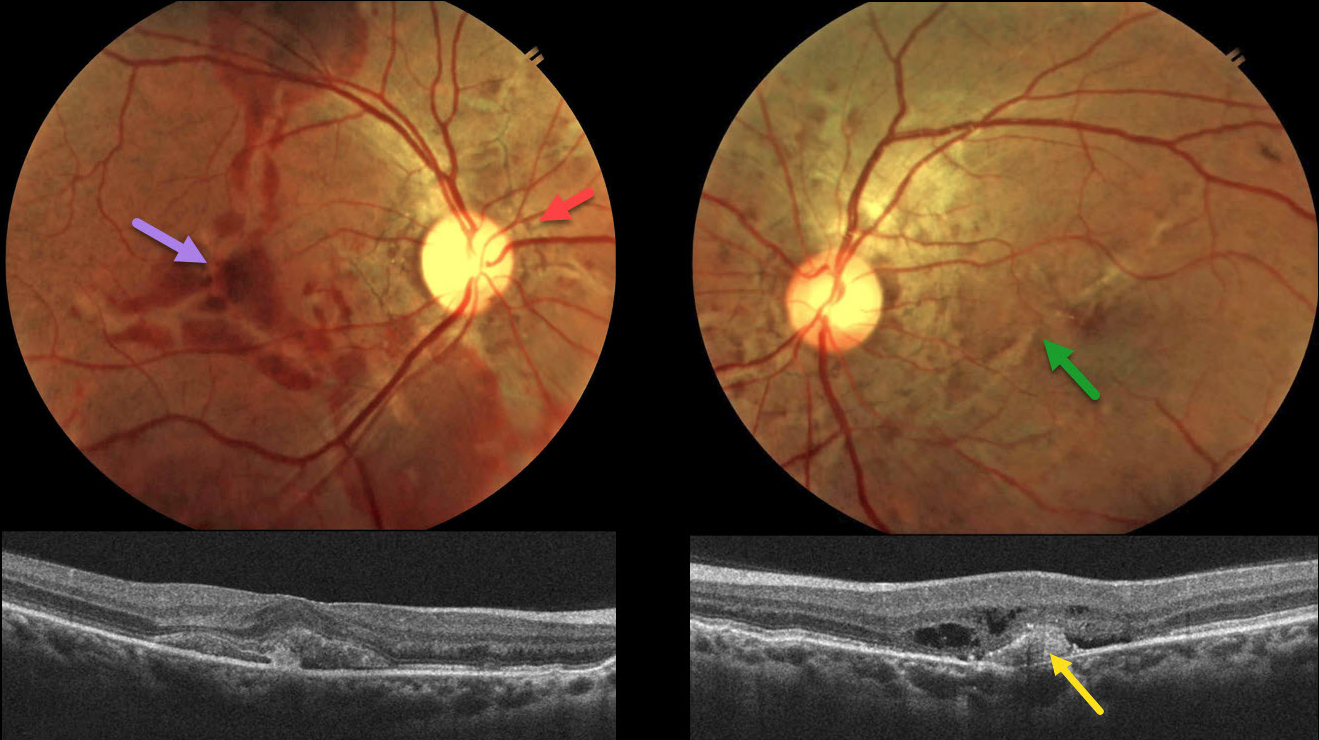
This 38YO male presented with 10 days of bilateral vision loss after being hit in the right eye with a fist. He denied any past medical history, and family history was negative for eye disease. Vision was 20/200 OD and 20/80 OS.
Color fundus photography shows irregular orange-gray lines radiating outwards from each nerve. Variable areas of subretinal blood are noted in the right macula, extending into the superior and inferior midperipheries. There is milder subretinal blood OS along with an irregular oblique macular angioid streak vs choroidal rupture. OCT OD shows subretinal hyperreflective material, likely blood. A possible type 1 (below the RPE) MNV with some outer retinal fluid is seen OS.
An anti-VEGF injection was given bilaterally, but unfortunately, the patient was immediately lost to follow-up.
Learning Points:
Initially described by Doyne in 1889, angioid streaks are orange-yellow, irregular, crack-like dehiscences in Bruch’s membrane associated with atrophic degeneration of the overlying RPE. The term “angioid” derives from its resemblance to blood vessels. Although they are historically mistakenly associated with numerous conditions, the only real association is with pseudoxanthoma elasticum (PXE, Nadelmann et al, Eye 2023;37:1596-1601). Patients are at significant risk for vision loss from MNV or pattern dystrophy-like changes (Murro et al, Graefe’s 2020;258:1881-1892).
Even minor trauma can cause choroidal ruptures with subretinal blood (Agrawal et al, JAMA Ophthalmology 2017;135(3):e165466). Unlike typical choroidal ruptures that are usually curvilinear with the nerve, the lesions from the brittle Bruch’s membrane in PXE appear as numerous irregular ruptures usually radiating from the nerve. Even though our patient sustained a direct injury to his right globe, the trauma was significant enough to cause choroidal ruptures in his left eye. The bilateral subretinal hemorrhages may be purely avascular, as their multifocality suggests.
CHOROIDAL RUPTURE
Originally posted on @retina.rocks September 23, 2024
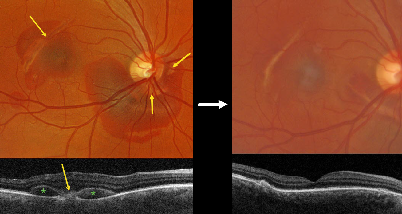
This 36YO male presented with one week of vision loss in his right eye immediately following a bungee cord injury. Vision was 20/60.
Optos color RG imaging shows areas of subretinal blood surrounding the inferior optic nerve and superotemporal macula. Multiple choroidal ruptures are evident. On Triton swept-source OCT, the superotemporal choroidal rupture appears as an outer retinal hyperreflective mound, and the blood as hyporeflective mounds between the outer retina and RPE.
Our patient was treated with observation, and one month later, all blood resolved with 20/100 vision.
Learning Points:
An acute submacular hemorrhage following blunt trauma almost always indicates an underlying choroidal rupture as the source of the blood. The final vision is usually good unless the rupture extends through the macular center. These patients need long-term follow-up due to the increased risk of macular neovascularization.
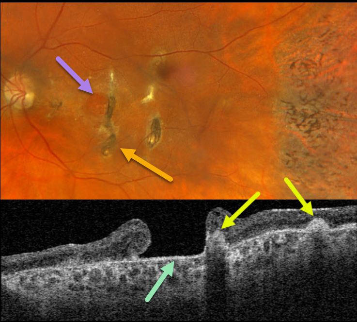
TRAUMATIC MACULAR HOLE + CHOROIDAL RUPTURE
Originally posted on @retina.rocks July 11, 2023
This 57YO female has a long history of vision loss in her left eye following blunt trauma years earlier. Vision was hand motion OS.
Optos color RGB imaging shows a large chronic macular hole with surrounding pigmentary changes. There are several hyperpigmented choroidal ruptures just temporal to the hole. Chorioretinal scarring with intraretinal pigment migration is noted in the temporal periphery.
Swept-source OCT shows a large macular hole. The choroidal ruptures appear as elevated hyperreflective subretinal mounds with posterior shadowing.
Learning Points:
This patient illustrates several sequelae of blunt trauma, including macular hole formation, choroidal rupture, and intraretinal pigment migration with a pseudo-sector retinitis pigmentosa appearance due to commotio-induced photoreceptor loss.
Since fresh traumatic holes can spontaneously flatten, especially in pediatric cases (Liu et al, AJO 2020;210:174-183), an initial period of observation is recommended before considering vitrectomy.
Given the chronicity of our patient’s hole, surgery was not considered.
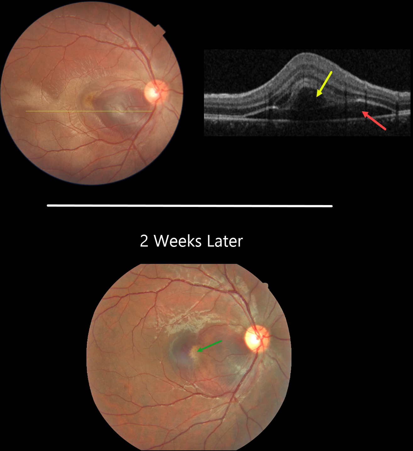
CHOROIDAL RUPTURE
Asma Samsudeen and Ashish Sharma
Originally posted on @retina.rocks July 6, 2023
This 21YO male presented with immediate vision loss after being hit with a cricket ball in his right eye one day earlier. Vision was 20/60.
Fundus photography shows a subretinal hemorrhage in the nasal macula, a small rim of surrounding subretinal fluid, and a more temporal ill-defined commotio retina.
OCT scanning shows hyporeflective retinal elevation from the subretinal blood and fluid, along with an overlying bacillary layer detachment.
Our patient shows several findings classic for acute blunt trauma, including commotio retinae and subretinal blood. This subretinal blood almost always indicates an underlying choroidal rupture, which did, in fact, reveal itself on follow-up 2 weeks later, when vision returned to 20/20. The visual prognosis for these choroidal ruptures is good as long as the rupture spares the foveal center, although patients need lifelong surveillance due to the risk of secondary macular neovascularization.
Learning Points:
A bacillary layer (cone and rod inner and outer segments) detachment (BLD) appears as a unique dome-shaped collection of intraretinal fluid from photoreceptor splitting. These detachments can be observed in an increasing number of conditions, most classically Vogt-Koyanagi-Harada disease (Cicinelli et al, Ophthalmology Retina 2020;4:454-456).
About 8% of eyes with acute non-penetrating ocular trauma have a BLD (Venkatesh et al, Can J Ophthalmol 2022;57:328-336). These resolve within 2-10 days without visual or anatomic sequelae.
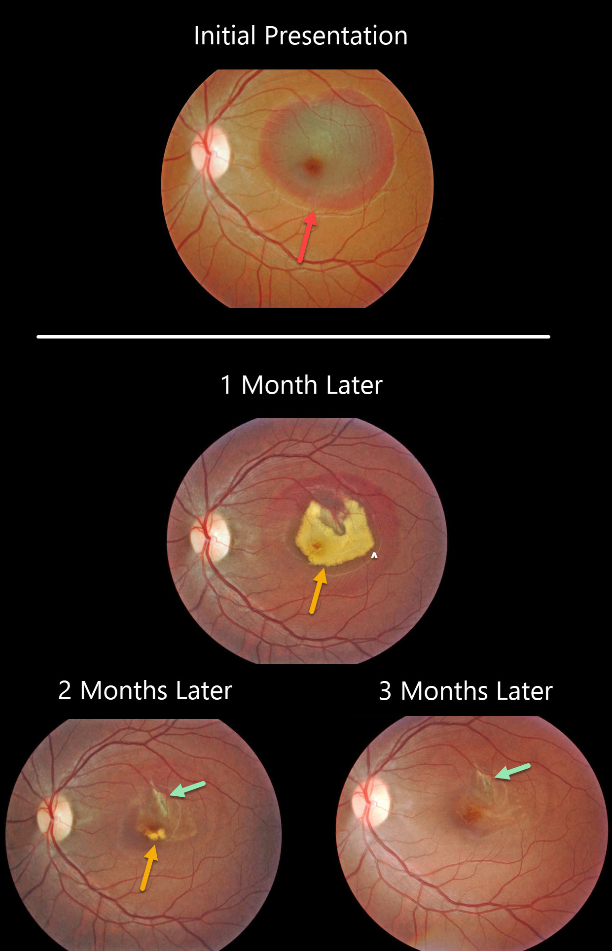
CHOROIDAL RUPTURE
Originally posted on @retina.rocks October 1, 2020
This patient presented with an acute submacular hemorrhage following a finger-to-eye injury.
The subretinal blood slowly resolved over the following three months. An underlying choroidal rupture is almost always the source of this traumatic blood.
Final vision was 20/400, either due to contusive foveal damage or possible toxicity from the subretinal blood.
Learning Point:
Choroidal ruptures are caused by blunt ocular trauma and are always circumferential with respect to the optic nerve.
These patients need to be followed since macular neovascularization can develop later in life.
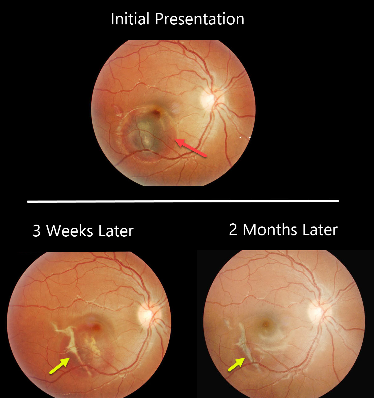
CHOROIDAL RUPTURE
Originally posted on @retina.rocks June 4, 2020
This patient presented with vision loss from an acute submacular hemorrhage following blunt trauma.
Subsequent images, 3 weeks later and 2 months later, show gradual resolution of the blood with the appearance of an underlying choroidal rupture.
Learning Point:
Choroidal ruptures are caused by blunt ocular trauma and are always circumferential with respect to the optic nerve. Subretinal blood following blunt trauma virtually always indicates the presence of an underlying choroidal rupture.
The initial visual prognosis is generally good if the rupture does not extend through the macular center, as was fortunately the case in this patient.
These patients need to be followed since macular neovascularization can develop later in life.


