

Choroideremia
<< Back to Cases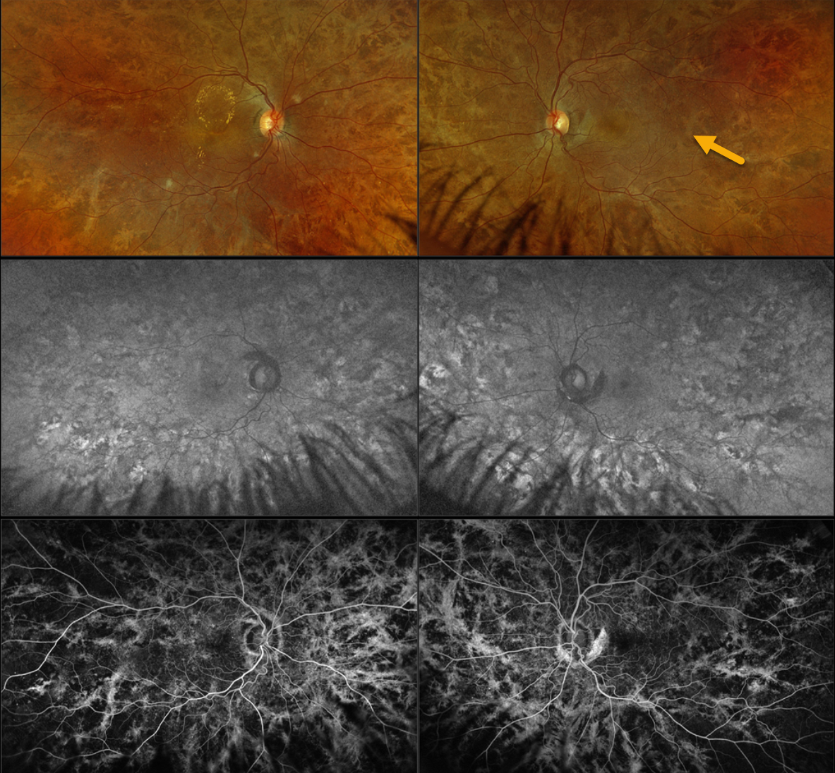
CHOROIDEREMIA CARRIER
Originally posted on @retina.rocks August 20, 2024
This 42YO female with type 2 diabetes was referred for asymptomatic diabetic macular edema (DME). There was no family history of ocular disease. Vision was 20/40 OD and 20/20 OS.
Optos color RGB imaging shows diffuse coarse subretinal hyperpigmentations throughout each posterior pole. Fundus autofluorescence (FAF) shows variable hyper- and hypo-FAF from these pigmentary changes, with fluorescein angiographic window defects. Genetic testing revealed a heterozygous pathogenic CHM mutation.
Learning Points:
Choroideremia is an X-linked recessive chorioretinal dystrophy caused by a mutation in the CHM gene located on the long arm of the X chromosome. Sons of female carriers have a 50% chance of developing choroideremia, and daughters have a 50% chance of becoming carriers. Some female carriers can develop asymptomatic pigmentary changes, as in our patient, or frank choroideremia due to irregular inactivation of the X chromosome (see Jauregui et al, AJO 2019;207:77-86).
CHOROIDEREMIA IN A FEMALE CARRIER
Yuenpang Cheung and Stephen Tsang
Originally posted on @retina.rocks August 6, 2024
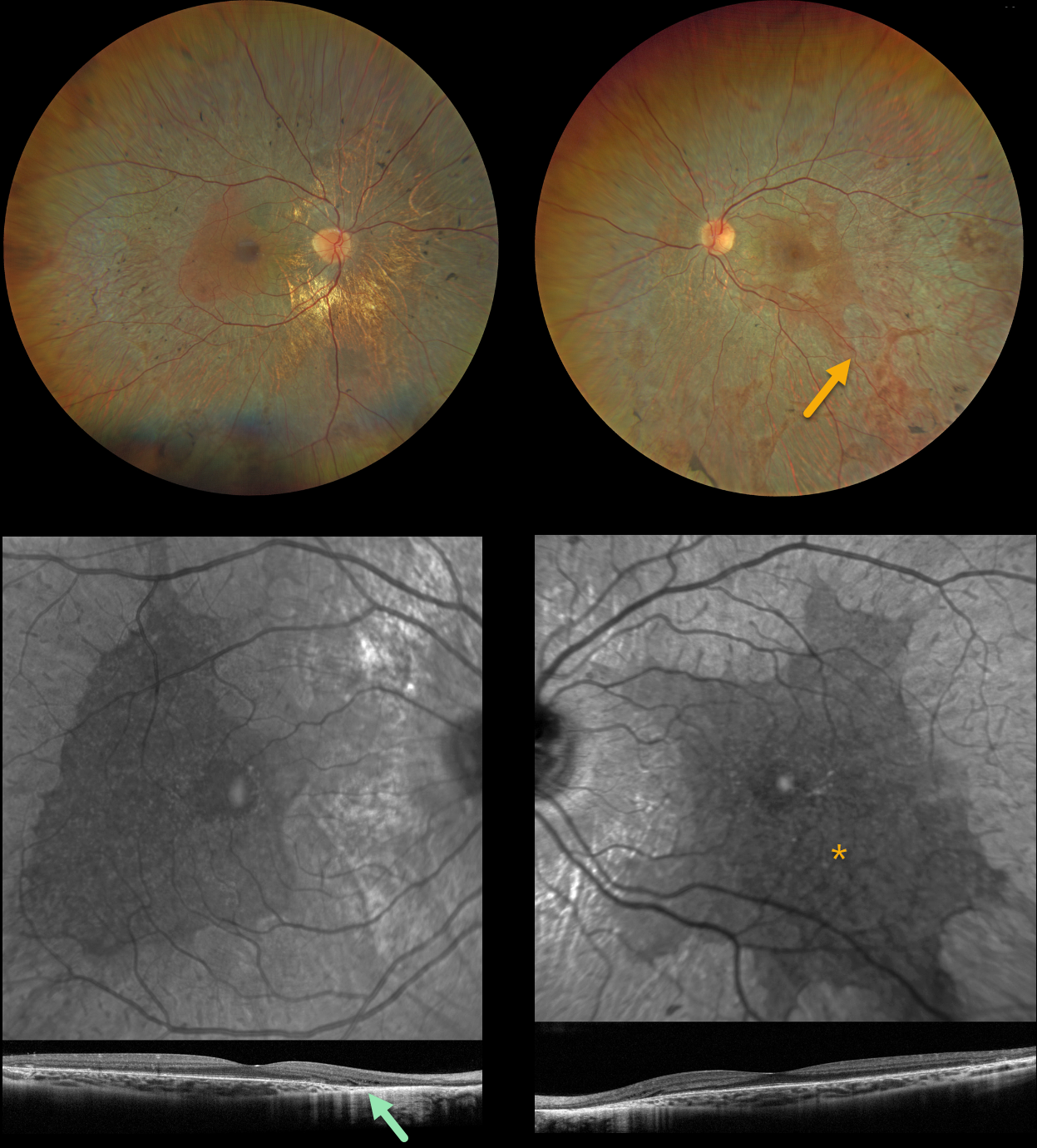
This 27YO female has a known history of choroideremia and is heterozygous for a pathogenic CHM mutation. Vision was 20/150 OD and 20/30 OS.
Color imaging shows large, symmetrically bilateral, scalloped areas of chorioretinal atrophy that spare the central maculae. Some peripheral areas of intraretinal pigment migration (bone spicules) are noted as well. OCT scanning shows variable outer retinal loss on B-scan with a scalloped preserved central macula on en face.
Learning Points:
Choroideremia is an X-linked recessive chorioretinal dystrophy caused by a mutation in the CHM gene located on the long arm of the X chromosome. Sons of female carriers have a 50% chance of developing choroideremia, and daughters have a 50% chance of becoming carriers. Some female carriers, as in our patient, can still develop choroideremia due to irregular inactivation of the X chromosome (see Jauregui et al, AJO 2019;207:77-86).
The onset of night blindness usually begins between the ages of 10 and 30, followed by peripheral visual field loss. Zones of patchy RPE and chorioretinal atrophy initially appear in the mid-periphery, gradually spreading anteriorly and posteriorly. Eventually, the patient is left with only a small island of vision, narrowed retinal vessels, and optic atrophy.
CHOROIDEREMIA IN A FEMALE CARRIER
Originally posted on @retina.rocks April 24, 2024
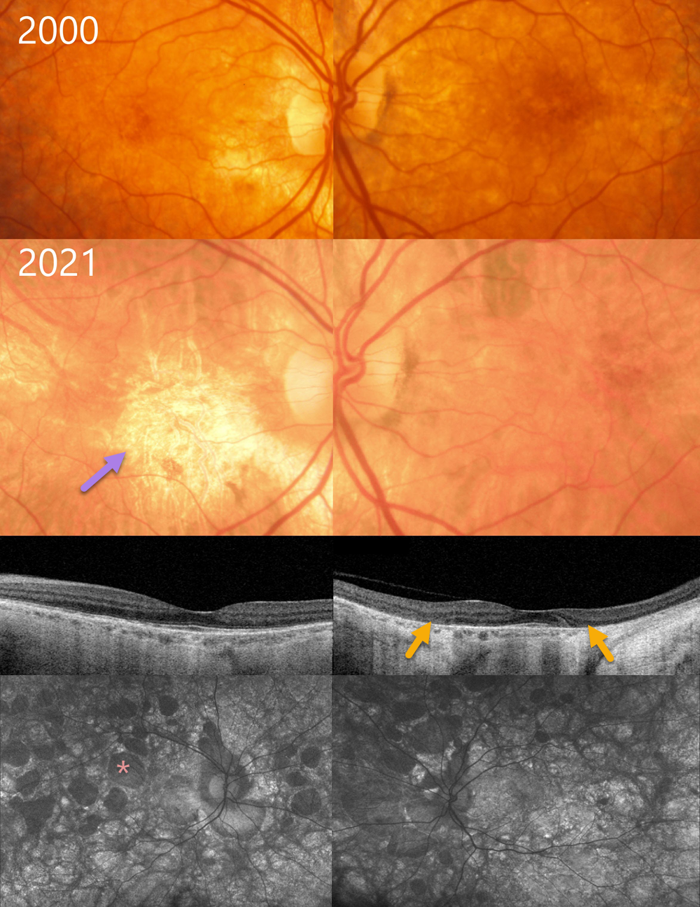
This 37YO female, whose son was diagnosed with choroideremia, was examined in 2000. Color photography shows some mild bilateral pigmentary changes.
Over the subsequent 20+ years, her ocular findings have progressed into more typical choroideremia. When last examined in 2021, vision was 20/30 OU.
The macula is now atrophic OD with minimal progressive changes OS. Swept-source OCT, however, shows fairly symmetrical outer retinal atrophy that spares each macular center. Optos autofluorescence reveals multifocal lobular areas of atrophy extending into each periphery. Genetic testing was heterozygous for a pathogenic CHM variant.
Learning Points:
Choroideremia is an X-linked recessive chorioretinal dystrophy caused by a mutation in the CHM gene located on the long arm of the X chromosome. Sons of female carriers have a 50% chance of developing choroideremia, and daughters have a 50% chance of becoming carriers. Some female carriers, as in our patient, can still develop choroideremia due to irregular inactivation of the X chromosome (see Jauregui et al, AJO 2019;207:77-86).
The onset of night blindness usually begins between the ages of 10 and 30, followed by peripheral visual field loss. Zones of patchy RPE and chorioretinal atrophy initially appear in the mid-periphery, gradually spreading anteriorly and posteriorly. Eventually, the patient is left with only a small island of vision, narrowed retinal vessels, and optic atrophy.
CHOROIDEREMIA
Originally posted on @retina.rocks August 31, 2022
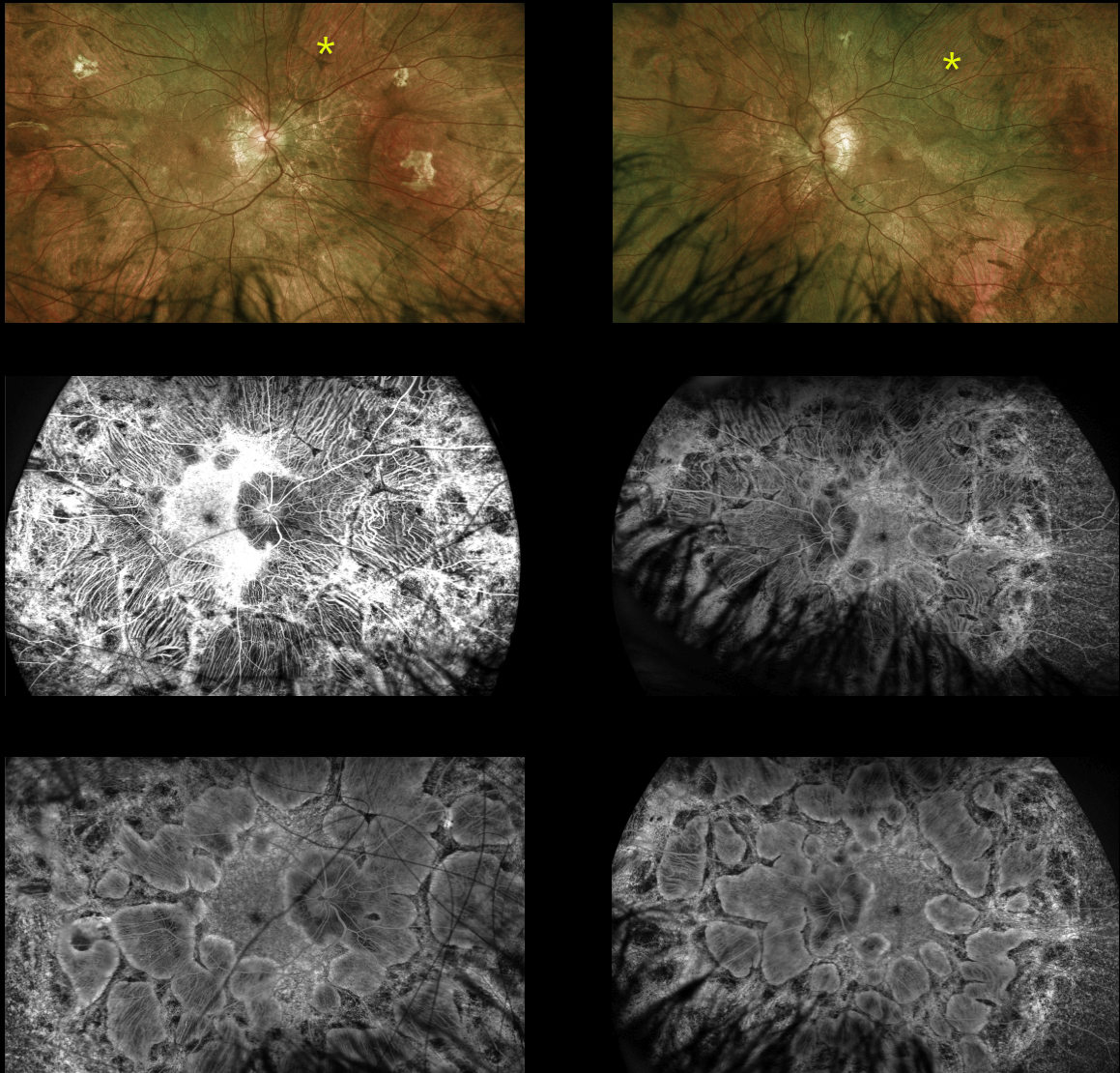
This 14YO male is surprisingly asymptomatic with visual acuity of 20/30 OD and 20/40 OS. His retinal findings were found on a routine exam.
Optos imaging shows large symmetrically bilateral scalloped areas of chorioretinal atrophy, which spare the central maculas. On fundus fluorescein angiography, the early phase (middle) shows exquisite detail of the choroidal vortex system and late phase (bottom) shows staining of the scalloped areas of atrophy.
The patient is adopted and is unaware of any family history of inherited retinal disease. Subsequent genetic testing revealed a pathogenic hemizygous CHM variant on the X chromosome.
Learning Points:
Choroideremia is an X-linked recessive chorioretinal dystrophy caused by a mutation in the CHM gene located on the long arm of the X chromosome. Sons of female carriers have a 50% chance of developing choroideremia, and daughters have a 50% chance of becoming carriers.
Some female carriers can still develop clinical disease due to irregular inactivation of the X-chromosome (see Jauregui et al, AJO 2019;207:77-86).
The onset of night blindness usually begins between the ages of 10 and 30, followed by peripheral visual field loss. Zones of patchy RPE and chorioretinal atrophy initially appear in the mid-periphery, gradually spreading anteriorly and posteriorly.
The choroidal vortex system is relatively resistant to most disorders, including choroideremia. This system is beautifully imaged angiographically in our patient since the RPE, which normally blocks the underlying choroid, is atrophic and no longer blocks these vessels.
Eventually, the patient is left with a small central island of vision, narrowed retinal vessels and optic atrophy.
For a great choroideremia review article, see Pennesi et al, Retina 2019;39:2019-2069.
CHOROIDEREMIA
Will Gibson
Originally posted on @retina.rocks June 27, 2022
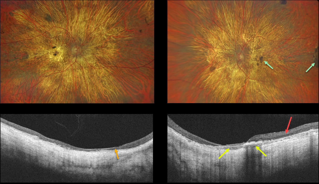
This 56YO male presented with a long history of vision loss and night blindness. Vision was counting fingers bilaterally.
Optos imaging shows severe, bilateral diffuse chorioretinal atrophy with some scattered large clumps of pigmentation. OCT scanning of the right eye shows diffuse thinned, disorganized retina and no choroidal structures. OCT scan of the left eye shows relatively preserved temporal retina and some preserved choroid.
Learning Points:
Choroideremia is an X-linked recessive chorioretinal dystrophy caused by a mutation in the CHM gene located in the long arm of the X chromosome. Sons of female carriers have a 50% chance of developing choroideremia, and daughters have a 50% chance of becoming carriers. Some female carriers can still develop choroideremia due to irregular inactivation of the X chromosome (see Jauregui et al, AJO 2019;207:77-86).
Night blindness usually begins between the ages of 10 and 30, followed by peripheral visual field loss. Zones of patchy RPE and chorioretinal atrophy initially appear in the mid-periphery, gradually spreading anteriorly and posteriorly. Eventually, the patient is left with only a small island of vision, narrowed retinal vessels, and optic atrophy.
For a great choroideremia review article, see Pennesi et al., Retina 2019;39:2019-2069.
CHOROIDEREMIA
Originally posted on @retina.rocks April 21, 2022
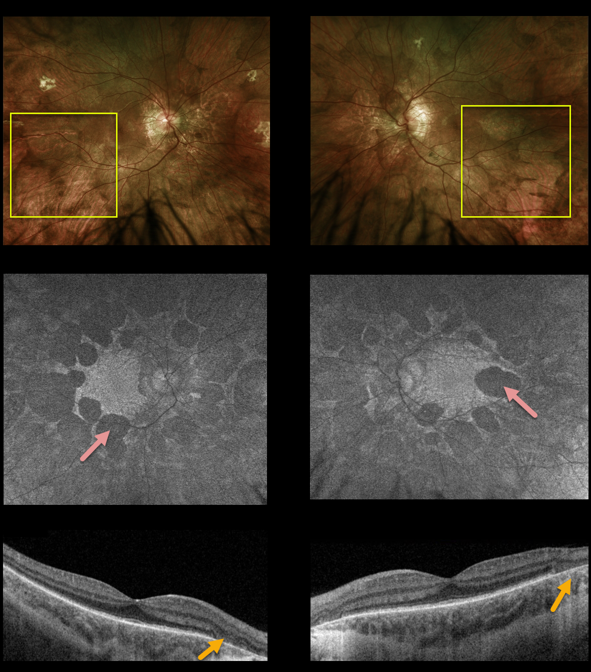
This 14YO male is surprisingly asymptomatic with visual acuity of 20/30 OD and 20/40 OS. His retinal findings were found on a routine exam.
Optos imaging shows large, symmetrically bilateral, scalloped areas of chorioretinal atrophy that spare the central maculae.
On fundus autofluorescence (FAF), the scalloped areas of atrophy show hypo-FAF.
Swept-source OCT shows outer retinal and RPE atrophy within the clinical areas of atrophy.
The patient is adopted and is unaware of any family history of inherited retinal disease. Subsequent genetic testing revealed a pathogenic hemizygous CHM variant on the X chromosome, consistent with choroideremia.
Learning Points:
Choroideremia is an X-linked recessive chorioretinal dystrophy caused by a mutation in the CHM gene located on the long arm of the X chromosome.
Sons of female carriers have a 50% chance of developing choroideremia, and daughters have a 50% chance of becoming carriers. Some female carriers can still develop clinical disease due to irregular inactivation of the X chromosome (see Jauregui et al, AJO 2019;207:77-86).
Night blindness usually begins between the ages of 10 and 30, followed by peripheral visual field loss.
Zones of patchy RPE and chorioretinal atrophy initially appear in the mid-periphery, gradually spreading anteriorly and posteriorly. Eventually, the patient is left with only a small central island of vision, narrowed retinal vessels, and optic atrophy.
For a great choroid plexus review article, see Pennesi et al., Retina 2019;39:2019-2069.
CHOROIDEREMIA IN A FEMALE CARRIER
Originally posted on @retina.rocks October 22, 2021
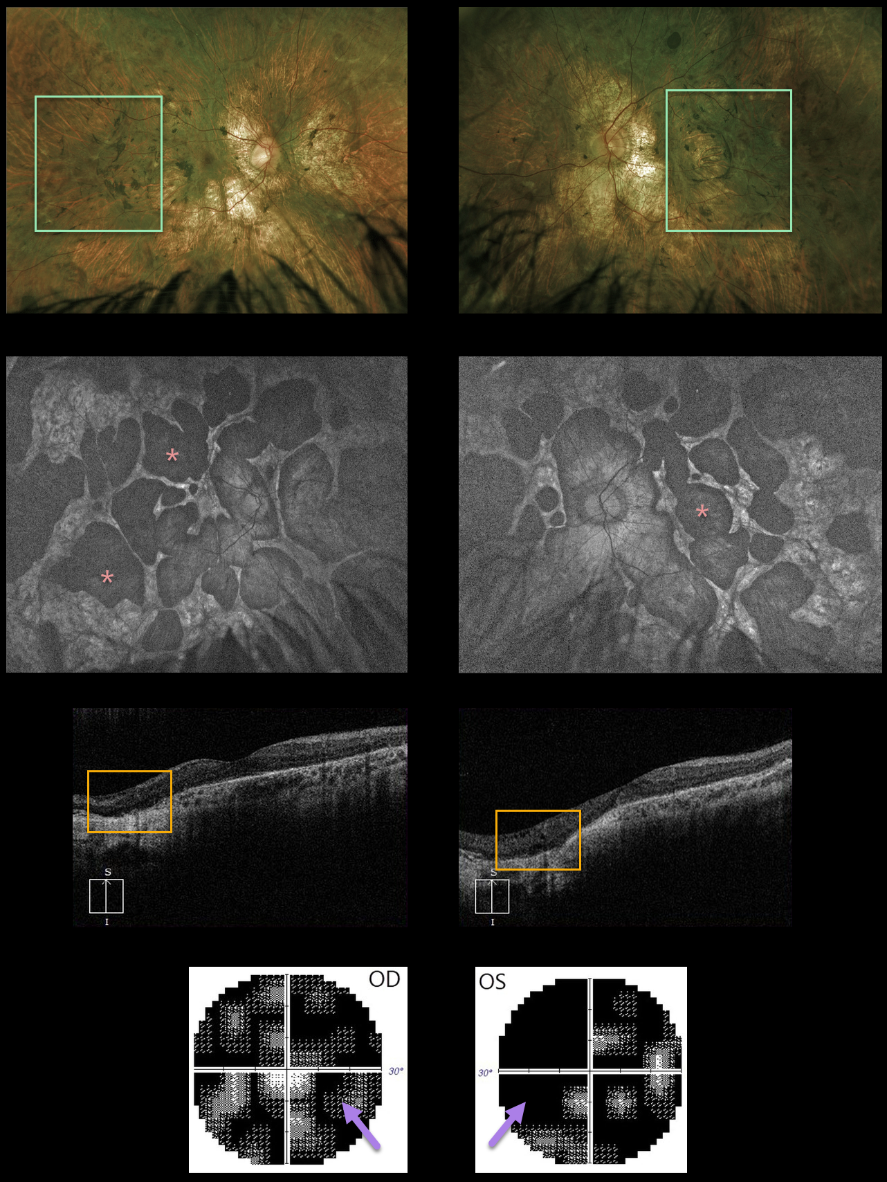
This 40YO female reported a 7-year history of reading difficulties and glare while driving at night.
Optos imaging shows severe diffuse chorioretinal scarring with intraretinal pigment migration extending from the optic nerves to the retinal peripheries.
The lobular nature of the chorioretinal changes is best seen with fundus autofluorescence. There was variable bilateral choroidal and outer retinal atrophy on OCT. Visual fields showed bilateral severe generalized depression.
Genetic testing showed that she was heterozygous for the pathogenic variant of the CHM gene. Vision remarkably was 20/20 OD and 20/25 OS due to sparing of the macular centers.
Learning Points:
Choroideremia is an X-linked recessive chorioretinal dystrophy caused by a mutation in the CHM gene located in the long arm of the X chromosome. Sons of female carriers have a 50% chance of developing choroideremia, and daughters have a 50% chance of becoming carriers. Some female carriers can still develop choroideremia due to irregular inactivation of the X chromosome (see Jauregui et al, AJO 2019;207:77-86).
Night blindness usually begins between the ages of 10 and 30, followed by peripheral visual field loss. Zones of patchy RPE and chorioretinal atrophy initially appear in the mid-periphery, gradually spreading anteriorly and posteriorly. Eventually, the patient is left with only a small island of vision, narrowed retinal vessels, and optic atrophy.
For a great choroideremia review article, see Pennesi et al, Retina 2019;39:2019-2069.
CHOROIDEREMIA
Originally posted on @retina.rocks January 28, 2021
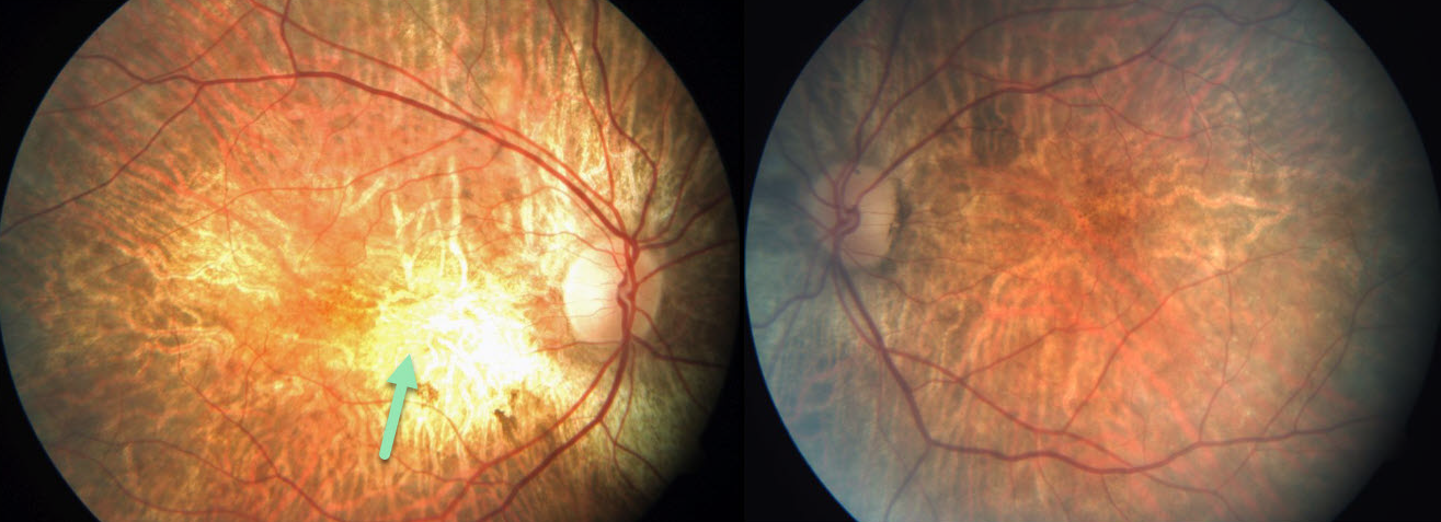
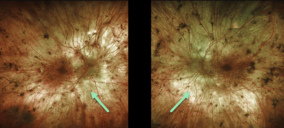
This patient has choroideremia and, luckily, has preserved central 20/30 vision bilaterally. Fundus photography shows patchy areas of RPE and chorioretinal atrophy.
Learning Points:
Choroideremia is an X-linked recessive chorioretinal dystrophy caused by a mutation in the CHM gene. Some female carriers can still develop choroideremia due to irregular inactivation of the X chromosome (lyonization).
Night blindness usually begins between the ages of 10 and 30, followed by peripheral visual field loss. Zones of patchy RPE and chorioretinal atrophy initially appear in the mid-periphery, gradually spreading anteriorly and posteriorly.
Eventually, the patient is left with only a small island of vision, narrowed retinal vessels, and optic atrophy.
CHOROIDEREMIA
Originally posted on @retina.rocks October 28, 2020
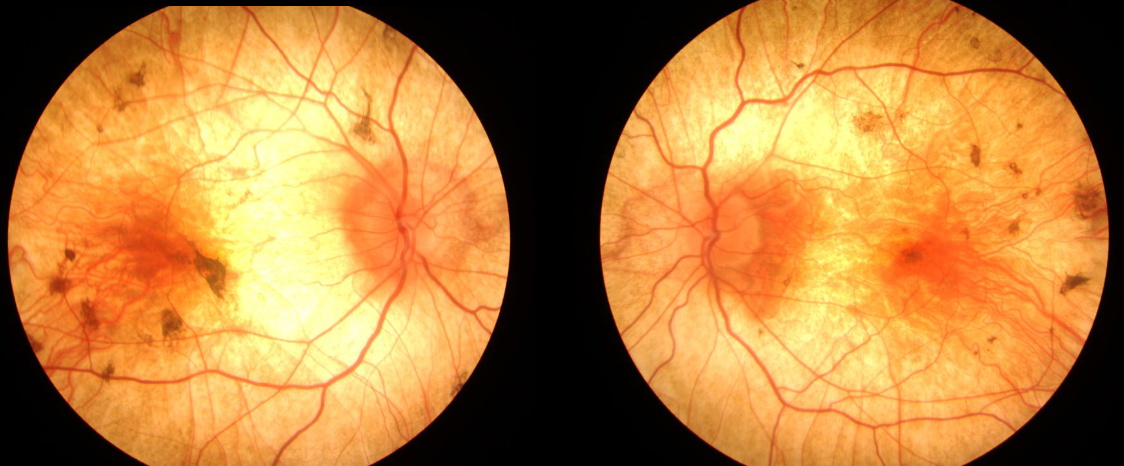
This is a 68YO male with a likely diagnosis of choroideremia. Vision is remarkably 20/60 OD and 20/50 OS. Note the areas of patchy RPE and chorioretinal atrophy in both eyes.
Learning Points:
Choroideremia is an X-linked recessive chorioretinal dystrophy caused by a mutation in the CHM gene. Some female carriers can still develop choroideremia due to irregular inactivation of the X chromosome (lyonization) later in life.
Night blindness usually begins between the ages of 10 and 30, followed by peripheral visual field loss. Patchy RPE and chorioretinal atrophy initially appear in the mid-periphery, gradually spreading anteriorly and posteriorly. Eventually, the patient is left with only a small island of vision, narrowed retinal vessels, and optic atrophy.
CHOROIDEREMIA
Originally posted on @retina.rocks March 6, 2020
This patient has choroideremia, an X-linked recessive chorioretinal dystrophy caused by mutation in the CHM gene.
It is characterized by a pale appearing fundus with RPE and choroidal atrophy initially in the mid-periphery that gradually spread anteriorly and posteriorly.
Learning Points:
Patients with choroideremia often develop severe central and peripheral vision loss with night blindness. During later stages only a small island of preserved foveal tissue remains.
Female carriers can present with a wide range of phenotypes, ranging from asymptomatic rubella-like pigmentary changes to severe disease.


