

Cilioretinal Artery
<< Back to Cases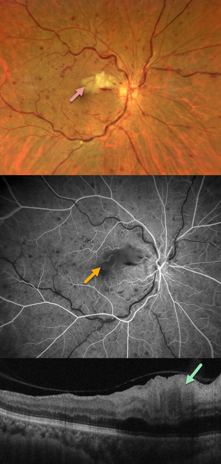
CRVO WITH SECONDARY CILIORETINAL ARTERY OCCLUSION
Francis Ezra Laxamana, Bianca Teresa Suarez, Jesus Jacinto Bautista and Carlo Ladores.
Originally posted on @retina.rocks September 7, 2026
This 58YO female with a history of hypertension and diabetes presented with sudden painless vision loss in her right eye. Vision was 20/25 OD and 20/20 in her normal OS.
Fundus photography shows a non-ischemic central retinal vein occlusion (CRVO) with dilated tortuous veins, mild scattered retinal hemorrhages, and a superonasal macular branch retinal artery occlusion (BRAO). The BRAO shows inner retinal hyperreflectivity on OCT. Fluorescein angiography shows good perfusion throughout the distribution of the CRVO, with a filling defect from the BRAO.
Learning Points:
Some patients with CRVO will develop a secondary non-embolic cilioretinal artery occlusion caused by either compression from optic nerve edema or from a sudden rise in intraluminal retinal capillary bed pressure (Hayreh et al, Retina 2008;28;581-594).
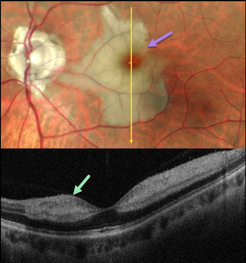
CILIORETINAL ARTERY OCCLUSION
Malvika Singh and Manish Nagpal
Originally posted on @retina.rocks December 30, 2025
This healthy 46YO male presented with an 8-day history of a central scotoma in his left eye. Vision was 20/20 in his normal OD and 20/400 OS.
Pseudocolor SLO imaging shows an acute cilioretinal artery occlusion with foveal sparing. OCT scanning shows marked inner retinal hyperreflectivity with posterior shadowing.
Learning Points:
The inner two-thirds of the neurosensory retina is supplied by the central retinal artery, with the choroid supplying the RPE and photoreceptor layer. Cilioretinal arteries, which arise from the posterior choroidal circulation, are present in up to 50% of individuals, and bilateral in about 25% (Schneider et al, Acta Ophthalmologica 2021;99:e310-e318).
Their clinical significance usually arises from foveal preservation in eyes with concomitant retinal artery occlusions (Brown and Shields, Arch Ophthalmology 1979;97:84-92). However, they can also be a rare cause of primary branch retinal artery occlusion, either in isolation (our patient) or in association with other entities, including central retinal vein occlusions (Hayreh et al, Retina 2008;28:581-594).
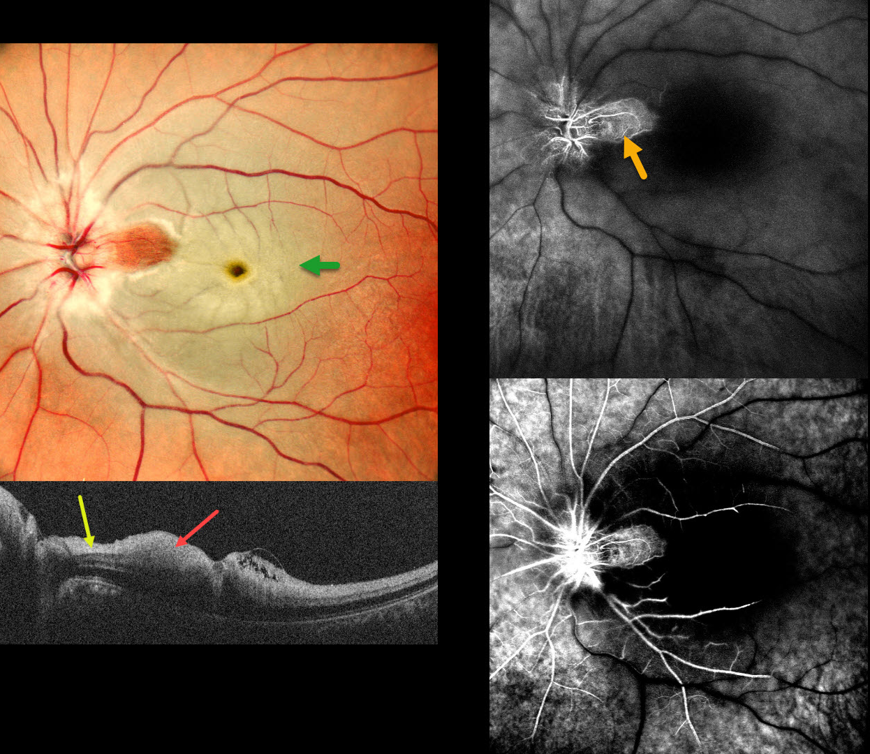
CRAO
Tejaswita Verma and Manish Nagpal
Originally posted on @retina.rocks September 1, 2025
This 37 YO male presented with acute vision loss in his left eye 3 days earlier. Vision was 20/20 in his normal OD and 20/200 in his OS.
Pseudocolor SLO imaging shows a fresh central retinal artery occlusion (CRAO) with sparing of a small area of temporal peripapillary retina supplied from a patent cilioretinal artery. OCT scanning shows opaque, hyperreflective, thickened inner retinal layers, except for a small area of more normal retina within the cilioretinal sparing.
Fluorescein angiography shows profound diffuse retinal nonperfusion and delayed filling except for the nasal macula. Workup elsewhere included an MRI showing left ICA and left MCA chronic infarcts.
Learning Points:
The inner two-thirds of the neurosensory retina is supplied by the central retinal artery, with the choroid supplying the RPE and photoreceptor layer. Acute CRAO, therefore, presents with a white edematous inner retina that spares the fovea. Cilioretinal arteries, which arise from the posterior choroidal circulation, are present in up to 50% of individuals, and bilateral in about 25% (Schneider et al, Acta Ophthalmologica 2021; 99:e310-e318).
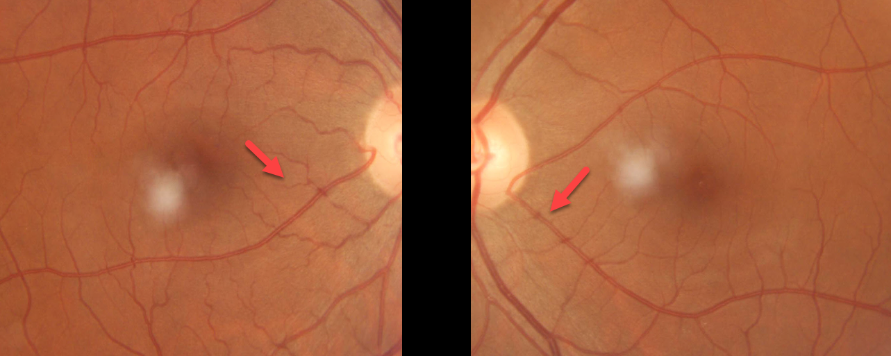
CILIORETINAL ARTERIES
Sajal Golchha and Sarthak Bafna
Originally posted on @retina.rocks December 3, 2024
This 46YO visually asymptomatic male presented with bilateral cilioretinal arteries.
The inner two-thirds of the neurosensory retina is supplied by the central retinal artery, with the choroid supplying the RPE and photoreceptor layer. Cilioretinal arteries, which arise from the posterior choroidal circulation, are present in up to 50% of individuals, and bilateral in about 25% (Schneider et al, Acta Ophthalmologica 2021;99:e310-e318).
Our patient is unique in that their left cilioretinal artery supplies virtually the entire macula. If he were unlucky enough to be the one in 50,000 people per year who develops a central retinal artery occlusion (Leavitt et al, AJO 2011;152:820-823), these vessels would most likely preserve macular function.
CILIORETINAL ARTERY + CENTRAL SEROUS CHORIORETINOPATHY
Mayelinne García, Luis Montero and Alberto Ruiz
Originally posted on @retina.rocks September 24, 2024
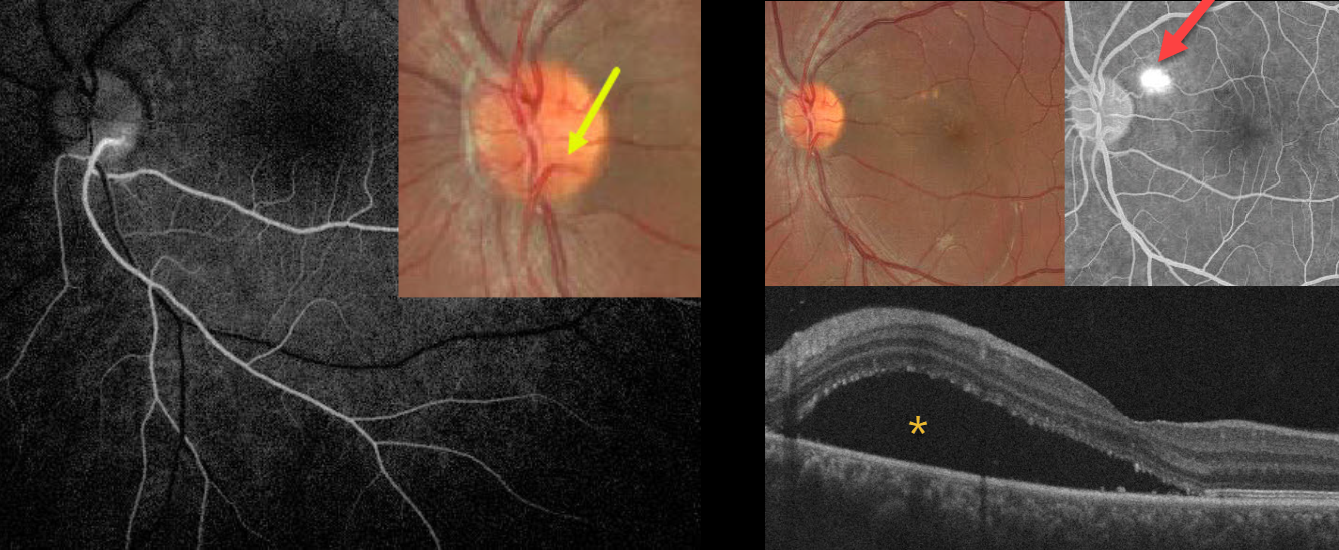
This healthy 25YO male presented with 3 weeks of vision loss in his left eye from active idiopathic central serous chorioretinopathy (ICSC). Vision was 20/20 in his normal OD and 20/110 OS.
A cilioretinal vessel originates within the inferotemporal aspect of the nerve and arches inferiorly. Fluorescein angiography reveals that this cilioretinal artery supplies the entire inferotemporal retina. Color imaging of the macula shows a large serous detachment with some drusen-like changes. This detachment is confirmed on OCT scanning. A blot leak is seen angiographically. Due to the patient’s symptoms, thermal laser was applied to the angiographic leak.
Learning Points:
The inner two-thirds of the neurosensory retina are supplied by the central retinal artery, with the choroid supplying the RPE and photoreceptor layer. Cilioretinal arteries, which arise from the posterior choroidal circulation, are present in up to 50% of individuals and are bilateral in about 25% (Schneider et al, Acta Ophthalmologica 2021;99:e310-e318).
CRVO + CILIORETINAL ARTERY OCCLUSION
Amit Nandan Tripathi and Vipin Rana
Originally posted on @retina.rocks June 18, 2024
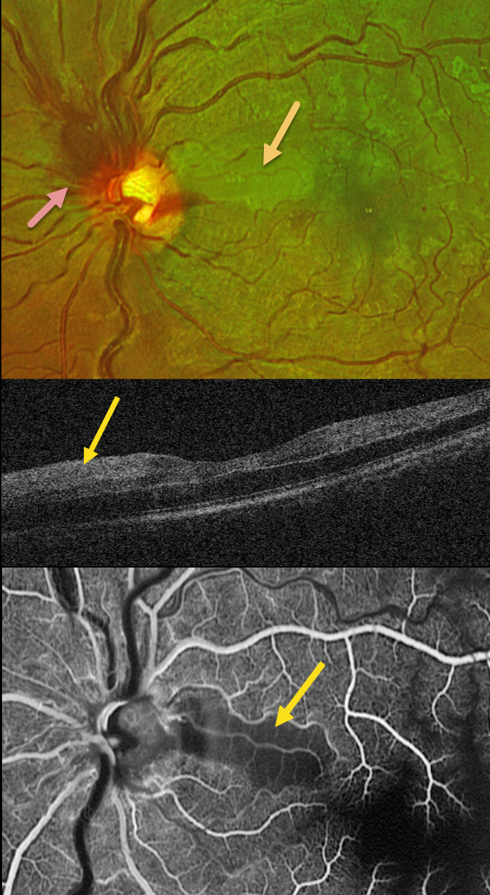
This healthy 30YO male presented with sudden vision loss in his left eye. Vision was 20/20 in his healthy right eye and counting fingers in his left eye.
Optos color RG imaging shows an acute non-ischemic central retinal vein occlusion (CRVO) with mild inner retinal hemorrhages scattered in all quadrants. The optic nerve is somewhat swollen superiorly, and there is an acute cilioretinal artery occlusion in the superior papillomacular bundle.
This area is hyperreflective on OCT. Fluorescein angiography shows a well-perfused CRVO except for a localized area of ischemia within the nasal occlusion.
Learning Points:
Some patients with CRVO will develop a secondary non-embolic cilioretinal artery occlusion caused by either compression from optic nerve edema or from a sudden rise in intraluminal retinal capillary bed pressure (Hayreh et al, Retina 2008;28:581-594). Observation was recommended, and vision improved to 20/60 five days after symptom onset.
CRAO WITH CILIORETINAL ARTERY SPARING
Anjana Mirajkar and Manish Nagpal
Originally posted on @retina.rocks November 2, 2023
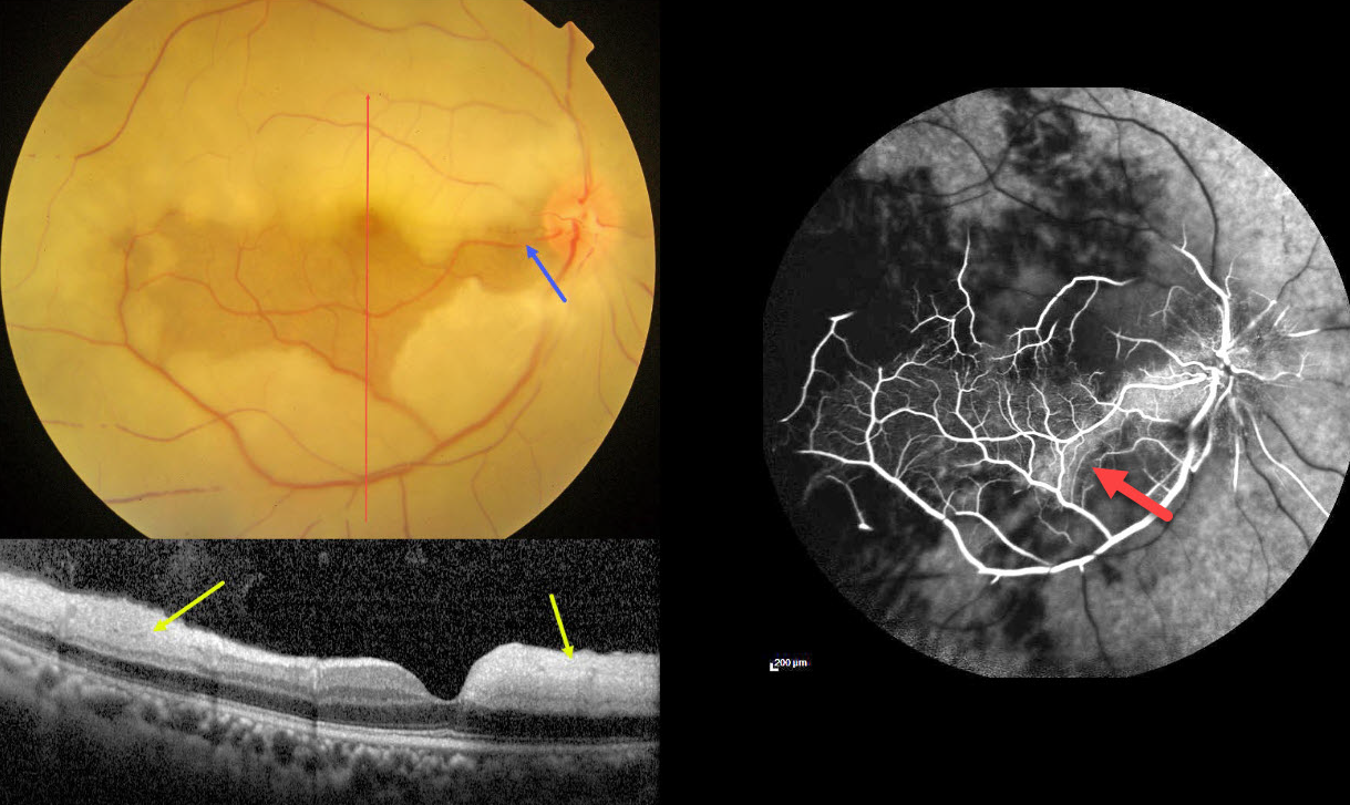
This 45YO male presented with acute vision loss in his right eye 4-5 days earlier. Vision was 20/80 in his right eye and 20/20 in his normal left eye.
Color photography shows an acute central retinal artery occlusion (CRAO) with sparing of the central and inferior macula due to a large cilioretinal artery.
The vertical OCT B-scan shows opaque hyperreflective paracentral inner retina with normal central and inferior foveal layers. Fluorescein angiography confirms preserved central and inferior macular arterial flow.
Workup elsewhere included a normal brain MRI and cardiac 2D Echo. Carotid Doppler showed a plaque in the right internal carotid artery.
Learning Points:
The inner two-thirds of the neurosensory retina is supplied by the central retinal artery, with the choroid supplying the RPE and photoreceptor layer. Acute CRAO, therefore, presents with a white edematous inner retina that spares the fovea.
Cilioretinal arteries, which arise from the posterior choroidal circulation, are present in up to 50% of individuals, and bilateral in about 25% (Schneider et al, Acta Ophthalmologica 2021;99:e310-e318). Our patient was quite fortunate to have a large cilioretinal vessel that relatively preserved central vision.
Patients with acute retinal ischemia (defined as transient monocular vision loss, acute BRAO, or acute CRAO) need to be emergently referred to a stroke center. This is especially urgent with an acute CRAO since about 75% of patients have already developed a recent stroke.
If the patient presents immediately after occlusion, measures to acutely lower intraocular pressure to move the causative embolus downstream can be performed, including ocular massage, paracentesis, and breathing into a bag. Unfortunately, most patients present outside this window (Shah et al, Ophthalmology Retina 2023;7:527-531), and even then, these measures are usually ineffective.
CILIORETINAL ARTERY
Originally posted on @retina.rocks December 27, 2022
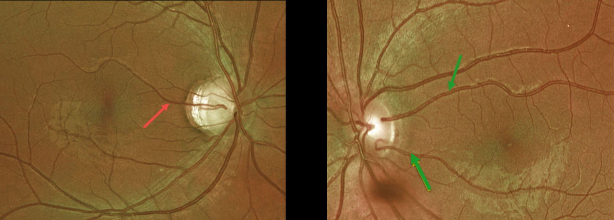
This healthy 23YO female presented with incidental findings of bilateral prominent cilioretinal arteries.
Optos imaging shows a superior cilioretinal vessel in her right eye. There are two cilioretinal vessels in her left eye, which collectively seem to supply the entire central macular region.
Learning Points:
The inner two-thirds of the neurosensory retina is supplied by the central retinal artery, with the choroid supplying the RPE and photoreceptor layer.
Cilioretinal arteries, which arise from the posterior choroidal circulation, are present in up to 50% of individuals, and bilateral in about 25% (Schneider et al, Acta Ophthalmologica 2021;99:e310-e318).
Our patient is unique in that she has multiple vessels in her left eye (Leavitt et al, AJO 2011;152:820-823), which seem to supply the arterial needs for her entire macula. If she were unlucky enough to be the one in 50,000 people per year to develop a central retinal artery occlusion (Leavitt et al, AJO 2011;152:820-823), these vessels would most likely preserve macular function.
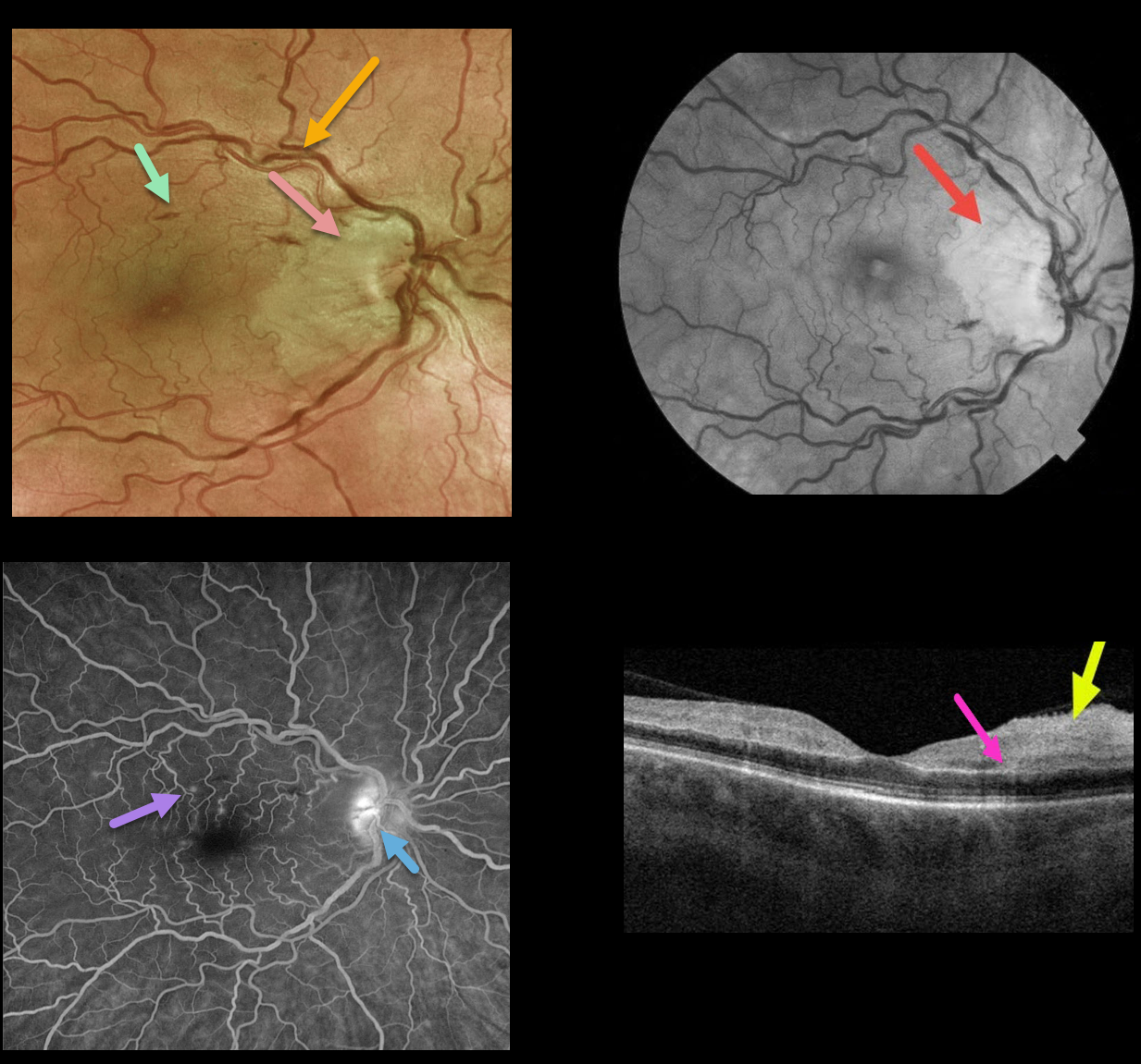
CRVO + RAO + PAMM
Originally posted on @retina.rocks March 4, 2022
This 50YO female presented with counting finger vision in her right eye due to an acute central retinal vein occlusion (CRVO) with a nasal macular retinal artery occlusion (RAO).
Optos fundus photography shows retinal vascular tortuosity, mild retinal hemorrhages, a mildly swollen nerve, and white ischemic nasal macular retina. The ischemic retinal changes are most apparent on the red-free photo.
Fluorescein angiography (FA) shows some mild focal areas of macular leakage with late staining of the temporal disc.
OCT shows areas of inner retinal edema and hyperreflectivity. There is also a small area of mid-retinal hyperreflectivity consistent with paracentral acute middle maculopathy (PAMM).
Learning Points:
Cilioretinal artery occlusion is usually non-embolic and seen in association with giant cell arteritis, secondary to a CRVO, or as an isolated event. The occlusion may be due in part to arterial compression from a swollen nerve.
Pichi et al felt that CRVO PAMM-type lesions, as seen in our patient, are due to hypoperfusion from increased downstream intraluminal pressure (Br J Ophthalmol 2019;103:1137-1145).
We couldn’t find a definite cilioretinal artery in our patient, although the ischemia was within the distribution of one if one were present. Interestingly, the nasal macular capillaries appeared perfused on FA. However, FA captures just the inner retinal capillary plexus.
Although the OCT shows evidence of acute inner capillary plexus ischemia, the angiographically perfused inner retina supports a more PAMM-type mechanism in our case.
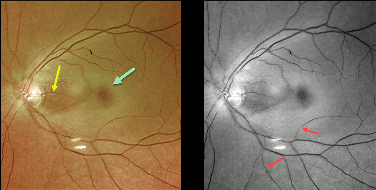
CRAO
Originally posted on @retina.rocks January 7, 2022
This 73YO female presented with counting-fingers vision secondary to an acute central retinal artery occlusion (CRAO).
The foveal retina is still perfused by the underlying choroid, resulting in the classic cherry red spot appearance. The nasal macula is also preserved due to the presence of a cilioretinal artery, which unfortunately does not supply the macular center. The Optos green channel best images the stagnant blood column.
Our patient’s MRI revealed several acute diffuse embolic strokes. Carotid duplex ultrasound showed severe bilateral carotid stenosis. In addition, her blood glucose was 357, and her erythrocyte sedimentation rate was 94. She was also started on systemic prednisone by the ER until giant cell arteritis could more definitively be ruled out.
Learning Points:
Patients with acute retinal ischemia (defined as transient monocular vision loss, acute BRAO, or acute CRAO) need to be emergently referred to a stroke center.
This is especially urgent with an acute CRAO since about 75% of patients have already developed a recent stroke.
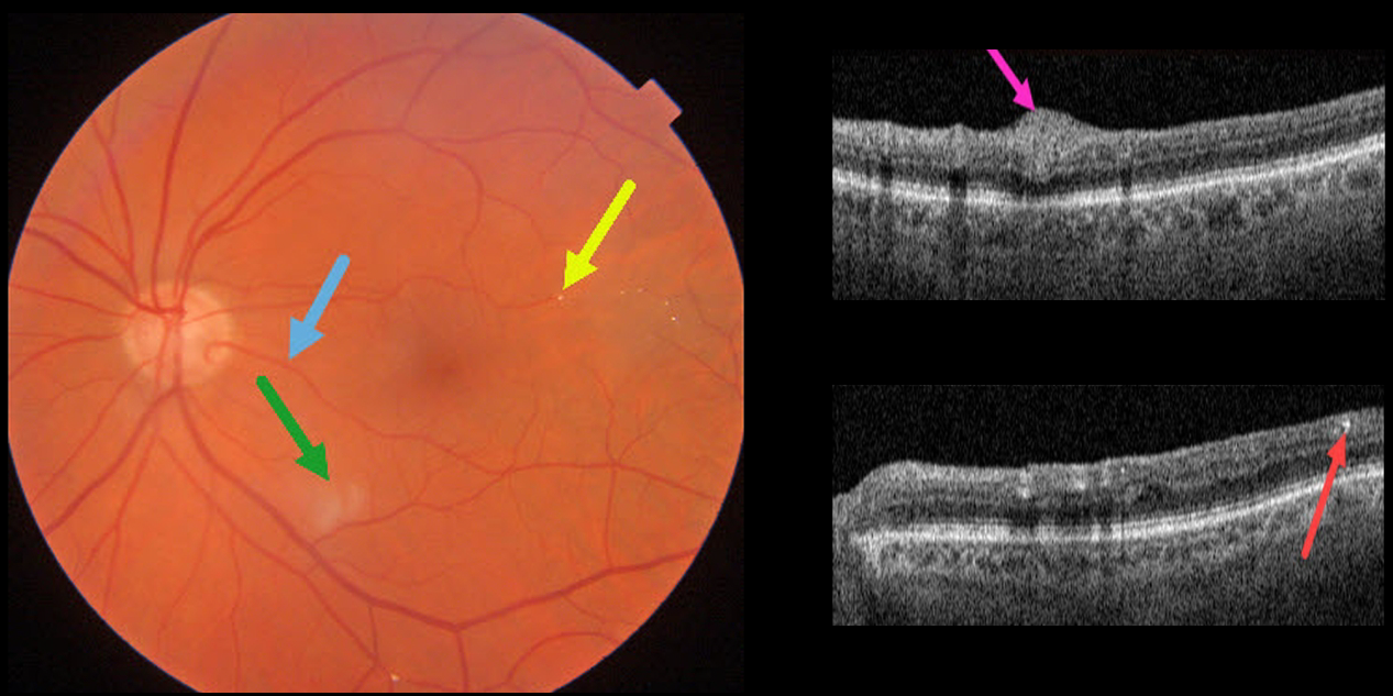
BRAO
Originally posted on @retina.rocks January 6, 2022
This 85YO male has 20/25 vision despite multiple retinal emboli and a resolving branch retinal artery occlusion (BRAO).
Triton swept-source OCT shows residual inner retinal edema and opacification. The embolus on Triton photography is also imaged in the OCT B-scan.
A cilioretinal artery may be responsible for the good vision.
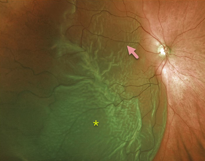
RHEGMATOGENOUS RETINAL DETACHMENT
Originally posted on @retina.rocks June 10, 2020
This patient has an acute, macula-off retinal detachment with a large cilioretinal artery.
Learning Points:
Cilioretinal arteries arise from the choroidal circulation. This results in cilioretinal sparing with central retinal artery occlusion.
Unfortunately, this vessel offers no visual protection against retinal detachment!


