

Coats Disease
<< Back to Cases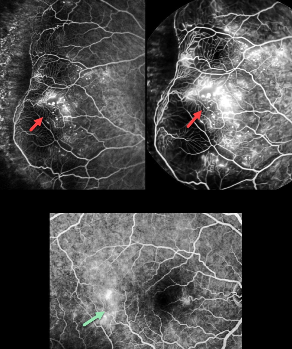
COATS DISEASE
Rajashree Salvi, Apoorva Ayachit, Guruprasad Ayachit, and Shrinivas Joshi
Originally posted on @retina.rocks May 15, 2026
This 32YO male complained of decreased vision in his right eye for 2 weeks. Vision was 20/20 OD and 20/20 in his normal OS.
Fundus fluorescein angiography shows a broad zone of temporal midperipheral leaking retinal telangiectasia with capillary dropout and distal temporal ischemia. Larger retinal vessel anastomoses are present at the border of the perfused and nonperfused retina. There is also late temporal macular leakage. Color photography is unfortunately not available, and our patient was immediately lost to follow-up.
Learning Points:
Described by George Coats in 1908, Coats disease is an idiopathic disorder, usually unilateral, in males. It is characterized by retinal vascular telangiectasia involving capillaries, arterioles, and venules. Patients can present with a wide range of findings, from minimal exudation to total exudative retinal detachment. The leaking vessels are usually treated to prevent exudative complications.
Thermal laser photocoagulation is usually the initial treatment modality, but anti-VEGF injections can also be used to stabilize the vascular leakage. Treatment involves a combination of focal laser to the leaking vascular abnormalities along with scatter laser into areas of ischemia. Patients will often require multiple rounds of treatment. See Sigler et al for a review of the current management (Survey Ophthalmology 2014;59:30-46).
COATS DISEASE
Originally posted on @retina.rocks March 12, 2026
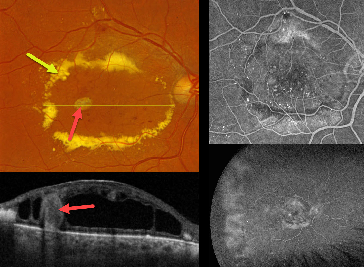
This 71YO male presented with a vague history of decreased vision in his right eye. He gave a history of diet-controlled type 2 diabetes. Vision was 20/200 OD and 20/20 in his normal OS.
Optos color RGB imaging shows an oval ring of subretinal lipid surrounding the central macula. Scattered telangiectatic vessels are noted. A large fibrosed macroaneurysm is seen temporally. On Triton swept-source OCT, this macroaneurysm spans nearly the entire retinal thickness and is surrounded by a large mid-retinal cystic cavity. Fundus fluorescein angiography (FFA) shows numerous macular microaneurysms with relative blockage from the subretinal lipid. There is diffuse leakage from the temporal vessels with ischemic retina more peripherally associated with some telangiectatic vessels. FFA was normal in his left eye (not shown). Intravitreal anti-VEGF therapy was started.
Learning Points:
Described by George Coats in 1908, Coats disease is an idiopathic disorder, usually unilateral, in males. It is characterized by retinal vascular telangiectasia involving capillaries, arterioles, and venules. Patients can present with a wide range of findings, from minimal exudation to total exudative retinal detachment. The leaking vessels are usually treated to prevent exudative complications.
Thermal laser photocoagulation is usually the initial treatment modality, but anti-VEGF injections can also be used to stabilize the vascular leakage. Treatment involves a combination of focal laser to the leaking vascular abnormalities along with scatter laser into areas of ischemia. Patients will often require multiple rounds of treatment. See Sigler et al for a review of the current management (Survey Ophthalmology 2014;59:30-46).
COATS DISEASE
Shraddha Raj Shrivastava and Manish Nagpal
Originally posted on @retina.rocks December 15, 2025
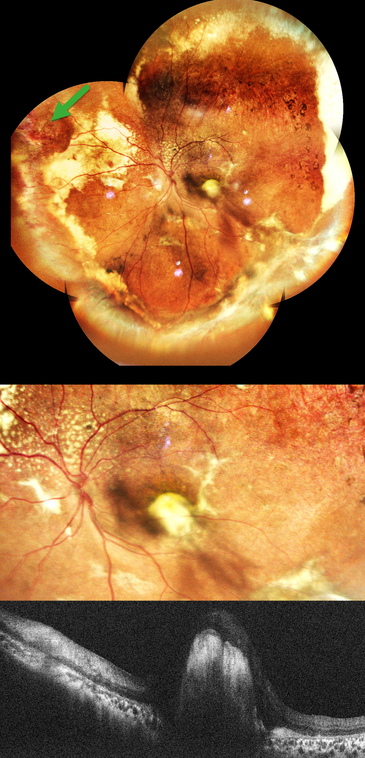
This healthy 11YO boy was brought in by his parents with a 1.5-year history of vision loss and outward deviation. Vision was 20/15 in his normal OD and light perception OS. Xanthocoria was noted on the pupillary light reflex.
Pseudocolor SLO imaging shows extensive peripheral subretinal lipid in all quadrants with telangiectatic and aneurysmal capillary dilations, most prominent in the nasal periphery. A superonasal orange-red elevated vasoproliferative mass is noted in the superonasal midperiphery. Pigmentary changes and RPE atrophy are observed in the superotemporal mid-peripheral retina. There is a large subfoveal lipid plaque that appears as a hyperreflective nodule on OCT. An exudative subtotal retinal detachment involving the inferior and nasal quadrants was noted clinically.
Learning Points:
Coats disease is characterized by vascular changes throughout the retinal vasculature, including the arteries, the capillary bed, and the veins. Larger dilated vessels in Coats can sometimes mimic angiomas, vascular tumors, or exudation from retinoblastoma. The presence of retinal telangiectasia can help establish the diagnosis of Coats in these cases.
Coats is usually unilateral in males. However, recent ultra-widefield angiographic studies indicate subtle vascular changes mostly in the temporal periphery of the fellow eye (Brockmann et al, BJO 2021;105:1444-1453).
Laser photocoagulation can be used to directly treat leaking telangiectatic vessels and ischemic retina. Intravitreal anti-VEGF injections can also be used as an adjunctive treatment. Pars plana vitrectomy (PPV) with external drainage of subretinal fluid plays an important role in the management of advanced Coats disease, particularly in cases with extensive exudative retinal detachment.
In our patient, pars plana vitrectomy with external drainage of subretinal fluid, along with endolaser/cryotherapy to leaking aneurysms, and an intravitreal anti-VEGF injection was advised.
COATS DISEASE
Ayushi Gupta and Vishal Agrawal
Originally posted on @retina.rocks August 26, 2025
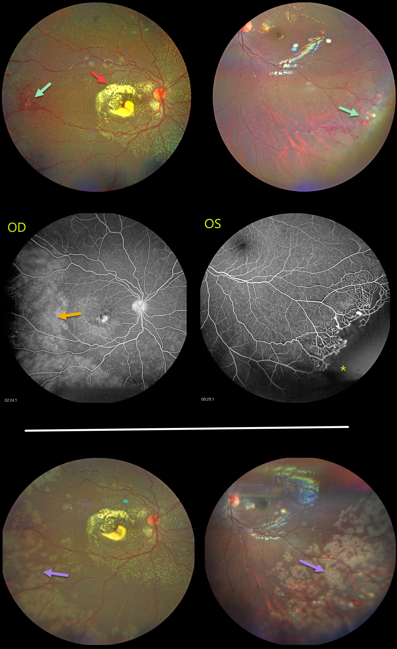
This healthy 6YO girl’s parents noted that her right eye had been deviating outward for the past several weeks. Vision was light perception OD and 20/20 OS.
Color photography shows fairly dense subfoveal lipid with more diffuse specks of subretinal lipid radiating outwards from the disc. Telangiectatic vessels are noted temporally OD and also in the left inferotemporal periphery. Fluorescein angiography shows diffuse leakage from these vessels OD, with distal retinal ischemia OS.
Scatter laser to the ischemic retina, along with focal treatment of the larger telangiectatic vessels, was applied. Nine months later, vision was unchanged, with consolidation of the subfoveal lipid scar OD along with persistent telangiectasia OS.
Learning Points:
Described by George Coats in 1908, Coats disease is an idiopathic disorder typically unilateral in males. It is characterized by retinal vascular telangiectasia involving capillaries, arterioles, and venules. Patients can present with a wide range of findings, from minimal exudation to total exudative retinal detachment. The leaking vessels are usually treated to prevent exudative complications. Thermal laser photocoagulation is usually the initial modality, but anti-VEGF injections can also be used to stabilize the vascular leakage. Patients will often require multiple rounds of treatment. See Sigler et al for a review of the current management (Survey Ophthalmology 2014;59:30-46).
Dyskeratosis congenita or familial exudative vitreoretinopathy should be considered for patients with frank bilateral disease (Teixeira et al, Arch Ophthalmol 2008;126:134-135), in women with Coats-like findings (Johnson et al, Genetics 2009;30:181-184), or in patients with a family history of similar findings. Our patient’s family history was negative, and her nail beds were normal. Genetic testing was not possible due to cost concerns.
COATS DISEASE
Tejaswita Verma and Manish Nagpal
Originally posted on @retina.rocks April 2, 2025
COATS DISEASE
Anjana Mirajkar and Manish Nagpal
Originally posted on @retina.rocks May 2, 2024
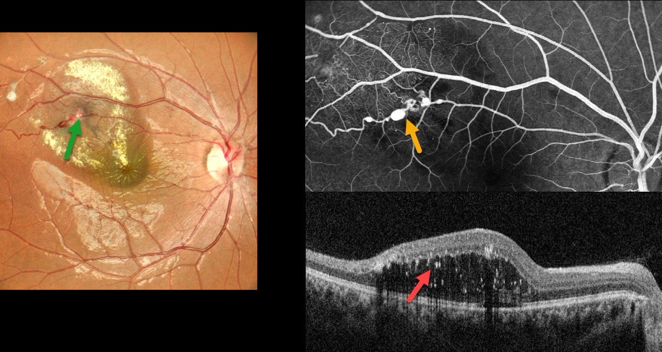
This healthy 19YO female presented with 1 week of blurred vision in her right eye. Vision was 20/80 OD and 20/20 in her normal OS.
Color imaging shows multiple superotemporal variably fibrosed macroaneurysms with surrounding lipid that extends towards the central macula. More subtle telangiectasia and lipid were seen in the superior periphery (not shown).
Fluorescein angiography shows multiple patent macroaneurysms along the superotemporal arteriole with more distal telangiectasia. OCT shows outer retinal fluid with numerous suspended hyperreflective lipid particles. Thermal laser followed by 3 monthly anti-VEGF injections was given.
Learning Points:
Described by George Coats in 1908, Coats disease is an idiopathic disorder that is usually unilateral in males. It is characterized by retinal vascular telangiectasia involving capillaries, arterioles, and venules. Patients can present with a wide variety of findings ranging from minimal exudation to total exudative retinal detachment. The leaking vessels are usually treated to prevent exudative complications.
Thermal laser photocoagulation is usually the initial modality, but anti-VEGF injections can also be used to stabilize the vascular leakage. Patients will often require multiple rounds of treatment. See Sigler et al for a review of the current management (Survey Ophthalmology 2014;59:30-46).
Dyskeratosis congenita or familial exudative vitreoretinopathy should be considered for patients with frank bilateral disease (Teixeira et al, Arch Ophthalmol 2008;126:134-135), in women with Coats-like findings (Johnson et al, Genetics 2009;30:181-184), or in patients with a family history of similar findings. Since our patient’s findings were unilateral and her nail beds were normal, we deferred genetic testing.
COATS DISEASE
Originally posted on @retina.rocks April 3, 2024
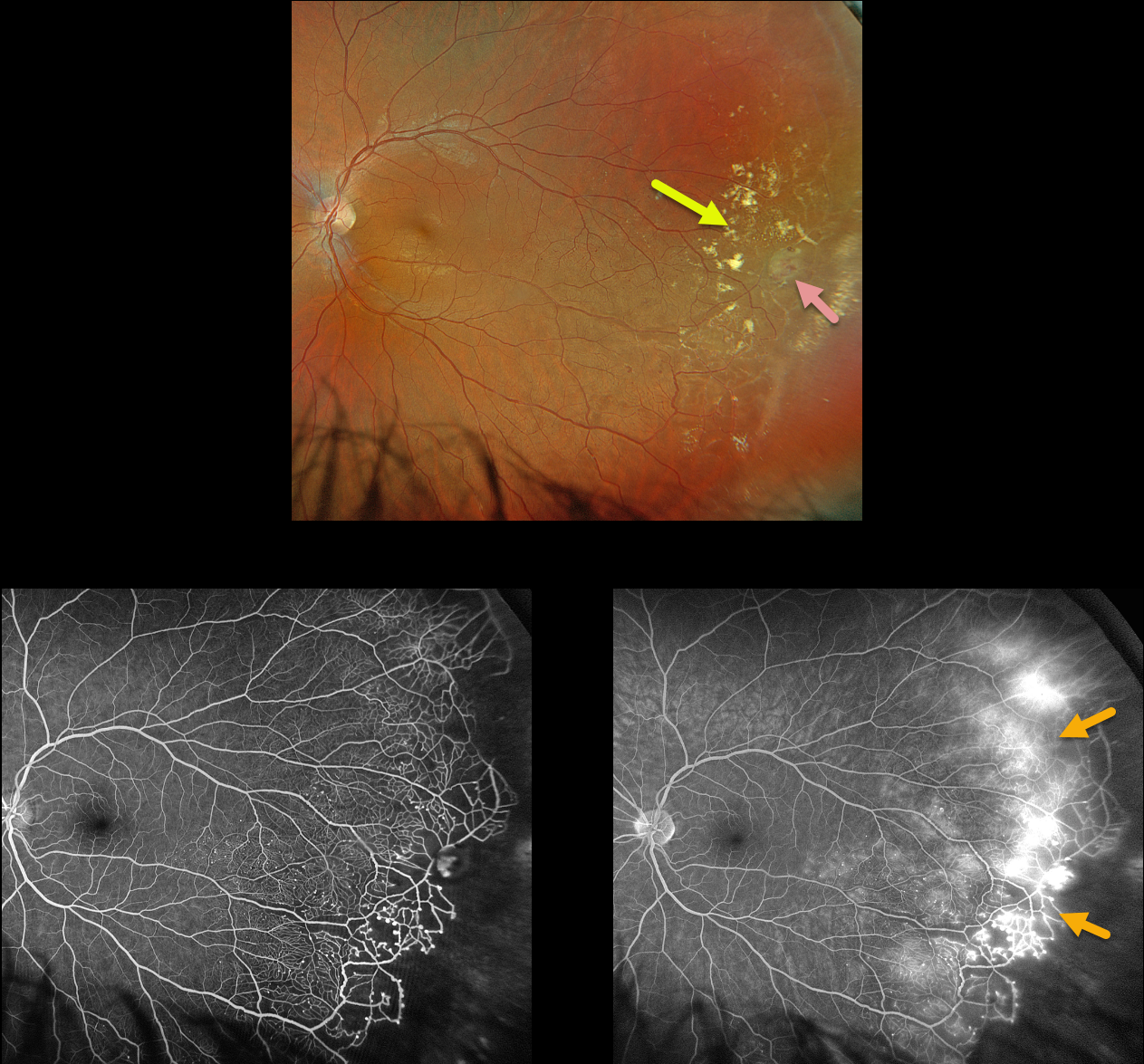
This 28YO healthy male was referred for asymptomatic findings on retinal examination in his left eye. Vision was 20/20 in his normal OD and 20/20 OS.
Optos color RGB imaging shows scattered temporal lipid and telangiectatic vessels. A large macroaneurysm is noted. The retina distal to the lipid is featureless and clinically ischemic.
Fluorescein angiography shows the telangiectasia becoming more prominent and ischemic as it extends from the distal temporal macula into the periphery. There is marked leakage at the junction of the perfused and ischemic retina. Scatter laser to the ischemic retina, along with a focal laser to the macroaneurysm, was scheduled.
Learning Points:
Described by George Coats in 1908, Coats disease is an idiopathic disorder that is usually unilateral in males. It is characterized by retinal vascular telangiectasia involving capillaries, arterioles, and venules. Patients can present with a wide variety of findings ranging from minimal exudation to total exudative retinal detachment.
The leaking vessels are usually treated to prevent exudative complications. Thermal laser photocoagulation is usually the initial modality, but anti-VEGF injections can also be used to stabilize the vascular leakage. Patients will often require multiple rounds of treatment.
See Sigler et al for a review of the current management (Survey Ophthalmology 2014;59:30-46).
COATS DISEASE
Will Gibson
Originally posted on @retina.rocks March 19, 2024
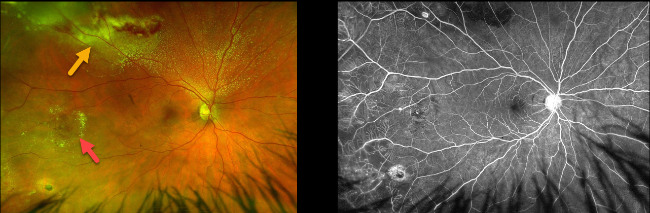
This 36YO male presented with asymptomatic findings on fundus examination in his right eye. Vision was 20/25 in his right eye and 20/20 in his normal left eye.
Optos color RG imaging shows telangiectatic vessels in the temporal and superotemporal midperipheries. Subretinal lipid is most dense superotemporally.
Fluorescein angiography beautifully shows the telangiectasia, including capillary loss, microaneurysms, and macroaneurysms. He was referred to a retinal specialist for further management.
Learning Points:
Described by George Coats in 1908, Coats disease is an idiopathic disorder that is usually unilateral in males. It is characterized by retinal vascular telangiectasia involving capillaries, arterioles, and venules. Patients can present with a wide variety of findings ranging from minimal exudation to total exudative retinal detachment. The leaking vessels are usually treated to prevent exudative complications.
Thermal laser photocoagulation is usually the initial modality, but anti-VEGF injections can also be used to stabilize the vascular leakage. Patients will often require multiple rounds of treatment.
See Sigler et al for a review of the current management (Survey Ophthalmology 2014;59:30-46).
Atypical cases, including positive family history, bilateral findings, or in females, should be considered for genetic testing to rule out inherited telomere disorders, including Coats plus syndrome (Teixeira et al, Arch Ophthalmology 2008;126:134-135) and dyskeratosis congenita (Johnson et al, Ophthalmic Genetics 2009;30:181-184).
TRIAMCINOLONE-MOXIFLOXACIN (TriMoxi)
Originally posted on @retina.rocks October 30, 2023
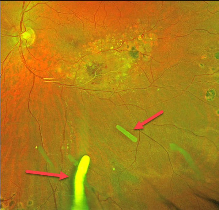
This 41YO male has been followed for years with Coats disease in his left eye. Numerous treatment modalities have been used, including thermal laser, intravitreal anti-VEGF, and Ozurdex (dexamethasone 0.7mg) injections. Most recently, he has received Ozurdex every 3-4 months for the past 2 years. Vision is stable at 20/30.
Optos color RG imaging shows multiple Ozurdex ghosts in variable stages of absorption.
Learning Points:
The Ozurdex implant slowly dissolves while releasing its dexamethasone. Although the clinical effect usually lasts about 2 to 3 months, remnants can remain in the eye for over a year (Kim et al, Retina 2020;40:2226-2231).
Approved for diabetic and retinal vein occlusion macular edema and posterior uveitis, it is also often used off-label for other causes of macular edema, including uveitic and pseudophakic CME. For our patient, Ozurdex has been the most successful modality at keeping his central macula dry.
COATS DISEASE
Originally posted on @retina.rocks October 19, 2023
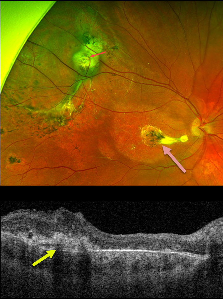
This 14YO boy received thermal laser photocoagulation for an exudative retinal detachment in his right eye from Coats disease about 10 years earlier. There were no new complaints, and vision was stable at 20/400. His normal left eye was 20/20.
Optos color RG imaging shows peripheral superotemporal scarring and subretinal fibrosis. A venous retinal-choroidal anastomosis is noted. Some variably pigmented laser scars are seen, and there are no active Coats lesions. A nematode-like area of subfoveal scarring is seen more centrally with variable outer retinal thinning and edema.
Learning Points:
Described by George Coats in 1908, Coats disease is an idiopathic disorder that is usually unilateral in males. It is characterized by retinal vascular telangiectasia involving capillaries, arterioles, and venules.
Patients can present with a wide variety of findings ranging from minimal exudation to total exudative retinal detachment. The leaking vessels are usually treated to prevent exudative complications.
Thermal laser photocoagulation is usually the initial modality, but anti-VEGF injections can also be used to stabilize the vascular leakage. Patients will often require multiple rounds of treatment.
See Sigler et al for a review of the current management (Survey Ophthalmology 2014;59:30-46).
Under normal conditions, the retinal and choroidal circulations remain separate and distinct. Rarely, the two circulations connect directly through a chorioretinal anastomosis. These can occur in chorioretinal scars, typically from inactive toxoplasmosis lesions, macular telangiectasia, and disciform scars.
COATS DISEASE
Asma Samsudeen and Ashish Sharma
Originally posted on @retina.rocks July 27, 2023
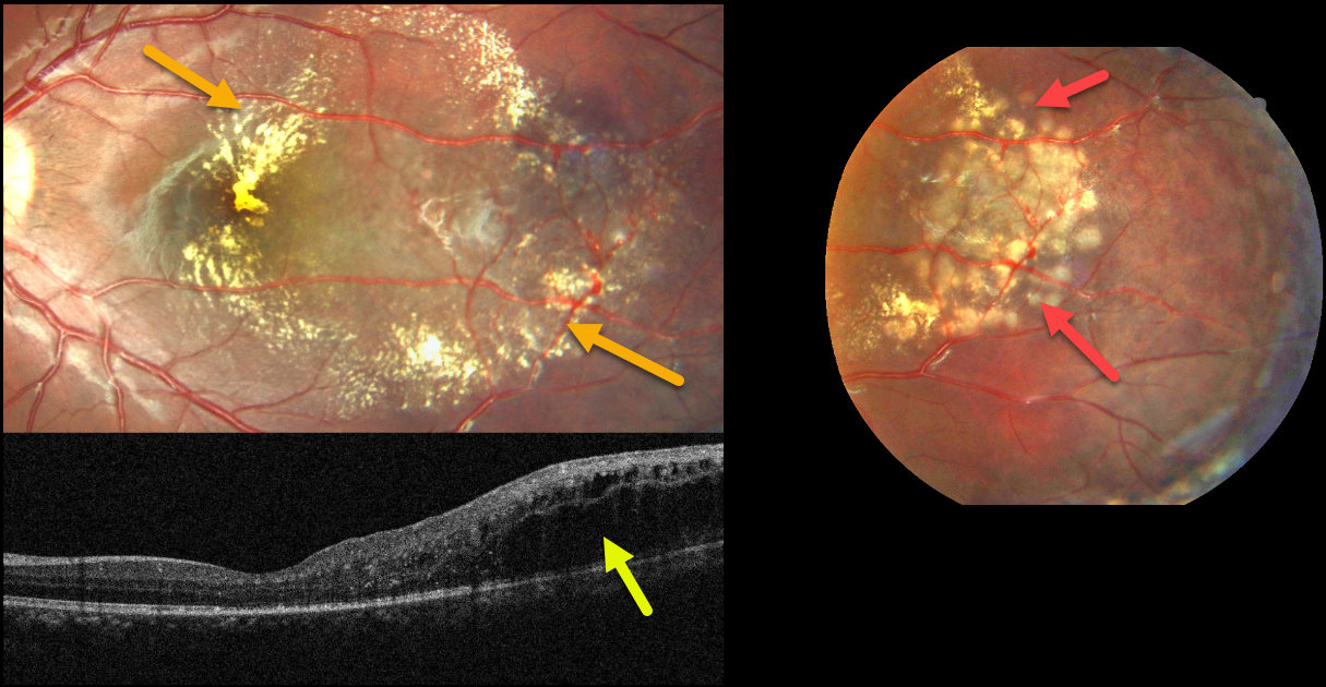
This healthy 8YO boy presented for a routine eye examination. Vision was 20/20 in his normal right eye and 20/30 in his left eye.
Fundus photography shows telangiectatic vessels in the temporal macula associated with surrounding lipid. The lipid is particularly dense centrally.
OCT scanning shows temporal, mostly outer retinal edema. The macular center looks remarkably normal given the fundoscopic appearance. Focal macular laser to the telangiectatic lesions was applied
Learning Points:
Described by George Coats in 1908, Coats disease is an idiopathic disorder typically unilateral in males. It is characterized by retinal vascular telangiectasia involving capillaries, arterioles, and venules.
Patients can present with a wide range of findings, from minimal exudation to total exudative retinal detachment. The leaking vessels are usually treated to prevent exudative complications.
Thermal laser photocoagulation is usually the initial modality, but anti-VEGF injections can also be used to stabilize the vascular leakage. Patients will often require multiple rounds of treatment.
See Sigler et al for a review of the current management (Survey Ophthalmology 2014;59:30-46).
COATS DISEASE
Marianna Kavalarak
Originally posted on @retina.rocks July 5, 2023
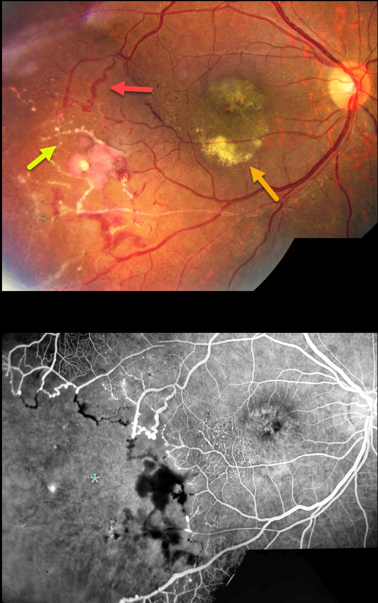
This 19YO female presented with a chronic history of unilateral vision loss in her right eye. Vision was 20/100 OD and 20/20 in her healthy left eye.
Color imaging shows extensive vascular changes in the inferotemporal retina, including retinal telangiectasia, macroaneurysms, vascular beading, sheathed vessels, and featureless ischemic retina. The central macula is hyperpigmented with variable subretinal lipid.
Fluorescein angiography shows marked distal capillary loss. The vessels bordering the perfused and ischemic retina are irregularly dilated with anastomoses. Fine telangiectatic vessels are noted in the inferotemporal macula.
Learning Points:
Coats disease is characterized by vascular changes throughout the retinal vasculature, including the arteries, the capillary bed, and the veins. Larger dilated vessels in Coats can sometimes mimic angiomas, vascular tumors, or exudation from retinoblastoma. The presence of retinal telangiectasia can help establish the diagnosis of Coats in these cases.
Coats are usually unilateral in males. However, recent ultra-widefield angiographic studies indicate subtle vascular changes mostly in the temporal periphery of the fellow eye (Brockmann et al, BJO 2021;105:1444-1453).
Dyskeratosis congenita or familial exudative vitreoretinopathy (FEVR) should be considered for patients with frank bilateral disease (Teixeira et al, Arch Ophthalmol 2008;126:134-135), in women with Coats-like findings (Johnson et al, Genetics 2009;30:181-184), or in patients with a family history of similar findings.
Laser photocoagulation can be used to directly treat leaking telangiectatic vessels, and a scatter laser can be placed into the ischemic retina. Anti-VEGF injections can also be used as primary or secondary treatment.
COATS DISEASE
Pramod Suman and Manish Nagpal
Originally posted on @retina.rocks September 5, 2022
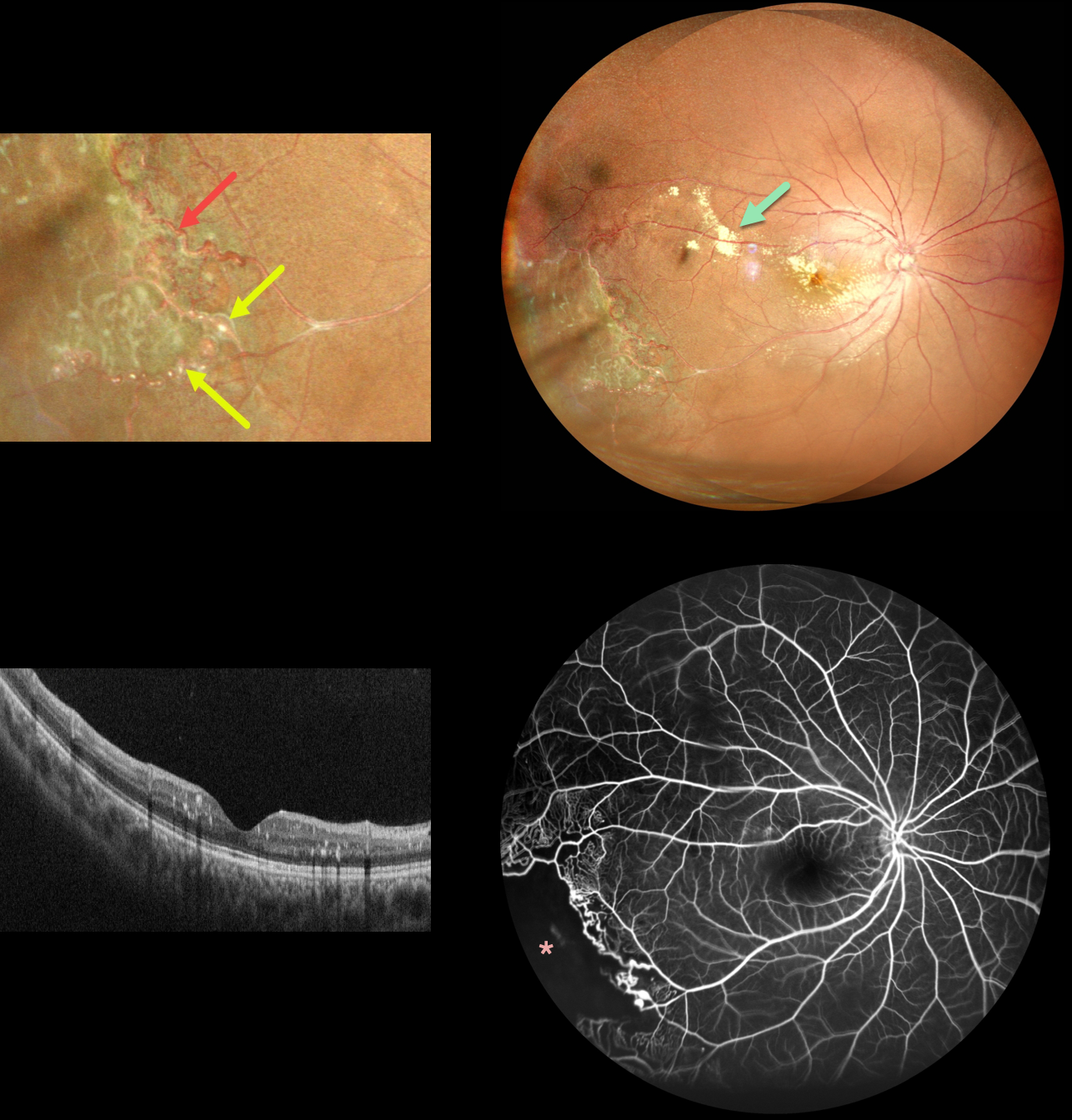
This 11YO boy presented with these classic unilateral findings for Coats disease. An ultrawidefield image of the right eye shows scattered submacular and subretinal lipid posteriorly with distal featureless ischemic retina inferotemporally.
More peripherally, there are both large vessel changes, including beading and multiple macroaneurysms, with small vessel telangiectatic changes.
OCT scanning is remarkably normal except for some scattered specks of hyperreflective intraretinal lipid. Fluorescein angiography shows peripheral temporal telangiectasia with more distal ischemia, along with some subtle superotemporal macular telangiectatic changes.
Sector scatter laser was recommended.
COATS DISEASE
Originally posted on @retina.rocks July 25, 2022
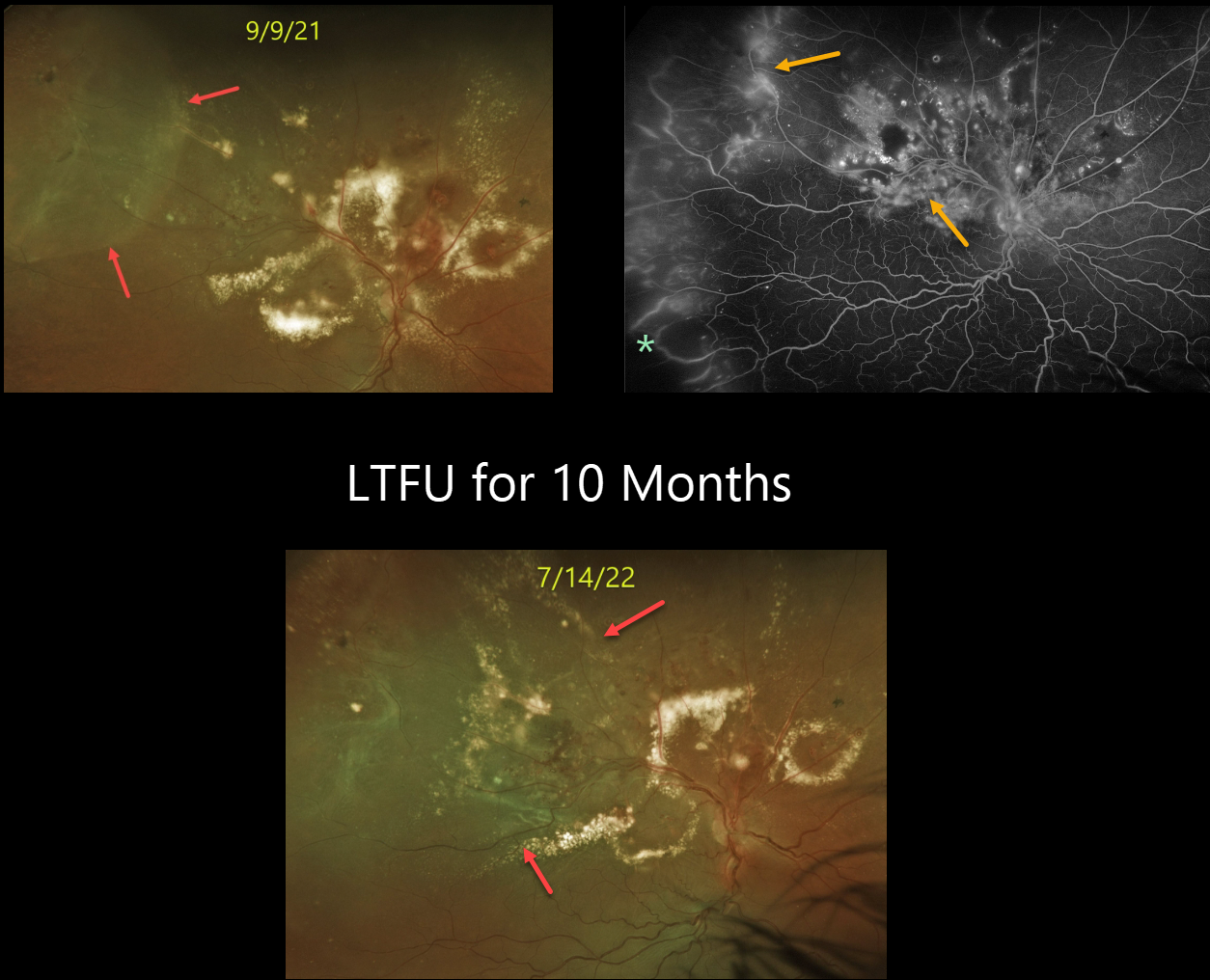
This 54YO male patient has a long history of Coats disease. Vision was 20/400 when he was referred to our practice on 9/9/21. Unfortunately, he was lost to follow-up for almost a year because of his fear of doctors. Vision decreased to hand motion.
Optos color imaging shows his funduscopic appearance at the initial and subsequent visits. Fluorescein angiography shows extensive leakage from the various vascular abnormalities, as well as peripheral temporal ischemia. Although the posterior pole lipid mostly decreased during the prior year, the exudative detachment enlarged, now spreading through the macula.
Vitrectomy is scheduled in hopes of preventing further vision loss.
COATS DISEASE
Originally posted on @retina.rocks May 23, 2022
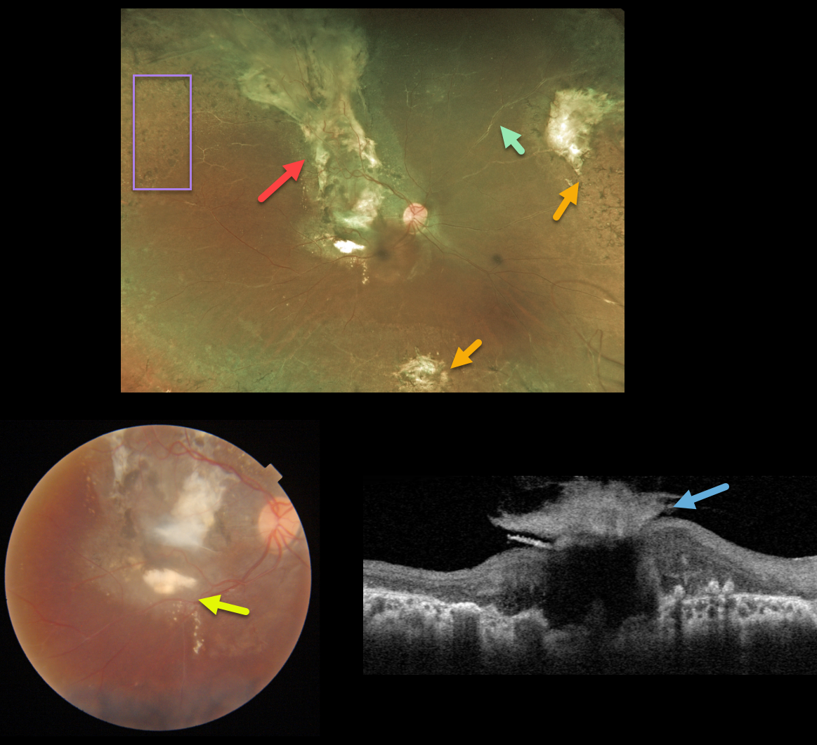
This 15YO female received prior thermal laser for Coats disease over 10 years earlier and was subsequently lost to follow-up. Right eye vision was counting fingers.
Optos color imaging shows subretinal scarring extending from the macula through the superior retinal periphery, nasal and inferior peripheral scarring, scattered peripheral retinal laser scarring, and sheathed vessels. There are no active Coats lesions.
Triton color imaging shows a magnified view of the macular scarring with some inferior macular subretinal lipid. Swept-source OCT shows a volcano-like vitreous component to the macular scar.
Learning Points:
Historically considered a unilateral disease, more recent ultra-widefield angiographic studies show subtle vascular changes, mostly in the temporal periphery (Brockmann et al., BJO 2021;105:1444-1453). Our patient’s left eye, however, was completely normal.
COATS DISEASE
Originally posted on @retina.rocks January 20, 2022
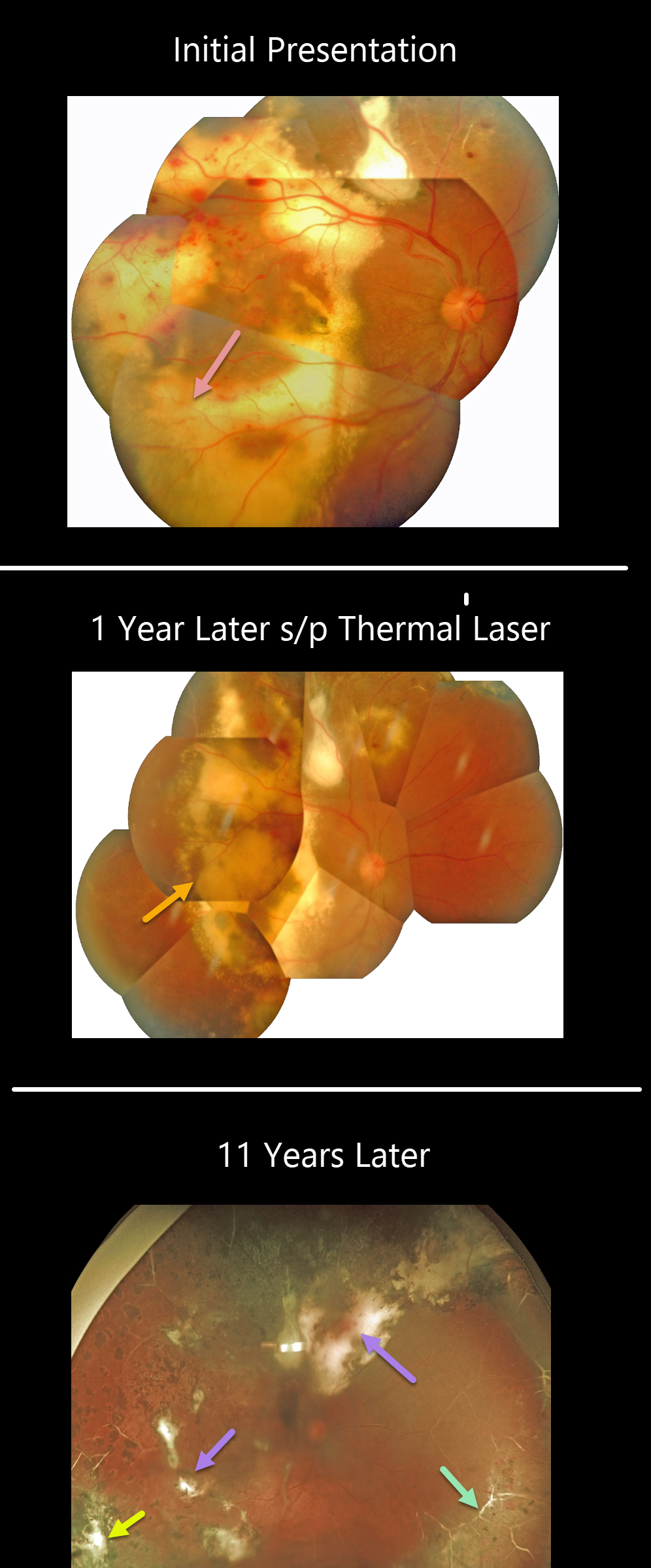
This patient was first seen in 2009 when he was 16 years old. Confluent subretinal lipid can be seen extending to the periphery. Several sessions of thermal laser were applied, and 1 year later, photos showed inactive Coats lesions with decreased subretinal lipid.
He was then lost to follow-up for 11 years when his vision was 20/400. Optos shows total resolution of all exudation and subretinal lipid with variable residual subretinal scarring. Peripheral scatter laser scarring is noted. The peripheral retinal vessels are white. There are no active Coats lesions.
Learning Points:
Historically considered a unilateral disease, more recent ultra-widefield angiographic studies show subtle vascular changes, mostly in the temporal periphery (Brockmann et al., BJO 2021;105:1444-1453).
Our patient’s left eye, however, was completely normal clinically and angiographically.
COATS DISEASE
Originally posted on @retina.rocks November 8, 2021
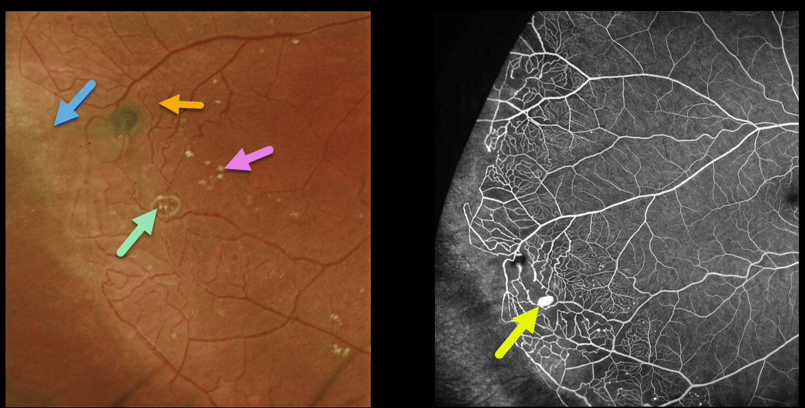
This healthy 13YO boy presented with these asymptomatic findings in his right eye. Vision was 20/20 OU.
Optos imaging shows classic findings for Coats disease in the temporal periphery, including telangiectatic vessels, a macroaneurysm, scattered mild lipid, and ischemic retina more distally. Fluorescein angiography more dramatically illustrates these vascular changes. The macroaneurysm stains.
Learning Points:
Historically considered a unilateral disease, more recent ultra-widefield angiographic studies show subtle vascular changes, mostly in the temporal periphery (Brockmann et al, BJO 2021;105:1444-1453). Our patient’s left eye, however, was completely normal clinically and angiographically.
Due to the lack of significant exudation, we are currently monitoring this patient twice yearly.
COATS DISEASE
Kumar Chugani
Originally posted on @retina.rocks September 16, 2021
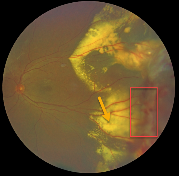
This 66YO male presented with classic Coats disease findings. Vision was 20/30.
Confluent subretinal lipid extends into the peripheral temporal macula. The source for this lipid is noted in the temporal midperiphery, with dilated, irregular telangiectatic vessels with aneurysmal dilations.
Learning Points:
Laser photocoagulation can be used to treat leaking macroaneurysms and telangiectatic vessels in Coats disease. Anti-VEGF injections can also be used to stabilize vascular leakage.
COATS DISEASE
Originally posted on @retina.rocks April 6, 2021
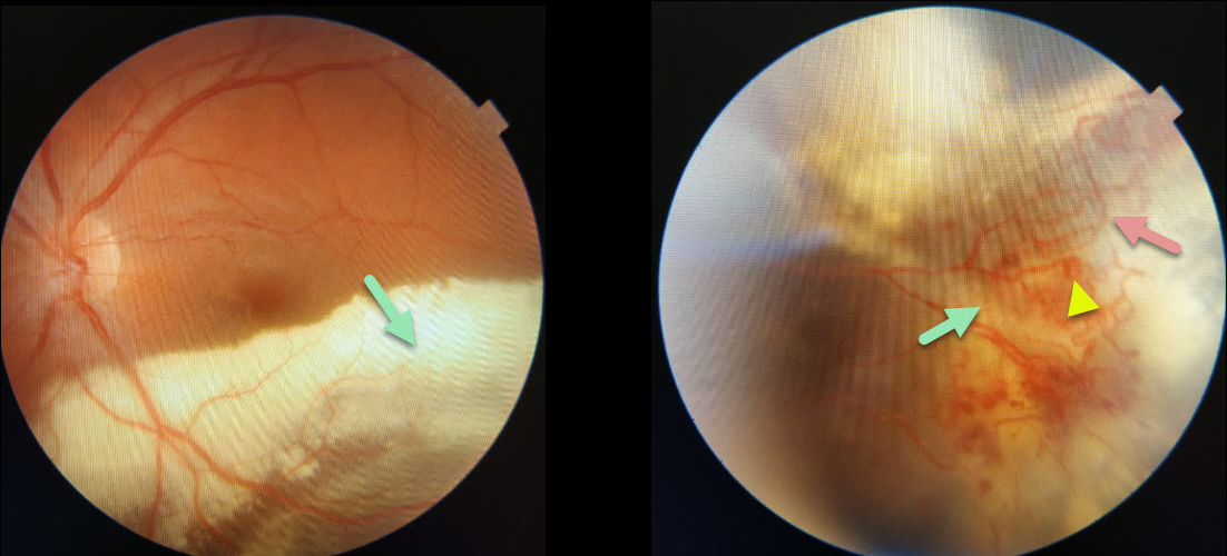
This 20YO male presented with classic Coats disease findings. Confluent subretinal lipid extends into the inferior macula, sparing the foveal center, allowing for 20/30 vision.
The source for this lipid is noted in the inferotemporal midperiphery with dilated, irregular vessels, aneurysmal dilations, and underlying subretinal fluid and lipid.
Learning Points:
Laser photocoagulation can be used to treat leaking macroaneurysms and telangiectatic vessels in Coats disease. Anti-VEGF injections can also be used to stabilize vascular leakage.
COATS DISEASE
Originally posted on @retina.rocks February 12, 2021
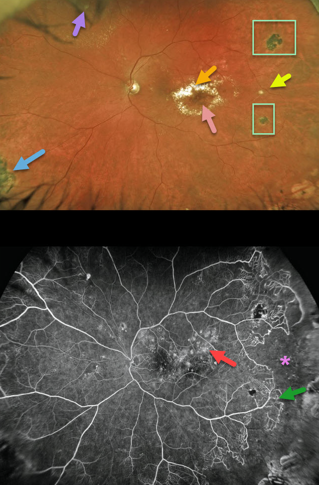
This 53YO has classic findings of Coats disease. A leaking macroaneurysm in the distal inferotemporal macula is causing exudation with lipid threatening the macular center. Prior macular laser scars are noted in the superior macula.
There is a fibrosed macroaneurysm in the temporal midperiphery, and some fibrosed telangiectatic lesions are visible in the superonasal periphery. Scattered peripheral hyperpigmented laser scars are also seen.
Fluorescein angiography nicely shows these findings and more, including macular telangiectasia with staining laser scars, some patches of mid-peripheral telangiectasia and capillary loss, and a large region of temporal capillary loss.
Learning Points:
Laser photocoagulation can be used to treat leaking macroaneurysms and telangiectatic vessels in Coats disease. Anti-VEGF injections can also be used to stabilize vascular leakage.
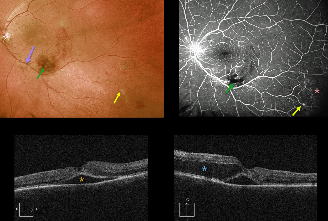
UNKNOWN DIAGNOSIS
Originally posted on @retina.rocks January 6, 2021
This 59YO female presented with acute vision loss of 20-100 in her left eye. At first glance, it looks like a macular branch retinal vein occlusion (BRVO) arising from an artery crossing over a vein more proximally. There is a likely macroaneurysm within the area of more confluent inferior macular blood.
But a more careful inspection shows some retinal telangiectasia more inferiorly and proximally, as well as more distal inferotemporal areas of retinal hemorrhages, telangiectasia, and an arterial macroaneurysm.
Fluorescein angiography better shows these vascular changes and broad peripheral areas of inferotemporal capillary loss.
OCT shows foveal subretinal fluid with a large pocket of outer intraretinal fluid.
Our best guess is that this patient has Coats disease, given the multiple macroaneurysms, multifocal areas of retinal vascular telangiectasia, and capillary loss. Although uncommon, Coats can be found in females.
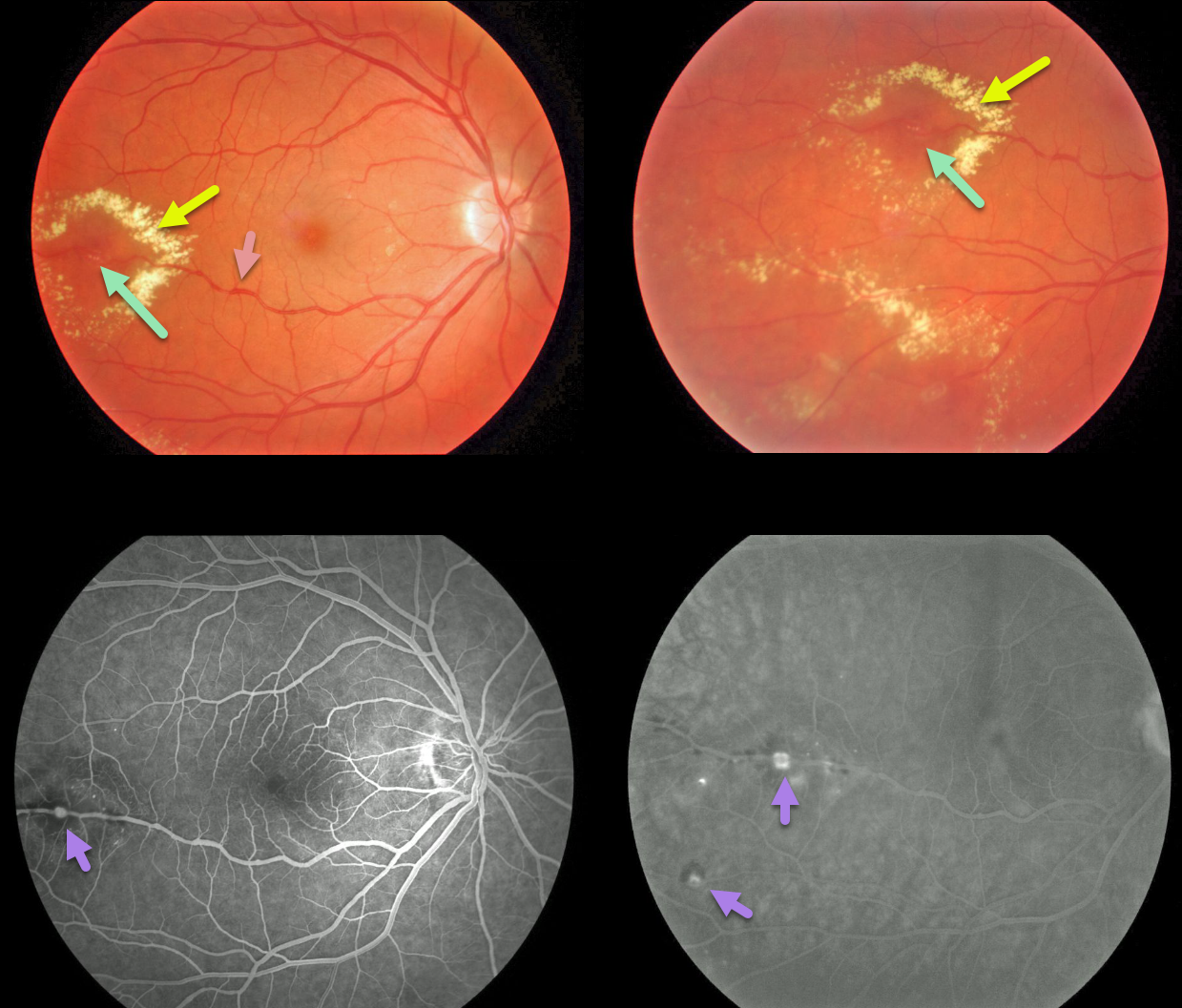
COATS DISEASE
Originally posted on @retina.rocks June 24, 2020
This is a classic case of Coats disease, with unilateral retinal vascular leakage from both the capillary bed and larger vessels.
Fundus images show dilated capillaries and retinal vessels. In addition, there are multiple arterial macroaneurysms with surrounding exudate.
Fluorescein angiography shows the leaking telangiectatic vessels.
Learning Points:
Laser photocoagulation can be used to treat leaking macroaneurysms and telangiectatic vessels in Coats disease. Anti-VEGF injections can also be used to stabilize vascular leakage. Patients will usually require multiple rounds of treatment.


