

Coloboma Non-Optic Nerve
<< Back to Cases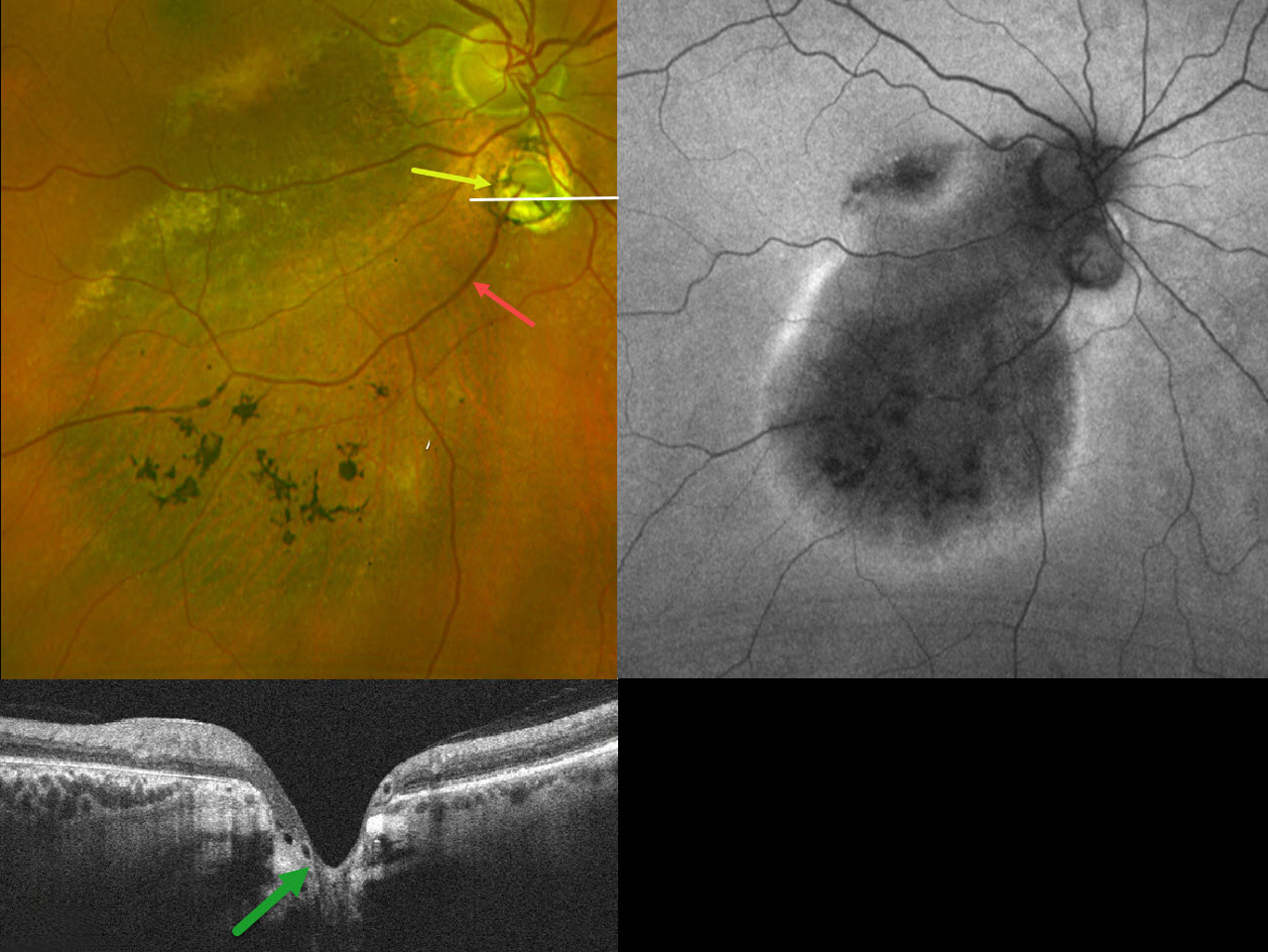
CHOROIDAL COLOBOMA WITH OPTIC NERVE PSEUDODUPLICATION
Originally posted on @retina.rocks October 8, 2025
This 60YO female was referred for asymptomatic unilateral fundus findings in her right eye. Vision was 20/30 OD.
Optos color RG imaging shows a choroidal coloboma just inferior to the disc, giving the appearance of a pseudoduplicated optic nerve. Coarse pigmentary changes with some pigment migration extend from the macula into the inferior midperiphery. An inferotemporal retinal vein drains directly into the choroid through the coloboma. Triton swept-source OCT shows that the coloboma overlies an area of ectatic sclera. Fundus autofluorescence (FAF) shows variable hypo-FAF within the area of clinical pigmentary changes, with a surrounding rim of hyper-FAF.
Learning Points:
Choroidal colobomas are most often due to failure of the optic vesicle and choroidal fissure to close during fetal development. Lesions like this may also be from other causes, including toxoplasmosis. Choroidal coloboma can present as solitary or multiple lesions. Visual prognosis depends on the location and involvement of the optic nerve and macula.
Pseudoduplication of the optic nerve occurs when a round choroidal coloboma or chorioretinal scar about the size of the optic nerve is associated with overlying radiating blood vessels (Bloom et al, Retinal Cases 2022;16:174-176). In our experience, it is most common for toxoplasmosis scars to be located near the optic nerve.
Under normal conditions, the retinal and choroidal circulations remain separate and distinct. Rarely, the two circulations connect directly through a chorioretinal anastomosis. These can occur in chorioretinal scars (typically from inactive toxoplasmosis lesions), in macular telangiectasia, and in disciform scars.
The prominent gutter of inferior pigmentary changes indicates a prior retinal detachment, most likely related to spontaneously resolved fluid associated with the coloboma (Tanaka et al, Ophthalmology Retina 2021;5:702-710). Similar pigmentary changes are seen in central serous retinopathy, although our patient’s macular OCTs showed no pachychoroid features (not shown). Finally, this may represent previously resolved pit-like macular fluid, which is rarely found in the absence of a pit or severe glaucomatous cupping (Fujimoto et al, Ophthalmology Retina 2023;7:811-818).
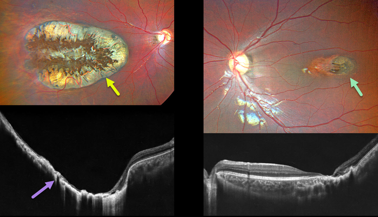
CHOROIDAL COLOBOMA
Ayushi Gupta and Vishal Agrawal
Originally posted on @retina.rocks August 4, 2025
This 7YO healthy boy has a history of esotropia in his amblyopic right eye. Vision was light perception OD and 20/40 OS.
Color photography of his right eye shows a large macular coloboma occupying nearly the entire macula. OCT scanning shows near complete atrophy and disorganization of the overlying RPE and neurosensory retina. A much smaller lesion is noted in the left temporal macula. Observation was recommended.
Learning Points:
Choroidal colobomas are most commonly caused by failure of the optic vesicle and choroidal fissure to close during embryonic development and are usually found inferonasally. They can present as solitary or multiple lesions. The differential diagnosis for congenital macular coloboma also includes post-inflammatory lesions (toxoplasmosis, cytomegalovirus, and Zika) and retinal dystrophies, including North Carolina macular dystrophy.
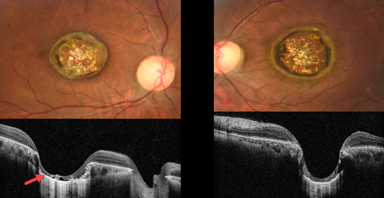
MACULAR COLOBOMA
Mauli Shah and Alay Banker
Originally posted on @retina.rocks May 6, 2025
This healthy 21YO male presented with a lifelong history of stable bilateral vision loss. There was no significant ocular family history, and ocular examination of family members revealed no abnormalities. Vision was 20/400 OU. There was bilateral horizontal nystagmus.
Color photography shows fairly symmetrical macular colobomas. Fine inner retinal striae radiate outwards from the coloboma OD. OCT scanning shows near-total choroidal loss within each coloboma, with a thinned, atrophic retina draped against bare sclera.
Learning Points:
Choroidal colobomas are most commonly due to failure of the optic vesicle and choroidal fissure to close during embryonic development and are usually found inferonasally. They can present as solitary or multiple lesions. Macular colobomas are most commonly either post-inflammatory (toxoplasmosis, cytomegalovirus, and Zika) or from an inherited retinal disorder (IRD), including North Carolina macular dystrophy (NCMD).
The symmetry of our patient’s lesions argues for an IRD. However, we doubt this is NCMD, given the lifelong history of poor vision and nystagmus. Due to financial constraints, the patient refused all blood work and genetic testing. It is
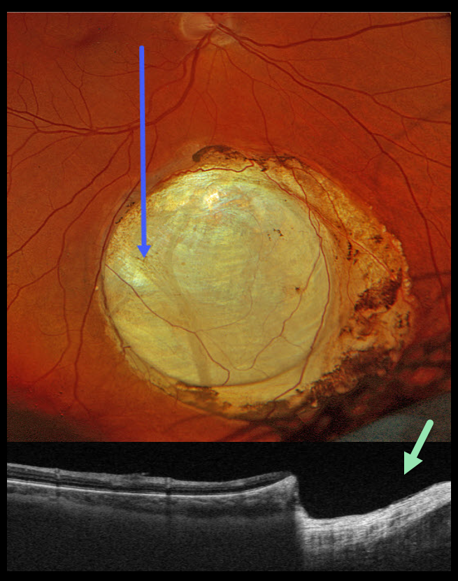
CHOROIDAL COLOBOMA
Originally posted on @retina.rocks April 8, 2025
This healthy 48YO male was referred for an asymptomatic lesion in his right eye. Vision was 20/20 OD and 20/20 in his healthy OS.
Optos color RGB imaging shows an oval choroidal coloboma in the inferior midperiphery. Variably pigmented chorioretinal atrophy surrounds the lesion. Triton swept-source OCT shows a fairly abrupt and near-complete loss of the neurosensory retina within the coloboma.
Learning Points:
Choroidal colobomas are most often due to failure of the optic vesicle and choroidal fissure to close during fetal development. Lesions like this may also be from other causes, including toxoplasmosis. Choroidal coloboma can present as solitary or multiple lesions. Visual prognosis depends on the location and involvement of the optic nerve and macula.
CHOROIDAL COLOBOMA
Evgenii Chernov
Originally posted on @retina.rocks March 18, 2025
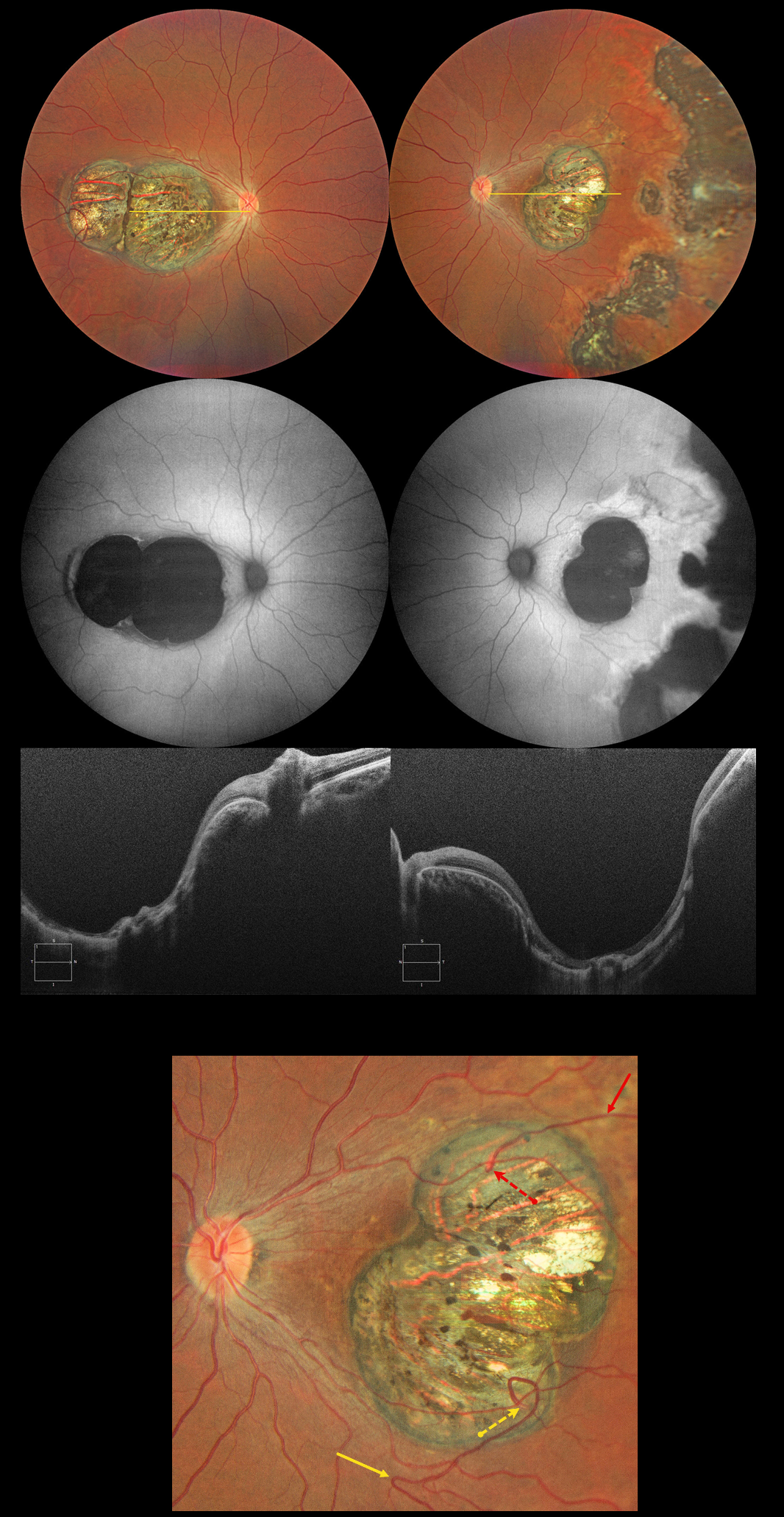
This 32YO female has a history of congenital nystagmus and poor vision since birth. Vision was 20/130 OU.
Color photography shows bilateral macular colobomas with more peripheral multifocal variably pigmented colobomatous scarring in her left eye. These lesions are hypoautofluorescent. OCT scanning through the macular lesions shows thinned hyperreflective retina draped across the posteriorly bowed sclera.
The right macular coloboma also contains several retinal choroidal anastomoses. The major inferotemporal vein acutely bends superotemporally to enter the coloboma and drain into the choroidal circulation. A superotemporal vein also enters the coloboma to drain into a visible choroidal vortex.
Learning Points:
Choroidal colobomas are most commonly caused by failure of the optic vesicle and choroidal fissure to close during embryonic development and are usually found inferonasally. They can present as solitary or multiple lesions. The differential diagnosis for congenital macular coloboma also includes post inflammatory lesions (toxoplasmosis, cytomegalovirus, and zika) and retinal dystrophies including North Carolina macular dystrophy. The retina within the coloboma is variably thinned, which increases the risk for rhegmatogenous retinal detachment.
Under normal conditions the retinal and choroidal circulations remain separate and distinct. Rarely, the two circulations directly connect with each other through a chorioretinal anastomosis. These can occur in chorioretinal scars (typically from inactive toxoplasmosis lesions), in macular telangiectasia, and in disciform scars.
CHOROIDAL COLOBOMA
Anand Temkar and Manish Nagpal
Originally posted on @retina.rocks January 16, 2025
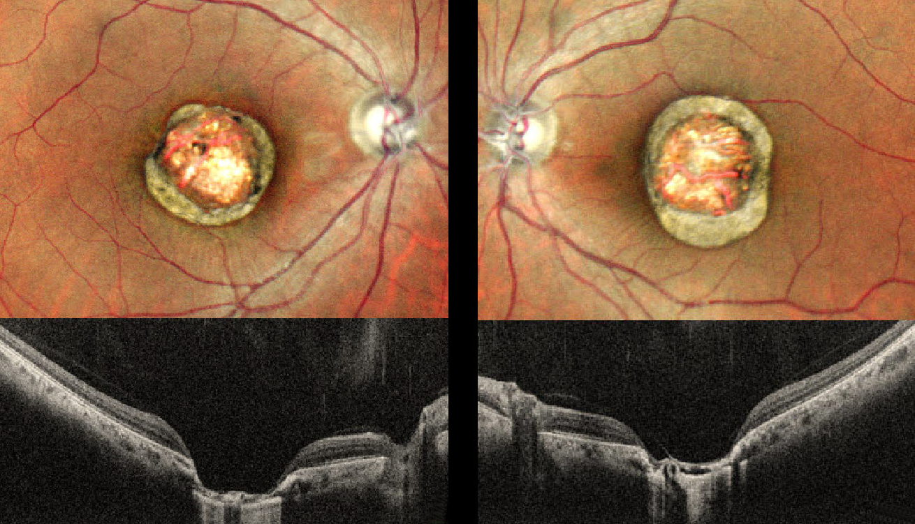
This healthy 24YO male presented with a lifelong history of stable bilateral vision loss. Vision was 20/80 OU.
Pseudocolor SLO imaging shows symmetrical macular colobomas. OCT scanning shows near-total choroidal loss within the coloboma, with a thinned, atrophic retina draped against bare sclera. Prior serologic testing elsewhere was negative for both toxoplasmosis and cytomegalovirus.
Learning Points:
Choroidal colobomas are most commonly due to failure of the optic vesicle and choroidal fissure to close during embryonic development and are usually found inferonasally. They can present as solitary or multiple lesions.
The differential diagnosis for congenital macular coloboma also includes post-inflammatory lesions (toxoplasmosis, cytomegalovirus, and Zika) and retinal dystrophies, including North Carolina macular dystrophy (NCMD). The retina within the coloboma is variably thinned, increasing the risk of rhegmatogenous retinal detachment (RRD).
The symmetry of the lesions argues for NCMD, but unfortunately, genetic testing was not performed due to cost.
RETINITIS PIGMENTOSA WITH MACULAR COLOBOMA
Ankit Jain and Manish Nagpal
Originally posted on @retina.rocks January 7, 2025
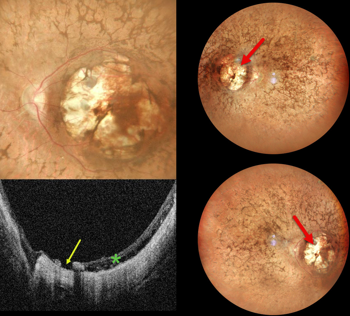
This 32YO female was born with severe vision loss and nystagmus. There was no family history of eye disease. Vision was counting fingers bilaterally.
Pseudocolor SLO imaging shows bilateral symmetric macular colobomas with diffuse retinitis pigmentosa (RP) findings, including vascular narrowing, diffuse pigmentary changes, and intraretinal pigment migration (bone spicules). OCT shows thinned and variably disorganized retinal bands within the colobomas, and a small macular hole is noted nasally OS.
Learning Points:
Macular colobomas are rarely associated with RP (Parmeggiani et al, Eye 2004;18:421-428). North Carolina macular dystrophy, caused by a mutation in the PRDM13 gene (Small et al, Ophthalmology 2016;123:9-18), is characterized by macular coloboma-like lesions. However, these eyes do not have peripheral RP findings. Genetic testing in our patient was not performed, and observation was recommended.
CHOROIDAL COLOBOMA
Kanwaljeet Harjot Madan
Originally posted on @retina.rocks October 30, 2024
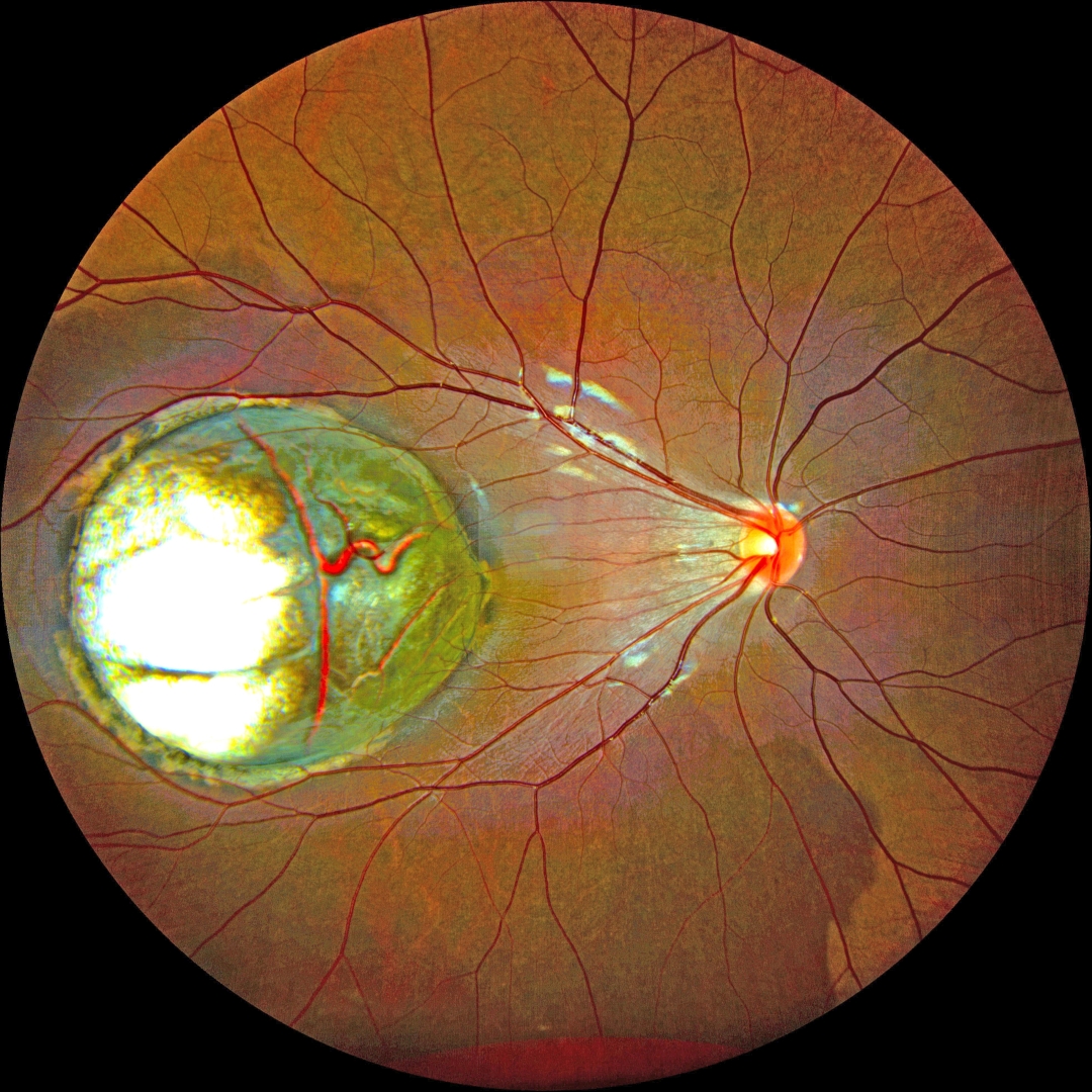
This healthy 5YO boy’s parents noticed that his right eye was wandering for a few weeks. Vision was 20/200 OD and 20/20 in the normal OS.
Color fundus photography shows a large choroidal coloboma occupying the central and temporal macula. The underlying outer choroidal vessels are visible. His systemic examination was normal. Serologic testing for toxoplasmosis and cytomegalovirus was negative.
Learning Points:
Choroidal colobomas are most commonly due to failure of the optic vesicle and choroidal fissure to close during embryonic development and are usually found inferonasally. They can present as solitary or multiple lesions. The differential diagnosis for congenital macular coloboma also includes post-inflammatory lesions (toxoplasmosis, cytomegalovirus, and Zika) and retinal dystrophies, including North Carolina macular dystrophy. The retina within the coloboma is variably thinned, increasing the risk of rhegmatogenous retinal detachment (RRD).
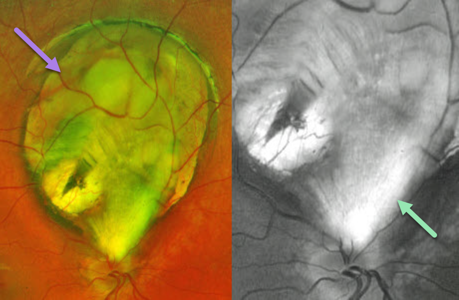
CHOROIDAL COLOBOMA + MYELINATED NERVE FIBER LAYER
Originally posted on @retina.rocks July 31, 2024
This healthy 8YO girl was referred for an asymptomatic retinal finding in her right eye. Vision was 20/30 in her right eye and 20/30 in her left eye. Anterior segments were normal.
Optos color RG imaging shows a superior choroidal coloboma that almost extends to the disc margin. An incidental patch of myelinated nerve fiber layer (NFL), best imaged in the green channel, extends from the superior nerve into the inferior aspect of the coloboma.
Learning Points:
Choroidal colobomas are caused by failure of the optic vesicle and choroidal fissure to close during fetal development. Choroidal coloboma can present as solitary or multiple lesions. Visual prognosis depends on the location and involvement of the optic nerve and macula. We will follow our patient yearly due to her increased risk of retinal detachment.
Myelinated NFL is a relatively uncommon and usually isolated finding present at birth. Most patients are asymptomatic, although some may have a corresponding relative scotoma. Myelination can progress in about 10% of cases and can also resolve following retinal ischemic events. Myelinated NFL occurs when retinal nerve fibers develop a myelin sheath, which usually stops posterior to the lamina cribrosa. The myelination may represent an oligodendrocytic choristoma (see Rao et al Retina 2019;39:1125-1132).
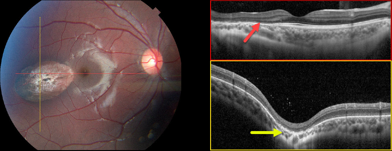
CHOROIDAL COLOBOMA
Neeket Patel
Originally posted on @retina.rocks March 18, 2024
This 12YO female complained of several years of daily frontal headaches associated with bilateral blurriness. Vision was 20/25 OU.
Color photography of her right eye shows an oval depigmented lesion in the temporal macula. Horizontal B-scan OCT shows outer retinal atrophy overlying the lesion. In the vertical B-scan, the lesion is colobomatous with outer retinal thinning. Some hyperreflective vitreous cells are noted. The left fundus was normal.
Learning Points:
Torpedo maculopathy was originally described by Gass as a solitary hypopigmented RPE nevus (Arch Ophthalmology 1992;110:1358-1359). These benign, unilateral lesions are tear-drop shaped and almost always located in the horizontal meridian in the temporal macula.
OCT scanning shows attenuation of the RPE and outer retinal layers. Vision is almost always normal, although there is a small risk for macular neovascularization. There are no known systemic associations.
Choroidal colobomas are most commonly due to failure of the optic vesicle and choroidal fissure to close during fetal development. Lesions like this may also be from other causes, including toxoplasmosis.
Although our patient’s lesion strongly resembles torpedo maculopathy, the coloboma would be highly atypical. Our best guess, given the vitreous cells, is that this lesion is due to toxoplasmosis.
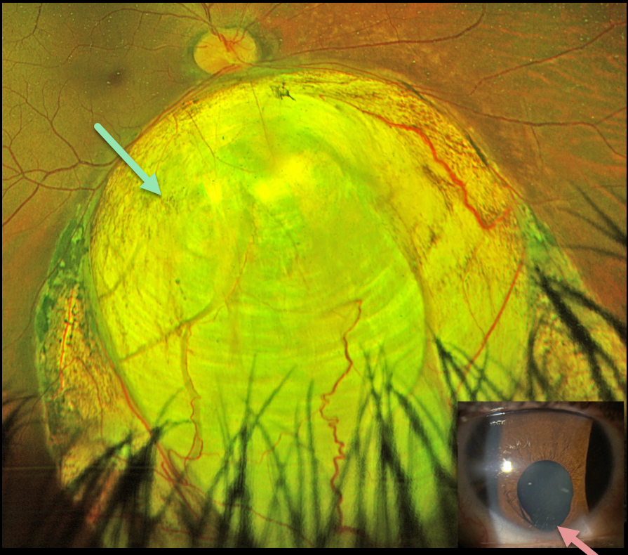
CHOROIDAL COLOBOMA
Austin Eckel
Originally posted on @retina.rocks December 14, 2023
This healthy 25YO female’s primary care doctor noticed an unusual finding in her right eye and referred her to us for an examination. She said that her right pupil had looked different.
Optos color RG imaging shows a large inferior choroidal coloboma extending from the inferior disc margin into the retinal periphery. An inferior iris coloboma is noted.
Learning Points:
Choroidal colobomas are caused by failure of the optic vesicle and choroidal fissure to close during fetal development. Choroidal coloboma can present as solitary or multiple lesions, as in this case. Visual prognosis depends on the location and involvement of the optic nerve and macula.
We will follow our patient yearly due to her increased risk of retinal detachment and expect her to do well, given the normal central macular appearance.
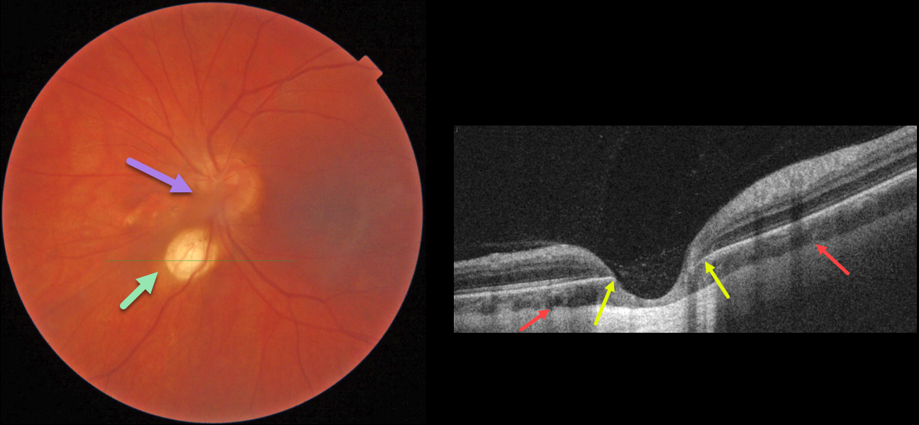
CHOROIDAL COLOBOMA + PERSISTENT FETAL VASCULATURE
Lesia Khrystych
Originally posted on @retina.rocks May 22, 2023
This 35YO female has a history of decreased vision in her left eye since childhood. Vision was 20/20 in her right eye and stable at 20/200 in her left eye. Anterior segments were normal except for a posterior polar cataract OS.
Color imaging of the left posterior pole shows a fairly round, one-disc-diameter depigmented lesion just inferonasal to the disc. An area of non-specific pigmentary changes is noted just nasal to the nerve, along with persistent fetal vasculature (hyaloid artery).
OCT through the depigmented lesion shows a choroidal coloboma. The underlying sclera is intact and slightly bowed posteriorly. Within the coloboma, there is loss of the outer retinal layers with draping of the inner retina against the bare sclera. The RPE is also absent within the coloboma.
Learning Points:
Choroidal colobomas result from a failure of closure of the optic vesicle and choroidal fissure during normal fetal development, but can also be caused by congenital toxoplasmosis and Zika virus. They can be solitary or multiple, and the visual potential depends on the extent of optic nerve and macular involvement. Patients with choroidal colobomas are at an increased risk for retinal detachment and macular neovascularization.
Embryologically, the hyaloid artery is vital to the proper development of the anterior segment, and usually regresses at about 10 weeks of gestation; however, it may persist in some cases. In severe cases, the eye may be microphthalmic with leukocoria and retinal traction. These eyes are typically amblyopic. A genetic association has not been identified.
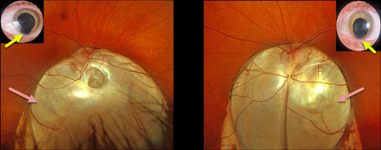
CHOROIDAL COLOBOMA
Originally posted on @retina.rocks May 15, 2023
This 30YO female presented with a 6-month history of vision loss in her right eye. Despite being born with bilateral iris and choroidal colobomas, her vision in each eye was excellent until recently. Vision was 20/200 OD and 20/40 OS.
Optos RGB imaging shows bilateral inferior choroidal colobomas extending from just superior to the optic nerves towards the inferior peripheries. A shallow macula-off retinal detachment extends from the temporal edge of the right coloboma into the 7 to 10 o’clock periphery. In the office, we could not identify any retinal breaks. At vitrectomy, a small anterior retinal tear was found at the superior edge of the detachment.
Learning Points:
Choroidal colobomas are caused by failure of the optic vesicle and choroidal fissure to close during fetal development. Choroidal coloboma can present as solitary or multiple lesions, as in this case. Visual prognosis depends on the location and involvement of the optic nerve and macula.
The retina within the coloboma is variably thinned, increasing the risk of rhegmatogenous retinal detachment (RRD). More centrally within the coloboma, the retina consists of a monolayer of atrophic glial structures and neurons, known as the intercalary membrane (ICM). Between the edge of the coloboma and the ICM is a region of thinned retina lacking RPE and choroid, the marginal ICM (MICM). Breaks within the MICM, which extend about 2 disc diameters from the coloboma margin, are usually the cause of RRD (Tanaka et al, Ophthalmology Retina 2021;5;702-710).
Fortunately, our patient had a more typical RRD that was independent of her coloboma. Laser treatment along the edge of the coloboma was therefore not performed, thereby avoiding the risk of iatrogenic paracentral vision loss. The retina remained attached 5 weeks postoperatively, and vision improved to 20/100.
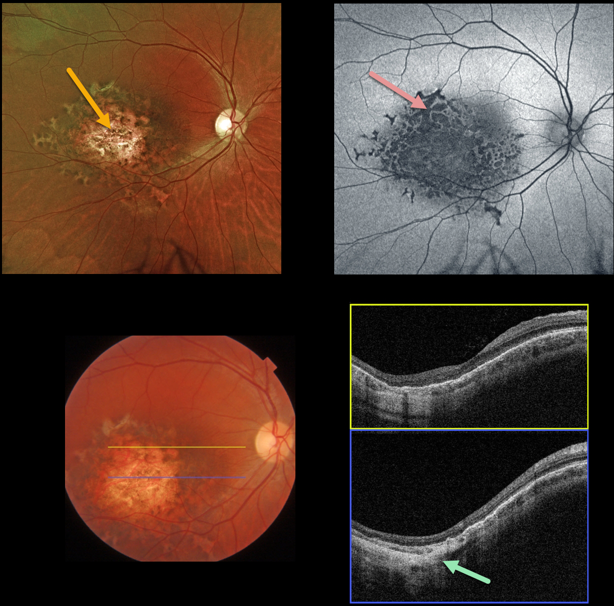
RPE DYSGENESIS WITH CHOROIDAL COLOBOMA
Originally posted on @retina.rocks October 31, 2022
This healthy 32YOF was referred for an asymptomatic lesion in her right macula. Vision was 20/30 OU, and her left fundus was normal.
Optos color imaging shows an atrophic, variably pigmented macular colobomatous lesion with a surrounding rim of scalloped pigmentation.
Fundus autofluorescence (FAF) shows variable, mostly hypo-FAF, indicating RPE dysfunction/loss.
Swept source OCT scanning shows outer retinal/RPE thinning with a colobomatous/staphylomatous outpouching.
Learning Points:
Retinal pigment epithelium dysgenesis (RPED) was first described by Cohen et al (Arch Ophthalmol 2002;120:512-516) as a unilateral, idiopathic lesion affecting the RPE and outer retina. These rare lesions have a pathognomonic scalloped margin with associated RPE atrophy and fibrosis.
Although initially described as a unilateral condition, it can also appear bilaterally (Renz et al, Arch Ophthalmol 2012;130:1341-1340). The lesions can enlarge with time and also carry a small risk for secondary macular neovascularization (Shimoyama et al, Case Rep Ophthalmol 2014;5:34-37) and presumed RPE tumor (Gal-Or et al, Retinal Cases & Brief Reports 2019;13:121-126).
To the best of our knowledge, our patient’s lesion is unique by the colobomatous appearance which has not been previously described.
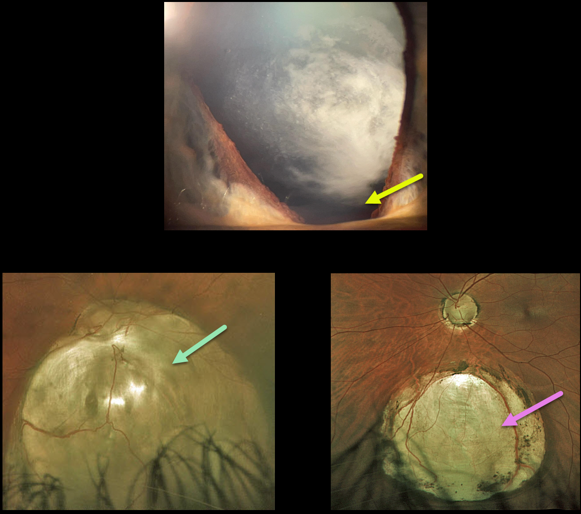
CHOROIDAL + IRIS COLOBOMA
Originally posted on @retina.rocks July 5, 2022
This 42YO state trooper presented with a few months of vision loss in his right eye, including significant glare with headlights. He reported a history of excellent vision throughout his life. Vision was 20/200 OD and 20/20 OS.
He had 3+ nuclear/cortical spoke cataracts consistent with this vision loss. Slit lamp examination shows an inferior iris coloboma OD, and it was normal OS.
Optos imaging of his right eye shows an inferior choroidal coloboma extending from the nerve inferiorly. The macula is normal, and OCT through the macular center is also normal (not shown). A smaller inferior midperipheral choroidal coloboma is noted in his left eye.
Cataract surgery is scheduled, and we expect his vision to return to normal, given his prior history and normal macular anatomy.
Learning Points:
Choroidal colobomas are most commonly due to failure of the optic vesicle and choroidal fissure to close during fetal development. Lesions like this may also be from other causes, including toxoplasmosis. Choroidal coloboma can present as solitary or multiple lesions. Visual prognosis depends on the location and involvement of the optic nerve and macula.
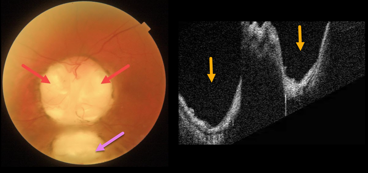
OPTIC NERVE COLOBOMA
Originally posted on @retina.rocks May 9, 2022
This 66YO male has mild lifelong decreased vision in his left eye. Vision was 20/70.
Triton imaging of the optic nerve coloboma shows optic nerve pits temporally and nasally, as well as a more inferior choroidal coloboma. Swept-source OCT dramatically reveals the pits.
We are following him yearly.
Learning Points:
Both optic nerve and choroidal colobomas can be unilateral or bilateral and are caused by incomplete closure of the embryonic fissure.
Patients with choroidal colobomas are at increased lifetime risk of rhegmatogenous retinal detachment due to breaks in the overlying thinned retina.
Optic nerve pits are also associated with serous macular detachment and macular neovascularization.
For a great review of cavitary optic nerve abnormalities, see Jain and Johnson, AJO 2014;158:423-435.
CHOROIDAL COLOBOMA
Originally posted on @retina.rocks September 10, 2021
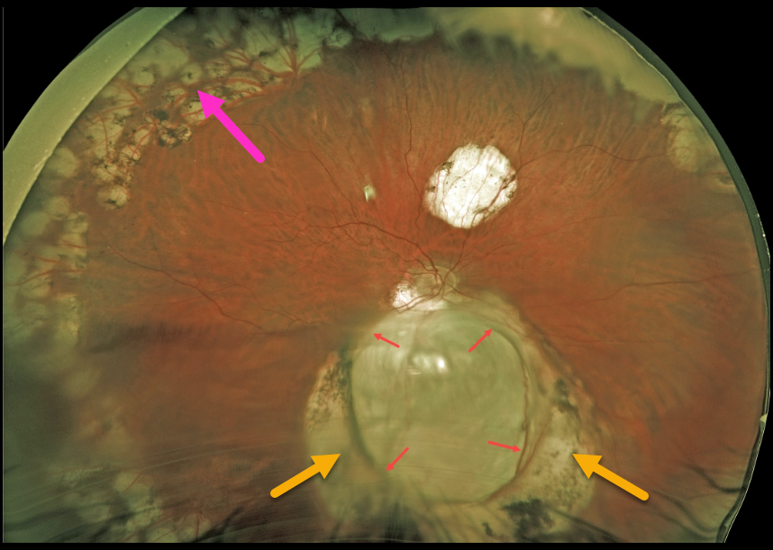
This 57YO male underwent successful vitrectomy surgery 8 years earlier for rhegmatogenous retinal detachment (RRD). Vision is now 20/40.
The detachment was caused by a retinal break associated with an inferior choroidal coloboma. The margins of the coloboma are surrounded by confluent depigmented scarring from prior laser photocoagulation. Prophylactic peripheral laser was also applied intraoperatively.
Learning Points:
Choroidal colobomas arise from incomplete closure of the embryonic fissure during the second month of embryonic development.
Patients are at lifelong risk of rhegmatogenous retinal detachment due to breaks in the overlying thinned retina.
TOXOPLASMOSIS
Originally posted on @retina.rocks August 12, 2021
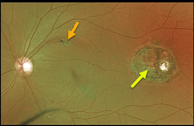
This 44YO male presented with 20/25 vision and a presumed inactive toxoplasmosis scar in the distal left macula. Although we do not have an OCT through this scar, it likely represents a colobomatous lesion.
This lesion somewhat resembles torpedo maculopathy, but the additional scar in the superior macula more suggests a prior inflammatory event.
Learning Points:
For a great discussion on how to differentiate these infectious macular colobomatous lesions (congenital toxoplasmosis, Zika, and cytomegalovirus) from inherited dystrophies like North Carolina Macular Dystrophy (NCMD), see Kumar and Mahalingam AJO 2019;200;47-56.
CHOROIDAL COLOBOMA
Originally posted on @retina.rocks May 24, 2021
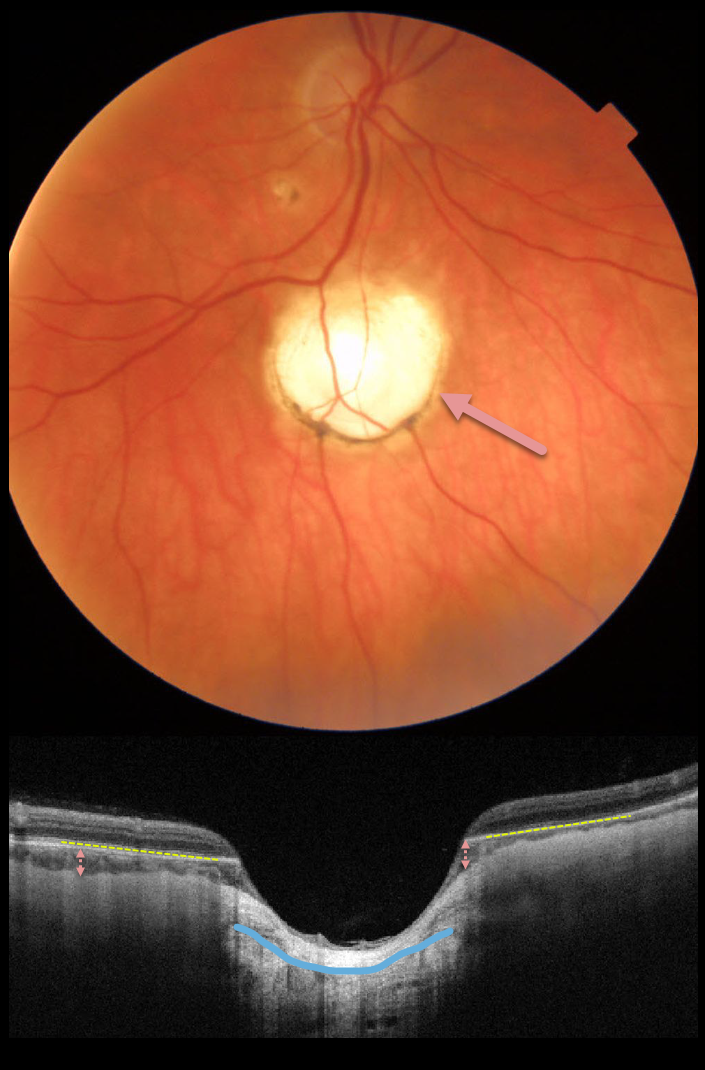
This 57yo female presented with an asymptomatic, unilateral choroidal coloboma inferior to the right optic nerve.
Triton swept-source OCT shows a total loss of the RPE and a near-complete absence of the choroid within the coloboma, with marked thinning of the overlying retina. There is also an outpouching of the underlying sclera.
Learning Points:
Choroidal colobomas are most commonly due to failure of the optic vesicle and choroidal fissure to close during fetal development. Lesions like this may also be from other causes, including toxoplasmosis. Choroidal coloboma can present as solitary or multiple lesions. Visual prognosis depends on the location and involvement of the optic nerve and macula.
TOXOPLASMOSIS
Originally posted on @retina.rocks March 26, 2021
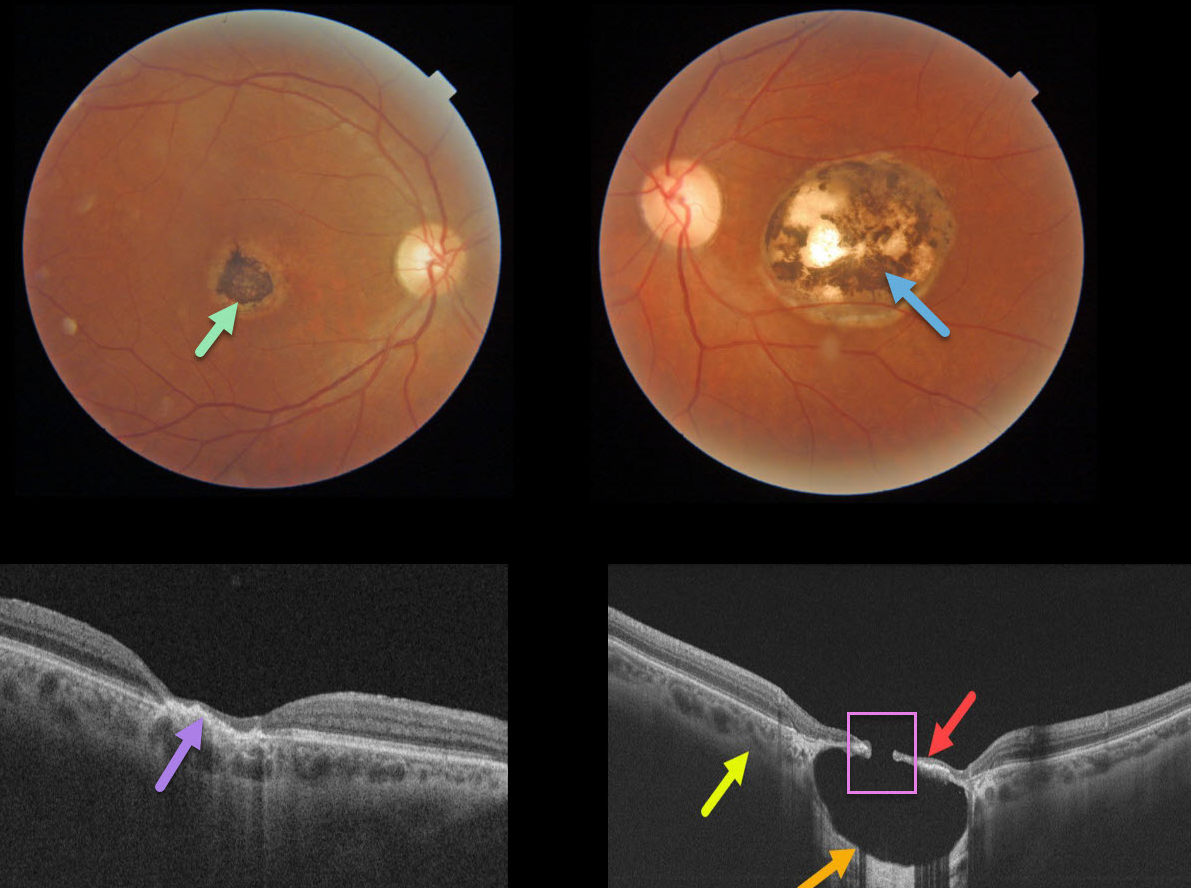
This 57YO male gave a history of lifelong poor vision in his left eye. Vision was 20/40 OD and 20/200 OS.
A small hyperpigmented foveal scar is noted in his right eye with neurosensory retinal atrophy and irregular elevation of the RPE.
A larger, round, variably pigmented, excavated macular scar is noted in his left eye. Triton swept-source OCT reveals a thinned atrophic retina and intact RPE suspended over an optically empty hyporeflective excavated colobomatous type lesion. The plane of the intact surrounding sclera is indicated by the yellow arrow. A small full-thickness retinal and RPE defect is noted centrally.
Learning Points:
For a great discussion on how to differentiate these infectious macular colobomatous lesions (congenital toxoplasmosis, Zika, and cytomegalovirus) from inherited dystrophies like North Carolina Macular Dystrophy (NCMD), see Kumar and Mahalingam AJO 2019;200;47-56.
In case you’re thinking our patient has NCMD, also see the first OCT description of congenital toxoplasmosis macular lesions (Garg et al, Retina 2009;29;631-637). Their Figures 4 and 6 look eerily similar to our patient’s right and left eyes!
TOXOPLASMOSIS
Originally posted on @retina.rocks January 12, 2021
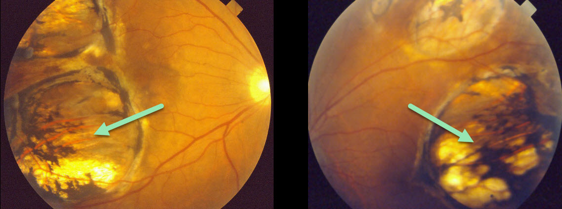
This patient presented in 1990 with what we assumed was bilateral inactive colobomatous congenital toxoplasmosis scarring.
Learning Points:
Choroidal colobomas are most commonly due to failure of the optic vesicle and choroidal fissure to close during fetal development. Multifocal lesions, as seen in our patient, can be caused by congenital toxoplasmosis and Zika.
For a comprehensive review of ocular coloboma, see Onwochei et al Surv Ophthalmol 2000;45:175-194.
CHOROIDAL COLOBOMA
Originally posted on @retina.rocks December 17, 2020
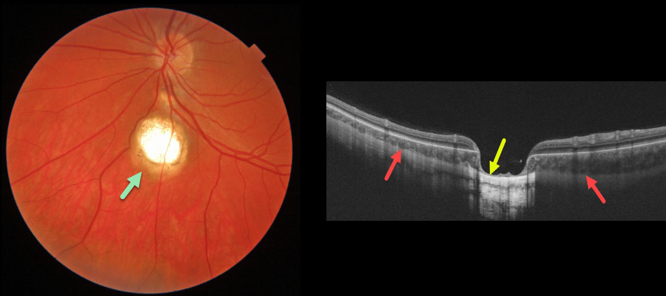
This 25yo was referred for this asymptomatic choroidal coloboma.
A B-scan OCT through our patient’s coloboma shows that the sclera is continuous and non-displaced within the coloboma.
A staphyloma, by definition, is an outpouching of the entire eye wall. Choroidal coloboma always involves a choroidal defect, but may or may not also involve the sclera.
Learning Points:
Choroidal colobomas are most commonly due to failure of the optic vesicle and choroidal fissure to close during fetal development. Lesions like this may also be from other causes, including toxoplasmosis. Choroidal coloboma can present as solitary or multiple lesions. Visual prognosis depends on the location and involvement of the optic nerve and macula.
CHOROIDAL COLOBOMA
Originally posted on @retina.rocks October 26, 2020
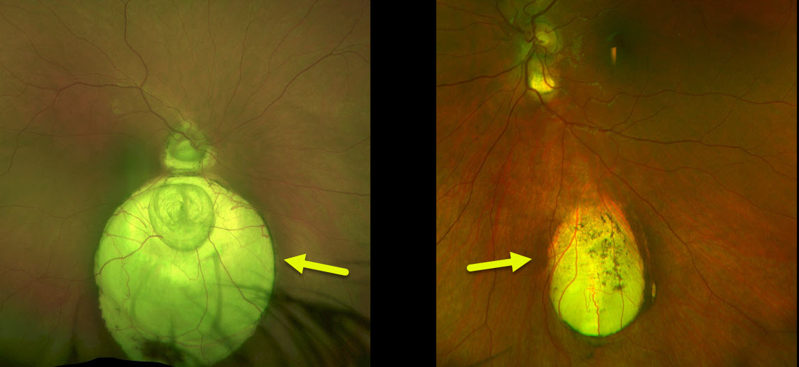
Our asymptomatic patient is being followed yearly for bilateral choroidal colobomas.
These patients are at risk for retinal detachment and choroidal neovascularization. Choroidal neovascularization has an increased tendency to develop at the superotemporal edge of the coloboma, which is thought to be caused by the abnormal architecture of the RPE and Bruch’s membrane.
Learning Points:
Choroidal colobomas are caused by failure of the optic vesicle and choroidal fissure to close during fetal development. Choroidal coloboma can present as solitary or multiple lesions as in this case. Visual prognosis depends on the location and involvement of the optic nerve and macula.
CHOROIDAL COLOBOMA
Originally posted on @retina.rocks July 30, 2020
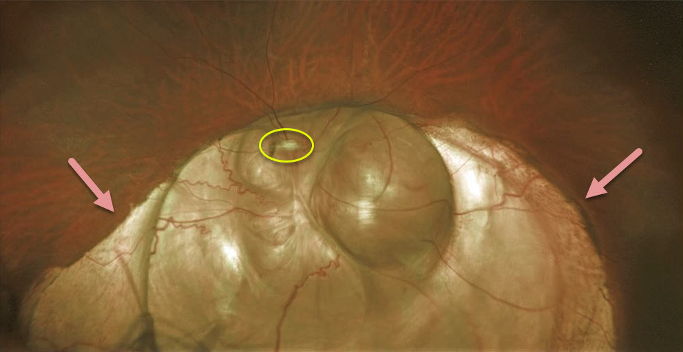
This patient has a large choroidal coloboma extending through the left optic nerve and macula. It’s so extensive that it hides the optic nerve.
These arise from incomplete closure of the embryonic fissure during the second month of embryonic development. Patients are at lifelong risk for rhegmatogenous retinal detachment due to breaks that occur in the overlying thinned retina.
Learning Points:
Defects in closure have a wide range of results such as microphthalmia, anophthalmia, iris coloboma, ciliary body coloboma, choroid coloboma, etc.
Colobomas of the iris or ciliary body result from failure of anterior closure while choroid, retina and optic nerve result from failure of posterior closure.
A staphyloma by definition is an outpouching of the entire eye wall. Choroidal coloboma, always involves a choroidal defect, but may or may not also involve the sclera (as in this case).


