

Combined Hamartoma of the Retina and RPE
<< Back to Cases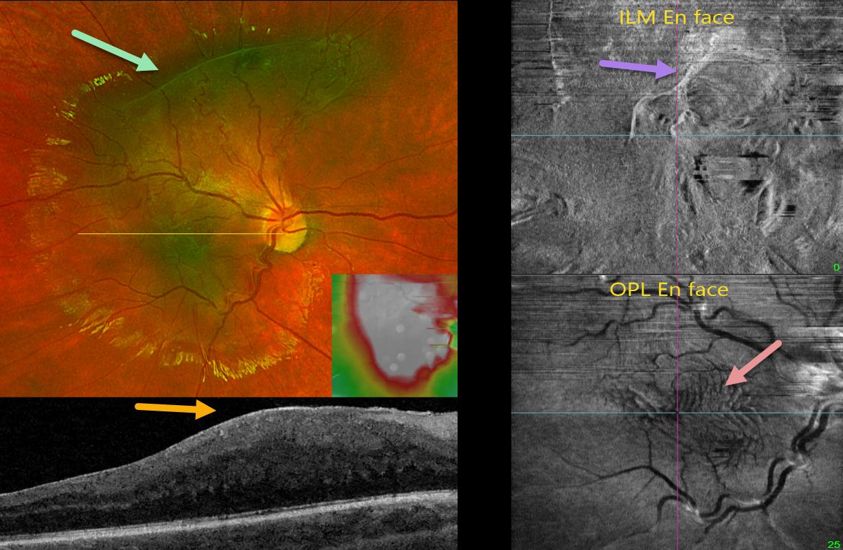
MACULAR PUCKER
Originally posted on @retina.rocks May 29, 2024
This healthy 9YO boy was referred for asymptomatic retinal changes in his right eye. Vision was 20/25 OU. The left eye was normal.
Optos color RG imaging shows a greenish hyperpigmented lesion in the macula extending into the superior midperiphery. Diffuse macular thickening is noted on swept-source OCT. En face imaging offers another multimodal perspective. En face of the internal limiting membrane/vitreous interface shows the epimacular fibrosis, while en face of the outer plexiform layer shows variable fingerprint-like whorls centrally.
Learning Points:
In 2017, Govetto et al described an OCT finding of ectopic inner foveal layers (EIFL) associated with epiretinal membranes (AJO 2017;175:99-113). The findings range from mild underlying retinal changes (stage 1, thin ERM with foveal depression), widening of the ONL and loss of foveal depression (stage 2), and ERM continuous with EIFL crossing the entire foveal area (stage 3). All retinal layers are clearly visualized in stages 1 through 3. In stage 4, the ERM is thick, and all retinal layers are disrupted. Vision is worse with increasing stage severity. It is also associated with worse postoperative visual recovery (Yang et al, Retina 2022;42:1472-1478).
The ‘fingerprint sign’ is a unique en face OCT finding described by Griffin et al (Retina 2021;41:381-386) that appears as concentric, fingerprint-like waves of Henle’s layer underlying epiretinal membranes. The authors found that the undulating OPL creates hyperreflective lines when oriented perpendicular to the incident OCT beam and hyporeflective lines when parallel to it.
Combined hamartomas are classically described as benign congenital lesions composed of glial cells, vascular tissue, and pigmented retinal pigment epithelial cells. However, more recent data indicate that these lesions primarily arise from the inner retinal layers (Chawla et al, AJO 2017;181:88-96).
Hamartomas and severe macular puckers can look very similar, even to some of the most seasoned retina specialists. OCT may be helpful in differentiating these entities (Ophthalmic Surg Lasers Imaging Retina 2017;48;122-125). In our experience, we feel that many ‘combined hamartomas’ described in the literature are most likely severe macular pucker with ectopic inner retina layers as in our case.
MACULAR PUCKER
Originally posted on @retina.rocks October 23, 2023
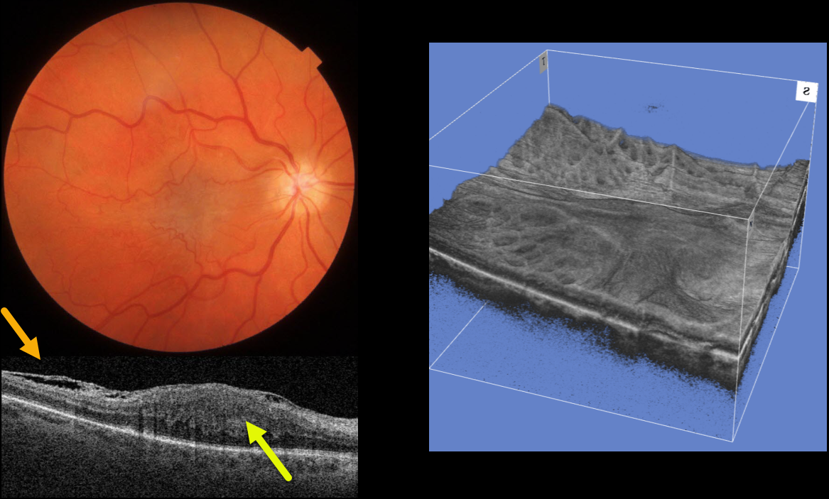
This 64YO female was referred for an asymptomatic macular pucker in her right eye. Vision was 20/200 OD and 20/20 OS.
Color imaging shows a severe central macular pucker. Triton swept-source OCT shows a variably adherent epiretinal membrane (ERM) with inner retinal hyperreflectivity and thickening, and complete loss of the normal inner retinal structures.
A 3D OCT reconstruction offers another view of the inner macular appearance, with unusual radiating dimples more peripherally.
Learning Points:
In 2017, Govetto et al described an OCT finding of ectopic inner foveal layers (EIFL) associated with epiretinal membranes (AJO 2017;175:99-113). The findings range from mild underlying retinal changes (stage 1, thin ERM with foveal depression), widening of the ONL and loss of foveal depression (stage 2), and ERM continuous with EIFL crossing the entire foveal area (stage 3). All retinal layers are clearly visualized in stages 1 through 3.
In stage 4, the ERM is thick, and all retinal layers are disrupted. Vision is worse with increasing stage severity. It is also associated with worse postoperative visual recovery (Yang et al, Retina 2022;42:1472-1478).
Combined hamartomas are classically described as benign congenital lesions composed of glial cells, vascular tissue, and pigmented retinal pigment epithelial cells. However, more recent data indicate that these lesions primarily arise from the inner retinal layers (see Chawla et al AJO 2017;181:88-96).
Combined hamartomas and severe macular puckers can look very similar, even to some of the most seasoned retina specialists. In our opinion, many cases of combined hamartomas in the literature most likely represent severe ERMs with EIFL.
Our patient has stage 4 EIFL. We recommended observation since she is binocularly asymptomatic.
COMBINED HAMARTOMA OF THE RETINA AND RPE
Originally posted on @retina.rocks December 30, 2020
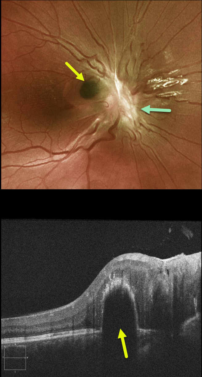
This case is either a combined hamartoma of the retina and RPE or an unusual macular pucker centered over the optic nerve. There is contracted fibrotic tissue overlying the optic nerve
We don’t know why a pigmented polyp-like RPE detachment developed, which was not present when we initially saw him about 5 years earlier.
Learning Points:
Combined hamartomas are classically described as benign congenital lesions composed of glial cells, vascular tissue, and pigmented retinal pigment epithelial cells. However, more recent data indicate that these lesions primarily arise from the inner retinal layer.
COMBINED HAMARTOMA OF THE RETINA AND RPE
Originally posted on @retina.rocks August 25, 2020
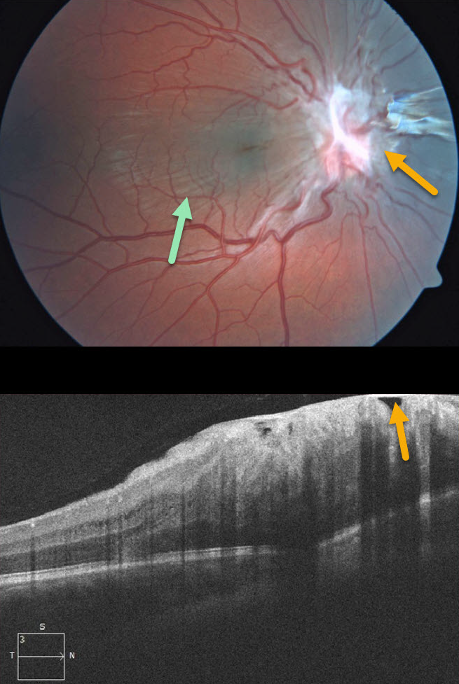
This 7YO boy presented without visual symptoms and 20/50 vision OD. The left eye was normal.
Contracted fibrotic tissue overlying the optic nerve is causing distortion of the surrounding retina, with striae visible through the macula. Some of this fibrosis may also be pigmented nasal to the nerve.
This case represents either a combined hamartoma of the retina and RPE or an unusual pucker centered over the optic nerve.
Learning Points:
Combined hamartomas are classically described as benign congenital lesions composed of glial cells, vascular tissue, and pigmented retinal pigment epithelial cells. However, more recent data indicate that these lesions primarily arise from the inner retinal layers.
MACULAR PUCKER (PSEUDO-COMBINED HAMARTOMA)
Originally posted on @retina.rocks June 25, 2020
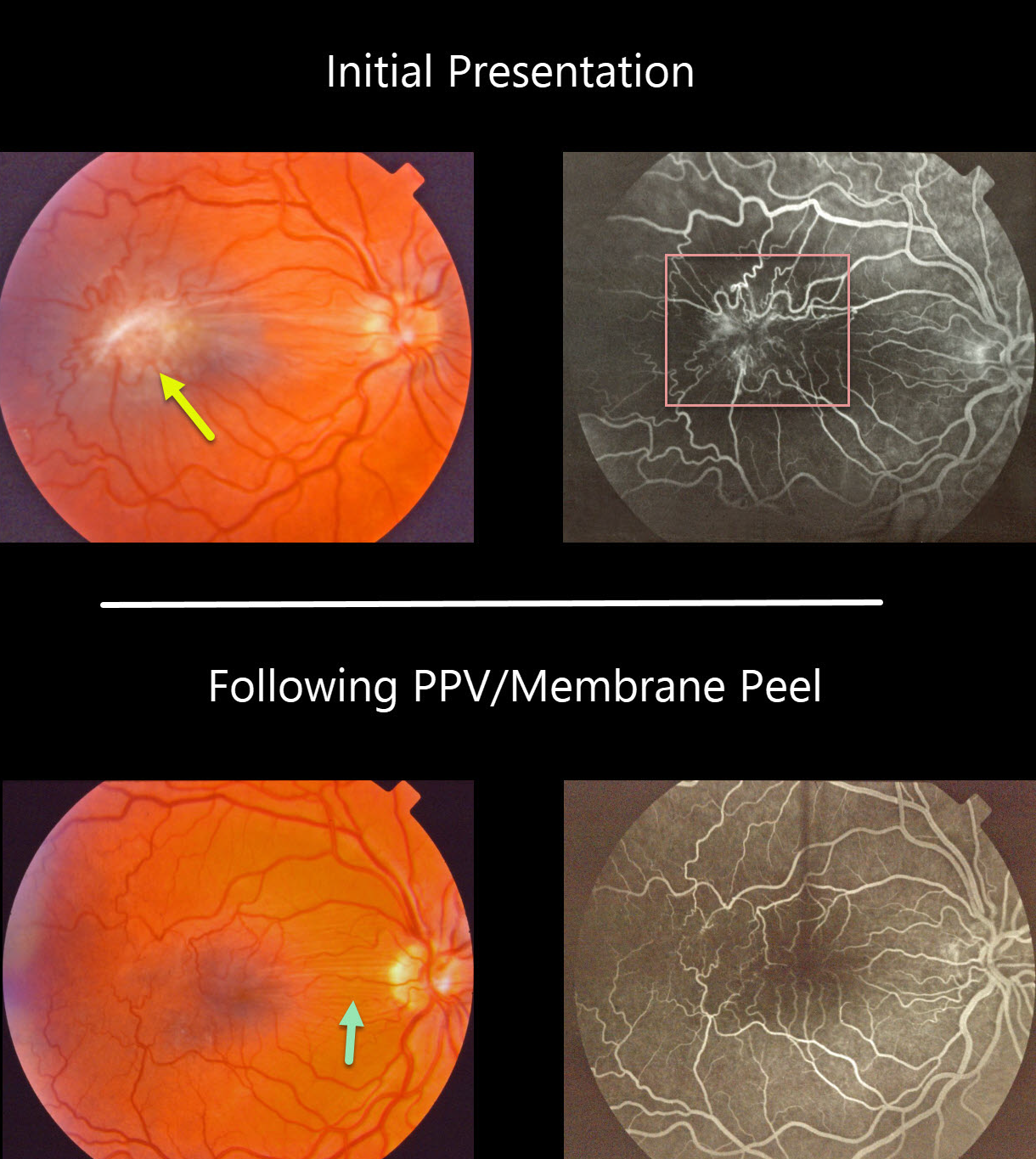
This patient had an isolated severe macular pucker. The fluorescein angiogram shows a vascular hamartomatous component due to severe retinal vascular distortion and leakage. This vascular component, along with the pigmented appearance of the pucker, helps give the false appearance that this was a combined hamartoma of the RPE.
The macular pucker was treated with vitrectomy and membrane peeling. Postoperatively, the retina appears nearly normal, with some residual peripapillary retinal folds and no vascular leakage.
Learning Points:
Hamartomas and severe macular puckers can look very similar, even to some of the most seasoned retina specialists. OCT may be helpful in differentiating these entities (see Ophthalmic Surg Lasers Imaging Retina 2017;48;122-125).
MACULAR PUCKER VS COMBINED HAMARTOMA
Originally posted on @retina.rocks May 25, 2020
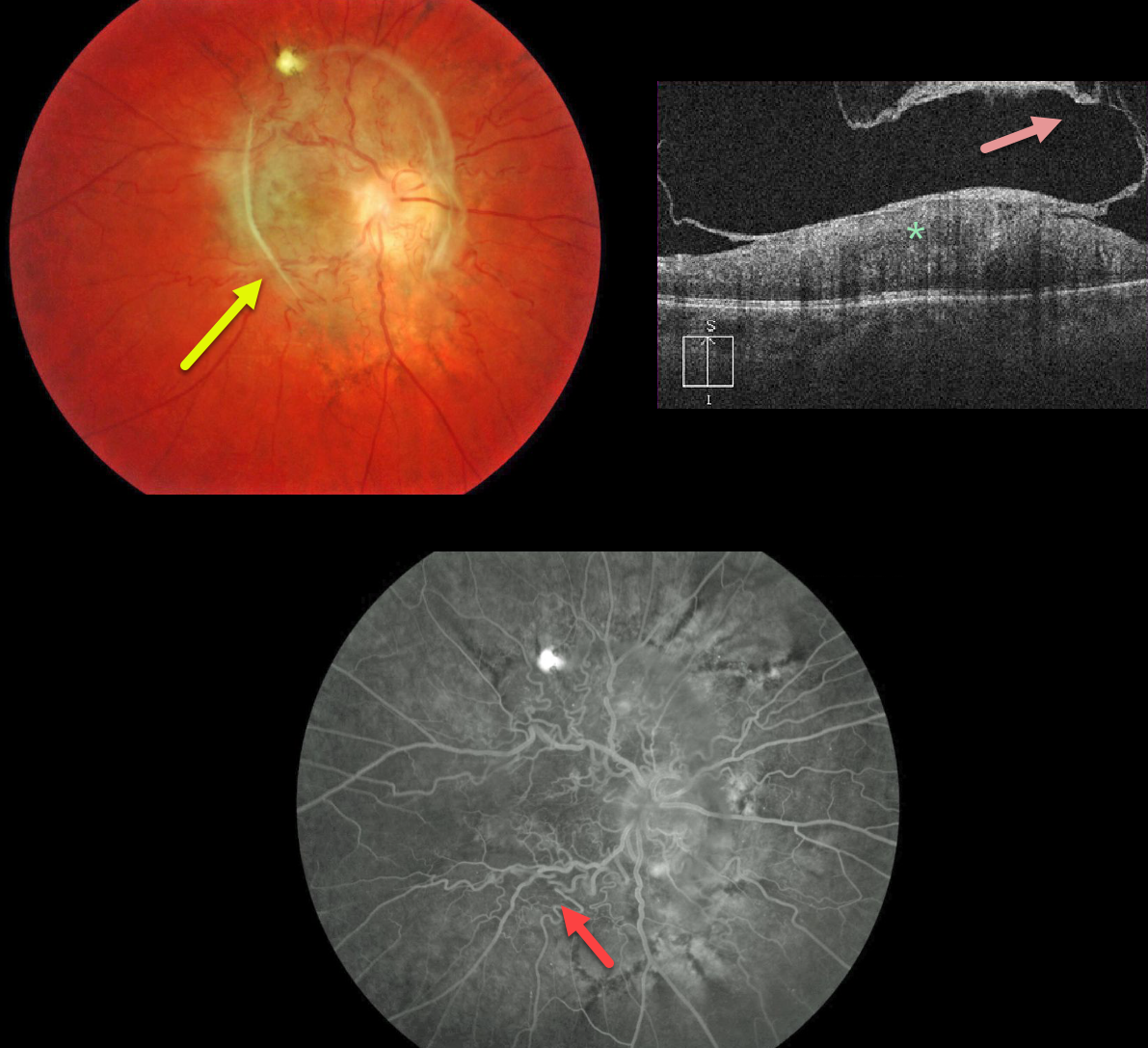
The fundus photo shows an unusual peripapillary pigmented epiretinal membrane distorting the underlying retina.
The OCT shows a partially detached epiretinal membrane and marked hyperreflectivity of the inner retina, with loss of normal retinal layer detail.
Fluorescein angiography shows a distorted retinal vasculature with late leakage.
Occasionally, vitrectomy with membrane peeling can be offered, but we are electing to observe this eye since vision has been poor since birth.
Learning Points:
A combined hamartoma of the RPE is a benign congenital lesion composed of glial cells, vascular tissue, and pigmented epithelial cells.
Even though we’re calling this a combined hamartoma, we’re not positive that this may not just be an unusual peripapillary/macular epiretinal membrane.


