

Commotio Retinae
<< Back to Cases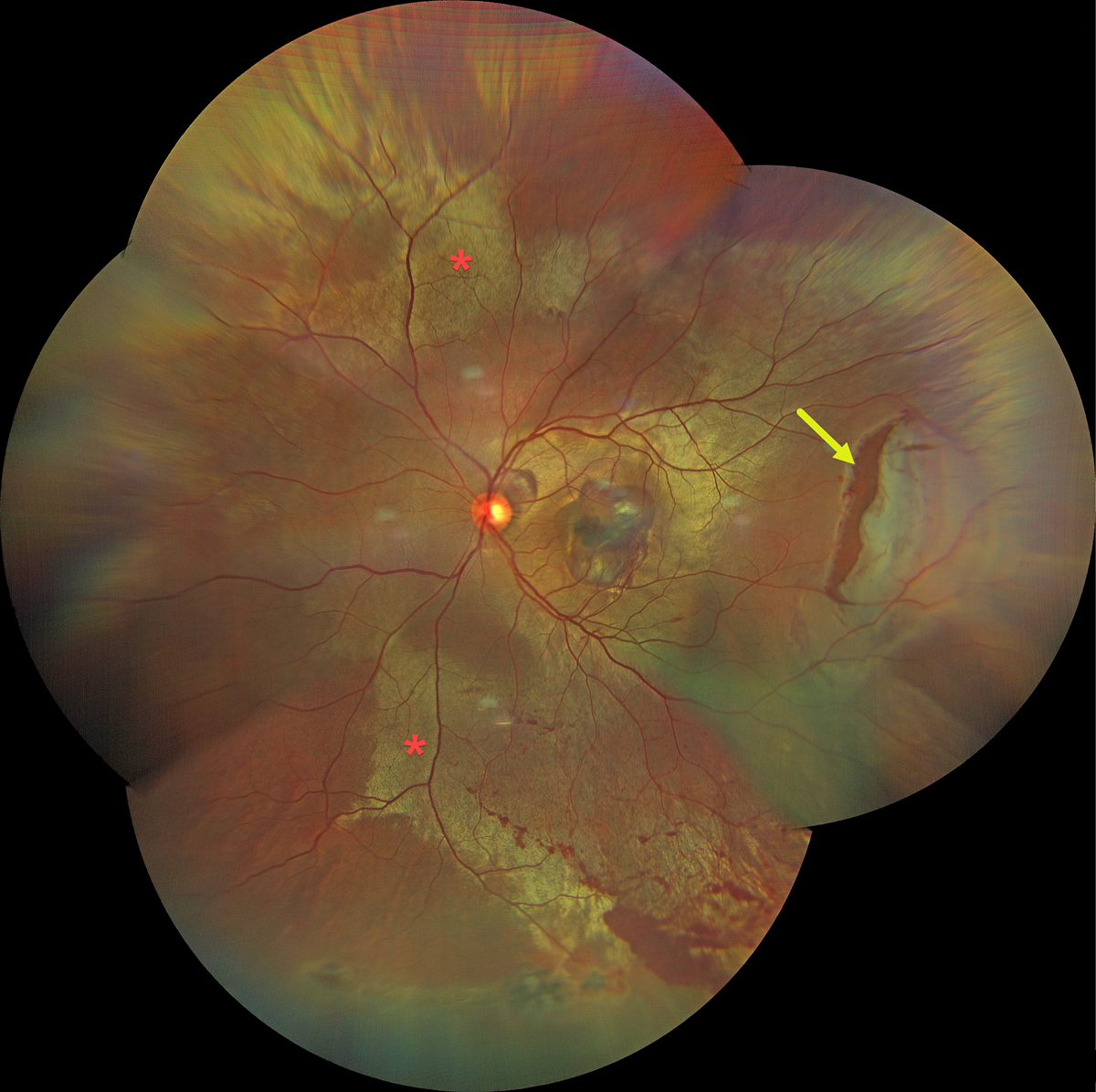
COMMOTIO RETINAE WITH TRAUMATIC RETINAL BREAK
Dibya Prabha
Originally posted on @retina.rocks August 7, 2026
This 38YO male presented with sudden vision loss in his left eye immediately following blunt trauma from a cricket bat 2 days earlier. Vision was 20/200.
Color fundus photography shows extensive commotio retinae throughout the posterior pole, along with a large retinal tear in the temporal midperiphery. Oral and topical steroids were prescribed, and the retinal break was surrounded with laser photocoagulation.
Learning Points:
Commotio retinae is caused by direct, blunt eye trauma. The outer retinal whitening is due to shearing of the photoreceptor outer segments. The term “Berlin’s edema” is a misnomer and should not be used, as the retina is not edematous. The acute white retinal changes usually resolve within several weeks. With resolution, the fundus appearance can normalize. With more severe injury, outer retinal loss can allow RPE cells to migrate into the retina, giving a pseudo-retinitis pigmentosa appearance. Patients can also develop retinal breaks and detachment.

COMMOTIO RETINAE WITH CONTUSIVE RPE FLUID AND CHOROIDAL RUPTURE
Brahim Alfonso Khouri Lopez, Julian Villarreal, Alexis Ramirez, and Angie Maldonado
Originally posted on @retina.rocks July 22, 2026
This 13YO boy presented 2 days following a slingshot injury to his right eye. Vision was counting fingers.
Color photography shows an opaque white discoloration below the retina throughout the posterior pole. Diffuse faint commotio retinae is also noted in the superior retinal periphery. There is a large choroidal rupture inferior to the macula, along with an inner retinal hemorrhage inferonasal to the nerve. OCT scanning shows variably opaque central subretinal fluid and marked cystic edema and disorganization of the nasal outer retinal layers external to the outer plexiform layer. Unfortunately, he was immediately lost to follow-up.
Learning Points:
Commotio retinae is caused by direct, blunt eye trauma. The outer retinal whitening is due to shearing of the photoreceptor outer segments. The term “Berlin’s edema” is a misnomer and should not be used, as the retina is not edematous. Our patient’s fluid is likely due to a contusive RPE injury that disrupted the outer blood-retinal barrier, allowing fluid to accumulate in the subretinal space and outer retina (Mishra et al, Journal of Vitreoretinal Diseases 2021;5:165-169).
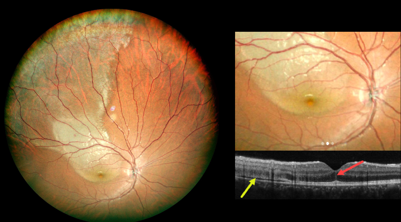
COMMOTIO RETINAE WITH FOVEAL CRACK SIGN
Andree Henaine-Berra and Gerardo Garcia-Aguirre
Originally posted on @retina.rocks July 7, 2026
This 15YO boy presented 1 day following blunt injury to his right eye from a soccer ball. Despite complaints of a central scotoma, vision was 2025.
Color photography shows a broad area of commotio retinae extending from the central macula into the superotemporal periphery. OCT scanning shows disruption of the outer retinal bands and shallow subretinal fluid temporally. A vertical hyperreflective line is present in the central outer nuclear layer.
Learning Points:
The foveal crack sign (FCS) is a central foveal hyperreflective vertical line, first described by Ishibashi et al. as an OCT biomarker for future macular hole development following vitrectomy for rhegmatogenous retinal detachment (AJO 2020;218:192-198). It has since been described in numerous other conditions, including macular pucker, diabetic macular edema, idiopathic macular telangiectasia type 2, vitreomacular traction, NAION with CRAO, AMD, non-infectious uveitis, Coats disease, and commotio retina (Kayabasi et al, Med Hypothesis Discov Innov Ophthalmol 2024;13:129-138).
Commotio retinae is caused by direct, blunt eye trauma. The outer retinal whitening is due to shearing of the photoreceptor outer segments. The term “Berlin’s edema” is a misnomer and should not be used, as the retina is not edematous. The acute white retinal changes usually resolve within several weeks. With resolution, the fundus appearance can normalize. With more severe injury, outer retinal loss can allow RPE cells to migrate into the retina, giving a pseudo-retinitis pigmentosa appearance. Patients can also develop retinal breaks and detachment.
We will follow our patient closely and hope that the FCS is not an omen for a future macular hole.
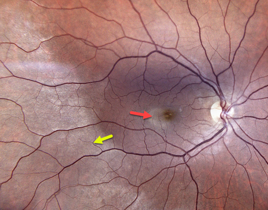
COMMOTIO RETINAE
The European VitreoRetinal Society (EVRS) and Gil Calvão-Santos
Originally posted on @retina.rocks March 20, 2026
This 30YO male presented two hours following a paddleball injury to his right eye. Vision had decreased from a baseline of 20/20 to 20/80 at presentation.
Color photography shows a large area of temporal and inferotemporal outer retinal whitening, along with a smaller area of temporal macula commotio.
Learning Points:
Commotio retinae is caused by direct, blunt eye trauma. The outer retinal whitening is due to shearing of the photoreceptor outer segments. The term “Berlin’s edema” is a misnomer and should not be used, as the retina is not edematous. The acute white retinal changes usually resolve within several weeks. With resolution, the fundus appearance can normalize. With more severe injury, outer retinal loss can allow RPE cells to migrate into the retina, giving a pseudo-retinitis pigmentosa appearance. Patients can also develop retinal breaks and detachment.
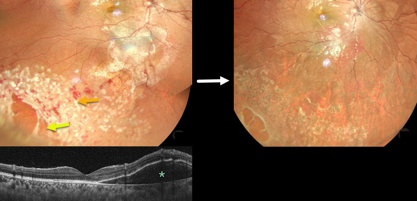
COMMOTIO RETINAE WITH RETINAL BREAKS AND CONTUSIVE SEROUS MACULAR DETACHMENT
Tejaswita Verma and Manish Nagpal
Originally posted on @retina.rocks September 24, 2025
This 13YO male was hit in his right eye with a cricket ball. When he presented later that day vision was 20/30.
Color photography shows a large area of commotio retinae with scattered retinal hemorrhages involving the inferotemporal fundus. There is a poor foveal reflex with possible nasal macular subretinal fluid, which is confirmed on OCT. Several atrophic retinal breaks are noted peripherally. Immediate prophylactic laser was applied around all breaks. Seven weeks later, the commotio and hemorrhages are resolved, and all breaks are well-surrounded with laser scarring. Vision was 20/20.
Although blunt trauma is a common cause of retinal breaks, acute or subsequent dissolution of the retina within areas of commotio with secondary holes is quite rare in our experience (Cox et al Arch Ophthalmol 1966;76:678-685 and Longstaff et al Br J Ophthalmology 1987;71:375-376). The acute white retinal changes usually resolve within several weeks. With resolution, the fundus appearance can normalize. With more severe injury, outer retinal loss can allow RPE cells to migrate into the retina, giving a pseudo-retinitis pigmentosa appearance.
Transient subretinal fluid is an uncommon finding with blunt trauma. Injury to the choroid and RPE allows fluid to accumulate in the subretinal space and outer retina (Mishra et al, Journal of Vitreoretinal Diseases 2021;5:165-169).
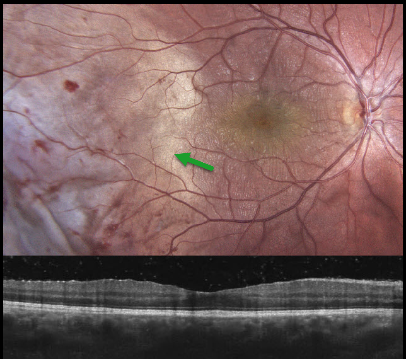
COMMOTIO RETINAE
Anastasia Petrus
Originally posted on @retina.rocks August 25, 2025
This 16YO male was chopping wood one day earlier when a branch recoiled and struck his right eye. Vision was 20/30 OD and 20/20 in his normal OS. The right anterior segment showed localized conjunctival chemosis with a subluxed crystalline lens.
Color photography shows temporal commotio retina extending into the temporal macula, along with scattered retinal hemorrhages. Although there appear to be fine inner macular striae, OCT scanning is fairly normal. The patient was referred for further management.
Learning Points:
Commotio retinae is caused by direct, blunt eye trauma. The outer retinal whitening is due to shearing of the photoreceptor outer segments. The term “Berlin’s edema” is a misnomer and should not be used, as the retina is not edematous. The acute white retinal changes usually resolve within several weeks. With resolution, the fundus appearance can normalize. With more severe injury, outer retinal loss can allow RPE cells to migrate into the retina, giving a pseudo-retinitis pigmentosa appearance. Patients can also develop retinal breaks and detachment.
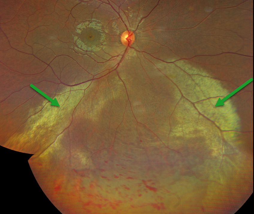
COMMOTIO RETINAE
Nilesh Kumar
Originally posted on @retina.rocks June 17, 2025
This 30YO male was hit in his right eye 2 days earlier with a cricket ball. He was visually asymptomatic, and vision was 20/20.
Color photography shows extensive commotio retinae in the inferior retina, with scattered intraretinal hemorrhages. There were no retinal breaks or detachment. Close observation was recommended.
Learning Points:
Commotio retinae is caused by direct, blunt eye trauma. The outer retinal whitening is due to shearing of the photoreceptor outer segments. The term “Berlin’s edema” is a misnomer and should not be used, as the retina is not edematous. The acute white retinal changes usually resolve within several weeks. With resolution, the fundus appearance can normalize. With more severe injury, outer retinal loss can allow RPE cells to migrate into the retina, giving a pseudo-retinitis pigmentosa appearance. Patients can also develop retinal breaks and detachment.
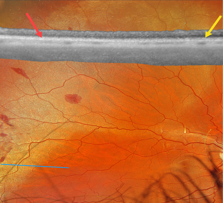
COMMOTIO RETINAE
Originally posted on @retina.rocks December 24, 2024
This healthy 19YO male was hit in his right eye while playing soccer one day earlier. Vision was 20/20.
Optos color RGB imaging shows subretinal whitening with associated retinal hemorrhages in the superotemporal periphery. Optos Silverstone swept-source OCT shows at least two outer retinal hyperreflective bands in the uninvolved retina, which merge into a single hyperreflective band within the commotio. The retina was attached without breaks. The retinal appearance and OCT completely normalized 1 month later (not shown).
Learning Points:
Commotio retinae is caused by direct, blunt eye trauma. The outer retinal whitening is due to shearing of the photoreceptor outer segments. The term “Berlin’s edema” is a misnomer and should not be used, as the retina is not edematous. The acute white retinal changes usually resolve within several weeks. With resolution, the fundus appearance can normalize. With more severe injury, outer retinal loss can allow RPE cells to migrate into the retina, giving a pseudo-retinitis pigmentosa appearance. Patients can also develop retinal breaks and detachment.
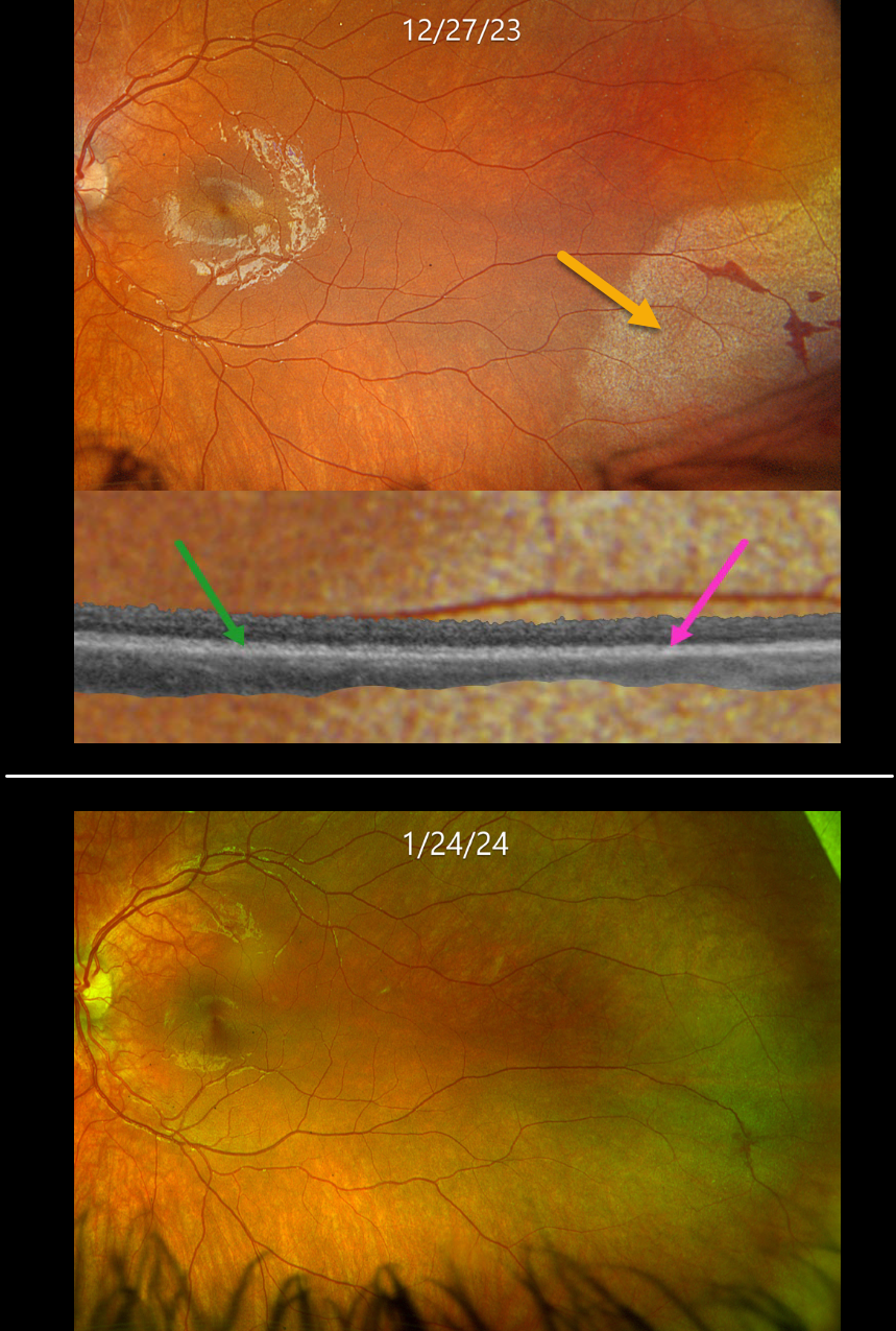
COMMOTIO RETINAE
Originally posted on @retina.rocks April 16, 2024
This healthy 10YO boy presented several hours after being hit in his left eye with a Nerf ball. Vision was 20/25.
Optos color RGB imaging shows subretinal whitening with associated retinal hemorrhages in the inferotemporal periphery. Optos Silverstone swept-source OCT shows at least two outer retinal hyperreflective bands in the uninvolved retina, which merge into a single hyperreflective band within the commotio. The retina was attached without breaks.
Observation was recommended, and the retinal findings normalized within several weeks.
Learning Points:
Commotio retinae is caused by direct, blunt eye trauma. The outer retinal whitening is due to shearing of the photoreceptor outer segments.
The term “Berlin’s edema” is a misnomer and should not be used, since the retina is not edematous. The acute white retinal changes usually resolve within several weeks. With resolution, the fundus appearance can normalize.
With more severe injury, outer retinal loss can allow RPE cells to migrate into the retina, giving a pseudo-retinitis pigmentosa appearance. Patients can also develop retinal breaks and detachment.
CHOROIDAL RUPTURE
Asma Samsudeen and Ashish Sharma
Originally posted on @retina.rocks July 6, 2023
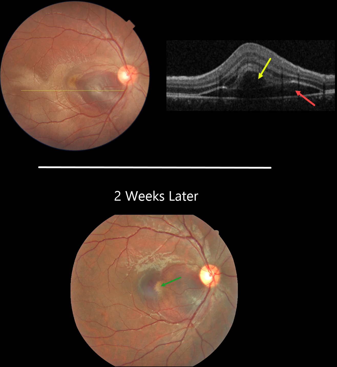
This 21YO male presented with immediate vision loss after being hit with a cricket ball in his right eye one day earlier. Vision was 20/60.
Fundus photography shows a subretinal hemorrhage in the nasal macula, a small rim of surrounding subretinal fluid, and a more temporal ill-defined commotio retina.
OCT scanning shows hyporeflective retinal elevation from the subretinal blood and fluid, along with an overlying bacillary layer detachment.
Our patient shows several findings classic for acute blunt trauma, including commotio retinae and subretinal blood. This subretinal blood almost always indicates an underlying choroidal rupture, which did, in fact, reveal itself on follow-up 2 weeks later, when vision returned to 20/20. The visual prognosis for these choroidal ruptures is good as long as the rupture spares the foveal center, although patients need lifelong surveillance due to the risk of secondary macular neovascularization.
Learning Points:
A bacillary layer (cone and rod inner and outer segments) detachment (BLD) appears as a unique dome-shaped collection of intraretinal fluid from photoreceptor splitting. These detachments can be observed in an increasing number of conditions, most classically Vogt-Koyanagi-Harada disease (Cicinelli et al, Ophthalmology Retina 2020;4:454-456).
About 8% of eyes with acute non-penetrating ocular trauma have a BLD (Venkatesh et al, Can J Ophthalmol 2022;57:328-336). These resolve within 2-10 days without visual or anatomic sequelae.
COMMOTIO RETINAE
Originally posted on @retina.rocks September 27, 2022
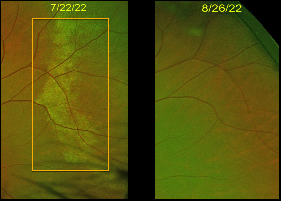
This 10YO boy was hit in the right eye with a Nerf ball and developed commotio retinae. One month later, the retinal appearance normalized.
Vision was 20/20 at both visits.
Learning Points:
Commotio retinae is caused by direct, blunt eye trauma. The outer retinal whitening is due to shearing of the photoreceptor outer segments.
The term “Berlin’s edema” is a misnomer and should not be used, as the retina is not edematous. The acute white retinal changes usually resolve within several weeks. With resolution, the fundus appearance can normalize, as in this patient.
With more severe injury, outer retinal loss can allow RPE cells to migrate into the retina, giving a pseudo-retinitis pigmentosa appearance. Patients can also develop retinal breaks and detachment.
COMMOTIO RETINAE
Originally posted on @retina.rocks July 12, 2022
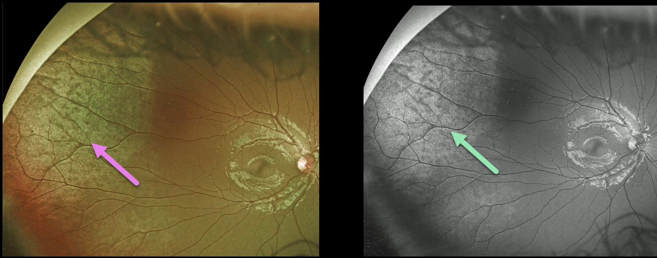
This 11YO male presented a few hours after being hit in his right eye with a toy gun. Optos color and green channel imaging show a broad area of commotio retina in the superotemporal periphery. Vision was 20/20.
Learning Points:
Commotio retinae is caused by direct, blunt eye trauma. The outer retinal whitening is due to shearing of the photoreceptor outer segments. The term “Berlin’s edema” is a misnomer and should not be used, since the retina is not edematous. The acute white retinal changes usually resolve within several weeks. With resolution, the fundus appearance can normalize.
With more severe injury, outer retinal loss can allow RPE cells to migrate into the retina, giving a pseudo-retinitis pigmentosa appearance. Patients can also develop retinal breaks and detachment.
COMMOTIO RETINAE
Originally posted on @retina.rocks August 9, 2021
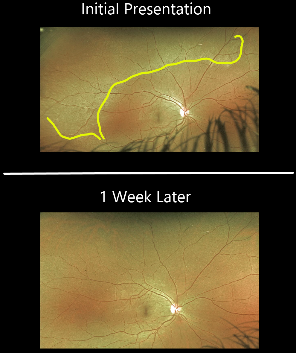
This 27YO female presented with eye pain, 3 days following falling down the stairs and hitting her right eye. Prior CT imaging revealed an associated right nasal fracture.
Optos imaging shows a broad area of commotio retinae superiorly. The commotio completely resolved 1 week later.
Learning Points:
Commotio retinae is caused by direct, blunt eye trauma. The outer retinal whitening is due to shearing of the photoreceptor outer segments.
The term “Berlin’s edema” is a misnomer and should not be used, since the retina is not edematous.
The acute white retinal changes usually resolve within several weeks. With resolution, the fundus appearance can normalize.
With more severe injury, outer retinal loss can allow RPE cells to migrate into the retina, giving a pseudo-retinitis pigmentosa appearance. Patients can also develop retinal breaks and detachment.
COMMOTIO RETINAE
Originally posted on @retina.rocks July 6, 2021
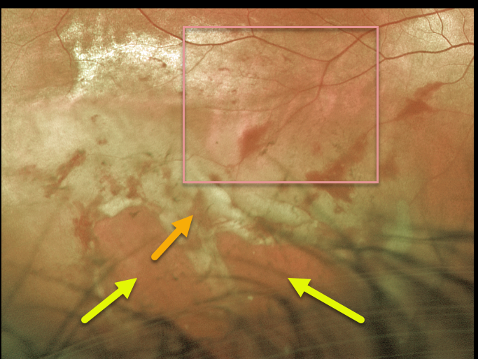
This 28YO male presented with 20/60 vision after being hit the night before in the right eye with a Nerf arrow.
Optos imaging shows outer retinal whitening and hemorrhages extending from the inferior macula into the inferotemporal periphery. Multiple ragged retinal breaks with a shallow retinal detachment are noted more peripherally.
The patient was taken to the operating room later that day for vitrectomy surgery.
Learning Points:
Commotio retinae follows direct, blunt eye trauma. The outer retinal whitening is due to shearing of the photoreceptor outer segments.
The term “Berlin’s edema” is a misnomer and should not be used, since the retina is not edematous.
COMMOTIO RETINAE
Originally posted on @retina.rocks November 27, 2020
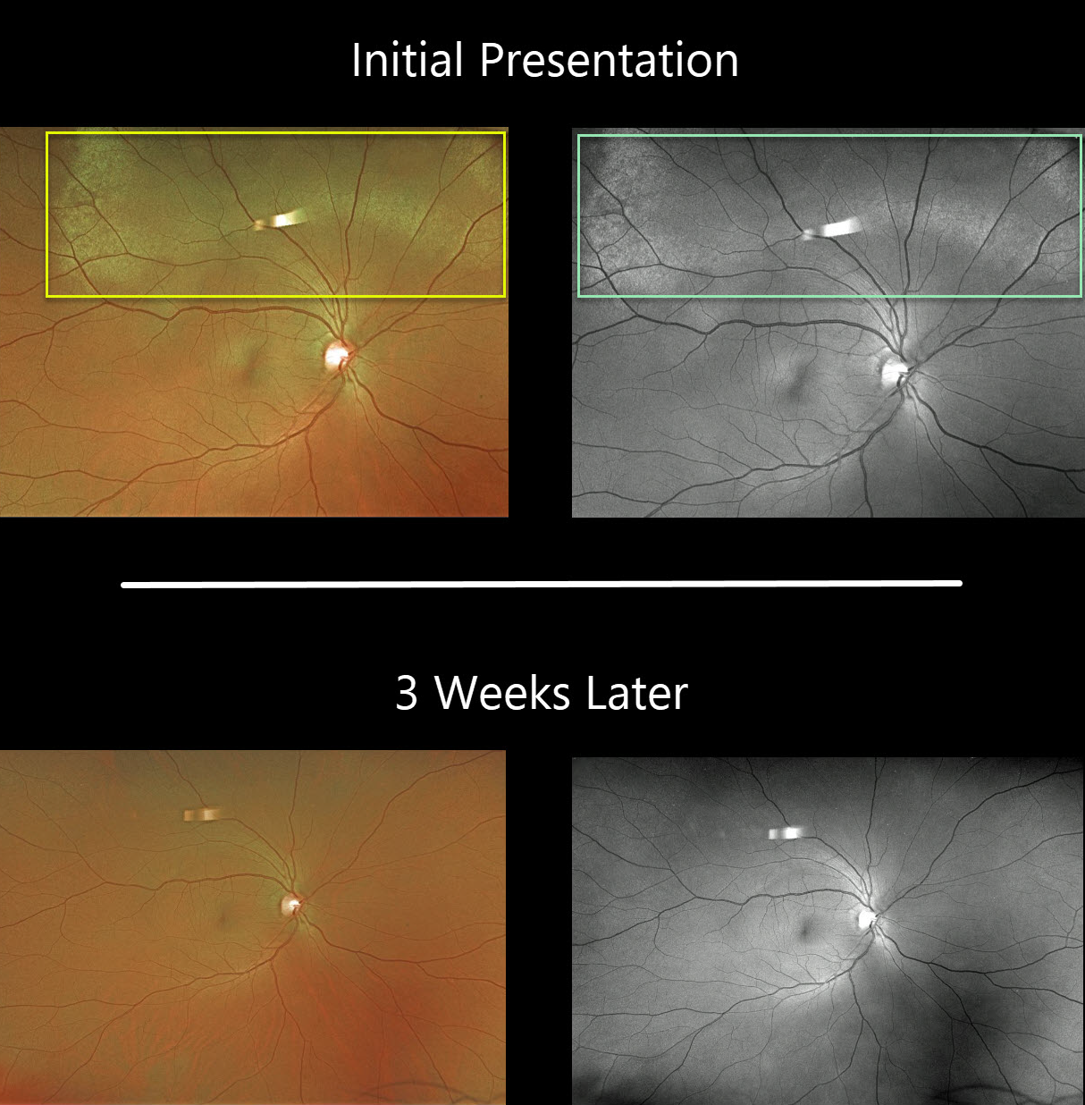
This patient presented with superior commotio retinae after being hit in the eye with a bungee cord. The outer retinal opacification was best visualized on the Optos green channel. Vision was 20/30.
Fortunately, the retina findings completely resolved 3 weeks later.
Learning Points:
Commotio retinae follows direct blunt eye trauma. Large areas of peripheral commotio may be associated with an increased risk for retinal detachment.
In some cases, the photoreceptors are permanently damaged, allowing RPE cells to migrate into the retina along capillaries, giving a pseudo-retinitis pigmentosa appearance.
The term “Berlin’s edema” is a misnomer and should not be used, since the retina is not edematous.
MACULAR HOLE
Originally posted on @retina.rocks November 6, 2020
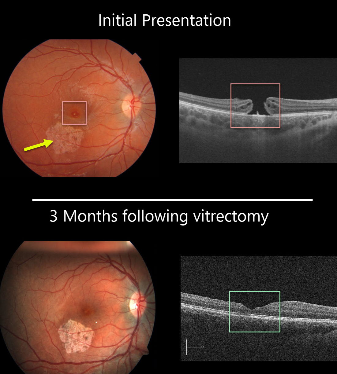
This 13YO girl was hit in her right eye while playing volleyball. She presented to us several months later with 20/200 vision, an inferotemporal macular scar from resolved commotio retina, and a full-thickness macular hole.
The macular hole was closed following successful vitrectomy, and she was 20/100 one month postoperatively. Vision improved to 20/40 three months following surgery. Since the scotoma from the macular scar was superior, she is now virtually completely visually asymptomatic and extremely lucky!
Learning Points:
Traumatic macular holes are more common in younger patients, often from recreational or sports injury, and tend to be more eccentric in shape.
The mechanism could be due to sudden vitreous separation or to stretching of the posterior pole resulting from anteroposterior ocular compression. The holes can spontaneously close and have a high surgical success rate, as in our patient (see Liu et al AJO 2020;210:174-183).
COMMOTIO RETINAE
Originally posted on @retina.rocks April 8, 2020
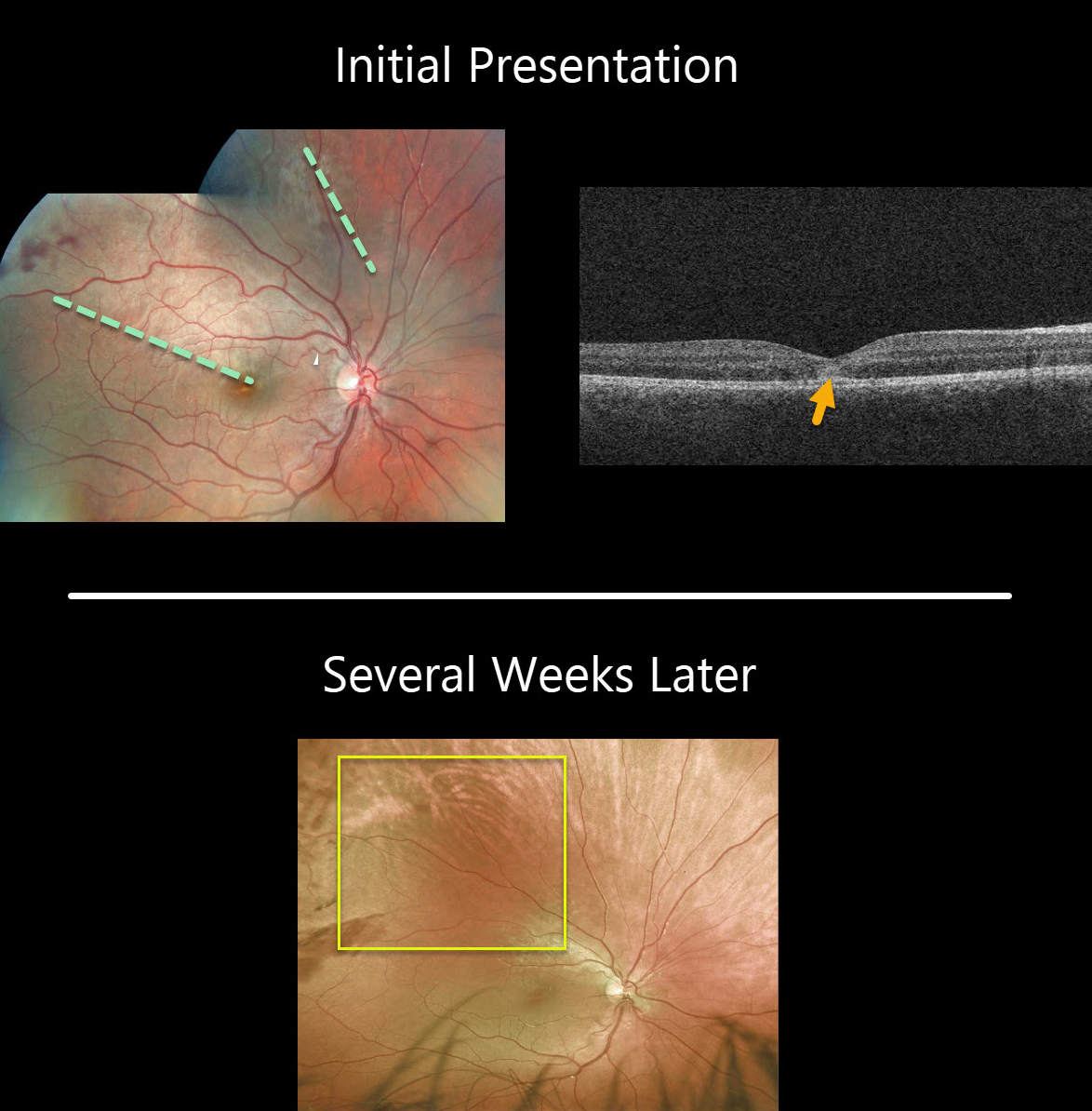
This patient was hit in the eye with a soccer ball and developed outer retinal whitening due to shearing of the outer segments. OCT reveals subfoveal ellipsoid zone (EZ) disruption and hyperreflectivity.
The retinal whitening usually disappears within a few weeks due to regeneration of the photoreceptors
Learning Points:
Commotio retinae follows direct blunt eye trauma. Large areas of peripheral commotio may be associated with an increased risk for retinal detachment.
In some cases, the photoreceptors are permanently damaged, allowing RPE cells to migrate into the retina along capillaries, giving a pseudo-retinitis pigmentosa appearance.
The term, Berlin’s edema, is a misnomer and should not be used, since the retina is not edematous.


