

Contusive RPE Subretinal Fluid
<< Back to Cases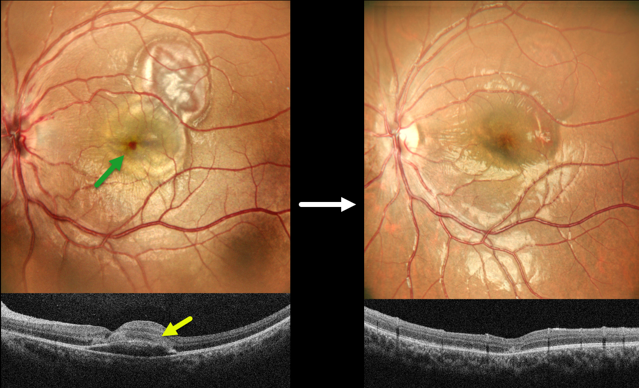
CHOROIDAL RUPTURE WITH LIKELY CONTUSIVE RPE FLUID
Akansha Sharma, Manish Nagpal and Navneet Mehrotra
Originally posted on @retina.rocks May 18, 2026
This 8YO boy presented with 2 days of vision loss in his left eye following a firecracker injury. Vision was 20/200 OS.
MultiColor SLO imaging shows a blister of foveal fluid with an overlying dot of foveal blood, and more superotemporal subretinal blood. OCT scanning shows foveal subretinal fluid with overlying hyperreflectivity from the blood. Intravenous methylprednisolone was given for 3 days, followed by a short course of oral steroids. One month later, vision improved to 20/60. The fluid and blood resolved with a choroidal rupture now visible temporally.
Learning Points:
An acute submacular hemorrhage following blunt trauma almost always indicates an underlying choroidal rupture as the source of the blood. The final vision is generally good unless the rupture extends through the macular center. These patients need long-term follow-up due to the increased risk of macular neovascularization.
Transient subretinal fluid is an uncommon finding with blunt trauma. Injury to the choroid and RPE allows fluid to accumulate in the subretinal space and outer retina (Mishra et al, Journal of Vitreoretinal Diseases 2021;5:165-169), as likely occurred in our patient.
COMMOTIO RETINAE WITH RETINAL BREAKS AND CONTUSIVE SEROUS MACULAR DETACHMENT
Tejaswita Verma and Manish Nagpal
Originally posted on @retina.rocks September 24, 2025
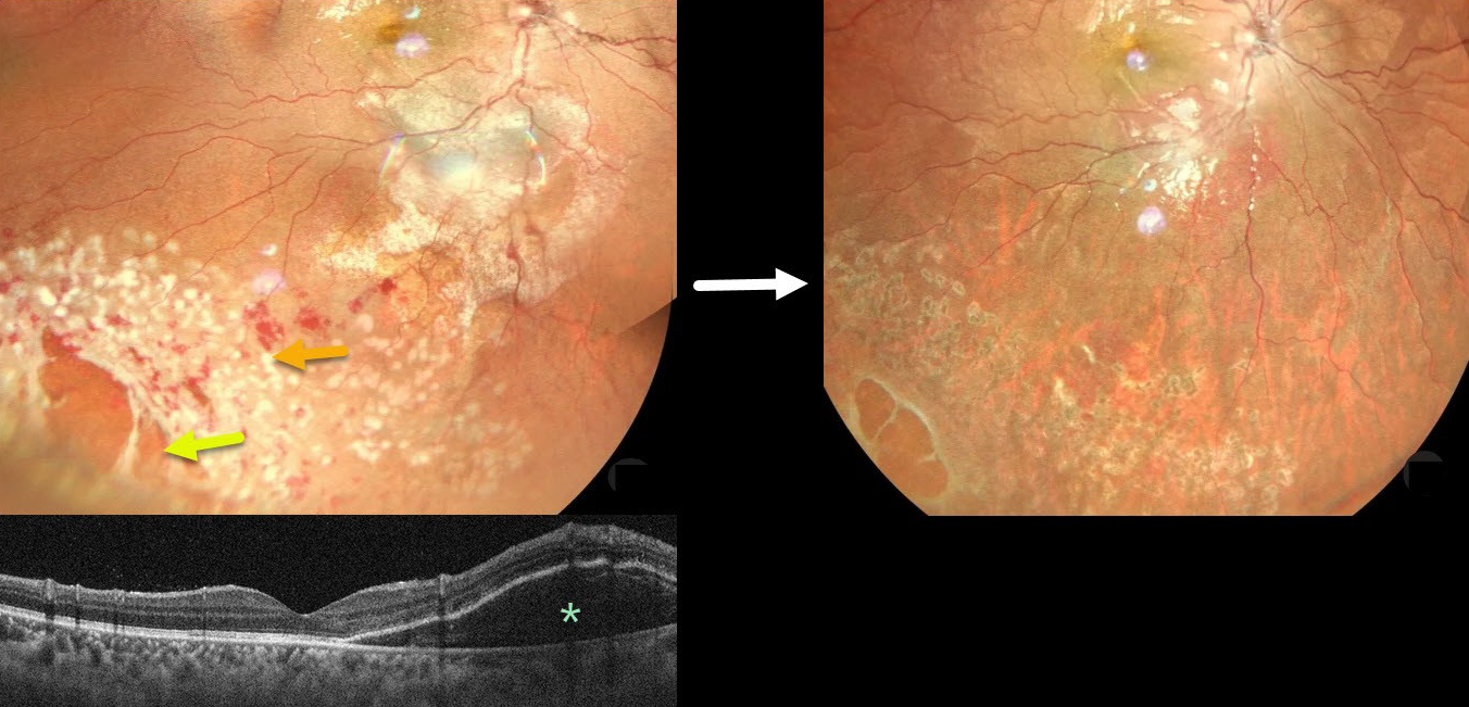
This 13YO male was hit in his right eye with a cricket ball. When he presented later that day vision was 20/30.
Color photography shows a large area of commotio retinae with scattered retinal hemorrhages involving the inferotemporal fundus. There is a poor foveal reflex with possible nasal macular subretinal fluid, which is confirmed on OCT. Several atrophic retinal breaks are noted peripherally. Immediate prophylactic laser was applied around all breaks. Seven weeks later, the commotio and hemorrhages are resolved, and all breaks are well-surrounded with laser scarring. Vision was 20/20.
Although blunt trauma is a common cause of retinal breaks, acute or subsequent dissolution of the retina within areas of commotio with secondary holes is quite rare in our experience (Cox et al Arch Ophthalmol 1966;76:678-685 and Longstaff et al Br J Ophthalmology 1987;71:375-376). The acute white retinal changes usually resolve within several weeks. With resolution, the fundus appearance can normalize. With more severe injury, outer retinal loss can allow RPE cells to migrate into the retina, giving a pseudo-retinitis pigmentosa appearance.
Transient subretinal fluid is an uncommon finding with blunt trauma. Injury to the choroid and RPE allows fluid to accumulate in the subretinal space and outer retina (Mishra et al, Journal of Vitreoretinal Diseases 2021;5:165-169).
CONTUSIVE TRAUMA WITH SEROUS MACULAR DETACHMENT
Originally posted on @retina.rocks January 16, 2023
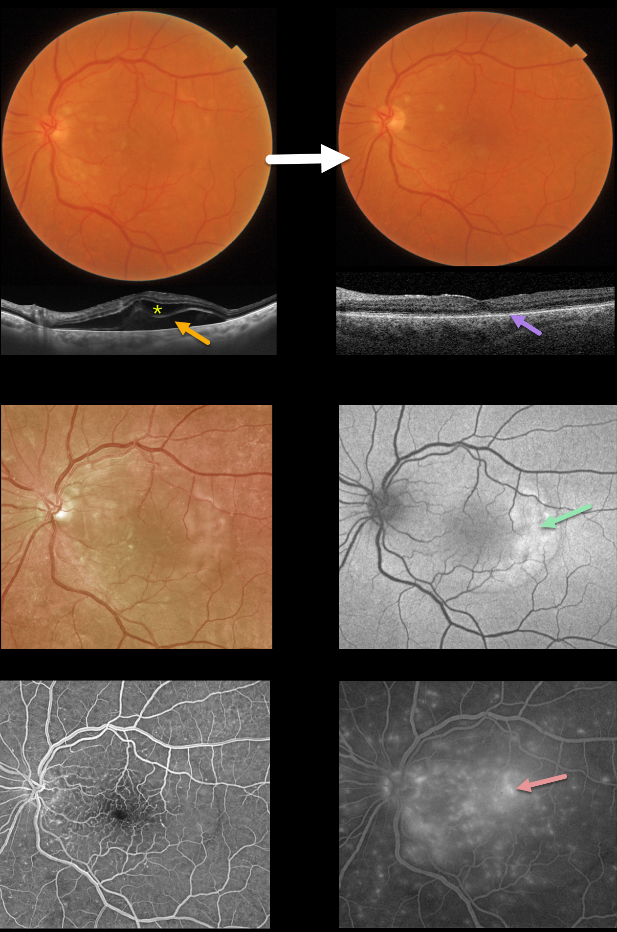
This 71YO male presented with decreased vision in his left eye one day following a motor vehicle accident with airbag deployment. Vision was 20/100.
Color photography shows variable faint patches of outer retinal whitening. Swept-source OCT shows outer retinal and subretinal fluid.
Optos fundus autofluorescence (FAF) shows temporal macular hyper-FAF. Fluorescein angiography shows early dots of subretinal hyperfluorescence with late deep leakage extending beyond the arcades.
Two weeks later, he was subjectively much improved, although he was bothered by metamorphopsia and some paracentral scotomas. Vision was 20/60. The macular pigmentary changes improved funduscopically. All macular fluid resolved, but the outer photoreceptor bands were disorganized and thinned.
Learning Points:
Posterior segment findings from blunt ocular trauma most commonly include commotio retinae or choroidal rupture.
Our patient’s unusual findings were from a contusive injury to the choroid and RPE. RPE dysfunction led to fluid accumulation in the subretinal space and the outer retina.
A strikingly similar case was reported by Mishra et al (Journal of Vitreoretinal Diseases 2021;5:165-169).


