

CRAO
<< Back to Cases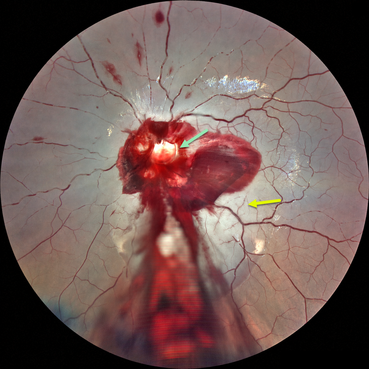
ACUTE OPTIC NERVE AVULSION WITH SECONDARY CRAO
Ayushi Gupta and Vishal Agrawal
Originally posted on @retina.rocks September 8, 2026
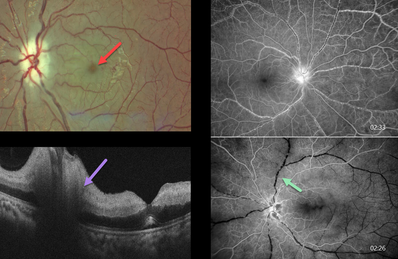
This 6YO boy developed sudden and complete loss of vision in his left eye immediately following blunt trauma. He was riding a bicycle as a pillion passenger when the bike stopped suddenly, throwing his face against the handlebar. Vision was no light perception. Anterior segments were normal.
Fundus photography shows preretinal blood surrounding the nerve, extending through the macula and into the inferior periphery. A pale-yellow cavity is noted where the nerve head should be. A fresh central retinal artery occlusion (CRAO) with a stagnant blood column is noted, along with some scattered inner retinal hemorrhages. There were no other physical or neurologic injuries.
Learning Points:
Traumatic optic nerve head avulsion is a rare and visually devastating traumatic event in which the optic nerve is forcibly separated from the globe at its junction with the sclera, typically resulting in immediate, severe, and irreversible vision loss. The mechanism involves sudden forceful rotation or anterior displacement of the globe following blunt periorbital trauma, including projectiles, falls, or finger-poke injuries, whereby the globe moves anteriorly or rotationally while the optic nerve remains relatively fixed, causing violent separation. Fundoscopic examination may reveal a partial or complete tear of the optic nerve head from the sclera (Foster et al, Arch Ophthalmol 1997;115:623-630). Avulsion can be partial or complete, and may be associated with a CRAO, as was noted in our patient.

CRAO FROM ATRIAL MYXOMA
Ayushi Gupta and Vishal Agrawal
Originally posted on @retina.rocks June 15, 2026
This previously healthy 29YO female presented with 2 days of sudden vision loss in her left eye. Vision was 20/20 in her normal OD and light perception OS.
Color photography shows an acute central retinal artery occlusion (CRAO) with diffuse retinal opacification and a prominent foveal cherry-red spot. The retinal arterioles are markedly attenuated with segmentation of the arterial and venous blood columns (box-carring). OCT shows inner retinal thickening and opacification except in the foveal center. Cardiac imaging revealed a left atrial myxoma, and she underwent immediate surgical excision.
Learning Points:
Atrial myxoma, the most common primary cardiac tumor, is typically attached to the interatrial septum at the fossa ovalis and can shed tumor fragments, thrombi, or surface debris into the systemic circulation (Griborio-Guzman et al, Heart 2022;108:827-833). Embolism occurs in 30-40% of patients, and the cerebral and retinal arteries are the most affected vascular beds. The vast majority of CRAOs occur in a more elderly population from carotid or valvular disease. Atrial myxomas are a rare but known cause, especially in younger patients.
Half of patients have an elevated erythrocyte sedimentation rate (ESR) due to tumor-produced interleukin-6 (Acebo et al, Chest 2003;123:1379-1385). These cytokines drive the systemic inflammatory response that produces constitutional symptoms (fever, fatigue, weight loss, arthralgias, myalgias) and laboratory abnormalities (elevated ESR/CRP, anemia, leukocytosis, elevated globulins). Elevated inflammatory markers can be a diagnostic pitfall, as they frequently lead to misdiagnosis of endocarditis, rheumatic fever, vasculitis, collagen vascular disease, or even polymyalgia rheumatica.
COMBINED CRAO-CRVO
Shraddha Raj Shrivastava and Manish Nagpal
Originally posted on @retina.rocks April 20, 2026
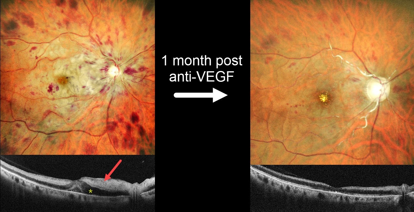
This 57YO female with a history of severe hypertension presented with one day of severe vision loss in her right eye. Blood pressure was 200/100 mmHg. Vision was hand motion OD and 20/20 in her normal OS.
MultiColor SLO imaging shows inner retinal opacification sparing the macular center, consistent with a fresh central retinal artery occlusion (CRAO). A central retinal vein occlusion (CRVO) is also noted with a swollen nerve and retinal hemorrhages in all quadrants. OCT scanning shows marked hyperreflectivity with loss of the band detail throughout the inner and mid-retinal layers. The peripapillary retina is edematous with fluid in the nasal outer nuclear layer and mild foveal subretinal fluid.
She was diagnosed with a combined CRAO-CRVO. An intravitreal anti-VEGF injection, and 1 month later, vision improved to 20/400. Repeat imaging shows marked improvement in the retinal hemorrhages, retinal opacification, and macular fluid, although new foveal lipid is noted. The nerve is flat and pale, and the major proximal arterioles are white without a blood column. The inner retina remains hyperreflective on OCT but is thinning with loss of the normal architecture. Panretinal photocoagulation was scheduled.
Learning Points:
Combined CRAO-CRVO is thankfully an extremely rare event, accounting for 0.3% of all retinal vascular occlusions (Raval et al, Indian J Ophthalmology 2020;68:2136-2142). Risk factors appear similar to those associated with isolated occlusions. The CRAO may be embolic or secondary to compression from a swollen optic nerve.
CRAO FOLLOWING DOUBLE PERFORATION FROM PERIBULBAR INJECTION
Ayushi Gupta and Vishal Agrawal
Originally posted on @retina.rocks April 6, 2026
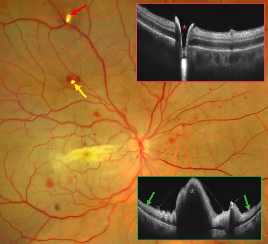
This 42YO female immediately lost vision in her right eye 4 days earlier following pterygium excision surgery performed under peribulbar anesthesia elsewhere. Vision in our office was light perception.
Fundus photography shows an opaque retina throughout the posterior pole consistent with an acute central retinal artery occlusion (CRAO). There is a horizontal retinal fold throughout the macula with scattered blot retinal hemorrhages. Entry and exit needle penetration sites are noted. OCT scan through the superior entry site confirms a full-thickness retinal break. Macular OCT shows diffuse inner retinal hyperreflectivity with elevation through the fold.
Learning Points:
Peribulbar anesthesia is considered to have a lower risk of inadvertent globe perforation, although this risk is not zero (Lim et al., J Clin Ophthalmol 2021;5:414-416). CRAO after such an injury is exceedingly rare and was reported by Lake et al. (J Cataract Refract Surg 2003;29:2234-2235), who felt the occlusion might have resulted from increased IOP due to intraocular anesthetic. A CRAO was also reported by Gyasi et al. following peribulbar anesthesia for pterygium excision, although they found no evidence of globe perforation (Ghana Medical Journal 2012;46:48-50).
CRAO FOLLOWING CRANIOTOMY
Fraser McKay
Originally posted on @retina.rocks March 17, 2026
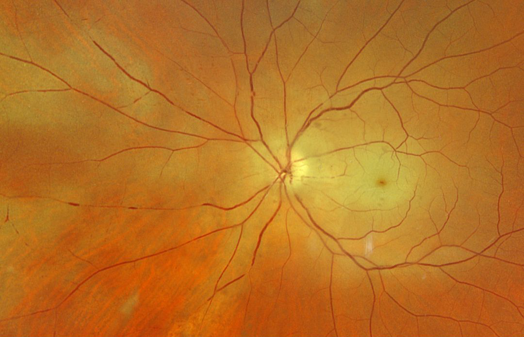
This 62YO male became hospitalized following a motor vehicle accident on 3/10/25. He underwent craniotomy on 4/16/25 and on 4/22/25. He woke from this last surgery blind in his right eye.
Optos color RGB imaging shows a fresh central retinal artery occlusion (CRAO) with a macular cherry red spot and patches of peripheral inner retinal whitening. Both the arteries and veins show focal areas of an absent blood column, indicating stagnant blood flow. Observation was recommended.
COMBINED CRAO-CRVO
Malvika Singh and Manish Nagpal
Originally posted on @retina.rocks March 3, 2026
This 60YO male presented with sudden vision loss in his left eye 1 month earlier. Vision was 20/20 in his normal OD and no light perception OS.
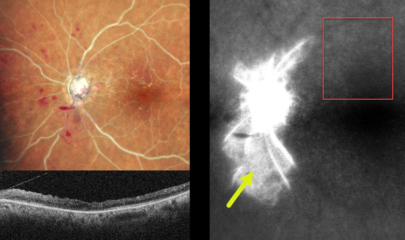
MultiColor SLO imaging shows a combined central retinal artery occlusion (CRAO) and central retinal vein occlusion (CRVO) with white arteries and veins with no blood column. Disc neovascularization is present inferotemporally, and a small preretinal hemorrhage is noted just inferonasal to the nerve. OCT shows a disorganized hyperreflective inner retina. Fluorescein angiography shows profound panretinal ischemia with a small island of preserved capillary bed inferior to the nerve.
Vasculitis workup was negative. An emergent stroke workup, including carotid Doppler, MRI, and neurologic consultation, was ordered. Given our patient’s vision, comfort measures for his left eye were recommended.
Learning Points:
Combined CRAO-CRVO is thankfully an extremely rare event, accounting for 0.3% of all retinal vascular occlusions (Raval et al, Indian J Ophthalmology 2020;68:2136-2142). Risk factors appear similar to those associated with isolated occlusions. The CRAO may be either embolic or secondary to compression from a swollen optic nerve.
HEMI-CRAO
Rohan Jain and Manish Nagpal
Originally posted on @retina.rocks January 26, 2026
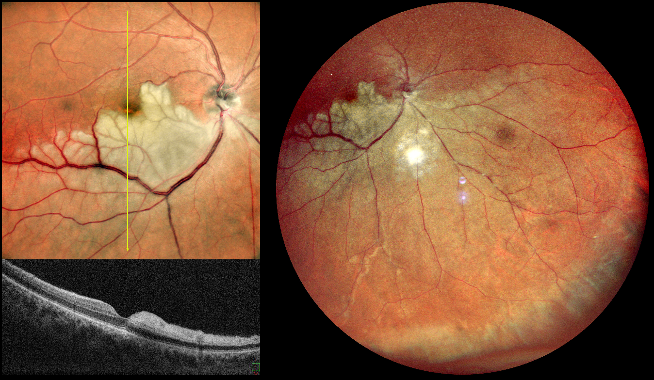
This is a healthy 42YO male presented with 7 days of blurred vision in his left eye. Vision was 20/90 OD and 20/20 in his normal OS.
Pseudocolor SLO imaging shows a white, opaque retina in the inferior retinal hemisphere, which spares the central macula. The vertical B-scan contrasts the normal superior retina with the ischemic inferior retina with marked inner retinal hyperreflectivity.
Learning Points:
Once the central retinal artery passes anteriorly through the lamina cribrosa, it branches into superior and inferior branches, which then further subdivide to supply the inner two-thirds of the neurosensory retina. Hemi-CRAO is a rare CRAO variant caused by the blockage of one of these superior or inferior branches. As with more typical CRAO, emboli or giant cell arteritis are the most common etiologies.
Our patient had acute retinal ischemia (defined as acute BRAO, central retinal artery occlusion, or transient monocular vision loss) and was immediately referred to the nearest stroke center. Unfortunately, he was immediately lost to follow up.
HEMI-CRAO
Ayushi Gupta and Vishal Agrawal
Originally posted on @retina.rocks December 2, 2025
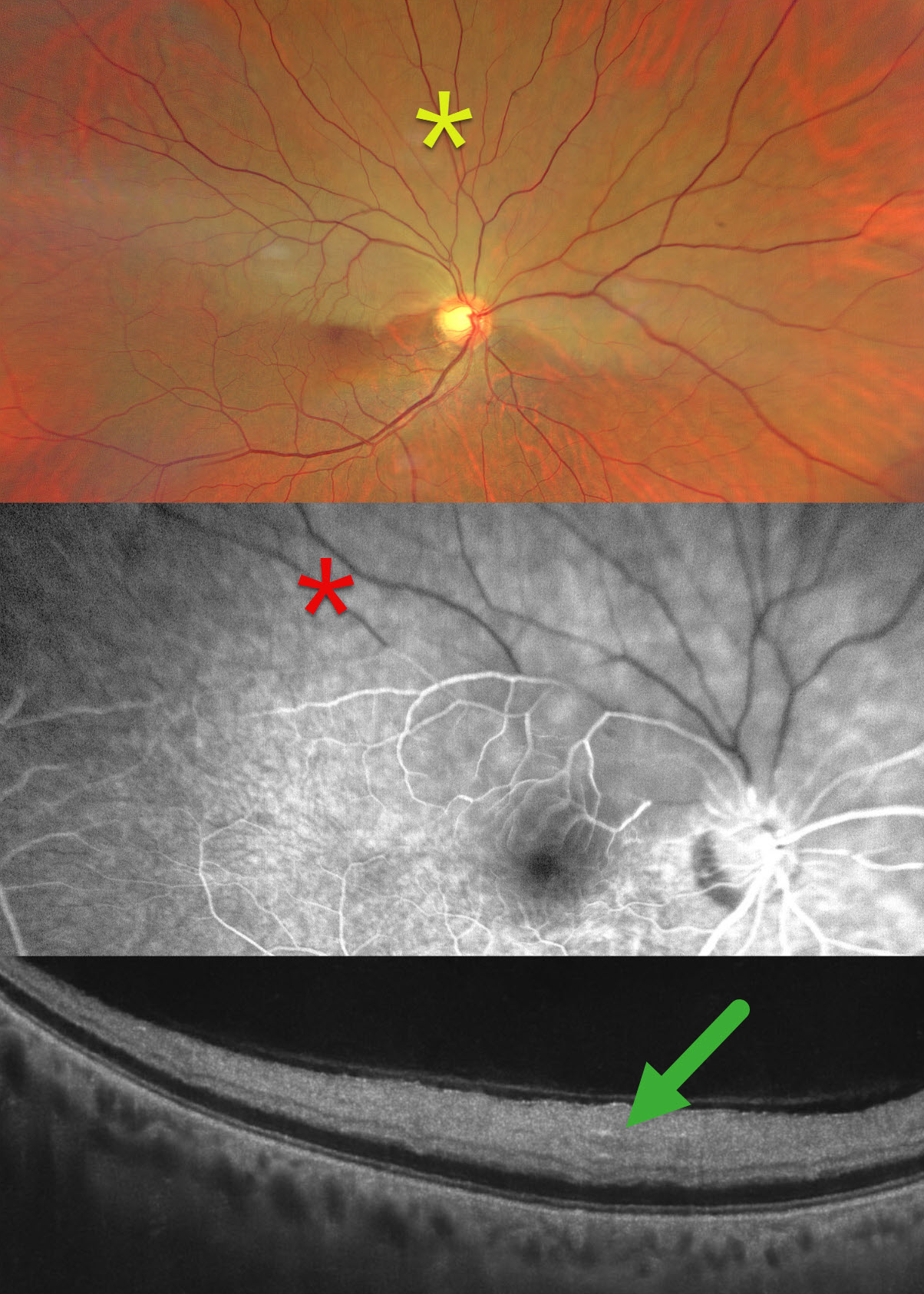
This healthy 65YO male presented with 2 days of sudden painless decreased vision in his right eye. Vision was 20/80 OD and 20/20 in his healthy OS.
Color photography shows an acute superior hemi-central retinal artery occlusion (CRAO) with opaque white retina. On fluorescein angiography, there is a marked absence of perfusion in the superior hemiretina, which persists 4 minutes into the study. On OCT, the retina is thickened and hyperreflective from the outer plexiform layer anterior to the internal limiting membrane.
Our patient was immediately referred to cardiology for a stroke protocol evaluation.
Learning Points:
Once the central retinal artery passes anteriorly through the lamina cribrosa, it branches into superior and inferior branches, which then further subdivide to supply the inner two-thirds of the neurosensory retina. Hemi-CRAO is a rare variant of CRAO caused by the blockage of one of these superior or inferior branches. As with more typical CRAO, emboli or giant cell arteritis are the most common etiologies.
CRAO
Tejaswita Verma and Manish Nagpal
Originally posted on @retina.rocks September 1, 2025
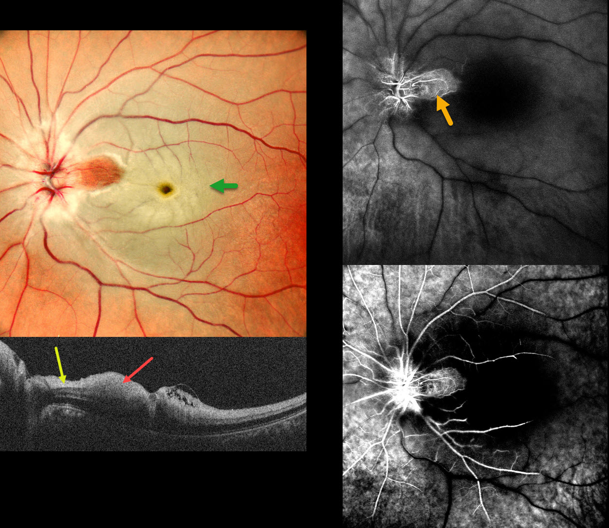
This 37 YO male presented with acute vision loss in his left eye 3 days earlier. Vision was 20/20 in his normal OD and 20/200 in his OS.
Pseudocolor SLO imaging shows a fresh central retinal artery occlusion (CRAO) with sparing of a small area of temporal peripapillary retina supplied from a patent cilioretinal artery. OCT scanning shows opaque, hyperreflective, thickened inner retinal layers, except for a small area of more normal retina within the cilioretinal sparing.
Fluorescein angiography shows profound diffuse retinal nonperfusion and delayed filling except for the nasal macula. Workup elsewhere included an MRI showing left ICA and left MCA chronic infarcts.
Learning Points:
The inner two-thirds of the neurosensory retina is supplied by the central retinal artery, with the choroid supplying the RPE and photoreceptor layer. Acute CRAO, therefore, presents with a white edematous inner retina that spares the fovea. Cilioretinal arteries, which arise from the posterior choroidal circulation, are present in up to 50% of individuals, and bilateral in about 25% (Schneider et al, Acta Ophthalmologica 2021; 99:e310-e318).
TAKAYASU VASCULITIS WITH CRAO
Ayushi Gupta and Vishal Agrawal
Originally posted on @retina.rocks April 29, 2025
This previously healthy 14YO girl presented with 10 days of vision loss in her right eye. She also gave a history of recurrent migraine-type headaches. She saw an outside eye doctor, where blood pressure was not checked, and spectacles were prescribed. At presentation, vision was 20/100 OD and 20/20 OS.
Funduscopic examinations revealed mild bilateral disc edema with venous dilation (not shown). Blood pressure could not be detected in any limb. We suspected Takayasu arteritis and urgently referred her to a rheumatologist and neurologist. Unfortunately, she presented 2 weeks later with sudden vision loss in her left eye. Vision was 20/100 OD and no light perception OS.
Color photography shows a cherry red spot and pale disc edema in her left eye. Fluorescein angiography shows markedly delayed venous filling in both eyes at 2.5 minutes following injection. OCT scanning shows a swollen disc and marked inner retinal hyperreflectivity with posterior shadowing. An anterior chamber paracentesis was performed with no change in her funduscopic appearance.
Medical workup between our initial and subsequent examinations revealed no pulses in the radial and dorsalis pedis arteries, with reduced left femoral and left carotid pulses. Renal artery Doppler revealed high-resistance flow bilaterally in the main renal and segmental arteries with 80-90% lumen narrowing in the infrarenal aorta. Brain MRI revealed an old infarct with encephalomalacia and gliosis in the left cerebellar hemisphere. Immediately following her second exam with us, she was emergently admitted by rheumatology and given a methylprednisolone infusion. She was immediately lost to follow-up.
Learning Points:
Takayasu arteritis is an idiopathic granulomatous vasculitis involving medium- to large-sized vessels throughout the body. Involvement of the aortic arch and its branches can cause ocular ischemia, including anterior segment ischemia with rubeosis and neovascular glaucoma.
Posterior segment findings include retinal ischemia, neovascularization, and vascular occlusions. Surprisingly, over 50% of patients present with ocular findings as the initial manifestation of their disease. For a wonderful review of the posterior segment findings in Takayasu, see Sharma et al, Indian J Ophthalmol 2024;72:637-647.
CRAO
Originally posted on @retina.rocks December 23, 2024
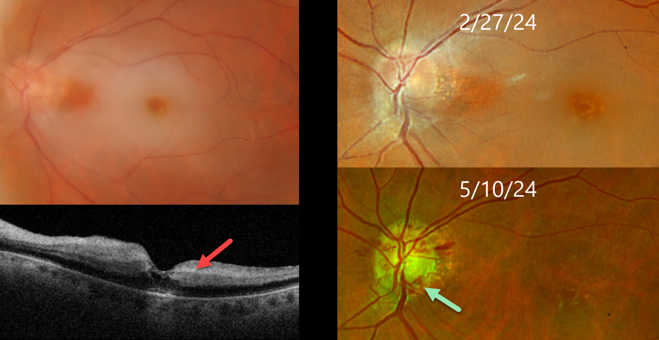
This 75YO female presented with an acute counting fingers central retinal artery occlusion. Triton color imaging shows a small area of papillomacular sparing, and swept-source OCT shows inner retinal hyperreflectivity. The retinal opacification completely resolved 10 weeks later with the development of extensive disc collaterals. Vision at counting fingers.
Learning Points:
Collateral vessels are dilated, pre-existing vessels that bypass an occlusion. These are seen in the posterior pole most commonly following retinal venous occlusions. Disc collaterals are exceedingly rare with arterial occlusions and were first described by Nettleship in 1891 (Ragge and Hoyt, Br J Ophthalmology 1992;76:186-188). The endothelial tight junctions are intact so they don’t leak angiographically. This helps to differentiate them from neovascularization which does leak.
CRAO
Ankit Jain and Manish Nagpal
Originally posted on @retina.rocks December 11, 2024
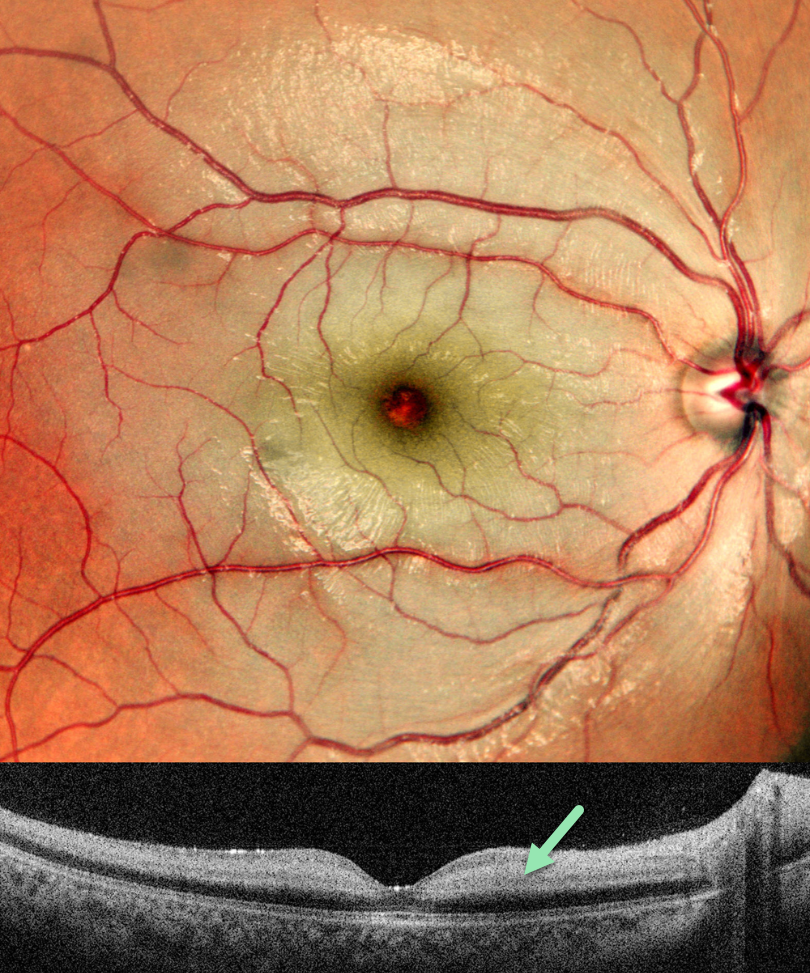
This previously healthy 8YO boy presented with sudden vision loss in his right eye 10 days earlier. Vision was counting fingers.
Pseudocolor SLO imaging shows diffuse inner retinal whitening, sparing the central macula. OCT scanning shows a somewhat thickened and markedly hyperreflective inner retina. Cardiac 2D Echo and MRA of the head and neck were normal. Anticardiolipin antibodies were elevated, and he was started on oral clopidogrel and aspirin.
Learning Points:
Central retinal artery occlusion (CRAO) in the pediatric population is exceedingly rare and requires an aggressive search for underlying clotting abnormalities. The antiphospholipid antibody syndrome, also known as anticardiolipin antibody syndrome or lupus anticoagulant syndrome, is caused by autoantibodies directed against phospholipid-bound proteins. These predispose patients to venous and arterial thrombi and increase the risk for pregnancy complications and miscarriage. The syndrome is more common in women and in those with other autoimmune disorders, including systemic lupus.
CRAO
Will Gibson
Originally posted on @retina.rocks August 29, 2024
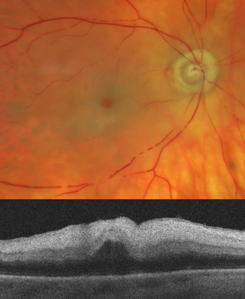
This 81YO female presented with acute vision loss in her right eye. Vision was light perception.
Optos color RG imaging shows an acute central retinal artery occlusion (CRAO) with opaque white retina surrounding the preserved central fovea (‘cherry red spot’). A stagnant blood column is noted inferiorly with box-carring. OCT scanning shows a thickened and markedly hyperreflective inner retina. She was immediately referred to the nearest stroke center.
CRAO
Originally posted on @retina.rocks July 16, 2024
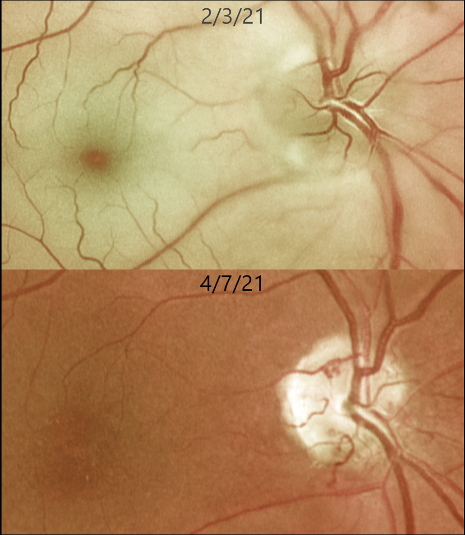
This 62YO female presented with an acute counting-fingers central retinal artery occlusion. Emergent stroke workup revealed a silent acute lacunar infarct. The retinal opacification completely resolved 2 months later. New fine collateral vessels were noted on the nerve. Vision remained at counting fingers.
Learning Points:
Collateral vessels are dilated, pre-existing vessels that bypass an occlusion. These are most commonly seen at the posterior pole following retinal venous occlusions and are exceedingly rare with arterial occlusions. The endothelial tight junctions are intact, so they don’t leak angiographically. This helps differentiate them from neovascularization, which does leak.
CRAO
Asma Samsudeen and Ashish Sharma
Originally posted on @retina.rocks June 6, 2024
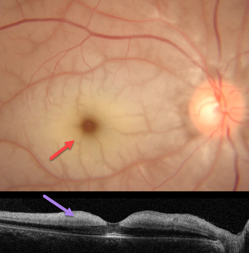
This 41YO male presented with a 2-day history of acute vision loss in his right eye. Vision was counting fingers OD and 20/20 in his normal OS.
Fundus photography shows an acute central retinal artery occlusion (CRAO) with a pale, opaque inner retina and a cherry-red spot. The retina immediately adjacent to the radiating perifoveal arterioles is relatively transparent, creating a fern-like appearance. OCT shows a slightly thickened and markedly hyperreflective inner retina. The patient was immediately sent to the nearest stroke center.
Learning Points:
Acute CRAO often exists along a spectrum from complete occlusion with a stagnant blood column to a more partial blockage with variable patchy ischemia. Our patient still has enough active flow to supply the neurosensory retina immediately adjacent to the smaller arterioles, but not enough to supply the more distal retinal tissue.
About 90% of CRAO are embolic, with the remainder most commonly due to giant cell arteritis. The lack of a visible embolus, as in our patient, does not preclude an embolic cause since the causative embolus can either be posterior to the visible disc or have already traveled further downstream.
CRAO
Originally posted on @retina.rocks April 18, 2024
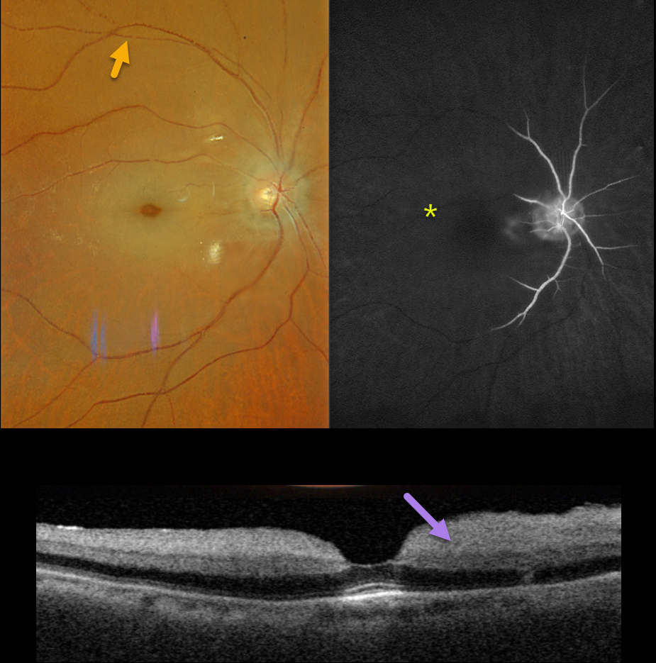
This 89YO male presented with 2 days of acute and severe vision loss in his right eye. Vision was no light perception.
Optos color RGB imaging shows an acute central retinal artery occlusion (CRAO) with a cherry-red spot and a stagnant blood column, best seen superiorly as box-carring of the column.
Fluorescein angiography shows profound ischemia, with no vascular filling beyond the peripapillary region.
Triton swept-source OCT shows a diffusely slightly thickened and markedly hyperreflective inner retina. He was immediately referred to the nearest stroke center.
Learning Points:
Patients with acute retinal ischemia (defined as transient monocular vision loss, acute branch retinal artery occlusion, or acute CRAO) require emergent referral to a stroke center. This is especially urgent with an acute CRAO since about 75% of patients have already developed a recent stroke.
If the patient presents immediately following the occlusion, measures to acutely lower the intraocular pressure to move the causative embolus downstream can be performed, including ocular massage, paracentesis, and breathing into a bag.
Unfortunately, most patients present outside this window (Shah et al, Ophthalmology Retina 2023;7:527-531) and even then, these measures are usually ineffective.
CRAO WITH CILIORETINAL ARTERY SPARING
Anjana Mirajkar and Manish Nagpal
Originally posted on @retina.rocks November 2, 2023
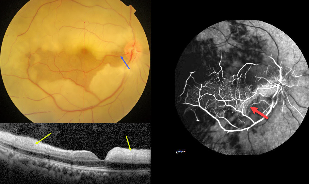
This 45YO male presented with acute vision loss in his right eye 4-5 days earlier. Vision was 20/80 in his right eye and 20/20 in his normal left eye.
Color photography shows an acute central retinal artery occlusion (CRAO) with sparing of the central and inferior macula due to a large cilioretinal artery.
The vertical OCT B-scan shows opaque hyperreflective paracentral inner retina with normal central and inferior foveal layers. Fluorescein angiography confirms preserved central and inferior macular arterial flow.
Workup elsewhere included a normal brain MRI and cardiac 2D Echo. Carotid Doppler showed a plaque in the right internal carotid artery.
Learning Points:
The inner two-thirds of the neurosensory retina is supplied by the central retinal artery, with the choroid supplying the RPE and photoreceptor layer. Acute CRAO, therefore, presents with a white edematous inner retina that spares the fovea.
Cilioretinal arteries, which arise from the posterior choroidal circulation, are present in up to 50% of individuals, and bilateral in about 25% (Schneider et al, Acta Ophthalmologica 2021;99:e310-e318). Our patient was quite fortunate to have a large cilioretinal vessel that relatively preserved central vision.
Patients with acute retinal ischemia (defined as transient monocular vision loss, acute BRAO, or acute CRAO) need to be emergently referred to a stroke center. This is especially urgent with an acute CRAO since about 75% of patients have already developed a recent stroke.
If the patient presents immediately after occlusion, measures to acutely lower intraocular pressure to move the causative embolus downstream can be performed, including ocular massage, paracentesis, and breathing into a bag. Unfortunately, most patients present outside this window (Shah et al, Ophthalmology Retina 2023;7:527-531), and even then, these measures are usually ineffective.
CRAO
Asma Samsudeen and Ashish Sharma
Originally posted on @retina.rocks August 10, 2023
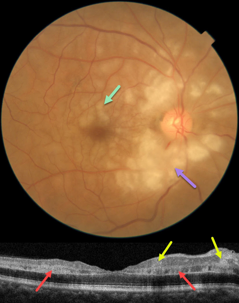
This 60YO male presented with an abrupt, painless loss of vision in his right eye 1 week earlier. There was no prior ocular history or recent history of trauma. Vision was 20/30 in his right eye, and 20/20 in his normal left eye.
Fundus photography shows patches of mostly inner-peripapillary ischemia, with deeper ischemia throughout the macula. This is shown on OCT as patches of inner and mid-retinal hyperreflectivity. Fluorescein angiography showed a markedly delayed transit time (not shown).
We immediately referred him to his cardiologist for an emergency stroke protocol evaluation.
Learning Points:
Purtscher retinopathy was first described by Otmar Purtscher in 1910. Although originally described in a man who fell from a tree with cranial trauma, these white patches of retinal ischemia and hemorrhages are found in numerous other etiologies, including pancreatitis, chest trauma, and collagen vascular diseases.
The findings are called Purtscher retinopathy when due to trauma, and Purtscher-like retinopathy when seen from other causes.
Our patient’s fundus appearance most resembles Purtscher-like retinopathy, with patches of inner- and middle-retinal ischemia. However, given the acute symptoms and delayed angiographic filling time, we believe our patient most likely has an incomplete central retinal artery occlusion (CRAO).
See Mangla et al for the OCT findings in CRAO of varying severities (Int J Retina Vitreous 2023;9;37).
CRAO
Ryan Kern
Originally posted on @retina.rocks January 10, 2023
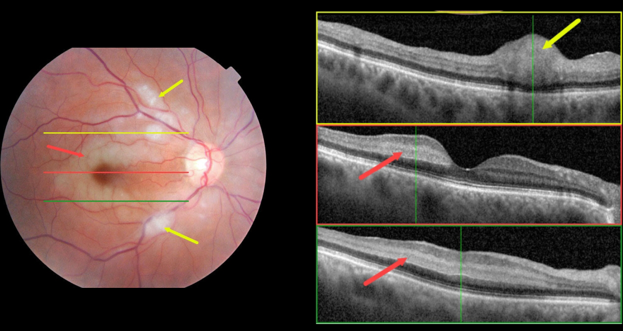
This 70 YO female presented with acute vision loss in her right eye starting 1 day earlier. Vision was 20/70 OD and 20/25 in her normal left eye.
Color fundus photography shows an incomplete central retinal artery occlusion (CRAO) with focal areas of acute inner retinal infarcts along with paracentral areas of deeper retinal opacification.
OCT scanning through the superior macula shows a focal area of retinal opacification and thickening that extends from the outer plexiform layer to the internal limiting membrane. Scans of the central and inferior macula show hyperreflectivity involving the middle retinal layers (outer plexiform, inner nuclear, and inner plexiform).
Blood pressure was 183/110. Emergent stroke evaluation was fortunately negative. Her blood pressure was stabilized, and baby aspirin was started. Visual acuity improved to 20/30 one month later.
This case was submitted by Ryan Kern.
Learning Points:
Paracentral acute macular maculopathy (PAMM) is a phenotype caused by acute mid-retinal ischemia due to obstruction of the deep (DCP) and intermediate (ICP) capillary plexi. PAMM is associated with an increasing constellation of conditions, including retinal vein and artery occlusions.
It is unclear if patients with a pure PAMM phenotype require an immediate stroke protocol referral. In our practice, we have seen several elderly patients who developed a complete CRAO within days of presenting with PAMM, so we do send these patients for emergent evaluation. We do not believe that typical PAMM in younger patients or found as a secondary finding in other ocular conditions requires a stroke workup.
Our patient’s fundus appearance has more of a Purtscher ’s-like appearance with focal areas of cotton wool spot-like inner retinal ischemia (although on OCT these involved the middle retinal layers as well) and mid-retinal ischemia.
However, given her age and history, acute onset, and unilateral presentation, her diagnosis is most consistent with an incomplete CRAO.
For a great recent PAMM review, see Scharf et al, Progress in Retinal and Eye Research 2021;81;100884.
CRAO
Originally posted on @retina.rocks November 21, 2022
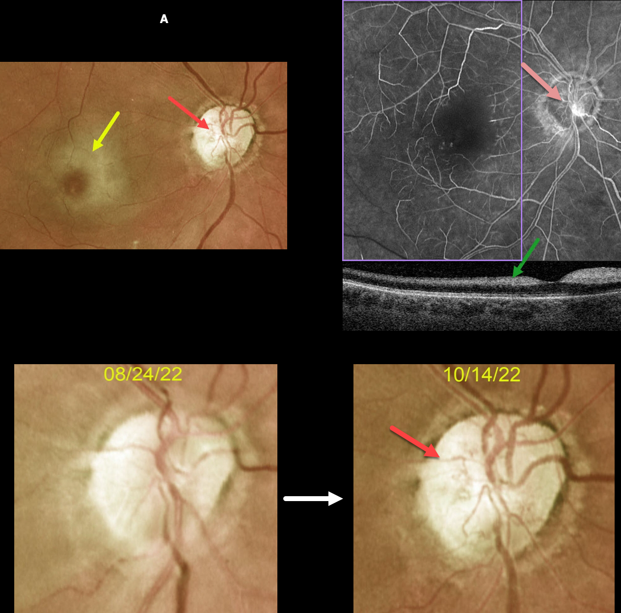
This 63YOF initially presented with an acute central retinal artery occlusion in her right eye. Emergency stroke referral revealed two prior strokes with a new asymptomatic stroke.
She returned two months later. There was subjective visual improvement, although vision remained at hand motion. Although the retinal opacification in general was improving, the perifoveal retina was more opaque with a pronounced cherry red spot.
New fine vessels were noted on the nerve. These vessels did not leak on fundus fluorescein angiography. Marked angiographic ischemia is also noted. OCT scanning shows increased reflectivity from the ischemic inner retina.
Learning Points:
Collateral vessels are dilated, pre-existing vessels that bypass an occlusion. These are seen in the posterior pole most commonly following retinal venous occlusions and are exceedingly rare with arterial occlusions.
The endothelial tight junctions are intact, so they don’t leak angiographically. This helps differentiate them from neovascularization, which does leak.
CRAO
Originally posted on @retina.rocks August 10, 2022
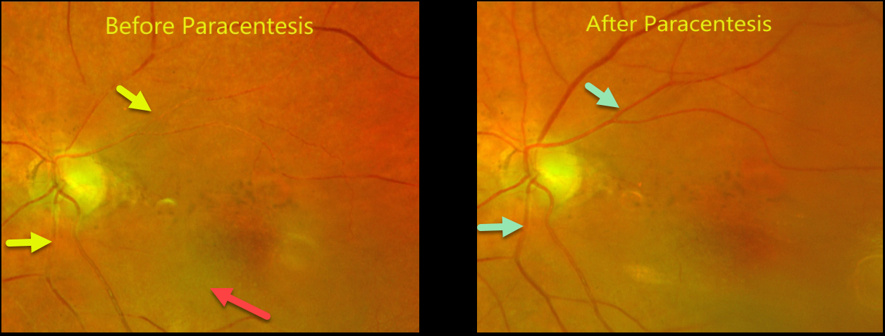
This 94YO female had a prior central retinal artery occlusion (CRAO) with vision that returned back to a baseline of 20/100. There was a history of multiple prior strokes, and she was not a surgical candidate.
Six months later, she came to see us emergently after waking with severe vision loss in her left eye. Vision was light perception.
A fresh CRAO was noted with a stagnant blood column and early inner retinal opacification. Blood flow was immediately restored following paracentesis.
The patient’s daughter reported sadly that the nursing home had made an error and not given her mom anticoagulants for months.
Learning Points:
Measures to relieve the occlusion by moving the causative embolus downstream include vasodilation by increasing the carbon dioxide concentration (rebreathing into a paper bag, carbogen) or rapid IOP lowering (paracentesis, ocular massage). Interestingly, a recent meta-analysis suggested that eyes treated with these measures had worse visual outcomes and recovery rates (Schrag et al, JAMA Neurology 2015;72:1148-1154).
Our patient hasn’t returned yet for us to determine if the paracentesis truly helped.
CRAO + MACULAR HOLE
Originally posted on @retina.rocks February 21, 2022
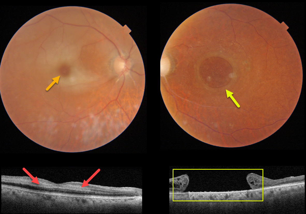
This 82YO female presented with counting fingers vision in her previously healthy right eye. Vision in her left eye had been counting fingers for many years due to a large chronic macular hole measuring 2,870 microns.
An acute central retinal artery occlusion (CRAO) was noted in her right eye. The foveal retina is still perfused by the underlying choroid, resulting in the classic cherry red spot.
Swept-source OCT shows hyperreflectivity from the opaque, ischemic inner retinal layers.
CRAO
Originally posted on @retina.rocks January 7, 2022
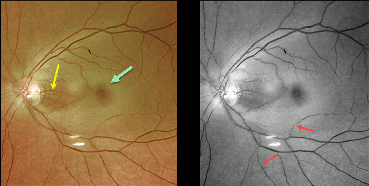
This 73YO female presented with counting-fingers vision secondary to an acute central retinal artery occlusion (CRAO).
The foveal retina is still perfused by the underlying choroid, resulting in the classic cherry red spot appearance. The nasal macula is also preserved due to the presence of a cilioretinal artery, which unfortunately does not supply the macular center. The Optos green channel best images the stagnant blood column.
Our patient’s MRI revealed several acute diffuse embolic strokes. Carotid duplex ultrasound showed severe bilateral carotid stenosis. In addition, her blood glucose was 357, and her erythrocyte sedimentation rate was 94. She was also started on systemic prednisone by the ER until giant cell arteritis could more definitively be ruled out.
Learning Points:
Patients with acute retinal ischemia (defined as transient monocular vision loss, acute BRAO, or acute CRAO) need to be emergently referred to a stroke center.
This is especially urgent with an acute CRAO since about 75% of patients have already developed a recent stroke.
CRAO
Originally posted on @retina.rocks February 26, 2021
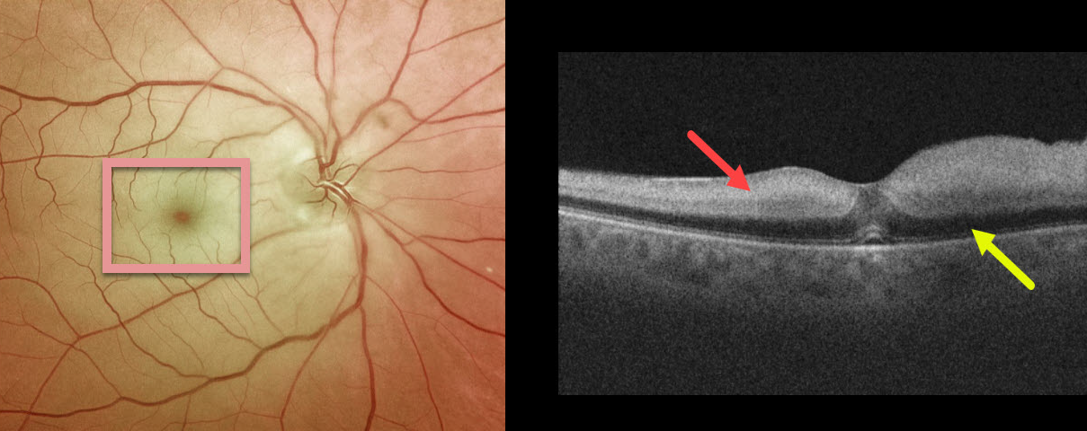
This patient presented with counting-finger vision and classic findings of a white retina and a cherry-red spot from an acute central retinal artery occlusion (CRAO). The cherry red spot is caused by the unaffected foveal retina (receiving intact choroidal blood flow) being surrounded by more distal white and ischemic retina no longer receiving circulation from the central retinal artery.
On Triton swept-source OCT, the inner two-thirds of the acutely ischemic retina were thickened with marked hyper-reflectivity. Less light reaches the deep retina, making it darker than normal and producing pseudo-edema of the outer nuclear layer.
She was immediately sent to the nearest stroke center, where she was found to have had an acute but asymptomatic lacunar stroke.
Learning Points:
Patients with acute retinal ischemia (acute CRAO, acute branch retinal artery occlusion, or transient monocular vision loss) should be sent immediately to the nearest stroke center due to the risk of a pre-existing or recent asymptomatic stroke or impending stroke (see Biousse et al, Ophthalmology 2018;125;1597-1607).
About 75% of patients with an acute CRAO will be found to have a concurrent, often asymptomatic recent stroke, as was the case with this patient.
CRAO
Originally posted on @retina.rocks December 24, 2020
This healthy 49YO female presented with severe, sudden vision loss in her right eye.
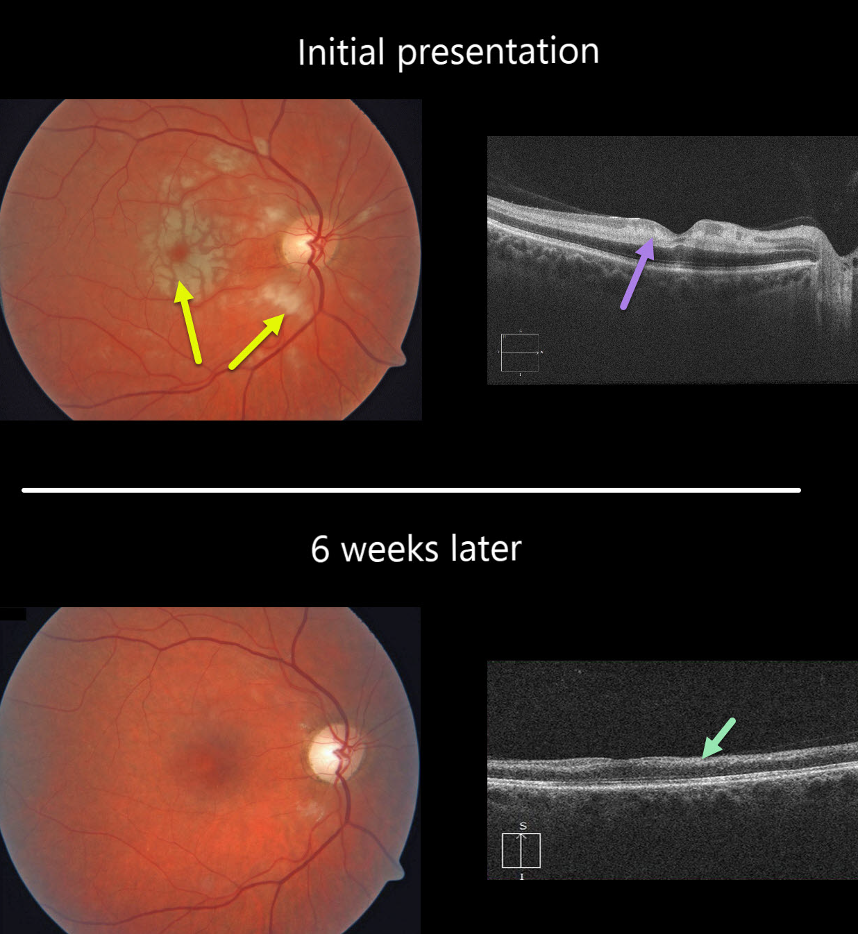
There were multiple white macular patches similar to Purtscher’s retinopathy, but without retinal hemorrhages.
Optical coherence tomography (OCT) showed multiple mid-retinal hyperreflective paracentral acute macular maculopathy (PAMM) lesions. Since we felt this could represent an incomplete central retinal artery occlusion (CRAO), we sent her immediately to the nearest stroke center for further assessment.
At her six-week follow-up, the retinal appearance virtually normalized. However, OCT showed severe diffuse thinning of the inner non-photoreceptor retinal layers consistent with a resolved CRAO. Her stroke workup was unremarkable.
Learning Points:
Purtscher’s retinopathy was first described by Otmar Purtscher in 1910. Although originally described in a man who fell from a tree with cranial trauma, these white patches of retinal ischemia and hemorrhages are found in numerous other etiologies, including pancreatitis, chest trauma, and collagen vascular diseases.
The findings are called Purtscher’s retinopathy when due to trauma, and Purtscher ‘s-like retinopathy when seen from other causes.
The underlying cause of Purtscher’s is unknown, but is thought to involve leukoembolization due to complement activation. The areas of ischemia include more superficial nerve fiber layer infarcts (cotton wool spots) and deeper ischemia involving the middle retinal layers (PAMM).
CRAO
Originally posted on @retina.rocks April 30, 2020
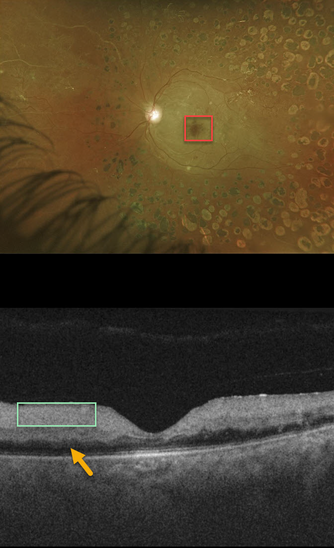
This patient presented with acute vision loss from a fresh central retinal artery occlusion (CRAO). Of note, this patient also has involuted PDR following full PRP.
The cherry red spot is caused by the unaffected foveal retina (receiving intact choroidal blood flow) being surrounded by more distal white and ischemic retina no longer receiving circulation from the central retinal artery.
Although the retina is somewhat thickened on OCT, the acutely ischemic inner two-thirds of the retina, which is supplied by the central retinal artery, is extremely hyperreflective. Less light is available to illuminate the deep retina, which is therefore darker than normal with “pseudo-edema.”
Learning Points:
Patients with acute retinal ischemia (acute CRAO, acute branch retinal artery occlusion or transient monocular vision loss) need to be sent immediately to the nearest stroke center due to the risk of pre-existing recent asymptomatic stroke or impending stroke (see Biousse et al, Ophthalmology 2018;125;1597-1607 or our post https://odsonfb.com/amaurosis-brao-and-crao-the-medical-emergency-many-eye-doctors-still-dont-know-about/).
CRAO
Originally posted on @retina.rocks February 24, 2020
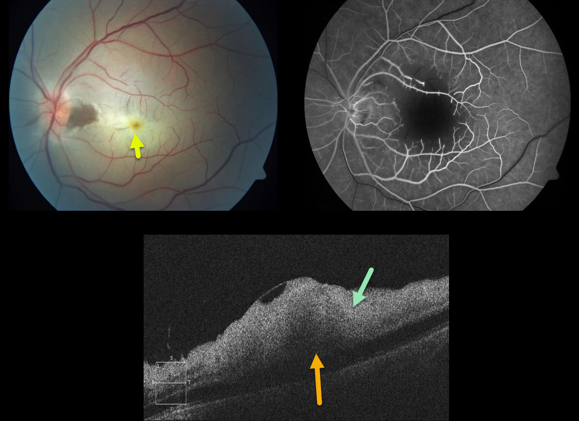
This is a 49 yo M who presented with acute 20/400 monocular vision loss and retinal whitening.
The OCT shows marked inner retinal hyperreflectivity from the acute ischemia, with pseudo-edema of the outer retina due to decreased transmission of light through the inner opaque retina.
The outer retina is still receiving oxygen from the choroid, as evidenced by sparing of the fovea (the yellow xanthophyll is prominently seen).
Learning Points:
This patient was sent to the emergency room for a stroke protocol workup (including electrocardiography, MRI, carotid non-invasive, cardiac echography, hypercoagulable labs, and inflammatory labs). The evaluation was totally negative.
This case looks very similar to gentamicin retinal toxicity. Intravitreal gentamicin can cause severe macular ischemic retinal necrosis. The macular capillaries have a characteristic cut-off appearance, as is seen in our patient who had no gentamicin exposure.


