

CRVO
<< Back to Cases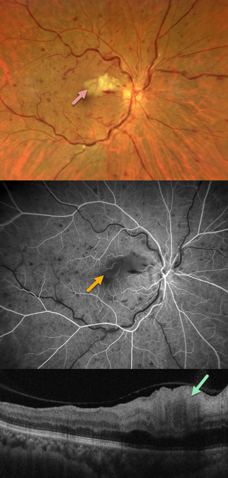
CRVO WITH SECONDARY CILIORETINAL ARTERY OCCLUSION
Francis Ezra Laxamana, Bianca Teresa Suarez, Jesus Jacinto Bautista and Carlo Ladores.
Originally posted on @retina.rocks September 7, 2026
This 58YO female with a history of hypertension and diabetes presented with sudden painless vision loss in her right eye. Vision was 20/25 OD and 20/20 in her normal OS.
Fundus photography shows a non-ischemic central retinal vein occlusion (CRVO) with dilated tortuous veins, mild scattered retinal hemorrhages, and a superonasal macular branch retinal artery occlusion (BRAO). The BRAO shows inner retinal hyperreflectivity on OCT. Fluorescein angiography shows good perfusion throughout the distribution of the CRVO, with a filling defect from the BRAO.
Learning Points:
Some patients with CRVO will develop a secondary non-embolic cilioretinal artery occlusion caused by either compression from optic nerve edema or from a sudden rise in intraluminal retinal capillary bed pressure (Hayreh et al, Retina 2008;28;581-594).
ISCHEMIC CRVO
Ayushi Gupta and Vishal Agrawal
Originally posted on @retina.rocks August 18, 2026
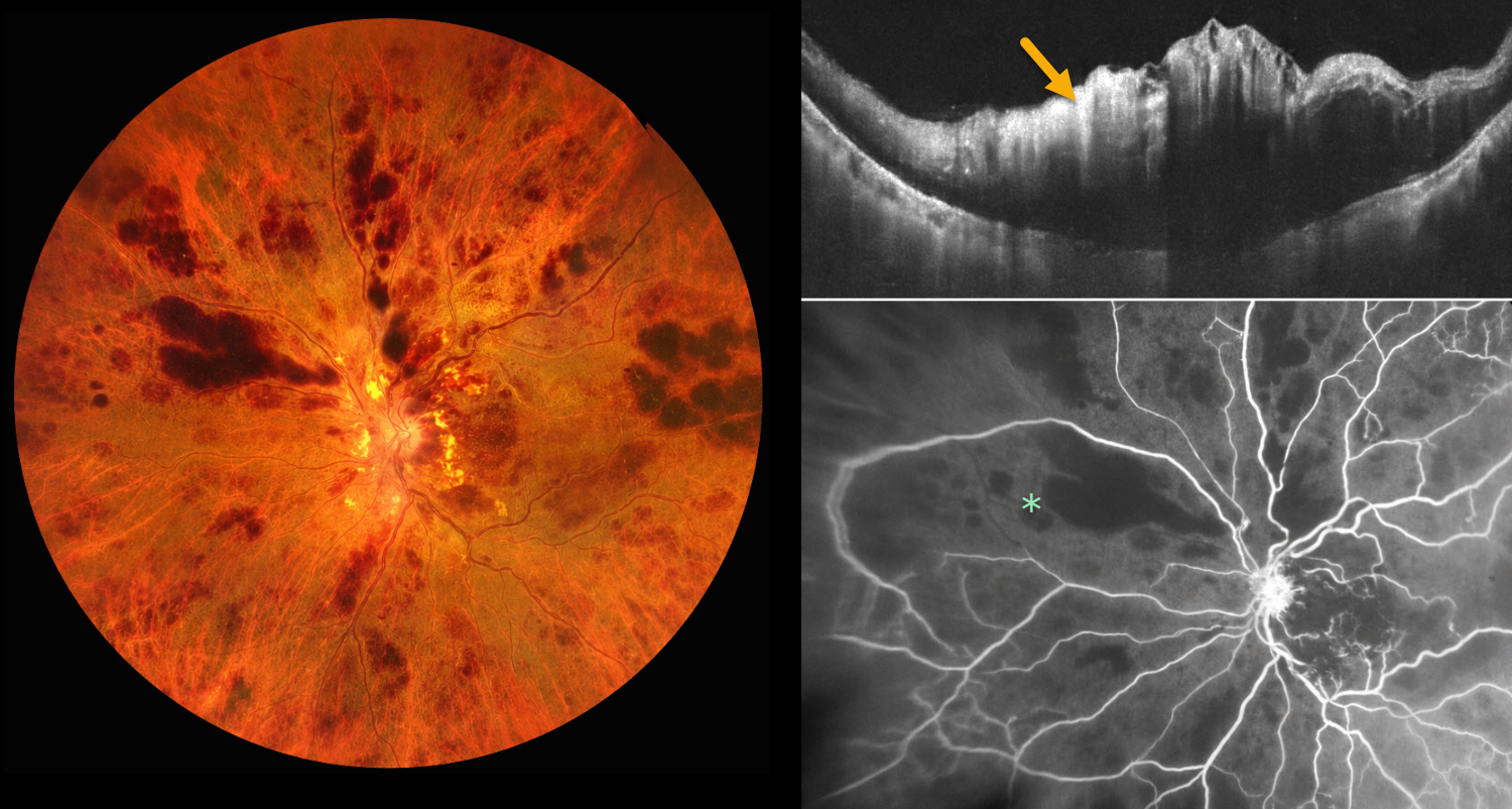
This 78YO male presented with 1 week of sudden, painless decreased vision in his left eye. Vision was 20/20 in his normal OD and light perception OS. Despite iris neovascularization, intraocular pressure was normal.
Color photography shows a hemorrhagic central retinal vein occlusion (CRVO) with variably confluent inner retinal hemorrhages and scattered nerve fiber layer infarcts. OCT scanning shows marked macular thickening with a hyperreflective inner retina. Fluorescein angiography shows profound diffuse retinal capillary loss. An intravitreal anti-VEGF injection was given. One week later, the rubeosis completely regressed, and full panretinal photocoagulation was performed.
Learning Points:
Ischemic CRVO, which accounts for 20% of eyes, is characterized by severe visual loss (typically worse than 20/200), a relative afferent pupillary defect, extensive retinal hemorrhages, cotton-wool spots, marked venous dilation and tortuosity, optic disc edema, and large areas of angiographic nonperfusion of the retinal capillaries.
Untreated iris neovascularization usually leads to neovascular glaucoma (NVG). Treatment involves an immediate anti-VEGF injection to allow for more rapid regression of neovascularization, followed by full panretinal photocoagulation. NVG is much less common in the modern anti-VEGF era, since the majority of these eyes also have macular edema that requires ongoing anti-VEGF therapy.
CRVO WITH CENTRAL BOUQUET HEMORRHAGE
Originally posted on @retina.rocks June 26, 2026
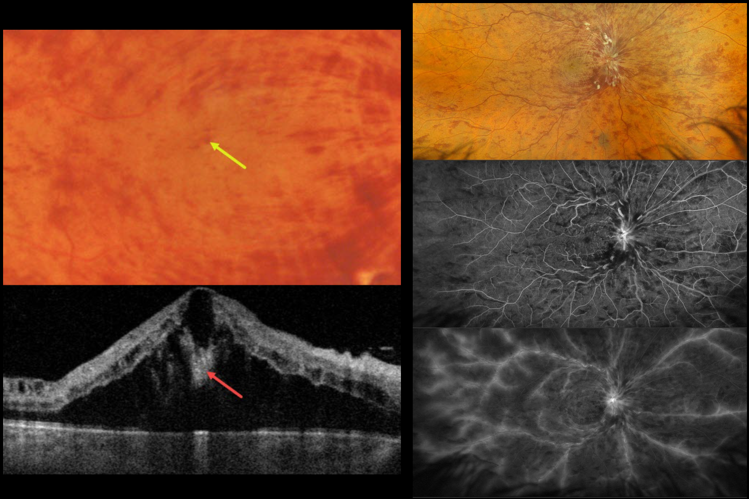
This healthy 52YO male presented with recent severe vision loss in his right eye. Vision was 20/400 OD and 20/20 in his normal OS.
Color photography and Optos RGB imaging show a central retinal vein occlusion (CRVO) with marked retinal hemorrhages and macular edema. Inner blood-filled retinal cysts are noted on funduscopy and swept-source OCT. On fluorescein angiography, the occlusion is fairly well perfused with late leakage from the nerve, macula, and retinal veins. Monthly anti-VEGF therapy was started.
Learning Points:
Cicinelli et al recently reported that central bouquet hemorrhages in retinal vein occlusion are associated with poor visual outcomes due to localized effects of elevated venous pressure and macular ischemia (IOVS 2025;66(12):62).
Prior to the introduction of intravitreal injections in the early 2000s, there were essentially no treatments for CRVO-related macular edema. Anti-VEGF injections are now generally the initial treatment of choice, with about 50-60% of eyes gaining at least 3 lines of vision. Intravitreal steroids, usually Ozurdex, are more often used as secondary treatment. Macular grid laser photocoagulation is not performed because it has been shown to confer no visual benefit in the Central Vein Occlusion Study Group (Ophthalmology 1995;102:1425-1433).
COMBINED CRAO-CRVO
Shraddha Raj Shrivastava and Manish Nagpal
Originally posted on @retina.rocks April 20, 2026
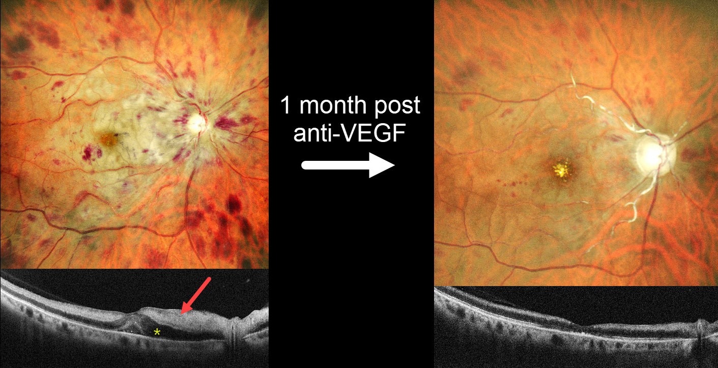
This 57YO female with a history of severe hypertension presented with one day of severe vision loss in her right eye. Blood pressure was 200/100 mmHg. Vision was hand motion OD and 20/20 in her normal OS.
MultiColor SLO imaging shows inner retinal opacification sparing the macular center, consistent with a fresh central retinal artery occlusion (CRAO). A central retinal vein occlusion (CRVO) is also noted with a swollen nerve and retinal hemorrhages in all quadrants. OCT scanning shows marked hyperreflectivity with loss of the band detail throughout the inner and mid-retinal layers. The peripapillary retina is edematous with fluid in the nasal outer nuclear layer and mild foveal subretinal fluid.
She was diagnosed with a combined CRAO-CRVO. An intravitreal anti-VEGF injection, and 1 month later, vision improved to 20/400. Repeat imaging shows marked improvement in the retinal hemorrhages, retinal opacification, and macular fluid, although new foveal lipid is noted. The nerve is flat and pale, and the major proximal arterioles are white without a blood column. The inner retina remains hyperreflective on OCT but is thinning with loss of the normal architecture. Panretinal photocoagulation was scheduled.
Learning Points:
Combined CRAO-CRVO is thankfully an extremely rare event, accounting for 0.3% of all retinal vascular occlusions (Raval et al, Indian J Ophthalmology 2020;68:2136-2142). Risk factors appear similar to those associated with isolated occlusions. The CRAO may be embolic or secondary to compression from a swollen optic nerve.
COMBINED CRAO-CRVO
Malvika Singh and Manish Nagpal
Originally posted on @retina.rocks March 3, 2026
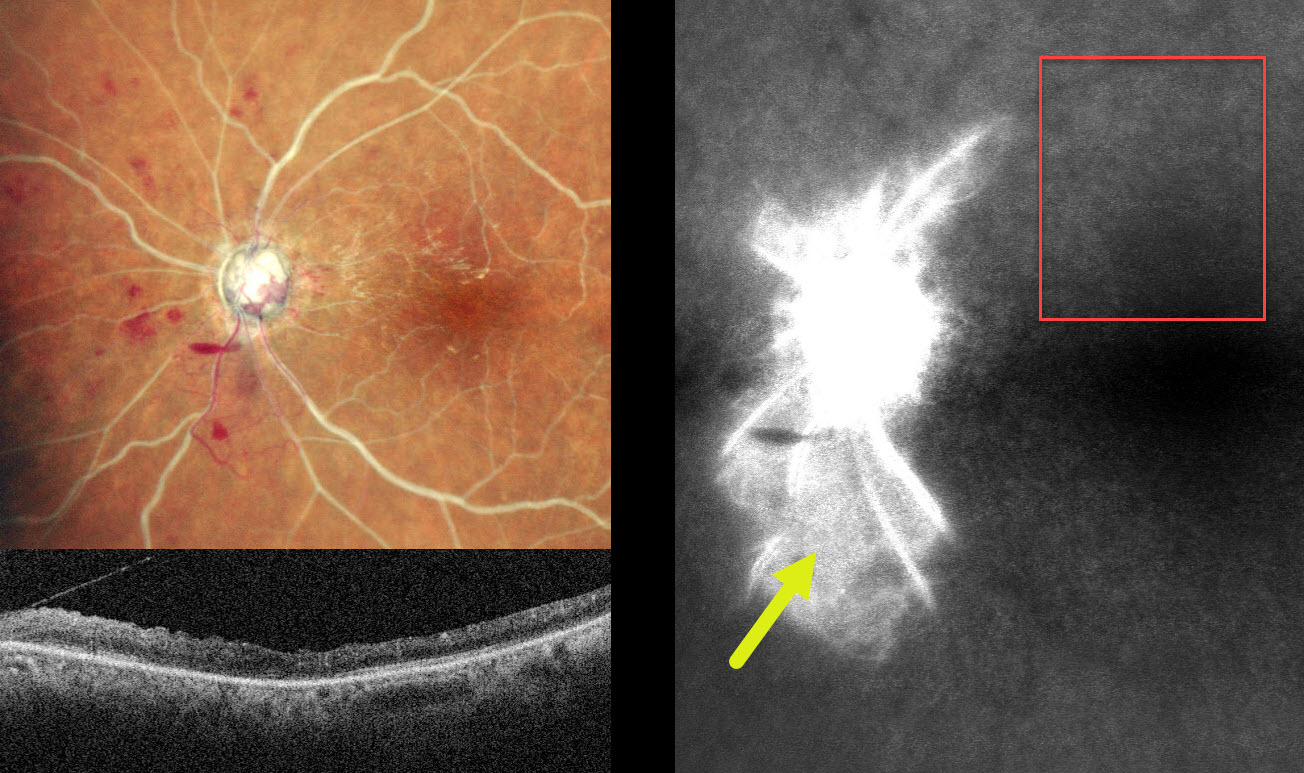
This 60YO male presented with sudden vision loss in his left eye 1 month earlier. Vision was 20/20 in his normal OD and no light perception OS.
MultiColor SLO imaging shows a combined central retinal artery occlusion (CRAO) and central retinal vein occlusion (CRVO) with white arteries and veins with no blood column. Disc neovascularization is present inferotemporally, and a small preretinal hemorrhage is noted just inferonasal to the nerve. OCT shows a disorganized hyperreflective inner retina. Fluorescein angiography shows profound panretinal ischemia with a small island of preserved capillary bed inferior to the nerve.
Vasculitis workup was negative. An emergent stroke workup, including carotid Doppler, MRI, and neurologic consultation, was ordered. Given our patient’s vision, comfort measures for his left eye were recommended.
Learning Points:
Combined CRAO-CRVO is thankfully an extremely rare event, accounting for 0.3% of all retinal vascular occlusions (Raval et al, Indian J Ophthalmology 2020;68:2136-2142). Risk factors appear similar to those associated with isolated occlusions. The CRAO may be either embolic or secondary to compression from a swollen optic nerve.
LEUKEMIA WITH PSEUDO-BILATERAL CRVO
Mattie Adams
Originally posted on @retina.rocks July 28, 2025
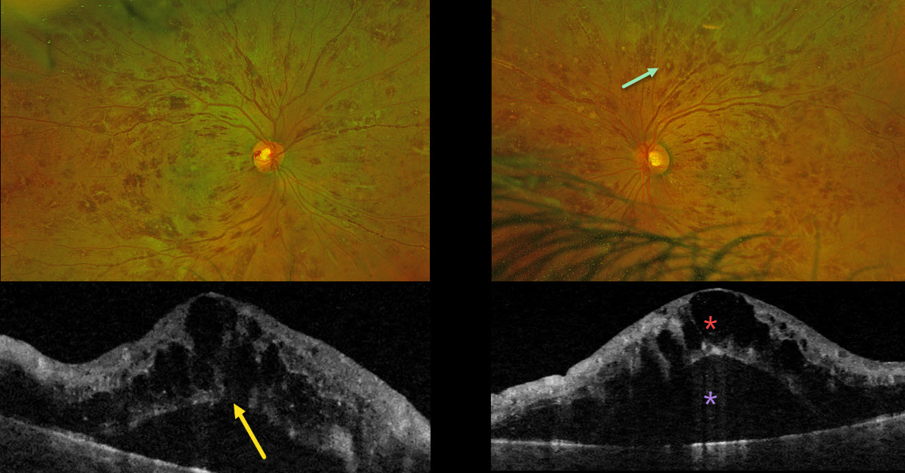
This previously healthy 38YO male presented with 1 month of bilateral blurriness. Vision was 20/60 OD and 20/80 OS.
Optos color RG imaging shows severe, bilateral, mostly white-centered retinal hemorrhages extending from the maculae into the retinal peripheries. Triton swept-source OCT shows marked bilateral cystic foveal swelling with subretinal fluid. An outer macular hole is probably present OD. His white blood cell count was markedly elevated at 206,400. Subsequent emergent oncologic workup led to the diagnosis of chronic myelogenous leukemia.
Learning Points:
The term “Roth spot” was named after Dr. Moritz Roth, who, in 1872, noted white-centered hemorrhages in patients with bacterial endocarditis. He felt that these white spots were septic emboli that originated from an infected cardiac valve. However, similar lesions can be seen in many other disorders, including leukemia, hyperviscosity, anemia, diabetes, and hypertension (Duane et al Ophthalmology 1980;87:66-69). The white material may represent a variety of causes, including fibrin. So, these lesions are best called white-centered hemorrhages, unless one is specifically referring to those found associated with bacterial endocarditis.
In our patient’s case, the severity of the retinal hemorrhages gave an appearance resembling bilateral central retinal vein occlusions (CRVO). However, CRVO hemorrhages are located predominantly in the nerve fiber layer (our patients’ hemorrhages were deep and white-centered), and the optic nerve is usually swollen with this degree of hemorrhage (our patients’ nerves were flat).
CRVO
Originally posted on @retina.rocks July 2, 2024
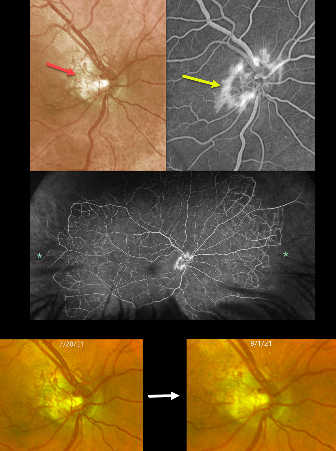
This 57YO male had a history of an old central retinal vein occlusion (CRVO) in his left eye with hand motion vision. After being lost to follow-up for 5 years, he presented on 7/28/21 with an asymptomatic CRVO with 20/25 vision in his right eye.
Optos color RG imaging shows diffuse disc neovascularization (NVD) that leaks on angiography. Ultrawidefield fluorescein angiography shows good posterior perfusion but with marked encircling peripheral ischemia. The neovascularization partially regressed 6 weeks after panretinal photocoagulation and completely regressed with subsequent laser and anti-VEGF injections (not shown).
Learning Points:
Anterior segment neovascularization is more commonly noted with central retinal vein occlusions, whereas posterior segment neovascularization is more common with branch retinal vein occlusions. Hayreh’s classic 1983 paper noted that only 5% of eyes with CRVO will develop NVD, as in our patient (Ophthalmology 1983;90:488-506).
The Central Vein Occlusion Study (CVOS) defined ischemic CRVO as having at least 10 disc areas of angiographic retinal nonperfusion as determined by standard photographic views with the Canon (60 degrees) or Topcon (45 degrees) wide-angle fundus camera (Ophthalmology 1995;102:1434-1444). Although clearly ischemic peripherally, our patient would have been classified by the CVOS as a perfused occlusion.
As with diabetic retinopathy (see Brucker, Retina 2021;41:461-463), ultrawidefield imaging and anti-VEGF injections have thrown a literal wrench into how we think about, classify, and treat retinal disorders.
CRVO + CILIORETINAL ARTERY OCCLUSION
Amit Nandan Tripathi and Vipin Rana
Originally posted on @retina.rocks June 18, 2024
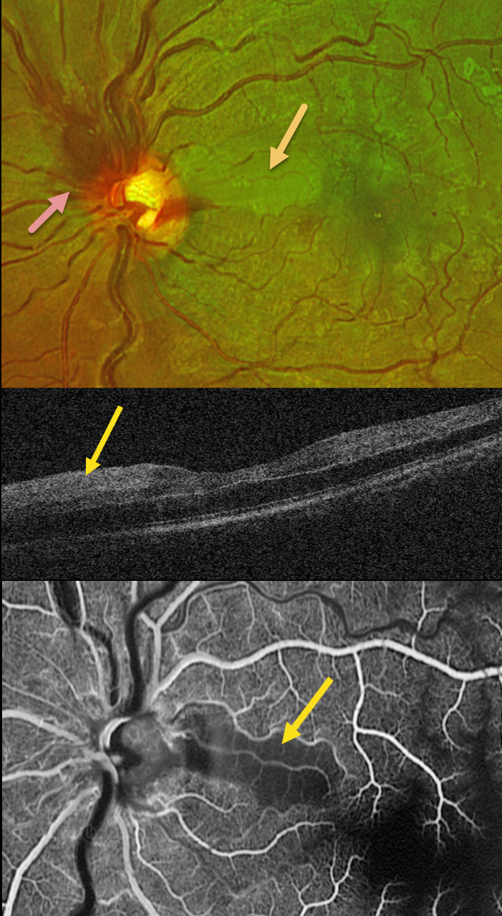
This healthy 30YO male presented with sudden vision loss in his left eye. Vision was 20/20 in his healthy right eye and counting fingers in his left eye.
Optos color RG imaging shows an acute non-ischemic central retinal vein occlusion (CRVO) with mild inner retinal hemorrhages scattered in all quadrants. The optic nerve is somewhat swollen superiorly, and there is an acute cilioretinal artery occlusion in the superior papillomacular bundle.
This area is hyperreflective on OCT. Fluorescein angiography shows a well-perfused CRVO except for a localized area of ischemia within the nasal occlusion.
Learning Points:
Some patients with CRVO will develop a secondary non-embolic cilioretinal artery occlusion caused by either compression from optic nerve edema or from a sudden rise in intraluminal retinal capillary bed pressure (Hayreh et al, Retina 2008;28:581-594). Observation was recommended, and vision improved to 20/60 five days after symptom onset.
CRVO + SECONDARY BRAO
Will Gibson
Originally posted on @retina.rocks April 15, 2024
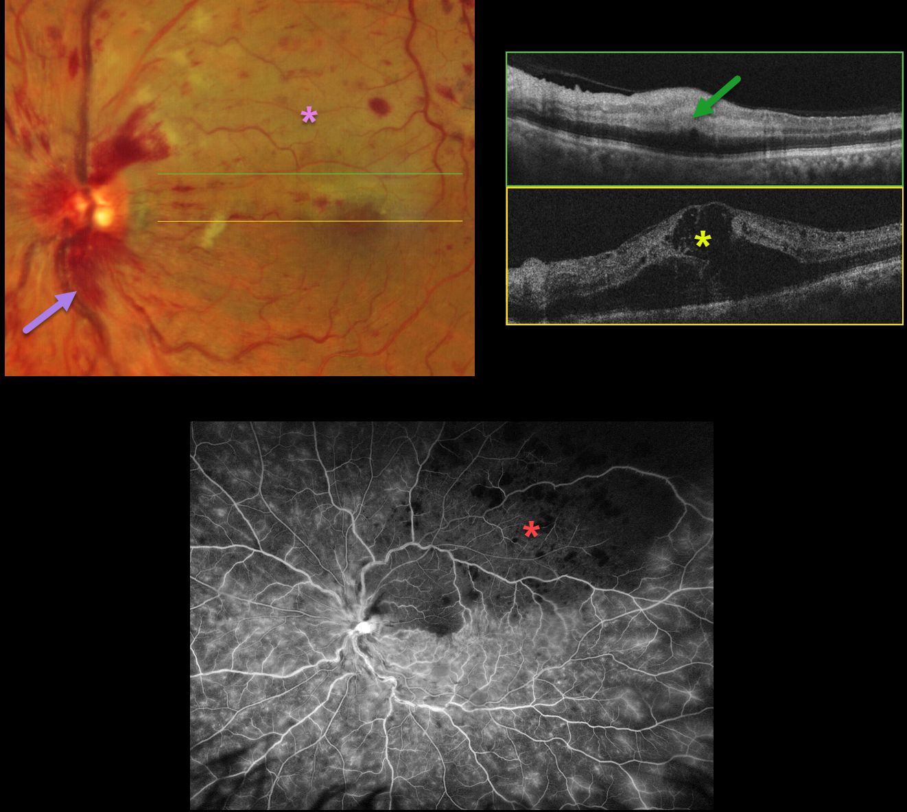
This 66YO male presented with sudden vision loss in his left eye. Vision was 20/30 in his normal right eye and counting fingers in his left eye.
Optos color RG imaging shows a central retinal vein occlusion (CRVO) with mild retinal hemorrhages in all quadrants, except for more moderate hemorrhages superotemporally. A secondary superior branch retinal artery occlusion (BRAO) bisects the fovea.
OCT scanning shows marked central edema, with hyperreflective and thickened inner retina within the BRAO. Fluorescein angiography shows a well-perfused CRVO, except for delayed arterial filling and ischemia within the BRAO.
Learning Points:
Some patients with CRVO will develop a secondary non-embolic BRAO caused by compression from optic nerve edema. However, our patient’s optic nerve did not appear that swollen. Our patient was started on anti-VEGF therapy for the severe macular edema.
CRVO
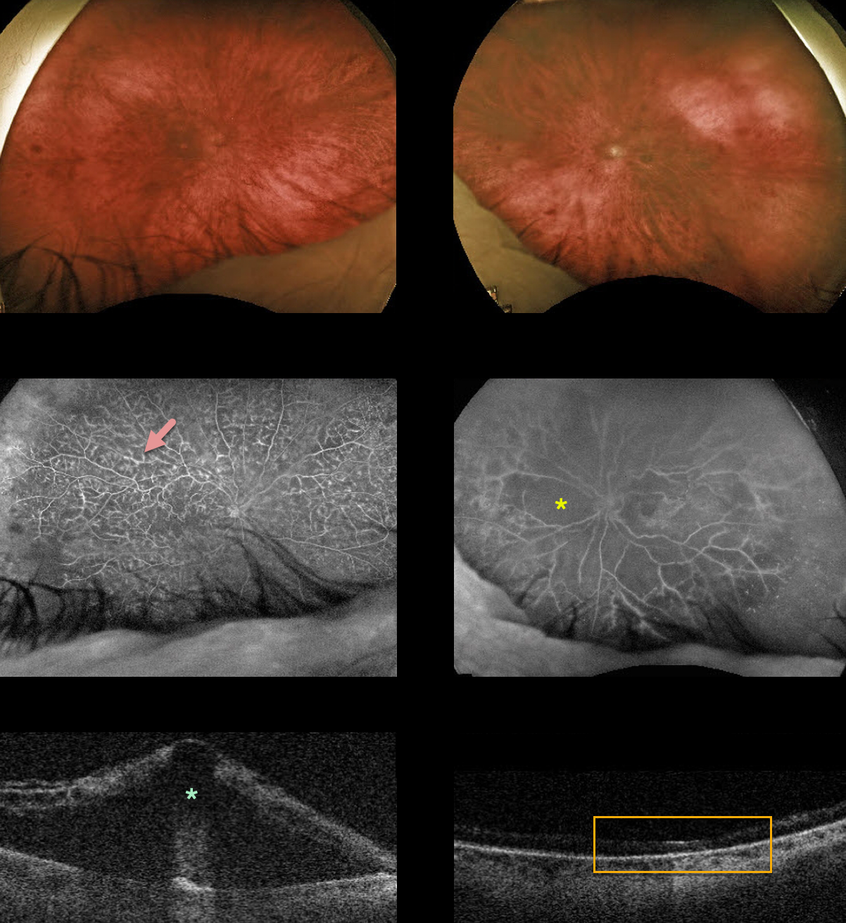
Originally posted on @retina.rocks December 22, 2022
This 78YO male presented with counting finger vision in his right eye. He had a prior CRVO in his left eye, causing hand motion vision.
Optos color imaging shows mild to moderate bilateral scattered retinal hemorrhages. Fluorescein angiography shows fairly good perfusion with leaking vessels OD with severe diffuse ischemia OS.
Swept-source OCT shows severe central macular edema OD with an atrophic left macula.
Anti-VEGF therapy was started for the edema OD.
Learning Points:
Prior to the introduction of intravitreal injections in the early 2000s, there were essentially no treatments for CRVO-related macular edema.
Anti-VEGF injections in general are now the initial treatment of choice, with about 50-60% of eyes gaining at least 3 lines of vision. Intravitreal steroids, usually Ozurdex, are more often used as secondary treatment.
Macular grid laser photocoagulation is not performed since it was shown to have no visual benefit in the Central Vein Occlusion Study Group (see Ophthalmology 1995;102;1425-1433).
CRVO + CHRPE + MYELINATED NERVE FIBER LAYER
Will Gibson
Originally posted on @retina.rocks October 27, 2022
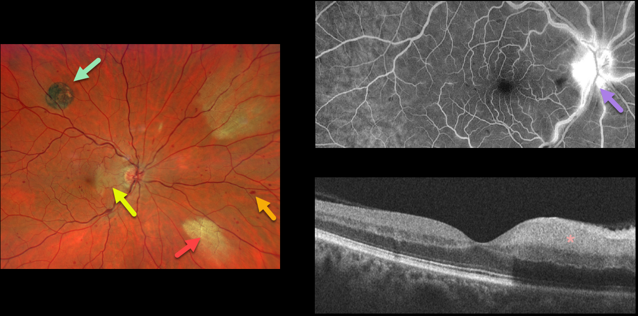
This 52YO male presented with acute visual symptoms in his right eye. Vision was 20/20.
Optos imaging shows a non-ischemic central retinal vein occlusion (CRVO) with mild scattered retinal hemorrhages in all quadrants. An acute nasal macular branch retinal artery occlusion (BRAO) is noted, along with a small round patch of congenital hypertrophy of the RPE (CHRPE) superotemporally, and two patches of myelinated nerve fiber layer (NFL) nasally.
Fluorescein angiography shows a well-perfused central retinal vein occlusion (CRVO) with late optic nerve leakage. OCT scanning shows hyperreflectivity of the inner nasal retina.
CRVO WITH SECONDARY BRAO
Originally posted on @retina.rocks October 18, 2022
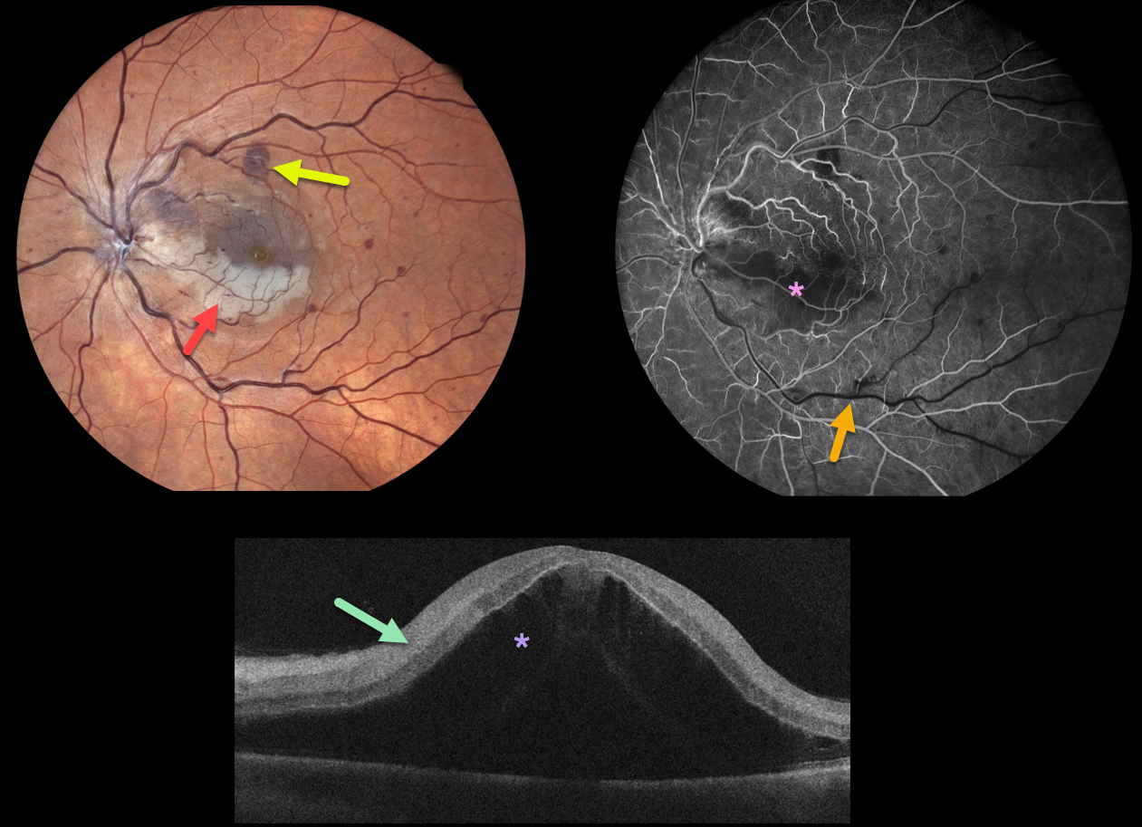
This 49YO male woke with counting finger vision in his left eye. Fundus photography shows a fresh central retinal vein occlusion (CRVO) with mild retinal hemorrhages. An acute inferior macular branch retinal arterial occlusion (BRAO) is also noted.
OCT B-scan shows marked inner retinal hyperreflectivity and severe macula edema.
Fluorescein angiography in the venous laminar phase shows inferior macular hypofluorescence from combined blockage from the ischemic white retina, along with a filling defect from the BRAO. The retinal perfusion elsewhere is good.
Some patients with CRVO will develop a secondary non-embolic BRAO caused by compression from optic nerve edema. However, our patient’s optic nerve did not appear that swollen. Anti-VEGF therapy was started for the severe macular edema.
ISCHEMIC CRVO
Nivesh Gupta
Originally posted on @retina.rocks June 28, 2022
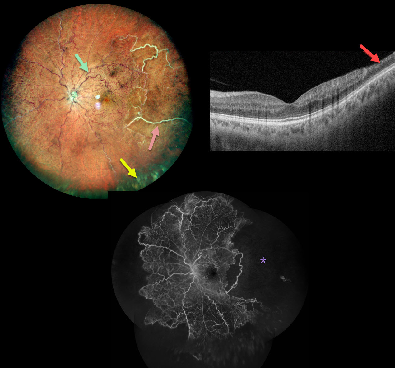
This healthy 34YO male presented with this relatively asymptomatic central retinal vein occlusion (CRVO). Vision was 20/20 OU.
Color imaging shows large temporal nonperfused retinal vessels with old inferior yellow vitreous hemorrhage. The retinal vessels are diffusely dilated and tortuous. OCT B-scan shows a remarkably normal central macula with some distal inner retinal thinning. Fluorescein angiography confirms widespread peripheral nonperfusion.
Anti-VEGF therapy, followed by panretinal photocoagulation, was administered.
Learning Points:
The Central Vein Occlusion Study (CVOS) defined ischemic CRVO as having at least 10 disc areas of angiographic retinal nonperfusion as determined by standard photographic views with the Canon (60 degrees) or Topcon (45 degrees) wide-angle fundus cameras (Ophthalmology 1995;102:1434-1444).
Although clearly ischemic, this patient might have been classified by the CVOS as a perfused occlusion, depending on the extent of peripheral ischemia imaged. As with diabetic retinopathy (see Brucker, Retina 2021;41:461-463), ultrawidefield imaging and anti-VEGF injections have thrown a literal wrench into how we think about, classify, and treat retinal disorders.
CRVO
Originally posted on @retina.rocks April 5, 2022
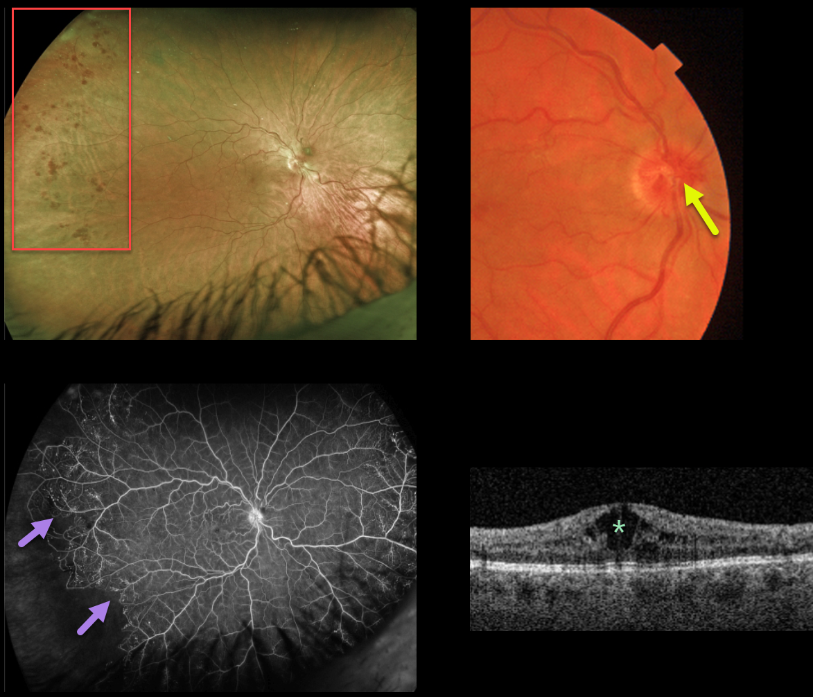
This 40YO female presented with 20/70 vision OD from a chronic central retinal vein occlusion (CRVO). The hemorrhages are virtually limited to the temporal periphery. Triton color imaging shows optic nerve collaterals.
Ultrawidefield Optos fluorescein angiography shows good posterior pole perfusion, nasal peripheral telangiectasia, and a sharp margin of infero-temporal peripheral ischemia.
Central cystoid edema is seen on swept-source OCT. Anti-VEGF injections were started for the macular edema.
Learning Points:
This sharply demarcated peripheral border of perfused ischemic retina is classically described in Eales and sickle retinopathy.
However, we have also found this to be a not uncommon finding in resolved CRVO, which has more classically been described as having vascular abnormalities extending outwards from the optic nerve. In this case, the optic disc collaterals point to the cause of the peripheral findings.
The Central Vein Occlusion Study (CVOS) defined ischemic CRVO as having at least 10 disc areas of angiographic retinal nonperfusion as determined by standard photographic views with the Canon (60 degrees) or Topcon (45 degrees) wide-angle fundus camera (Ophthalmology 1995;102:1434-1444).
Although clearly ischemic, this patient would have been classified by the CVOS as a perfused occlusion.
As with diabetic retinopathy (see Brucker, Retina 2021;41:461-463), ultrawidefield imaging and anti-VEGF injections have thrown a literal wrench into how we think about, classify, and treat retinal disorders.
CRVO + RAO + PAMM
Originally posted on @retina.rocks March 4, 2022
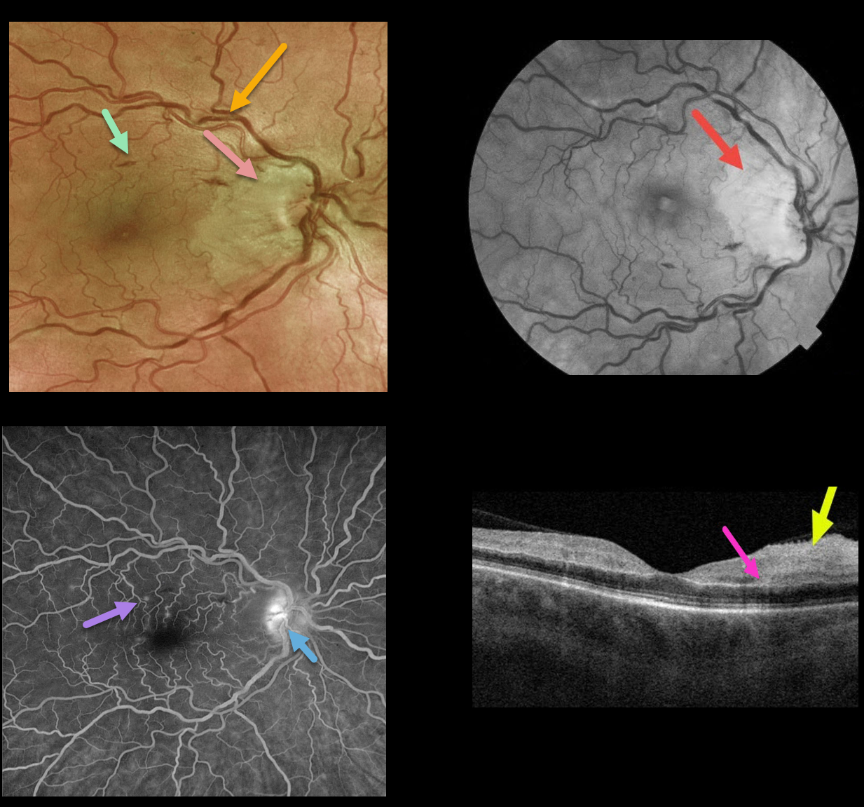
This 50YO female presented with counting finger vision in her right eye due to an acute central retinal vein occlusion (CRVO) with a nasal macular retinal artery occlusion (RAO).
Optos fundus photography shows retinal vascular tortuosity, mild retinal hemorrhages, a mildly swollen nerve, and white ischemic nasal macular retina. The ischemic retinal changes are most apparent on the red-free photo.
Fluorescein angiography (FA) shows some mild focal areas of macular leakage with late staining of the temporal disc.
OCT shows areas of inner retinal edema and hyperreflectivity. There is also a small area of mid-retinal hyperreflectivity consistent with paracentral acute middle maculopathy (PAMM).
Learning Points:
Cilioretinal artery occlusion is usually non-embolic and seen in association with giant cell arteritis, secondary to a CRVO, or as an isolated event. The occlusion may be due in part to arterial compression from a swollen nerve.
Pichi et al felt that CRVO PAMM-type lesions, as seen in our patient, are due to hypoperfusion from increased downstream intraluminal pressure (Br J Ophthalmol 2019;103:1137-1145).
We couldn’t find a definite cilioretinal artery in our patient, although the ischemia was within the distribution of one if one were present. Interestingly, the nasal macular capillaries appeared perfused on FA. However, FA captures just the inner retinal capillary plexus.
Although the OCT shows evidence of acute inner capillary plexus ischemia, the angiographically perfused inner retina supports a more PAMM-type mechanism in our case.
CRVO
Originally posted on @retina.rocks December 1, 2021
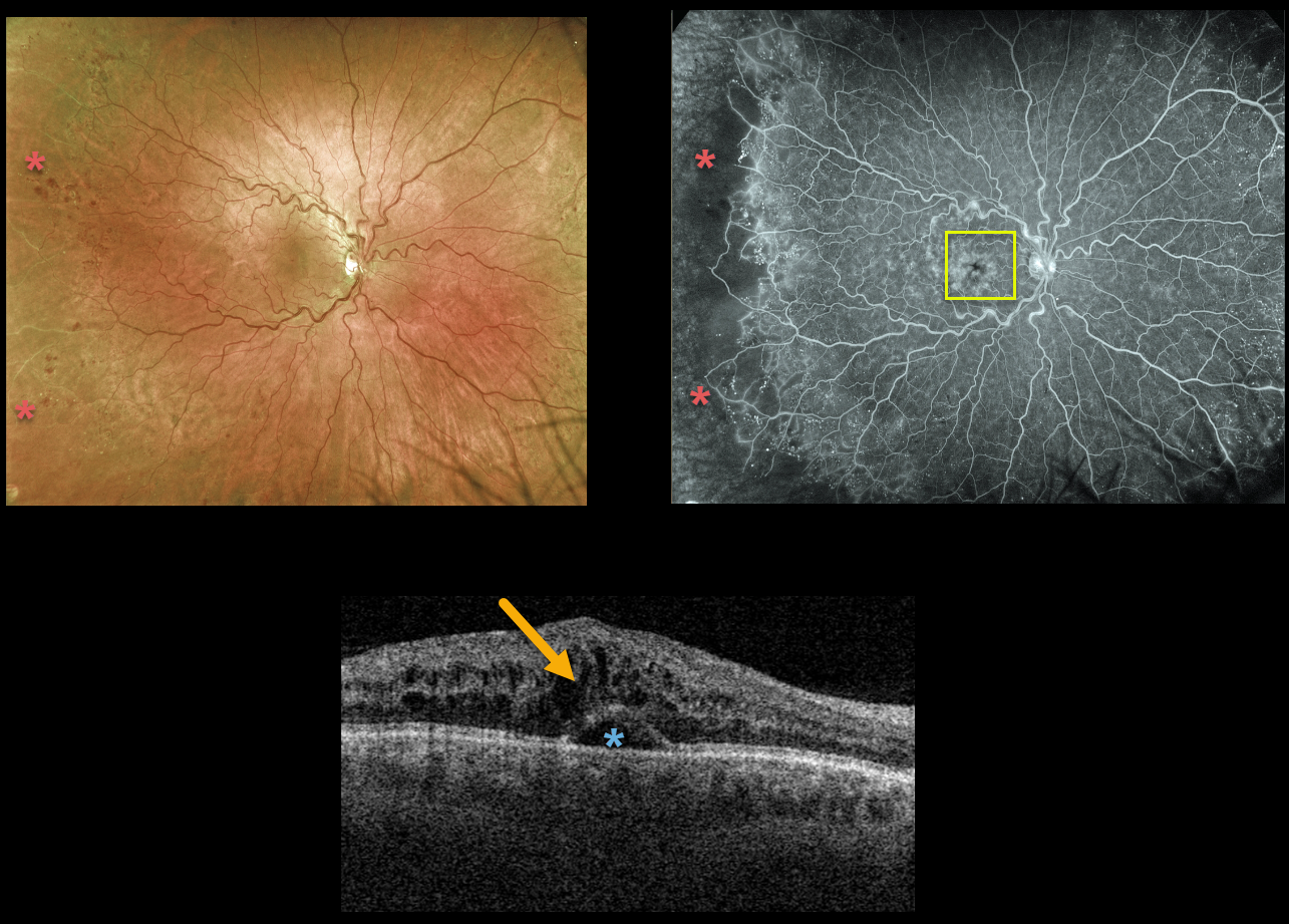
This 51YO male presented with 20/200 vision from an acute central retinal vein occlusion (CRVO).
There are virtually no posterior pole hemorrhages. Ultrawidefield Optos fluorescein angiography shows good posterior pole perfusion, although peripheral temporal ischemia is noted. Angiographic cystoid leakage is seen.
Triton swept-source OCT confirmed cystoid edema with some subfoveal fluid. Intravitreal anti-VEGF injections were started.
Learning Points:
The Central Vein Occlusion Study (CVOS) defined ischemic CRVO as having at least 10 disc areas of angiographic retinal nonperfusion as determined by standard photographic views with the Canon (60 degrees) or Topcon (45 degrees) wide-angle fundus camera (Ophthalmology 1995;102:1434-1444).
Although clearly ischemic, our patient would have been classified by the CVOS as a perfused occlusion.
As with diabetic retinopathy (see Brucker, Retina 2021;41:461-463), ultrawidefield imaging and anti-VEGF injections have thrown a literal wrench into how we think about, classify, and treat retinal disorders.
CRVO
Originally posted on @retina.rocks October 25, 2021
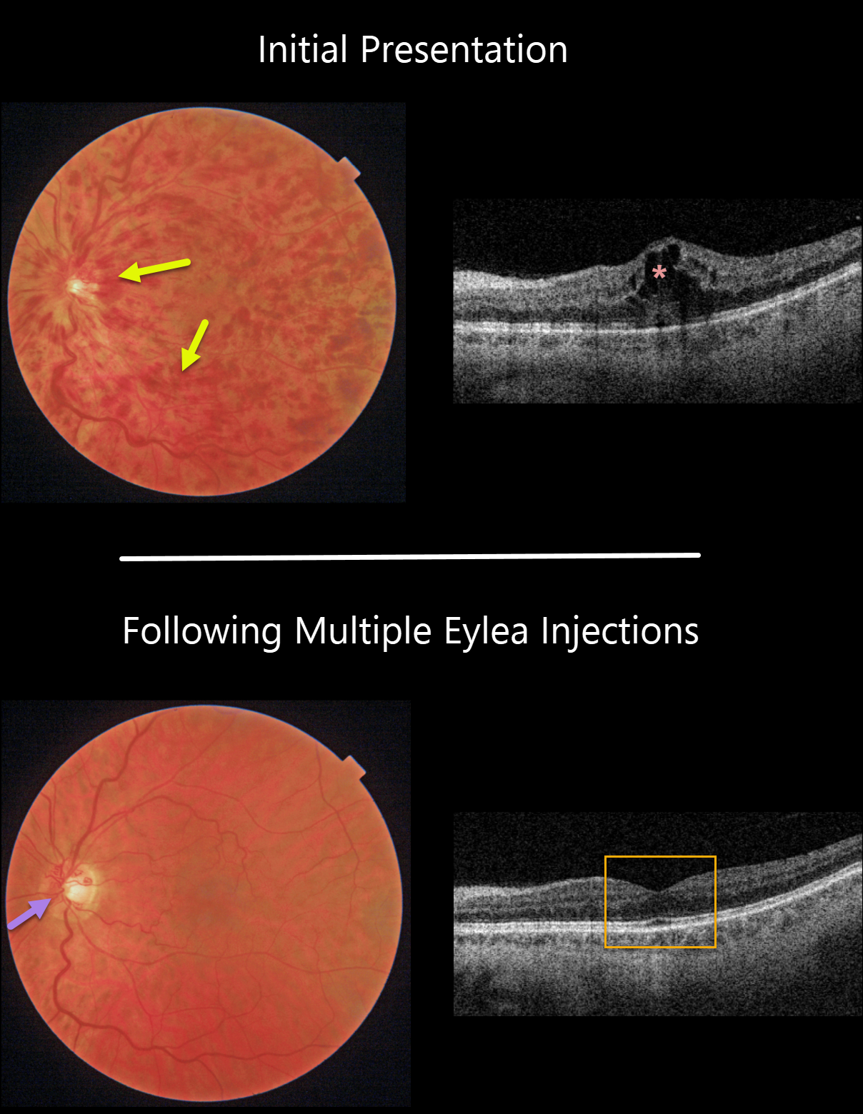
This 61YO male patient presented with a non-ischemic central retinal vein occlusion (CRVO), 20/30 vision, and macular edema.
With ongoing Eylea treat-extend protocol, the hemorrhages and macular edema completely resolved. Prominent optic nerve collaterals were noted.
Learning Points:
Before the introduction of intravitreal injections in the early 2000s, there were essentially no treatments for CRVO-related macular edema.
Anti-VEGF injections are now generally the initial treatment of choice, with about 50-60% of eyes gaining at least 3 lines of vision. Intravitreal steroids, usually Ozurdex, are more often used as secondary treatment.
Macular grid laser photocoagulation is not performed because it has been shown to confer no visual benefit in the Central Vein Occlusion Study Group (see Ophthalmology 1995;102;1425-1433).
CRVO
Originally posted on @retina.rocks June 30, 2021
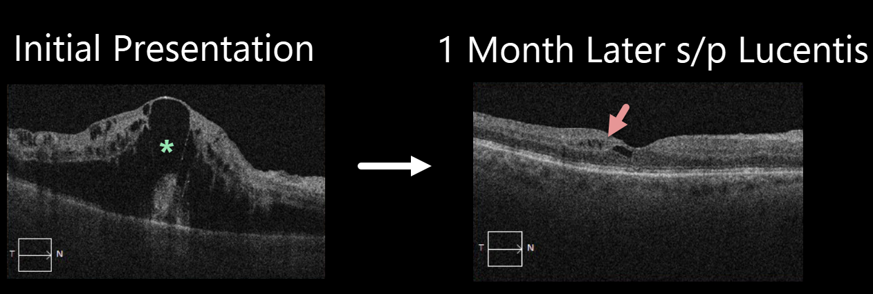
This 72YO male presented with marked macular edema and 20/150 vision from a nonischemic central retinal vein occlusion (CRVO).
Four weeks later, following a single Lucentis injection, vision improved to 20/70, and the edema almost completely resolved. Although most patients respond favorably to anti-VEGF therapy, our patient’s dramatic, rapid improvement following a single injection is quite unusual.
CRVO macular edema usually requires ongoing injections, and we are currently following a treat-and-extend protocol to maintain this improvement while minimizing the treatment burden.
Learning Points:
Prior to the introduction of intravitreal injections in the early 2000s, there were essentially no treatments for CRVO-related macular edema. Anti-VEGF injections in general are now the initial treatment of choice, followed by intravitreal steroids (usually Ozurdex).
Macular grid laser photocoagulation is not performed because it has been shown to confer no visual benefit in the Central Vein Occlusion Study Group (see Ophthalmology 1995;102;1425-1433).
CRVO
Originally posted on @retina.rocks May 5, 2021
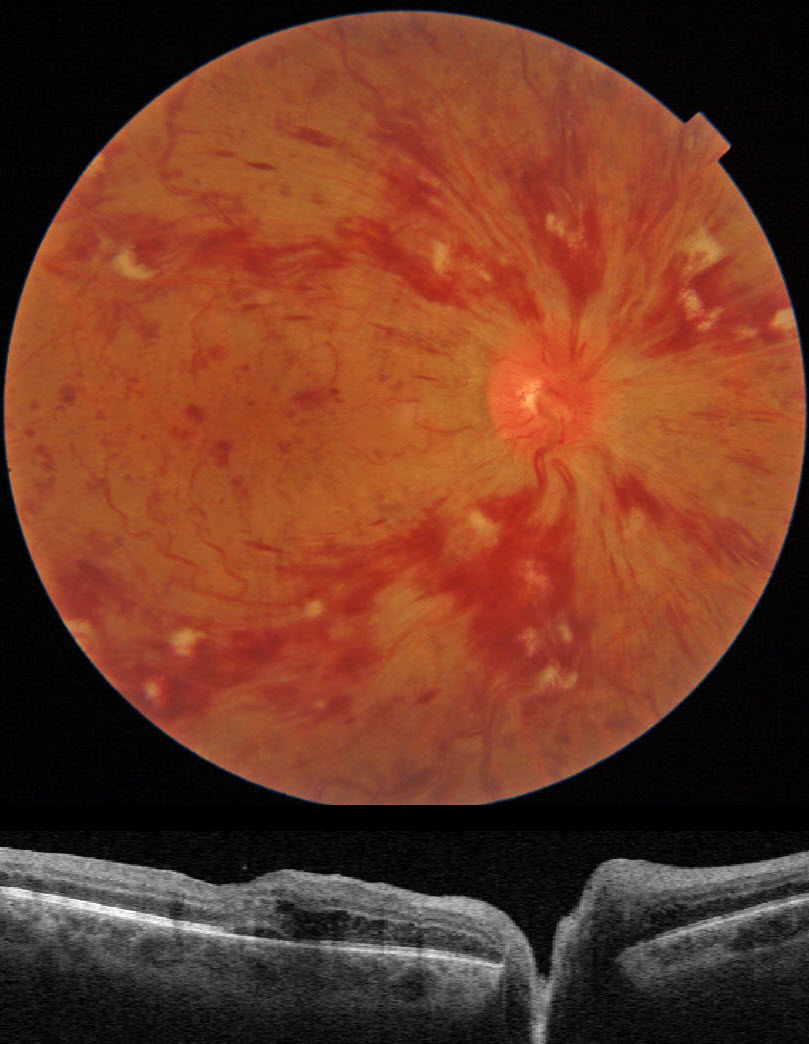
This patient presented with 20/70 vision from an acute central retinal vein occlusion (CRVO) with mild macular edema.
The mild vision reduction and the well-perfused fluorescein angiogram support the diagnosis of a non-ischemic CRVO.
He is currently undergoing intravitreal injections for the macular edema.
Learning Points:
Patients with CRVO are at risk of vision loss due to macular edema or anterior segment neovascularization. Macular edema is common in both non-ischemic and ischemic occlusions, although rubeosis is found almost exclusively with ischemic occlusions.
Iris neovascularization with neovascular glaucoma develops in about two-thirds of patients with untreated ischemic occlusions. Rubeosis is initially treated with an anti-VEGF injection, followed by aggressive, complete panretinal photocoagulation.
Some patients will need a glaucoma tube or a cyclodestructive procedure if the IOP remains too high.
CRVO
Originally posted on @retina.rocks April 22, 2021
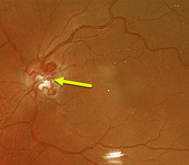
This 74YO female has large optic nerve collaterals from an old non-ischemic central retinal vein occlusion. Vision was 20/30.
Learning Points:
Collateral vessels are dilated, pre-existing vessels that bypass an occlusion. These are most commonly seen in the posterior pole, following retinal venous occlusions, often on the optic disc.
In collateral vessels, the endothelial tight junctions are intact, so they don’t leak angiographically. This helps differentiate them from neovascularization, which does leak.
CRVO
Originally posted on @retina.rocks March 23, 2021
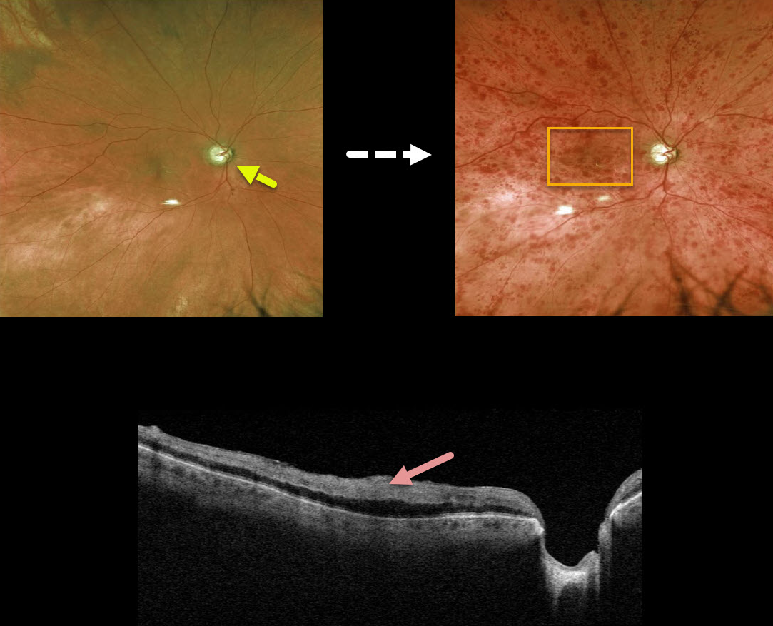
This 79YO male with a history of chronic open-angle glaucoma (COAG) presented with counting fingers vision from an acute nonischemic central retinal vein occlusion.
Even though his COAG was well-controlled with topical therapy, we felt that his glaucoma was a significant risk factor for the occlusion. A fundus photo from before the CRVO shows that significant glaucomatous cupping developed after the CRVO.
Triton swept-source OCT scanning shows marked inner retinal hyperreflectivity from the confluent macular blood.
Learning Points:
Patients with CRVO are at risk of vision loss due to macular edema or anterior segment neovascularization.
Macular edema is common in both non-ischemic and ischemic occlusions, although rubeosis is found almost exclusively with ischemic occlusions. Inner macular blood, as in this patient, can also cause vision loss.
Iris neovascularization with neovascular glaucoma develops in about two-thirds of patients with untreated ischemic occlusions. Rubeosis is treated initially with an anti-VEGF injection, followed by aggressive, complete panretinal photocoagulation.
Some patients will need a glaucoma tube or a cyclodestructive procedure if the IOP remains too high.
CRVO
Originally posted on @retina.rocks December 21, 2020
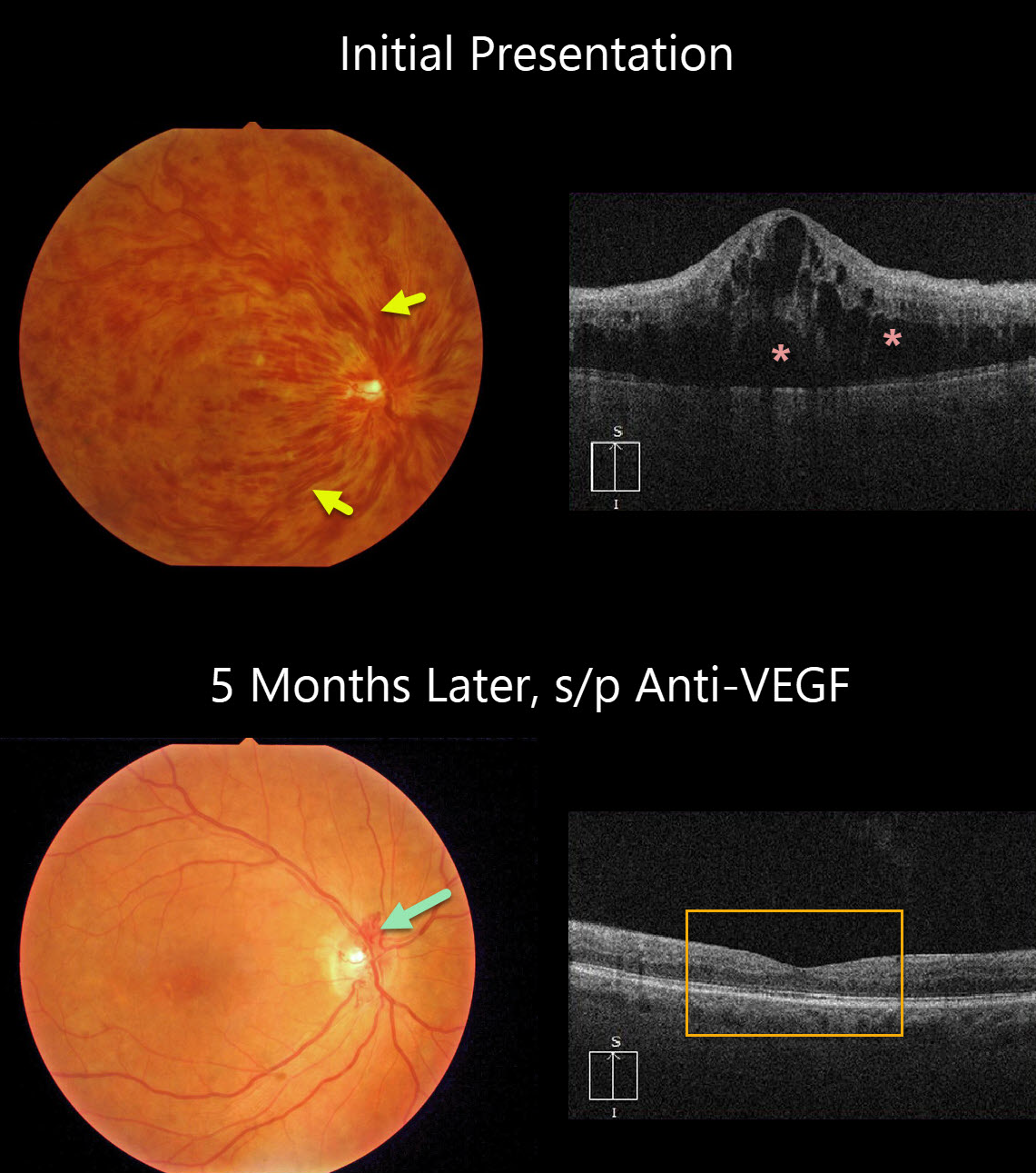
This patient presented with counting-finger vision from an acute hemorrhagic central retinal vein occlusion (CRVO) with marked macular edema. The poor vision and extensive retinal hemorrhages suggest that this is an ischemic CRVO.
After five monthly anti-VEGF injections, vision improved to 20/50, the retinal hemorrhages resolved dramatically, optic disc collaterals formed, and OCT showed complete resolution of all macular fluid. This degree of improvement is quite unusual.
Learning Points:
Patients with CRVO are at risk of vision loss due to macular edema or anterior segment neovascularization. Macular edema is common in both non-ischemic and ischemic occlusions, and this responds best to anti-VEGF therapy.
Rubeosis is found almost exclusively with ischemic occlusions, developing in about two-thirds of patients with untreated ischemic occlusions. This was common in the pre-anti-VEGF days.
However, since most patients with CRVO have macular edema and are receiving ongoing anti-VEGF therapy, neovascularization is now rarely seen unless patients present with untreated CRVO or therapy is discontinued.
CRVO
Originally posted on @retina.rocks July 22, 2020
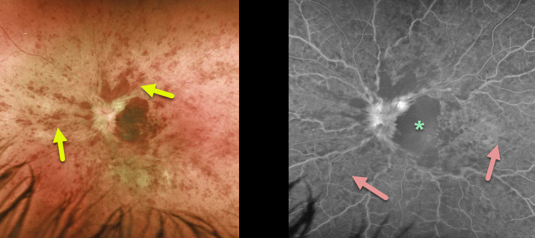
This patient is a 28YO who presented with hand motion vision from an acute central retinal vein occlusion (CRVO).
The poor vision and extent of retinal hemorrhages would suggest an ischemic occlusion. However, fluorescein angiography shows good peripheral perfusion, indicating a non-ischemic CRVO.
Angiography also shows marked foveal hypofluorescence, which may be due to a filling defect (ischemia) or a blockage. The contour of the dark, hypofluorescent macula precisely corresponds to the macular hemorrhage noted on the color photograph, indicating that this is due to blockage rather than ischemia.
Learning Points:
Although CRVOs can occur in younger patients, it is sometimes prudent to look for other systemic causes, including hypercoagulable conditions and hyperviscosity syndromes.
See Liu et al. Retina 2018;38;1175-1179 and Bremond-Gignac et al Retina 2020;40;1339-1343 for further discussion of the debate over the need for a medical workup in younger patients with CRVO.
CRVO
Originally posted on @retina.rocks June 15, 2020
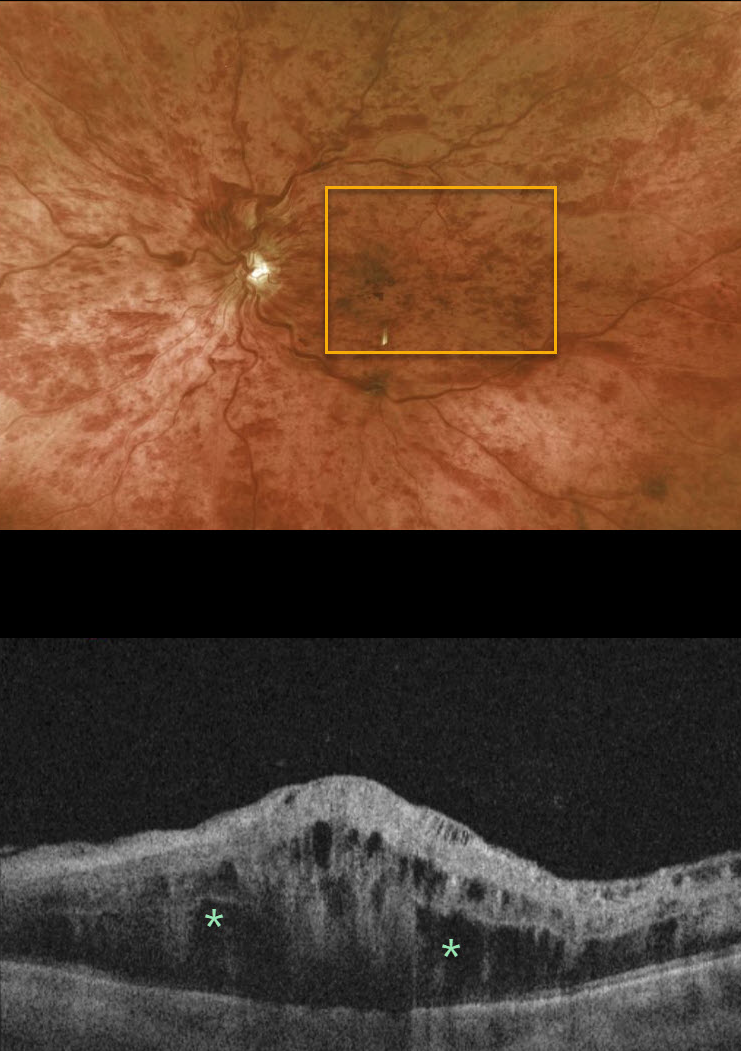
This 76YO male presented with 20/200 vision, the presence of an afferent pupillary defect, and extensive retinal hemorrhages. He had an acute ischemic central retinal vein occlusion (CRVO) with macular edema.
Patients with CRVO are at risk of vision loss due to macular edema or anterior segment neovascularization. Macular edema is common in both non-ischemic and ischemic occlusions, although rubeosis is found almost exclusively with ischemic occlusions.
Learning Points:
Intravitreal therapy in the early 2000s brought about the first successful treatment for CRVO-related macular edema. Anti-VEGF drugs are the first-line therapy, with the majority of patients gaining several lines of vision. Treatment is often ongoing, as it is for wet macular degeneration.
Although rubeosis was a common finding in the past (and is still seen if patients present with an untreated ischemic occlusion), this is now a rare occurrence since the vast majority of these patients have macular edema that is treated with ongoing anti-VEGF injections. Interestingly, if the injections are stopped several years later, rubeosis can still develop.
Iris neovascularization with neovascular glaucoma develops in about two-thirds of patients with untreated ischemic occlusions. Rubeosis is treated initially with an anti-VEGF injection, followed by aggressive, complete panretinal photocoagulation. Some patients will need a glaucoma tube or a cyclodestructive procedure if the IOP remains too high.
CRVO
Originally posted on @retina.rocks May 12, 2020
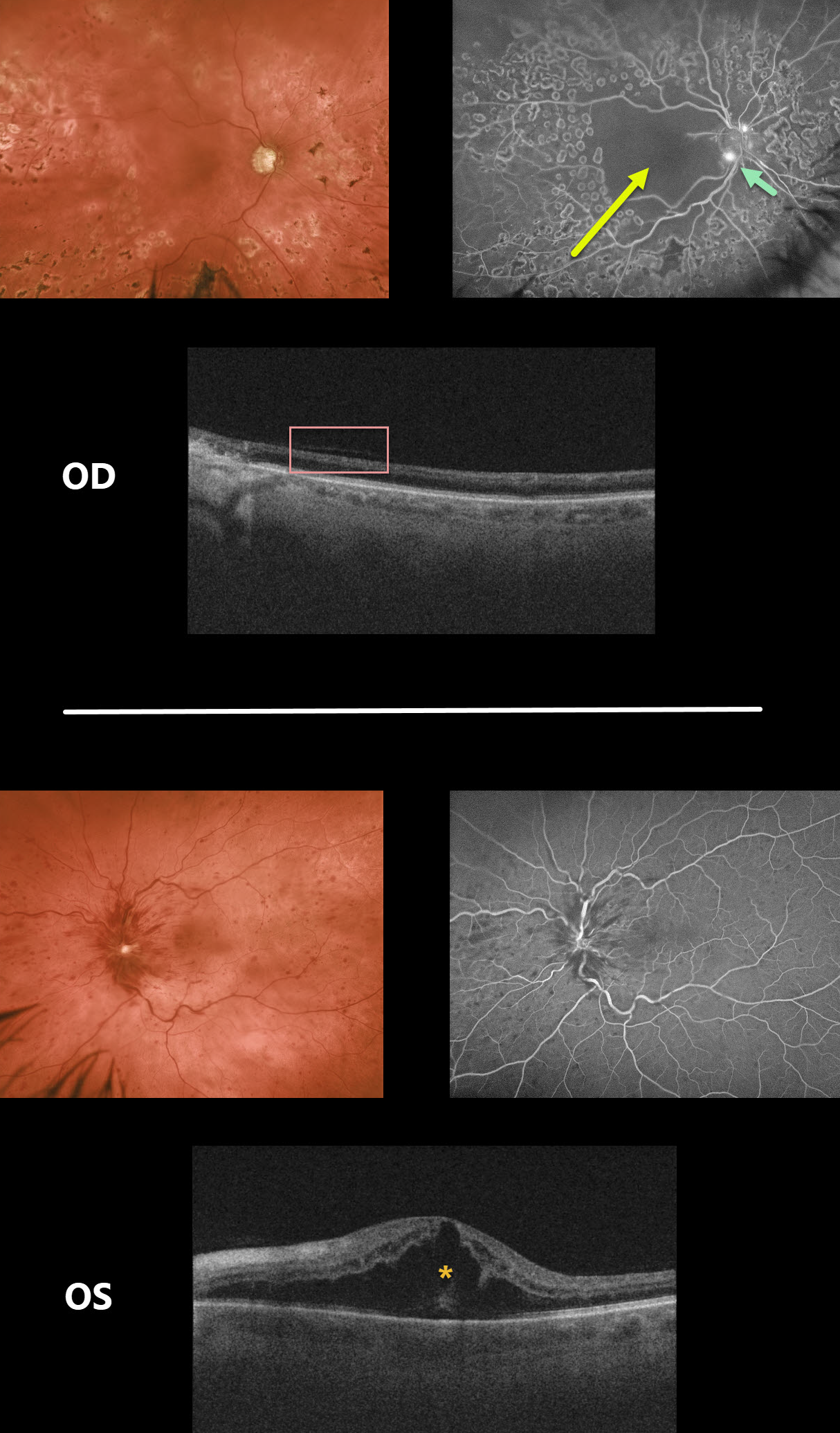
This patient had counting fingers vision OD due to a longstanding ischemic CRVO complicated by macular ischemia, disc neovascularization and neovascular glaucoma.
Fluorescein angiography shows profound capillary loss throughout the macula along with leaking disc neovascularization.
Due to the profound retinal ischemia there is marked OCT thinning of the inner two-thirds of the retina, giving a similar appearance to that seen with central retinal arterial occlusion.
She recently developed a new non-ischemic CRVO in her left eye with macular edema and 20/80 vision.
Learning Points:
This patient highlights several common and uncommon findings with CRVO. About 10% of patients will develop bilateral occlusions.
Eighty percent of occlusions are non-ischemic, although about one-third of these will convert to ischemic occlusions within several years. About 60% of ischemic occlusions will develop anterior segment neovascularization with neovascular glaucoma.
For some reason, posterior segment neovascularization rarely occurs (see the classic Hayreh 1983 paper Ophthalmology 1983;90;488-506).
CRVO
Originally posted on @retina.rocks January 7, 2020
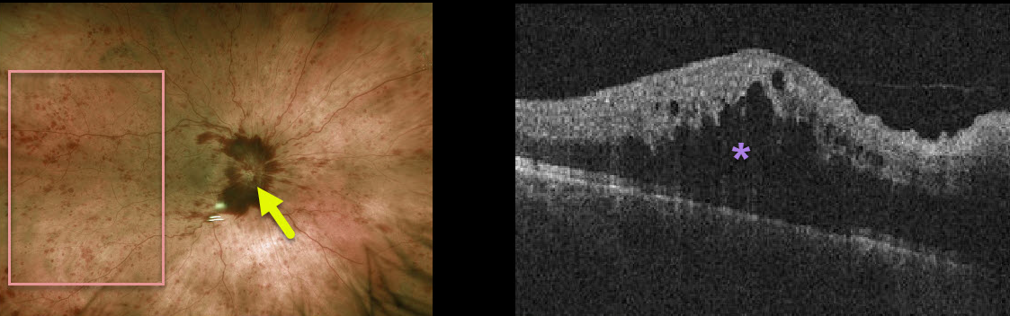
This patient has a non-ischemic central retinal vein occlusion (CRVO) with macular edema.
The fundus photo shows diffuse intraretinal hemorrhages, disc hemorrhages, and optic disc edema of the right eye. The OCT demonstrates marked macular edema.
Learning Points:
About 10% of patients with CRVO will develop bilateral occlusions. Eighty percent of occlusions are non-ischemic, although about one-third of these will convert to ischemic occlusions within several years.
About 60% of ischemic occlusions will develop anterior segment neovascularization with neovascular glaucoma. For some reason, posterior segment neovascularization rarely occurs (see the classic Hayreh 1983 paper Ophthalmology 1983;90;488-506).


