

Cysticercosis
<< Back to Cases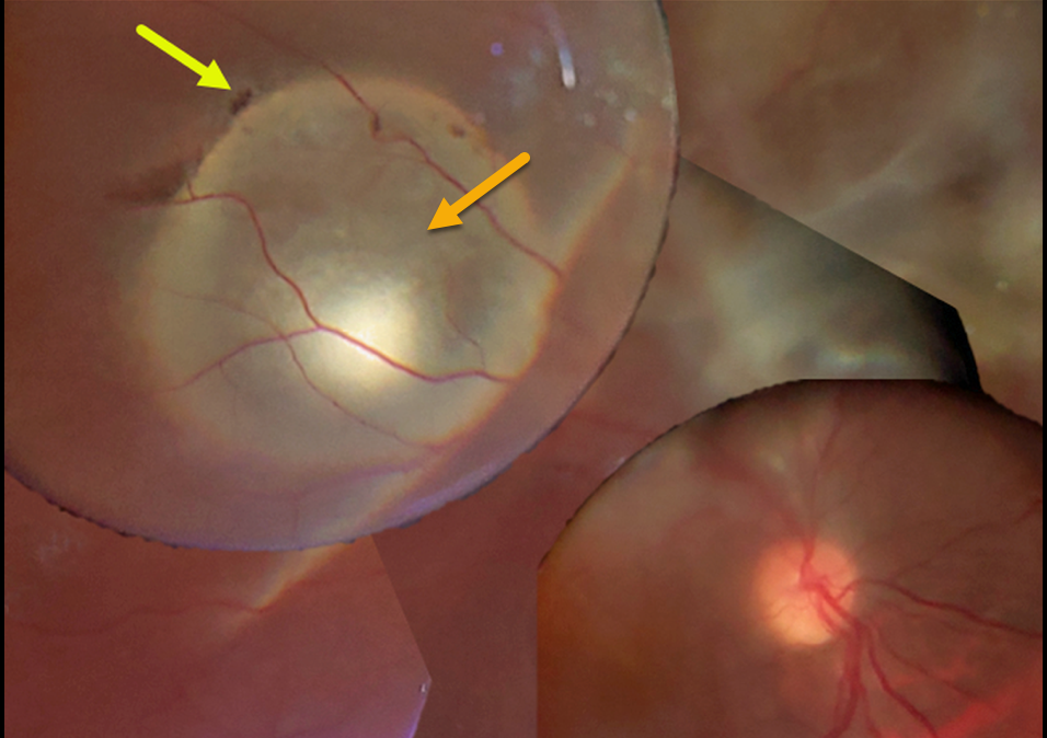
CYSTICERCOSIS WITH RHEGMATOGENOUS RETINAL DETACHMENT
Chhaya Bharti
Originally posted on @retina.rocks September 14, 2022
This 15YO boy presented with 20/200 vision. A large subretinal cysticercosis cyst is noted in the right distal superotemporal macula.
There is an overlying rhegmatogenous retinal detachment caused by a small retinal break. Unfortunately, the patient was subsequently lost to follow-up.
Learning Points:
Ocular cysticercosis is a rare parasitic eye infection caused by a pork tapeworm (larval form of the cestode, Taenia solium).
The ocular adnexa are most commonly affected, but the encysted organism can appear within the eye, including the anterior segment, vitreous cavity, and the subretinal space, as in this patient.
Vitrectomy with complete removal of the subretinal cyst is required since the dead organism can cause a severe inflammatory response.
For a great recent cysticercosis in ophthalmology review article, see Pujari et al, Surv Ophthalmology 2022;67:544-569.


