

Cystoid Macular Edema (CME)
<< Back to Cases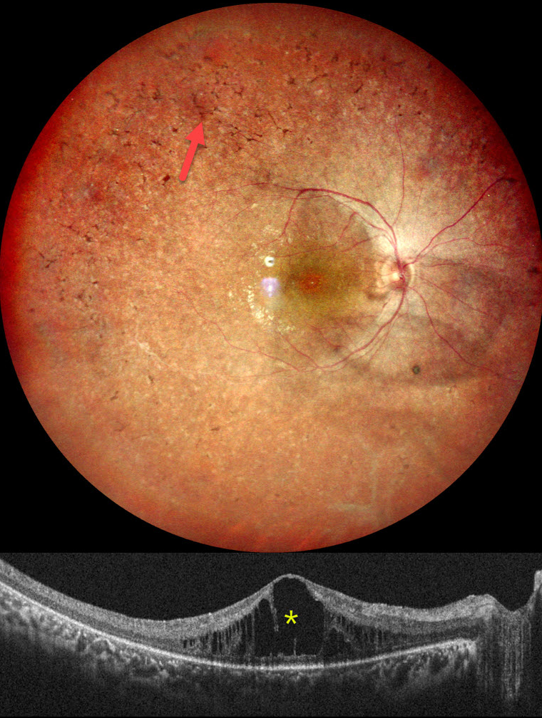
RETINITIS PIGMENTOSA WITH CME
Rohan Jain and Manish Nagpal
Originally posted on @retina.rocks September 15, 2025
This healthy 18YO female complained of night blindness for a few years. Her mother and sister have a history of retinitis pigmentosa (RP). Vision was 20/90 OU.
Pseudocolor SLO imaging of her right eye shows typical RP scarring with intraretinal pigment migration extending from the macula into the retinal periphery. There is loss of the foveal reflex and cystoid macular edema (CME). Identical findings were noted in the OS (not shown).
Learning Points:
RP is a clinically and genetically heterogeneous group of inherited retinal disorders, which can present sporadically or with any inheritance pattern (autosomal dominant, autosomal recessive, sex-linked, or mitochondrial). It is characterized by diffuse, progressive dysfunction of predominantly rod photoreceptors, followed by RPE degeneration with intraretinal pigment migration. Visual impairment usually presents as night blindness and progressive visual field loss.
CME is observed in up to 20% of RP patients, often without angiographic leakage. Pathological mechanisms include vitreous traction/epiretinal membranes or a breakdown of the blood-retinal barrier due to inflammation or RPE/Muller cell dysfunction (Gaudric et al, Progress in Retinal and Eye Research 2022;91:101092). First-line treatment is topical or oral carbonic anhydrase inhibitors (CAIs). Other options for refractory CME include steroids (topical, periocular, or intravitreal), anti-VEGF injections, or vitrectomy for a tractional component. Our patient was given topical CAIs but was immediately lost to follow-up.
BITEMPORAL HEMIANOPIA WITH RETINITIS PIGMENTOSA
Mattie Adams
Originally posted on @retina.rocks August 12, 2024
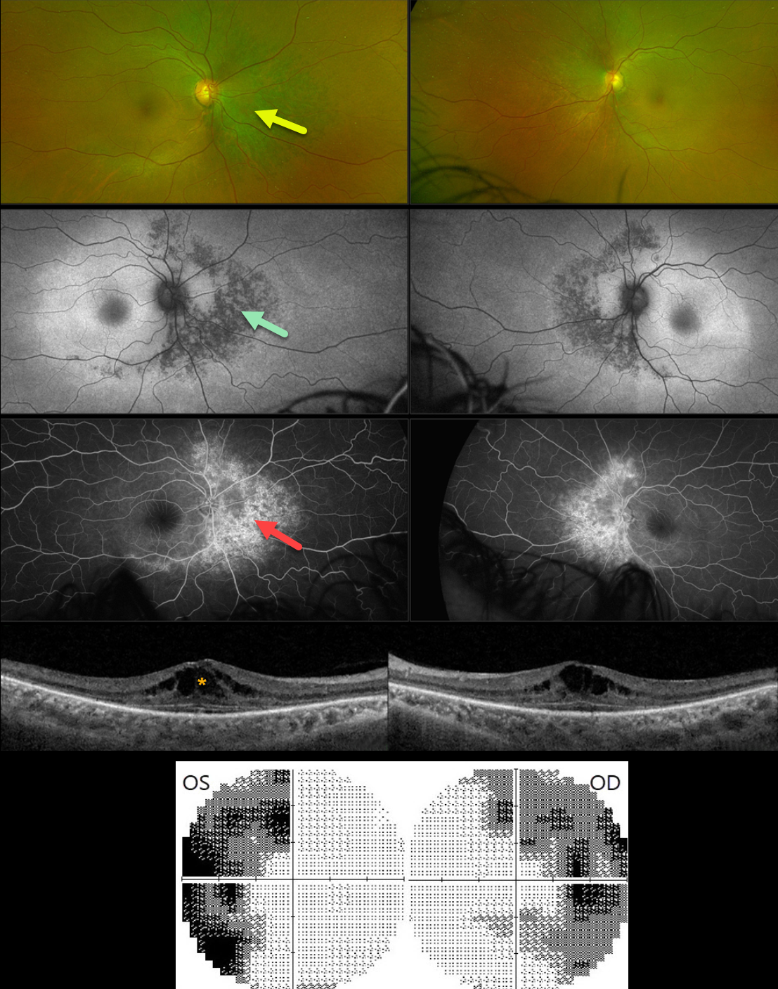
This 54YO female was referred for asymptomatic retinal changes. Vision was 20/30 OD and 20/20 OS.
Visual fields showed bitemporal hemianopia. What is your diagnosis?
Optos color RG imaging shows pigmentary changes with intraretinal pigment migration extending superiorly, nasally, and inferiorly from the optic nerves. These changes are hypoautofluorescent and appear as angiographic window defects. Bilateral cystoid edema is seen on Triton swept-source OCT.
Learning Points:
Although bitemporal hemianopia usually signifies retrobulbar pathology at the optic chiasm, retinal disease can sometimes also cause similar field loss. Our patient’s retinitis pigmentosa (RP) changes were centered around each optic nerve, causing the hemianopia. Genetic testing revealed double heterozygous pathogenic ARSG mutations. This genotype has been associated with atypical Usher syndrome (Fowler et al, Ophthalmic Genetics 2021;42:338-343).
CHOROIDAL DETACHMENT
Originally posted on @retina.rocks November 30, 2023
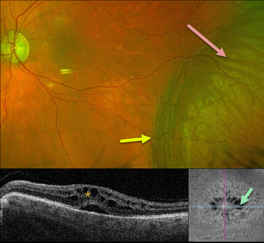
This 84YO male has a long history of advanced chronic open-angle glaucoma with numerous surgical procedures. For several years, his left eye has had stable hypotony, with an intraocular pressure (IOP) of about 3-6 mmHg. Vision is 20/60.
Optos color RG imaging shows an inferotemporal choroidal detachment with chorioretinal folds along its posterior margin. Triton swept-source OCT shows cystoid edema with subretinal fluid, and en face imaging shows radiating mid-retina foveal cysts.
Since his symptoms, vision, IOP, and retinal findings remain stable, we are continuing to follow him without further intervention.
IDIOPATHIC CENTRAL SEROUS CHORIORETINOPATHY
Originally posted on @retina.rocks May 25, 2022
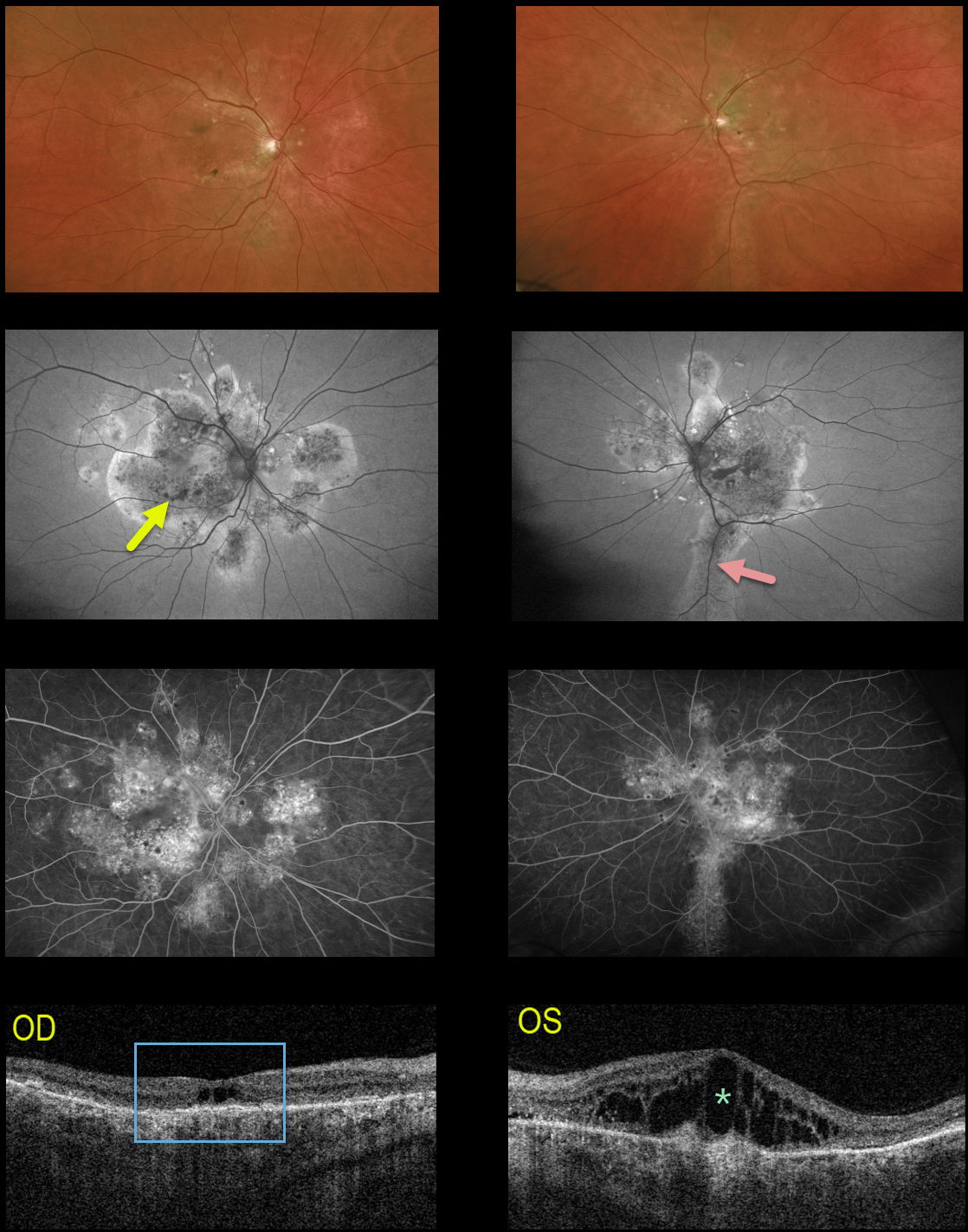
This 64YO male received prior photodynamic therapy in one of his eyes for idiopathic central serous chorioretinopathy (ICCS) and was subsequently lost to follow-up for 10-15 years. He presented with 20/400 vision bilaterally.
Multimodal imaging shows classic findings for chronic ICSC. Optos color imaging shows macular and peripapillary pigmentary changes with a suggestion of an inferior gutter OS.
Fundus autofluorescence better shows the bilateral pigmentary changes and inferior gutter OS, and fluorescein angiography shows variable chorioretinal staining.
Triton swept-source OCT shows bilateral thickened choroid, dry central cavitation-like changes OD, and marked cystoid macular edema OS.
Photodynamic therapy was performed for the left eye, although we are not certain this will improve his vision, given the chronicity of these findings.
Learning Points:
Macular edema is an uncommon finding with typical ICSC, likely due at least in part to the external limiting membrane acting as a barrier to subretinal fluid migrating into the retina. Eyes with chronic ICSC, however, can develop retinal edema as in this case.
Gutters are commonly seen in pathology with chronic subretinal leakage, including central serous retinopathy, choroidal hemangioma, choroidal nevus, and uveal melanoma. These pigmentary changes are usually best imaged with FAF.
CYSTOID MACULAR EDEMA
Alex Hynes
Originally posted on @retina.rocks November 23, 2021
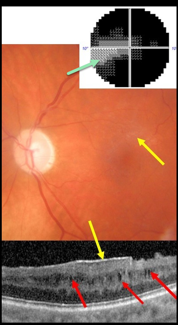
This 61YO male presented with end-stage glaucoma OS, 20/30 vision, and an IOP of 8 mmHg following successful trabeculectomy ten years earlier. He had prior cataract surgery in this eye 15 years ago with no documented history of pseudophakic cystoid macular edema.
There was a moderate macular pucker noted clinically and on OCT, along with multiple inner nuclear layer (INL) microcysts on OCT scanning. He has a tiny island remaining in his visual field.
Learning Points:
Microcystic macular changes have been associated with both advanced primary open-angle glaucoma and idiopathic macular pucker (see Govetto et al, AJO 2017;181:156-165).
Macular pucker-induced mechanical stress, coupled with retrograde trans-synaptic degeneration of bipolar cells secondary to retinal ganglion cell loss, likely predisposes towards the accumulation of fluid within the inner retinas of these patients.
Relative to non-glaucomatous eyes, microcystic macular changes in glaucomatous eyes tend to be more persistent following membrane peeling. This is likely due to retrograde trans-synaptic degeneration.
It is important that these microcystic spaces in eyes with optic neuropathy are not mistaken for inflammatory edema following membrane peeling, which could lead to unnecessary treatment.
Finally, disproportionate preservation of overall retinal thickness despite severe thinning of the ganglion cell and/or nerve fiber layers should not be surprising in glaucomatous eyes.
PACLITAXEL MACULOPATHY
Originally posted on @retina.rocks September 2, 2021
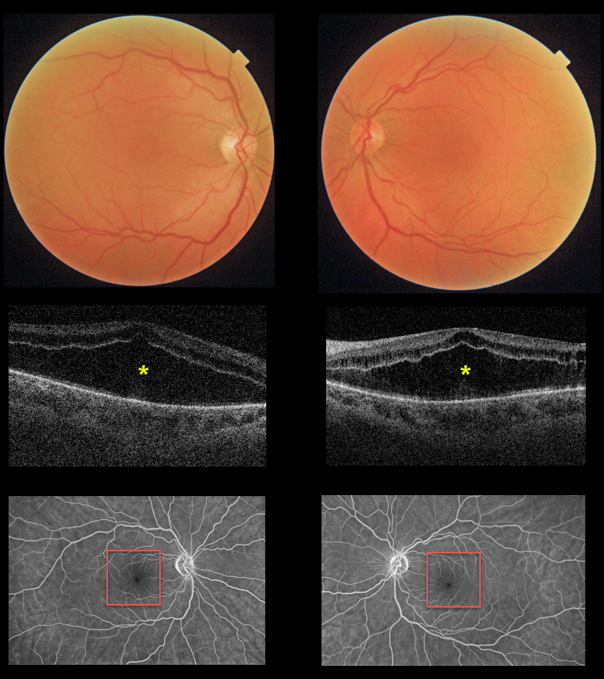
This 55YO female presented with an 8-month history of bilateral vision loss. She had been treated for metastatic breast cancer with Taxol (paclitaxel) for 1 year prior to this exam. Vision was 20/200 OU.
Triton imaging and swept-source OCT show severe symmetrical bilateral cystoid changes in each macula. The cystoid changes are most pronounced in the outer nuclear layer and less so in the inner nuclear layer. Fluorescein angiography shows no leakage.
We have consulted with her oncologist, and expect him to discontinue the paclitaxel. The macular changes and vision should improve within several months.
Learning Points:
Paclitaxel is a member of the taxane family of microtubule-stabilizing agents that is used to treat various solid malignant tumors. Their major adverse effects are bone marrow toxicity and peripheral neuropathy. Ophthalmic complications are rare, including taxane-related cystoid macular edema (CME).
Unlike more common exudative causes for CME, there is no angiographic leakage, and OCT shows intact and continuous inner and outer plexiform layers (see Perez et al, Graefe’s 2020;258:1607-1615).
The cause for CME is uncertain but may be due to Müller cell dysfunction (see Nakao et al, Ophthalmic Surg Lasers Retina 2016;47:81-84). There is no treatment for the CME other than stopping the causative drug.
BABY YODA MACULOPATHY
Originally posted on @retina.rocks May 4, 2021
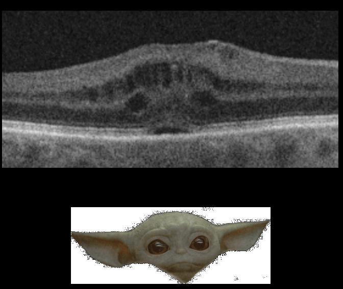
In a galaxy far, far away, this wife of Retina Rock’s founder had an unexpected visitor following her cataract surgery.
Grogu, otherwise known as Baby Yoda or The Child, decided to take refuge within her retina as cystoid macular edema (CME). Thankfully, for the mental health of both the patient and her retinal specialist husband, the CME resolved following topical steroids and non-steroidals.
UVEAL MELANOMA
Originally posted on @retina.rocks March 29, 2021
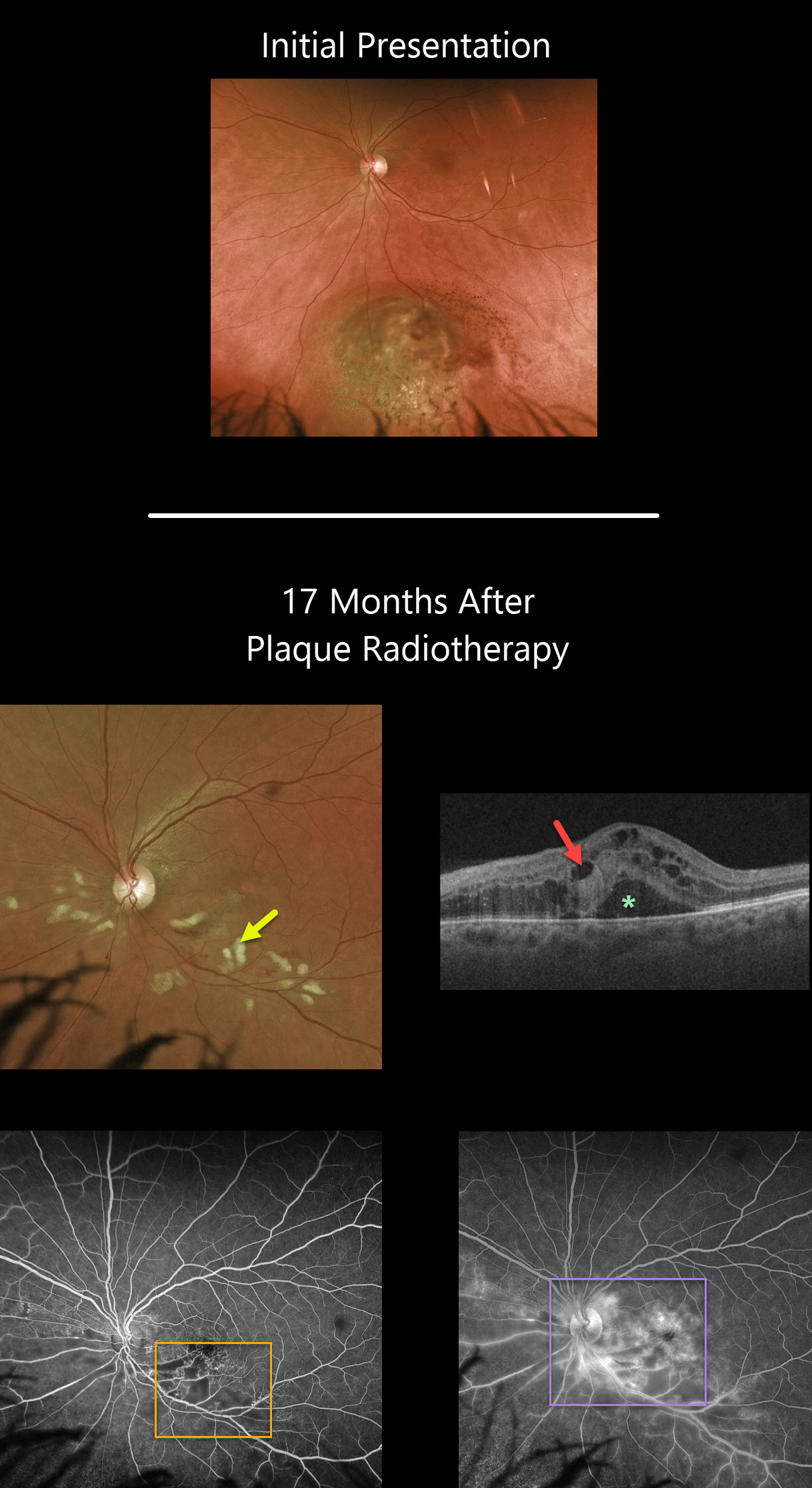
Seventeen months following plaque radiotherapy for a uveal malignant melanoma, this patient developed radiation retinopathy with scattered inner retinal hemorrhages and nerve fiber layer infarcts (cotton wool spots). Vision was counting fingers, although it had been 20/40 ten days following plaque therapy a year and a half earlier.
Cystoid macular edema with subretinal fluid is noted on Triton swept-source OCT. Fluorescein angiography shows areas of capillary dropout, telangiectasias, and hyperfluorescent leakage secondary to cystoid macular edema.
Learning Points:
Radiation retinopathy usually occurs 3 months to 3 years after external beam radiation (e.g., for facial or orbital cancers) or plaque radiotherapy. The ocular findings closely resemble those seen with diabetic retinopathy, as in our patient, who is receiving Avastin injections for the macular edema.
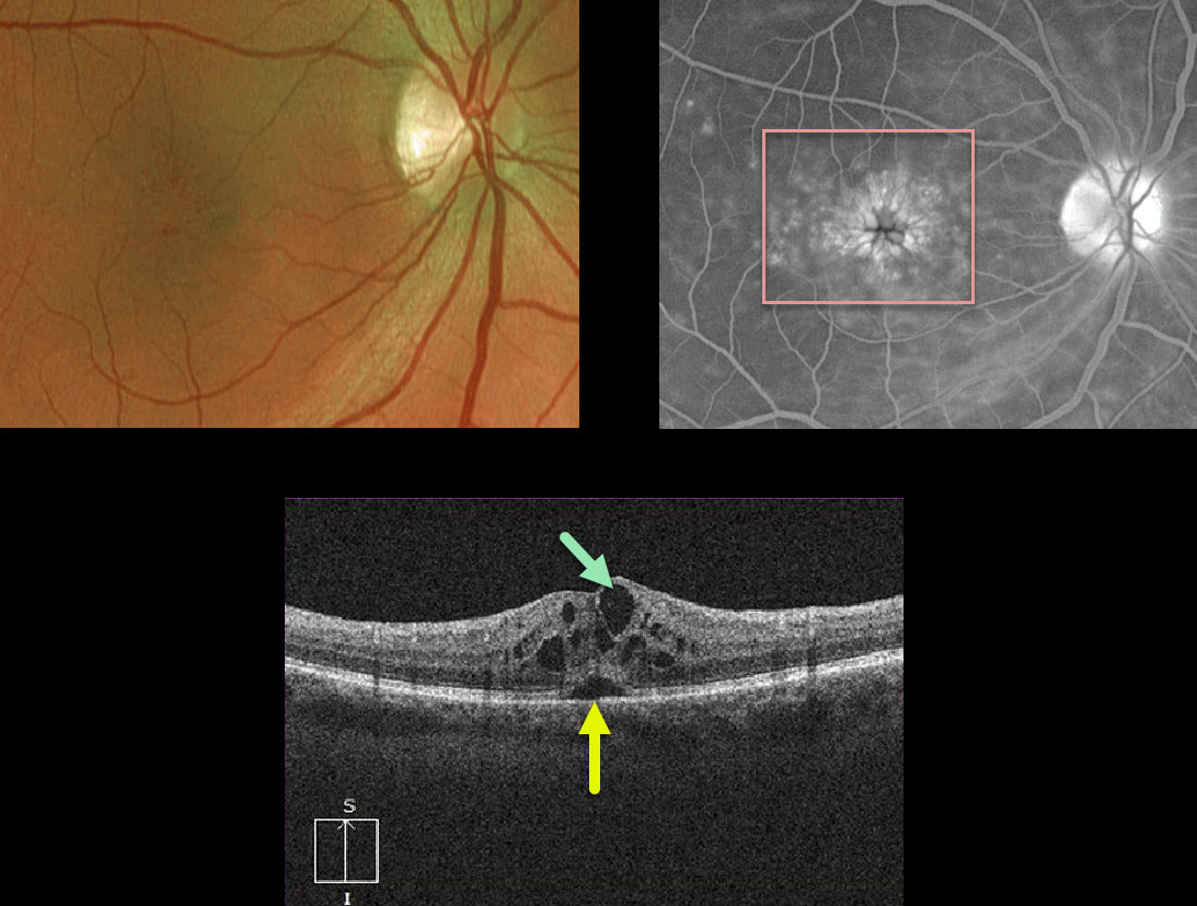
CYSTOID MACULAR EDEMA
Originally posted on @retina.rocks December 10, 2020
This is a classic case of cystoid macular edema (CME) following cataract surgery, otherwise known as the Irvine-Gass Syndrome.
The decompensated inner blood retinal barrier of the perifoveal capillaries allows fluid to leak into the surrounding outer plexiform (Henle’s) layer, causing the classic radiating petaloid pooling on fluorescein angiography.
OCT shows intraretinal edema with cystic spaces and some foveal subretinal fluid.
Learning Points:
Dr. Irvine was the first to describe the condition in 1953 (AJO 1953;36:599-619), and Dr. Gass expanded on this entity in his classic paper (Arch Ophthalmology 1966;76:646-661).
Clinically, at the slit lamp, the cystic changes are best visualized with a contact lens examination (this was how Drs. Irvine and Gass figured things out 60-70 years ago!), although this is rarely currently utilized due to the wide availability and superiority of OCT.
For a great explanation for differentiating exudative from tractional cystic spaces with multimodal imaging, see Govetto et al AJO 2020;212:43-56.


