

Dengue and Epidemic (Post-Fever) Retinitis
<< Back to Cases
DENGUE RETINITIS
Rohan Jain and Manish Nagpal
Originally posted on @retina.rocks September 9, 2026
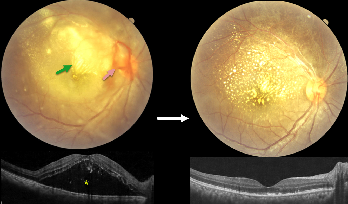
This 63YO male presented with 1 month of bilateral decreased vision following dengue fever, with positive Dengue NS1 antigen during the acute illness. Vision was 20/200 OD and counting fingers OS.
Pseudocolor SLO imaging shows extensive peripapillary cotton wool spots, retinal whitening, and significant macular edema. The fundus detail is partially obscured by overlying vitreous debris from vitritis. OCT scanning neurosensory retinal detachment with subretinal hyperreflective material, intraretinal cystoid edema, and subretinal fluid involving the macula in both eyes, consistent with post-dengue inflammatory retinitis. A possible bacillary layer detachment is also noted in the left eye.
Topical steroids and a 9-week course of tapering oral steroids, as well as a 3-week course of oral doxycycline, were started. Four months later, vision improved to 20/20 OD and 20/30 OS. The patches of white inner retinal ischemia are fading, and some lipid flecks have precipitated out in the mid-retina.
Learning Points:
Epidemic (post-fever) retinitis is an infectious or para-infectious entity that develops in immunocompetent individuals 2-6 weeks following a bacterial, viral, or protozoal infection. Rickettsiosis is the most identified cause, though a large proportion of cases remain idiopathic despite serologic testing; dengue, chikungunya, West Nile, Zika, leptospirosis, typhoid, and Ebola have also been implicated. Patients present with sudden painless vision loss. Fundus findings include unilateral or bilateral unifocal or multifocal patches of retinitis with possible nerve involvement, neuroretinitis with macular star, serous macular detachment, cystoid macular edema, and vasculitis or frosted branch angiitis.
Management is stratified by etiology and lacks controlled data: bacterial causes are treated with systemic antibiotics with or without corticosteroids, viral causes with corticosteroids alone, and many authors advocate observation, as the disease often resolves on its own. Corticosteroids should not be used as sole therapy while infectious causes (including TB, syphilis, and toxoplasmosis) remain in the differential. See Mahendradas et al (Indian J Ophthalmol 2020;68:1775-1786) for an excellent post-fever retinitis review.
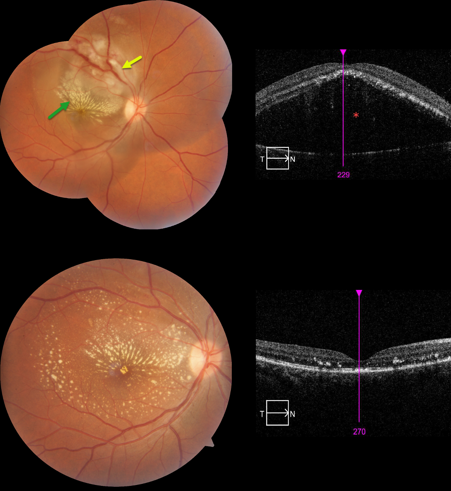
POST-FEVER RETINITIS
Vedant Gambhir
Originally posted on @retina.rocks August 3, 2026
This 31YO female presented with 1 week of decreased vision in her right eye following a febrile illness. Vision was 20/200 OD and 20/20 in her normal OS.
Color photography shows opaque, white inner and deep retinal whitening centered in the superonasal macula, with surrounding subretinal fluid extending into the temporal and inferior macula. A lipid star radiates superiorly and nasally from the foveal center. OCT scanning confirms a highly elevated serous detachment.
An extensive infectious workup was negative for common bacterial, viral, and parasitic etiologies. Given the history of antecedent fever and characteristic retinal findings, a diagnosis of post-fever (epidemic) retinitis was favored. She was treated with oral corticosteroids and doxycycline. At follow-up, visual acuity improved dramatically from 20/200 to 20/30.
Learning Points:
Epidemic (post-fever) retinitis is an infectious or para-infectious entity that develops in immunocompetent individuals several weeks following a bacterial, viral, or protozoal infection. Causes include Dengue, Rickettsiosis, West Nile, Leptospirosis, Chikungunya, Typhoid, and Ebola. Serologic workup is negative in about 60% of cases (Kawali et al, Indian J Ophthalmol 2020;68:1916-1919), due to patients often presenting when acute-phase serologies are difficult to interpret, and viremia has resolved; limitations of available serologic testing; heterogeneous and overlapping etiologies; and a potential immune-mediated response.
Patients present with sudden, painless vision loss. Fundus findings include unilateral or bilateral unifocal or multifocal patches of retinitis with possible nerve involvement, serous macular detachment, and vasculitis or frosted branch angiitis. A macular lipid star in post-fever retinitis is uncommon, found in only 3.3% of affected eyes in rickettsial retinitis (the best-studied cause).
The exact pathogenesis remains uncertain; however, a post-infectious immune-mediated mechanism has been proposed. There is usually a favorable response to steroids and treating the underlying infectious cause with appropriate systemic therapy. See Mahendradas et al (Indian J Ophthalmol 2020;68:1775-1786) for an excellent post-fever retinitis review.
SCRUB TYPHUS POST-FEVER RETINITIS
Anand Temkar, Surendra Pal, Vasumathi Vijay, and Manorama Baheti
Originally posted on @retina.rocks June 3, 2026
This 28YO female presented with 4 days of blurred vision in her right eye. For the past 2 weeks, she has complained of generalized weakness, malaise, and body pain. Vision was counting fingers OD and 20/20 in her normal OS.
Fundus photography shows variable, dense subretinal lipid, particularly confluent in the superonasal macula. There is some preretinal blood around the nerve, and the retina is somewhat yellow and opaque temporally and inferiorly. Patches of inner retinal opacification are noted outside the macula superiorly. OCT shows hyperreflective dots from lipid exudate in the outer retina, with a large serous detachment. Laboratory testing, including CBC, ESR, syphilis, TB, HIV, TORCH profile, and chest X-ray, was negative. We started a 1-week tapering course of oral prednisone 40mg and doxycycline 100mg BID. Three days after our initial examination, she mentioned that she had 2 weeks of mild intermittent fever. Weil-Felix testing was positive, and she was subsequently diagnosed with scrub typhus and asked to continue the doxycycline for a full 2-week course.
The clinical picture gradually improved, and about 6 weeks following presentation, vision improved to 20/40 despite significant residual lipid. OCT scanning through the macular center was mostly normalized. We continue to follow her closely.
Learning Points:
Rickettsial infections cause posterior segment involvement in a substantial proportion of patients, often asymptomatic despite significant fundoscopic findings. The most common rickettsial organisms causing retinal disease include Rickettsia rickettsii (Rocky Mountain spotted fever), Rickettsia conorii (Mediterranean spotted fever), and Rickettsia typhi (murine typhus).
Scrub typhus, caused by Orientia tsutsugamushi and transmitted by a larval mite (chigger) bite, has ocular involvement in about 20% of patients. Retinal findings include retinal hemorrhages, branch retinal vein occlusion, retinal vasculitis/retinitis, and papillitis (Ganekal et al, Ind J Ophthalmol 2021;69:1167-1171).
The ocular and systemic findings of vasculitis and perivasculitis result from both direct endothelial cell destruction by the organism and exaggerated immune responses. Doxycycline is the drug of choice; treatment should last at least 7 days and continue for at least 3 days after fever has subsided and clinical improvement is evident. Visual prognosis is generally favorable with appropriate treatment.
POST-FEVER RETINITIS
Tejaswita Verma,Manish Nagpal and Anjana Mirajkar
Originally posted on @retina.rocks September 9, 2025
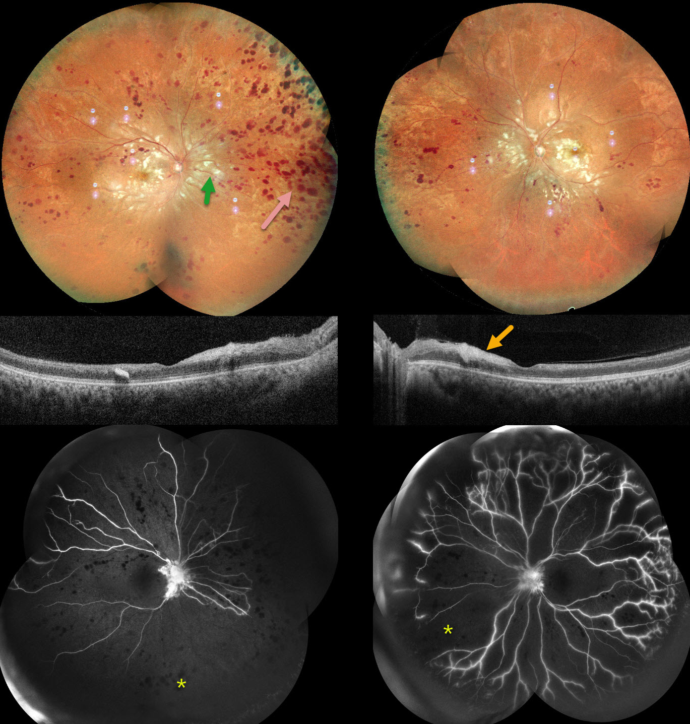
This previously healthy 32YO male presented with a 3-month history of bilateral vision loss following a viral illness. He was treated elsewhere for post-fever retinitis with intravitreal triamcinolone and oral steroids with antivirals. He also had a right lobe pneumonia. Testing was positive for CMV IgG and IgM and negative for TB.
When examined in our office, vision was 20/200 OU. Anterior segments were normal, with mild bilateral vitreous cells.
Pseudocolor SLO imaging shows extensive cotton-wool spots throughout each posterior pole, with more distal retinal hemorrhages that extend into the peripheries. OCT scanning shows possible vitreous cells with variable inner retinal thickened/thinned hyperreflectivity. Fluorescein angiography shows profound bilateral ischemia and disc leakage. We advised bilateral panretinal photocoagulation, but he was unfortunately immediately lost to follow-up.
Learning Points:
Epidemic (post-fever) retinitis is an infectious or para-infectious entity that develops in immunocompetent individuals several weeks following a bacterial, viral, or protozoal infection. Causes include Dengue, Rickettsiosis, West Nile, Leptospirosis, Chikungunya, Typhoid, and Ebola (none of which were tested in our patient).
Patients present with sudden, painless vision loss. Fundus findings include unilateral or bilateral unifocal or multifocal patches of retinitis with possible nerve involvement, serous macular detachment, and vasculitis or frosted branch angiitis. There is usually a favorable response to steroids and treating the underlying infectious cause with appropriate systemic therapy. See Mahendradas et al (Indian J Ophthalmol 2020;68:1775-1786) for an excellent post-fever retinitis review.
POSSIBLE EPIDEMIC RETINITIS
Sharat Hegde
Originally posted on @retina.rocks June 2, 2025
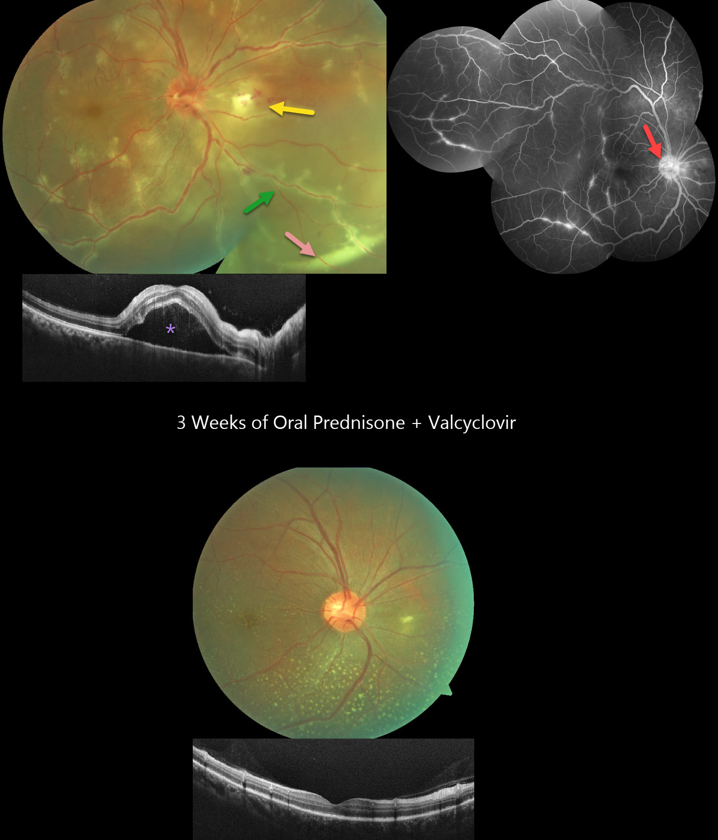
This healthy 36YO male presented with 3 days of floaters and decreased vision in his right eye. Vision was 20/60 OD and 20/20 in his healthy OS. The right eye contained mild anterior chamber cells and moderate vitreous cells.
Fundus photography shows a swollen nerve, a frosted branch pattern of vasculitis involving the retinal veins, an area of retinitis just nasal to the nerve, and a subretinal hypopyon in the inferonasal midperiphery. Some multifocal areas of outer retinal inflammation are noted, along with some retinal hemorrhages. OCT scanning shows variable hyperreflective foveal subretinal fluid. Fluorescein angiography shows variable venous staining and leakage, including from the disc and in nasal retinitis.
The workup included elevated ESR and CRP levels, along with an increased IgG titer for Varicella zoster virus. Mantoux testing, syphilis, Bartonella, typhus, HIV, and chest X-ray were all negative. Although very atypical for viral uveitis, we started oral prednisone and valaciclovir. After one week, the hypopyon resolved, with decreased vasculitis and new scattered flecks of subretinal lipid throughout the posterior pole. By three weeks, vision improved to 20/20 with continued funduscopic improvement.
Learning Points:
Epidemic (post-fever) retinitis is an infectious or para-infectious entity that develops in immunocompetent individuals several weeks following a bacterial, viral, or protozoal infection. Causes include Dengue, Rickettsiosis, West Nile, Leptospirosis, Chikungunya, Typhoid, and Ebola. Patients present with sudden, painless vision loss.
Fundus findings include unilateral or bilateral unifocal or multifocal patches of retinitis with possible nerve involvement, serous macular detachment, and vasculitis or frosted branch angiitis. There is usually a favorable response to steroids and treating the underlying infectious cause with appropriate systemic therapy. See Mahendradas et al (Indian J Ophthalmol 2020;68:1775-1786) for an excellent post-fever retinitis review.
Frosted branch angiitis is a retinal vasculitis that gets its name from the involved vessels resembling branches of a tree that are “frosted” with snow (Kleiner et al, AJO 1988;106:27-34). Although these findings may be isolated, they are often considered a phenotype found in many inflammatory conditions, most commonly CMV retinitis. The inflammation is usually highly responsive to steroids, and the visual prognosis is generally good, although some may develop secondary neovascularization in the anterior and posterior segments due to widespread ischemia.
Subretinal hypopyon is characterized by yellowish inflammatory material settling inferiorly in the subretinal space. It has been documented in infectious (bacterial and fungal endophthalmitis, acute retinal necrosis, tuberculosis, syphilis), inflammatory (sympathetic ophthalmia), and
POST-FEVER RETINITIS
Tejaswita Verma, Navneet Mehrotra and Manish Nagpal
Originally posted on @retina.rocks September 12, 2024
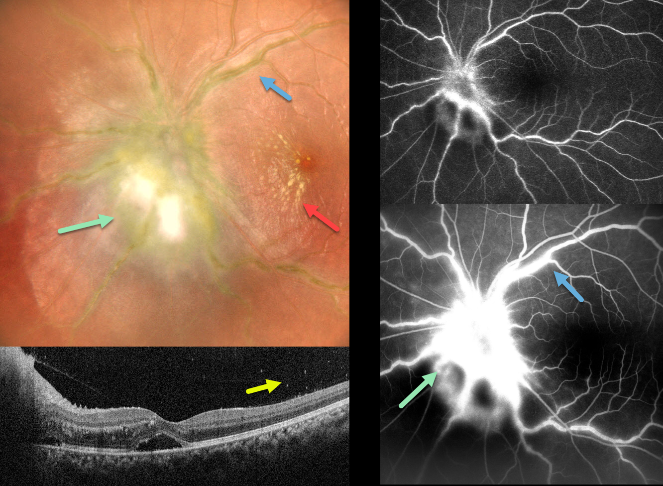
This 20YO female presented with sudden vision loss in her left eye that followed a 1-week febrile illness. Vision was 20/20 in her normal right eye and 20/30 in her left eye.
Multicolor imaging shows marked disc edema with surrounding retinitis, frosted branch vasculitis, and a nasal macular star. OCT shows marked nasal macular retinal thickening and foveal subretinal fluid. Vitritis was noted clinically, and vitreous cells were also noted on the OCT. Fluorescein angiography shows marked optic nerve leakage and vascular staining.
Oral steroids and doxycycline were recommended, along with Weill-Felix testing to rule out typhoid fever, but unfortunately, she was immediately lost to follow-up.
Learning Points:
Epidemic (post-fever) retinitis is an infectious or para-infectious entity that develops in immunocompetent individuals several weeks following a bacterial, viral, or protozoal infection. Causes include Dengue, Rickettsiosis, West Nile, Leptospirosis, Chikungunya, Typhoid, and Ebola. Patients present with sudden, painless vision loss.
Fundus findings include unilateral or bilateral unifocal or multifocal patches of retinitis with possible nerve involvement, serous macular detachment, and vasculitis or frosted branch angiitis. There is usually a favorable response to steroids and treating the underlying infectious cause with appropriate systemic therapy.
See Mahendradas et al (Indian J Ophthalmol 2020;68:1775-1786) for an excellent post-fever retinitis review.
POST-FEVER RETINITIS
Asma Samsudeen and Ashish Sharma
Originally posted on @retina.rocks June 19, 2024
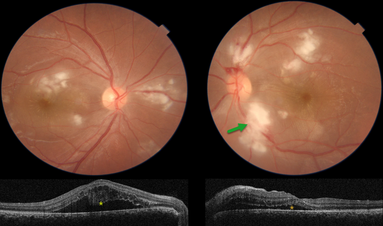
This 33YO male started to complain of blurred vision 10 days earlier. He was hospitalized two months prior with severe headaches, fever, and chills. Vision was 20/30 OU.
Color imaging shows bilateral Purtscher ‘s-like acute inner macular ischemic whitening with a few retinal hemorrhages. Faint radiating foveal lipid is also present.
OCT scanning shows outer retinal cystic edema and subretinal fluid. Hyperreflective material is adherent to the overlying detached right macula. He was referred for viral bloodwork, but unfortunately was subsequently lost to follow-up.
Learning Points:
Epidemic (post-fever) retinitis was first described by Kawali et al (Ocular Immunology & Inflammation 2019;27:571-577) as an infectious or para-infectious uveitis days to weeks following a febrile illness with Dengue, Rickettsiosis, West Nile, Leptospirosis, Chikungunya, Typhoid, or Ebola. It appears as a usually bilateral Purtscher ’s-like retinitis involving the posterior pole with a favorable response to steroids. It is most common in tropical countries like India.
See Mahendradas et al (Indian J Ophthalmol 2020;68:1775-1786) for an excellent post-fever retinitis review.
POST-FEVER DENGUE RETINITIS
Vidhya Nadahalli, Asma Samsudeen and Ashish Sharma
Originally posted on @retina.rocks June 8, 2023
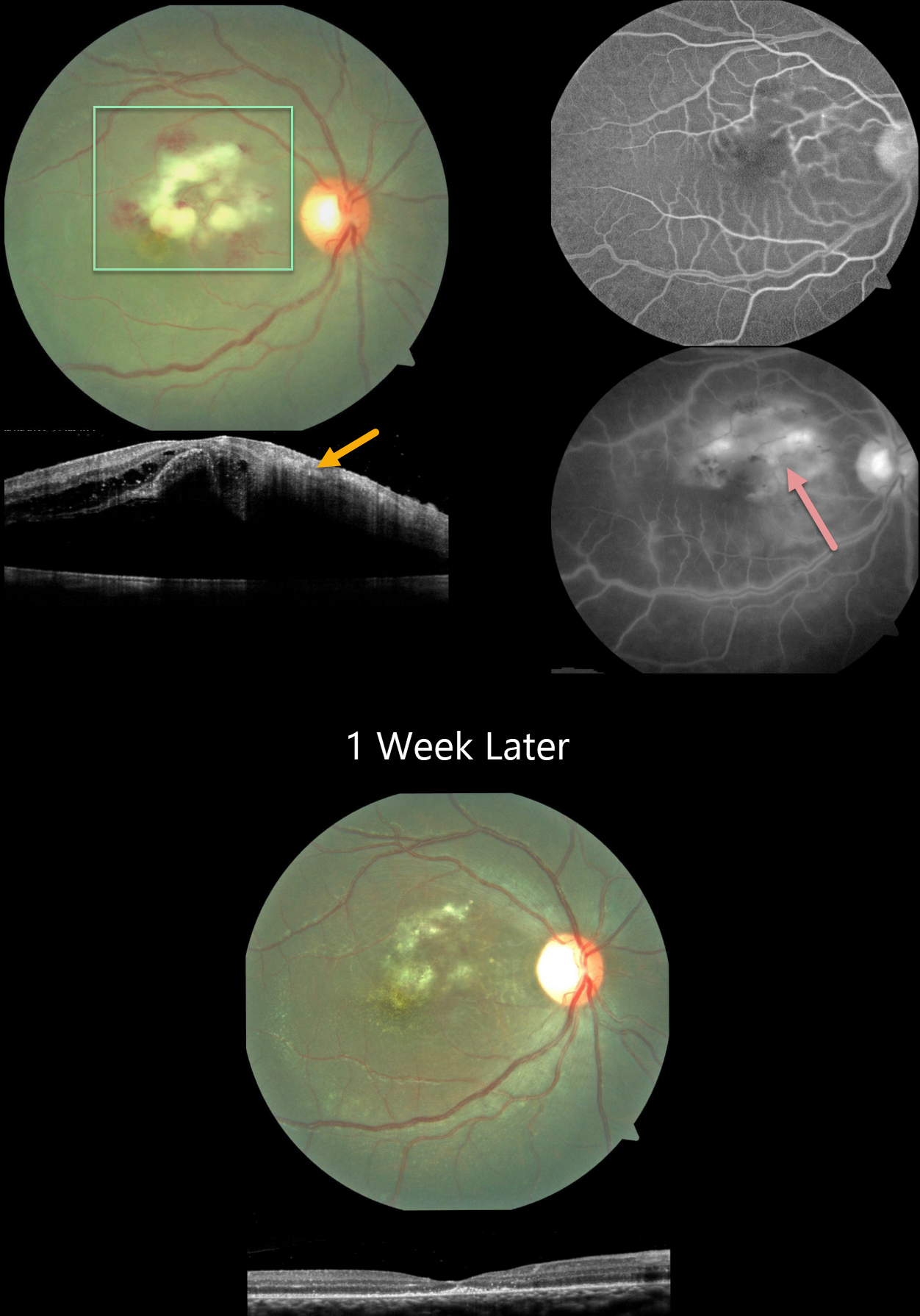
This 15YO boy had a 3-week history of Dengue fever followed by severe vision loss in his right eye. Vision was counting fingers OD and 20/20 in his normal left eye.
Color imaging shows Purtscher-like central macular findings of acute inner macular ischemic whitening with retinal hemorrhages. OCT scanning shows retinal thickening with increased inner retinal hyperreflectivity. Fluorescein angiography shows leakage and staining from the ischemic tissue.
Dengue testing was positive for IgG and negative for IgM antibodies. Weil Felix testing was positive for OX2 and OX19, indicating a likely rickettsial coinfection. Oral antibiotics and steroids were started.
One week later, vision improved to 20/120. The macular findings were resolving. OCT scanning shows complete resolution of the prior edema with secondary thinning and disorganization of the outer foveal layers.
Learning Points:
Epidemic (post-fever) retinitis was described by Kawali et al (Ocular Immunology & Inflammation 2019;27:571-577) as an infectious or para-infectious uveitis occurring days to weeks following a febrile illness due to Dengue, Rickettsiosis, West Nile, Leptospirosis, Chikungunya, Typhoid, and Ebola. It appears as a usually bilateral Purtscher ’s-like retinitis involving the posterior pole with a favorable response to steroids. It is most common in tropical countries like India. See Mahendradas et al (Indian J Ophthalmol 2020;68:1775-1786) for an excellent post-fever retinitis review.


