

Devitalized Blood
<< Back to Cases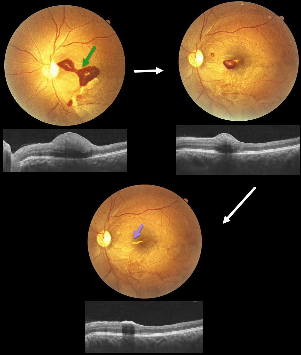
TERSON SYNDROME
Anand Temkar, Surendra Pal, Nagamani Gunasekar
Originally posted on @retina.rocks January 12, 2026
This 32YO male presented with 20 days of vision loss in his left eye. He underwent cardiovascular surgery 3 weeks earlier to remove a clot, followed 1 week later by a subarachnoid hemorrhage. Vision was 20/20 in his normal OD and 20/400 in his OS.
Fundus photography shows a subinternal limiting membrane (ILM) hemorrhage extending from the central macula, superonasally, towards the disc. This blood is hyperreflective on OCT. Observation was recommended.
One month later, vision improved to 20/70, with a marked decrease in sub-ILM blood. One month later, vision improved to 20/30, with near-complete resolution of the blood, except for a small amount of devitalized yellow blood extending nasally from the foveal center.
Learning Points:
Terson syndrome consists of intraocular hemorrhage (vitreous, subhyaloid, or retinal) associated with spontaneous or traumatic subarachnoid hemorrhage. Traditionally, the cause was attributed to elevated intracranial pressure (ICP), which caused cerebrospinal fluid effusion into the optic nerve sheath, compressing the central retinal vein and leading to subsequent rupture of retinal vessels. More recent evidence suggests glymphatic reflux, in which raised ICP causes subarachnoid blood to reflux through glymphatic channels into the globe (Kumaria et al, J Neurology 2022;269:1264-1271).
BRVO WITH RED AND YELLOW DEVITALIZED BLOOD
Originally posted on @retina.rocks September 29, 2025
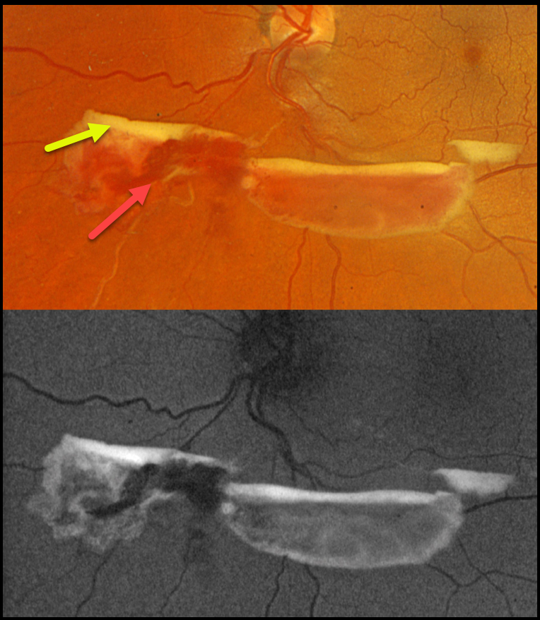
This 48YO female was referred for asymptomatic retinal findings. Vision was 20/20 in the normal OD and 20/20 OS.
Optos color RGB imaging shows red and yellow preretinal blood just inferior to the nerve due to an ischemic extramacular inferonasal branch retinal vein occlusion (BRVO). On fundus autofluorescence (FAF), the red blood is hypo-FAF, and the devitalized yellow blood is markedly hyper-FAF. Intravitreal Avastin was given, and scatter laser was scheduled, although she was immediately lost to follow-up.
Learning Points:
Porphyrins are organic molecules that are part of the heme molecule in hemoglobin. When these compounds break down, the devitalized blood appears yellow and becomes intensely hyperautofluorescent (Bloom and Spaide, Retinal Cases & Brief Reports 2022;16:401-402).
RETINAL ARTERIAL MACROANEURYSM
Originally posted on @retina.rocks September 22, 2025
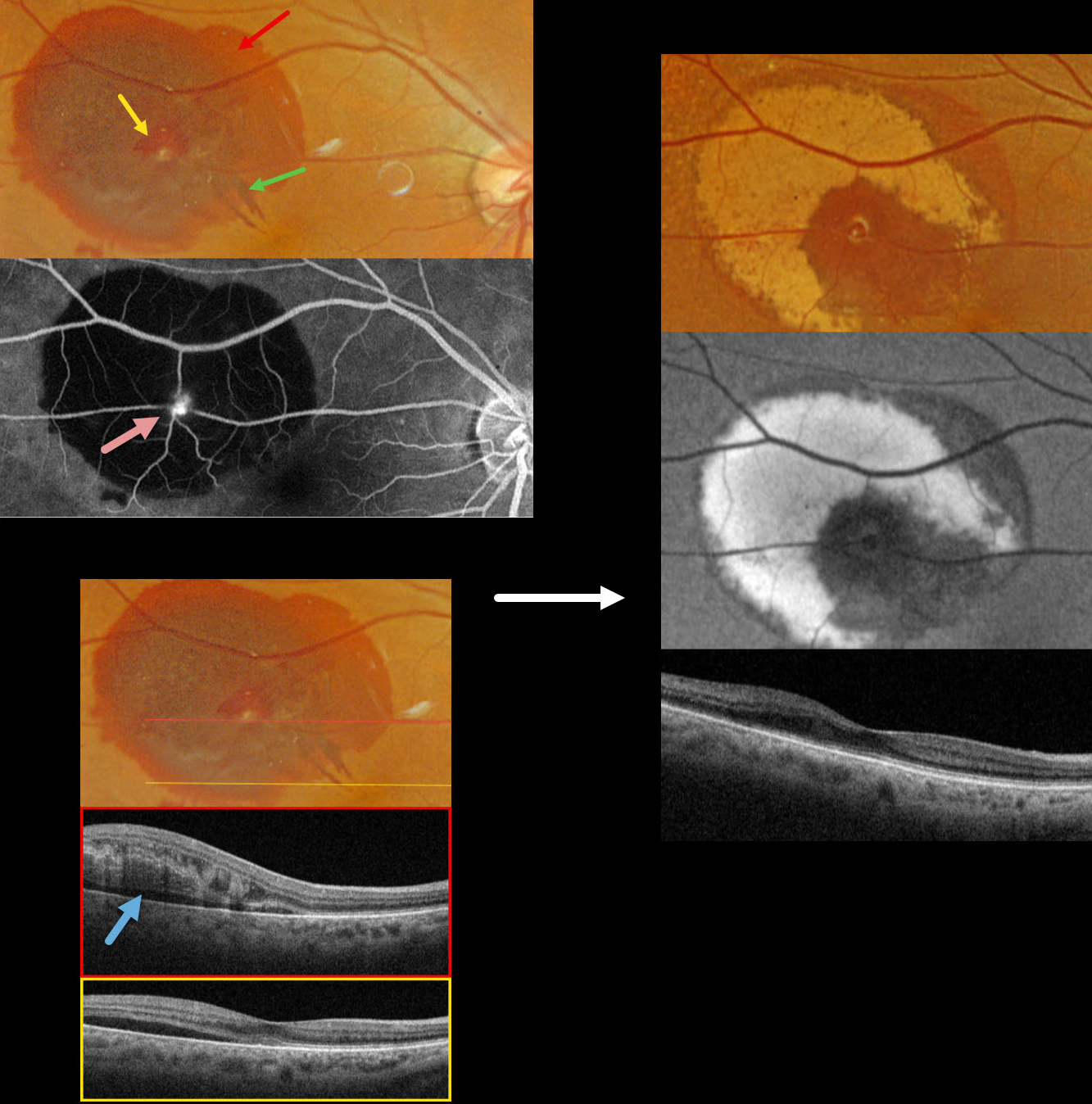
This 83YO female presented with recent blurred vision in her right eye. Vision was 20/40.
Optos color RGB imaging shows subretinal blood in the superotemporal macula. A small amount of inner vs preretinal blood surrounds a small yellow-white retinal arterial macroaneurysm (RAM). In addition, some radiating outer plexiform blood radiates towards the macular center. Fluorescein angiography shows a hyperreflective, leaking RAM with blockage by surrounding subretinal blood. Swept source OCT shows variable outer retinal edema, hyperreflective blood, and subretinal fluid.
Observation was recommended. Six weeks later, the blood is absorbing and turning yellow superiorly and temporally. This devitalized blood is markedly hyper-autofluorescent. The foveal fluid resolved, and vision improved to 20/30.
Learning Points:
A RAM is thought to occur in a weakened arterial wall from arteriosclerosis and appears as a yellow-white dilated lesion along a retinal arterial within the first 3 bifurcations from the optic nerve. There are 2 distinct presentations, hemorrhagic or exudative. The hemorrhagic type often presents, as in this case, with pathognomonic multiple layers of blood. Acute macular exudation can be treated with observation, anti-VEGF injections, or thermal laser
RHEGMATOGENOUS RETINAL DETACHMENT WITH SUB-ILM HEMORRHAGE
Originally posted on @retina.rocks February 12, 2025
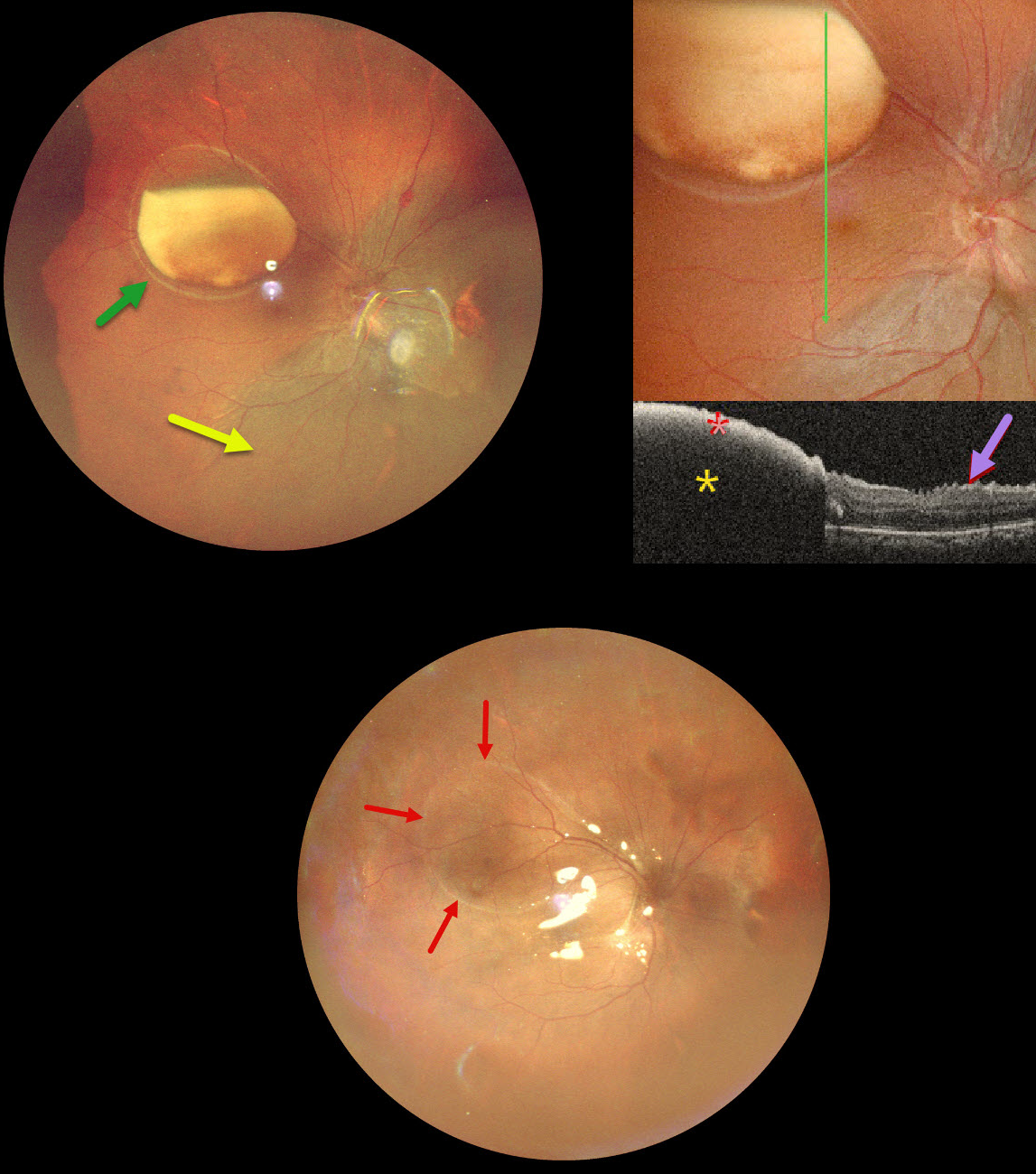
This 39YO male underwent vitrectomy OD elsewhere for a vitreous hemorrhage one month earlier. The treating surgeon noticed a sub-internal limiting membrane (ILM) hemorrhage at a postoperative visit and referred him to us for further evaluation. Vision was 20/30 OD and 20/20 in his normal OS.
Ultrawidefield pseudocolor SLO imaging shows a large partially layered sub-ILM hemorrhage with devitalized yellow blood in the superotemporal midperiphery. A retinal detachment is noted inferiorly and nasally. Inner retinal folds are visible at higher magnification. The blood is highly reflective on OCT scanning with total posterior shadowing. The inner retinal folds are noted inferiorly.
We don’t have a good explanation for our patient’s sub-ILM hemorrhage following vitrectomy for a vitreous hemorrhage. The yellow devitalized blood must have been present preoperatively, even though the initial surgeon told us he just noticed it one month postoperatively. Possibly our patient developed a breakthrough vitreous hemorrhage from a Valsalva maneuver, but without further history, this is just speculative.
We repaired the detachment with vitrectomy and silicone oil, and drained the sub-ILM blood intraoperatively. The retina was completely attached 2 weeks later. A high-water mark defines the margins of the prior ILM detachment.
NEOVASCULAR AMD
Originally posted on @retina.rocks February 5, 2025
This 79YO female underwent vitrectomy for a breakthrough vitreous hemorrhage from neovascular AMD. Vision was 20/400.
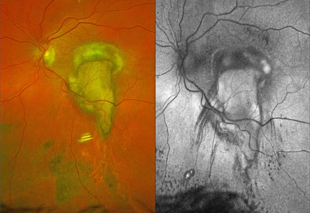
Postoperative Optos color RGB imaging shows an oval fibrotic macular scar with a tongue-like projection of organized devitalized yellow blood extending inferiorly. Coarse subretinal pigment extends into the inferior midperiphery. On fundus autofluorescence (FAF), the fibrotic portion of the scar and inferior subretinal pigment are hypo-FAF, and the devitalized blood is hyper-FAF. Steaks of inferiorly radiating hypo-FAF also extend from the tongue-like yellow blood.
Learning Points:
This complex healing neovascular lesion has many signs of chronicity. There is no fresh, red blood. The remaining blood is yellow and devitalized, developing as heme products degrade. The coarse peripheral subretinal pigment is a marker of prior resolved subretinal blood and appears on OCT as subretinal hyperreflective material. Finally, the subretinal fibrosis where the breakthrough blood originated represents the usual endpoint of untreated neovascular disease, which is thankfully much less common since the advent of anti-VEGF therapy in the mid-2000s.
VALSALVA RETINOPATHY
Nilesh Kumar
Originally posted on @retina.rocks September 26, 2024
This 43YO male presented with a 1-week history of sudden decreased vision in his right eye. He denied any Valsalva maneuver. Vision was 20/70 in his right eye and 20/30 in his left eye.
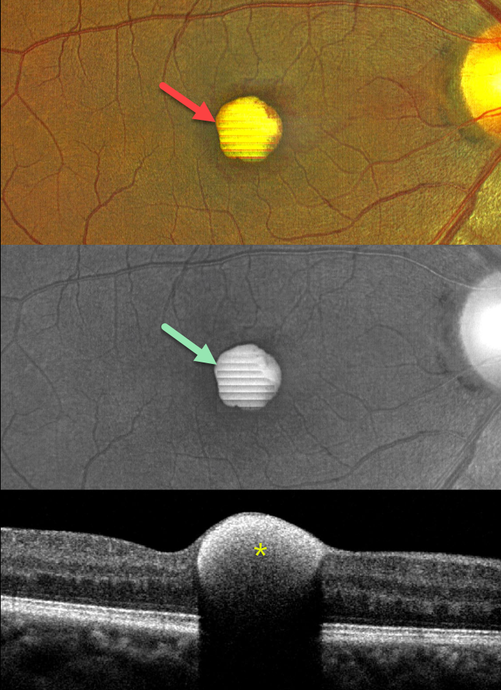
Color imaging shows a foveal yellow sub-internal limiting membrane (ILM) hemorrhage. Fundus autofluorescence (FAF) shows intense hyper-FAF. Spectral domain OCT shows a hyperreflective sub-ILM lesion that shadows posteriorly.
Our patient was treated with observation, and the blood levels were greatly decreased 3 weeks later (not shown).
Learning Points:
Valsalva retinopathy was first described by Thomas Duane (Trans Am Ophthalmol Soc 1972; 70:298-313). It is characterized by usually unilateral retinal and preretinal blood caused by raised intrathoracic or intra-abdominal pressure. Various causes include coughing, heavy lifting, and vomiting. However, as in our case, sometimes patients present with these findings, denying any Valsalva maneuver (Forshaw et al, Acta Ophthalmologica 2024;102:122-123)
These hemorrhages virtually always resolve without sequelae, although the preretinal blood in severe cases can be drained into the inferior vitreous by creating a small ILM opening using either a thermal or Nd:YAG laser (Durukan et al, Eye 2008;22:214-218).
Porphyrins are organic molecules that are part of the heme molecule in hemoglobin. When these compounds break down, the devitalized blood appears yellow and becomes intensely hyperautofluorescent (Bloom and Spaide, Retinal Cases & Brief Reports 2022;16:401-402).
PROLIFERATIVE DIABETIC RETINOPATHY (PDR)
Originally posted on @retina.rocks September 25, 2024
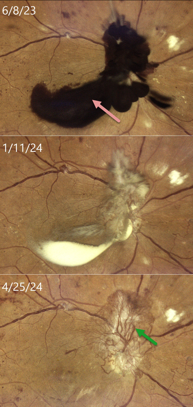
This 33YO female with type 2 diabetes was in the process of being treated with intravitreal Eylea, macular laser, and panretinal photocoagulation (PRP) for bilateral center-involved diabetic macular edema and proliferative diabetic retinopathy (PDR). She presented on 6/8/23 with recent vision loss OD and was overdue for this visit because of numerous medical issues. Vision had decreased to 20/400 OD.
Color imaging shows dark red preretinal blood obscuring the optic nerve and central macula. After several monthly Eylea injections and completion of the PRP, the preretinal blood became yellow and devitalized on 1/11/24, before finally completely absorbing, as noted at last examination on 4/25/24, with stable 20/400 vision. Despite full PRP, diffuse variably fibrosed neovascularization persists, along with venous beading. OCT scanning revealed no edema with central atrophy (not shown).
Our patient was initially in no rush for vitrectomy surgery, as vision in her fellow eye was 20/50. Conservative management with intravitreal injections and PRP was thankfully successful. This approach was also found to be safe and effective by Brar et al in a retrospective series of 22 eyes with foveal preretinal blood from PDR (Ind J Ophthalmol 2024;72:687-691).
LEUKEMIA
Kanwaljeet Harjot Madan
Originally posted on @retina.rocks August 28, 2024
This 42YO previously healthy male presented with one week of bilateral decreased vision. Vision was 20/100 OD and 20/200 OS.
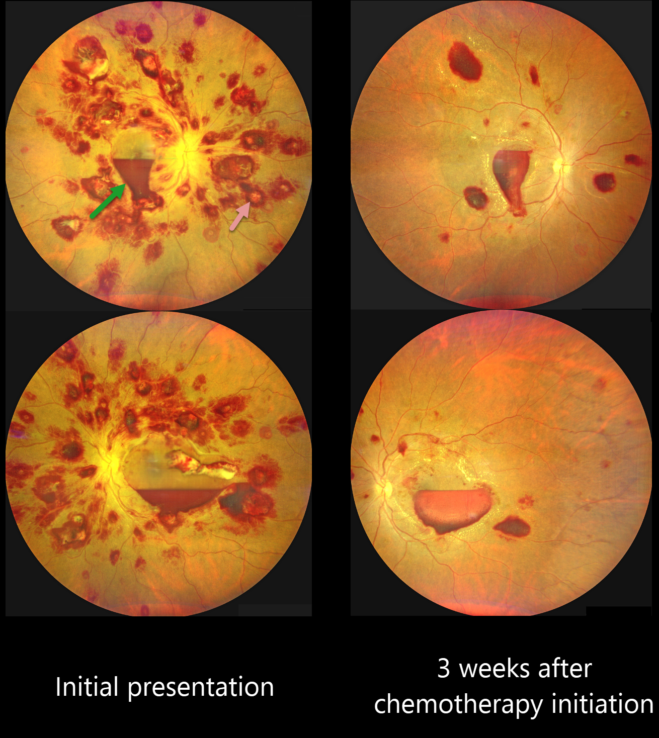
Color photography shows an extensive bilateral nerve fiber layer and deep white-centered retinal hemorrhages. Layered macular sub-internal limiting membrane (ILM) hemorrhages are noted as well, with some yellow devitalized blood in the temporal left macula. Hematologic workup revealed elevated blast cells, severe anemia (HgB 5.4) and thrombocytopenia.
He was diagnosed with acute myeloid leukemia and referred to oncology. He started chemotherapy, and 3 weeks later, his vision improved to 20/70 OU. The HgB was 8.2 with decreased blast cells. The bilateral retinal hemorrhages were markedly decreased.
This case was submitted by Kanwaljeet Harjot Madan.
Learning Points:
The term “Roth spot” was named after Dr. Moritz Roth, who, in 1872, noted white-centered hemorrhages in patients with bacterial endocarditis. He felt that these white spots were septic emboli that originated from an infected cardiac valve. However, similar lesions can be seen in many other disorders, including leukemia, hyperviscosity, anemia, diabetes, and hypertension (Duane et al Ophthalmology 1980;87:66-69). The white material may represent a variety of causes, including fibrin. So, these lesions are best called white-centered hemorrhages, unless one is specifically referring to those found associated with bacterial endocarditis.
Ocular leukemic manifestations are primarily caused by accompanying hematologic abnormalities, including hyperviscosity, anemia, and thrombocytopenia (Soman et al, Ophthalmology Retina 2018;2:17-23). Posterior segment findings include retinal hemorrhages (sub-ILM, nerve fiber layer or deeper, white-centered), cotton-wool spots, dilated retinal veins, and serous retinal detachment. Leukemic infiltration of the optic nerve, retina, and choroid can also occur. The sub-ILM hemorrhages are usually self-limiting and resolve within several months of treating the underlying disease (Ophthalmology Retina 2018;2:494-501).
RETINAL ARTERIAL MACROANEURYSM (RAM)
Asma Samsudeen and Ashish Sharma
Originally posted on @retina.rocks October 26, 2023
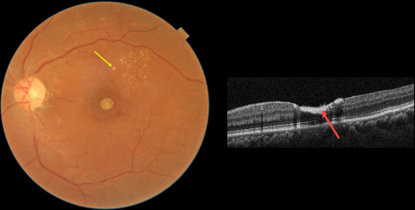
This 54YO female presented with 3 weeks of vision loss in her left eye. Vision was 20/20 in her normal OD and 20/200 OS.
Fundus photography shows devitalized yellow prefoveal blood, some lipid flecks centered in the superotemporal macula, and a tiny fibrosed superior retinal arterial macroaneurysm (RAM).
OCT scanning through the fovea confirms the sub-internal limiting membrane (ILM) location of the hyperreflective blood.
This case was submitted by Asma Samsudeen and Ashish Sharma.
Learning Points:
A RAM is thought to occur in a weakened arterial wall from arteriosclerosis, and appears as a yellow-white dilated lesion along a retinal artery within the first 3 bifurcations from the optic nerve.
There are 2 distinct presentations, hemorrhagic or exudative, and our patient has features of both. Although she reported only a several-week history of vision loss, the retinal findings suggest a much longer course.
The devitalized blood usually takes months to turn yellow, and was likely much larger in extent originally. The blood elsewhere has largely resolved. There was also likely macular thickening and possibly subretinal fluid, which spontaneously resolved, leaving behind residual peripheral macular lipid.
Observation was recommended, and we expect her vision to significantly improve, as the retina appears structurally fairly normal on OCT deep to the blood. Subretinal blood, especially devitalized, is toxic to the outer retina, which is why removal and/or displacement of thick subretinal blood is often recommended. Blood in the sub-ILM or preretinal space does not seem to damage the retina, so it can more safely be observed (see Bloom and Spaide, Retinal Cases 2022;16;401-402).
RETINAL ARTERIAL MACROANEURYSM (RAM)
Originally posted on @retina.rocks September 1, 2022
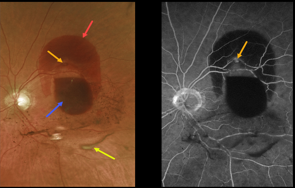
This 85 YO female presented with a retinal arterial macroaneurysm (RAM) with counting fingers vision.
The macroaneurysm has ruptured, causing multiple levels of blood shown on fundus photography and fluorescein angiography. The blood is subretinal superiorly, subhyaloid more centrally, and in the vitreous (both red and devitalized). We are observing this patient.
Learning Points:
A RAM is thought to occur in a weakened arterial wall from arteriosclerosis, and appears as a yellow-white dilated lesion along a retinal arterial within the first 3 bifurcations from the optic nerve.
There are 2 distinct presentations, hemorrhagic or exudative. The hemorrhagic type often presents, as in this case, with pathognomonic multiple layers of blood.
Acute macular hemorrhage can be treated with observation, anti-VEGF injections, or thermal laser.
PROLIFERATIVE DIABETIC RETINOPATHY (PDR)
Originally posted on @retina.rocks August 17, 2022
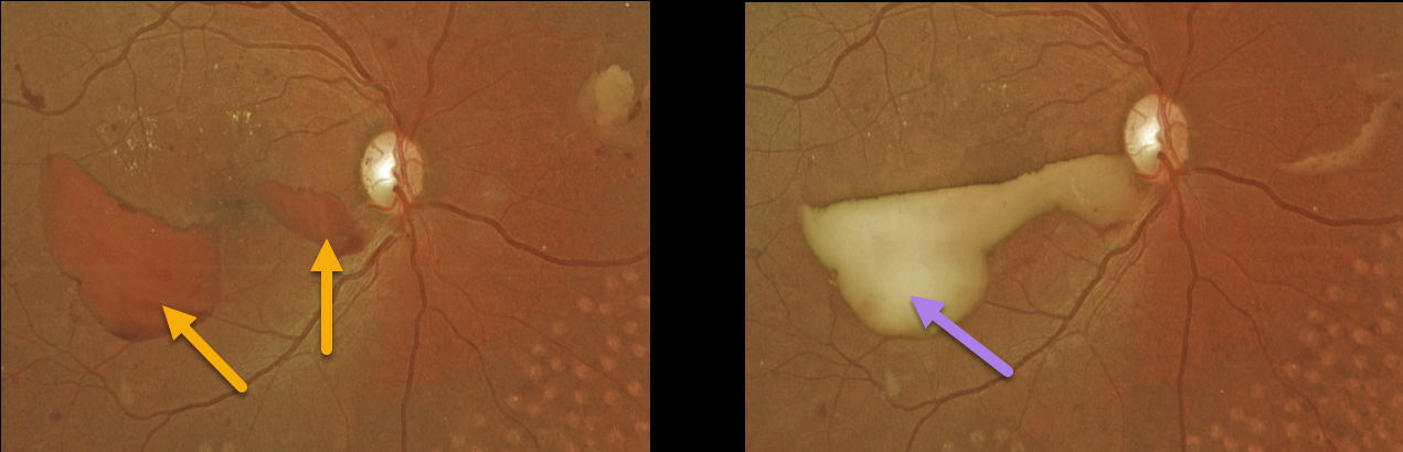
This 37YO female presented with recent vision loss in her right eye. She had a history of prior laser treatment elsewhere several years ago. Vision was 20/30 OD and 20/25 OS.
Red preretinal blood was seen in the inferotemporal right macula with partial PRP scarring. One month following a single Avastin injection, the devitalized preretinal blood was yellowing. Bilateral PRP is planned.
Learning Points:
PRP has been around since the 1960s and still remains the mainstay for treating proliferative disease. Often considered a “one and done”, patients can still develop break-through hemorrhage or progressive neovascular growth and traction.
Intravitreal anti-VEGF injections are often helpful, but some will require vitrectomy.
In our patient’s case, the new preretinal blood was most likely due to her having been incompletely treated rather than a PRP failure.
PERIPHERAL EXUDATIVE HEMORRHAGIC CHORIORETINOPATHY
Originally posted on @retina.rocks May 26, 2022
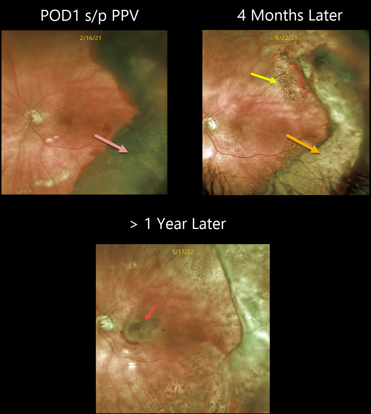
This 80YO female with a known history of dry AMD presented with a 3-week history of poor vision in her left eye. Vision was hand motion from a dense vitreous hemorrhage. B-scan ultrasonography showed a thickened temporal subretinal lesion without retinal detachment (not shown).
One day following vitrectomy, vision improved to 20/100. The temporal subretinal and RPE blood somewhat resembled a uveal malignant melanoma. However, the well-demarcated location and shape of the lesion, along with the posterior rim of subretinal blood, helped establish the diagnosis of a breakthrough vitreous hemorrhage from ectopic choroidal neovascularization (CNV), also known as peripheral exudative hemorrhagic chorioretinopathy (PEHCR).
Four months later, the resolved devitalized blood was turning yellow. The coarse subretinal pigmentation, most prominent superotemporally, is pathognomonic for prior subretinal blood.
She was subsequently lost to follow-up until she presented with a 6-month history of decreased vision. Although the PEHCR ridge continued to contract, vision was counting fingers due to a new macular neovascularization. Anti-VEGF therapy was started.
Learning Points:
PEHCR is an exudative process similar to wet AMD but located in the retinal periphery. Ectopic CNV can often masquerade as a choroidal mass or uveal melanoma.
It can also cause extensive choroidal/suprachoroidal, subretinal, and vitreous hemorrhage, more commonly in patients who are on blood thinners.
The subretinal blood often spontaneously regresses without treatment, although anti-VEGF injections can be used for symptomatic or increasing exudation.
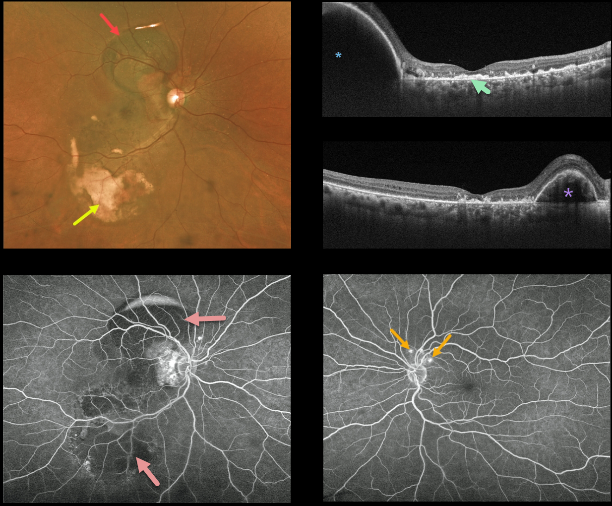
POLYPOIDAL CHOROIDAL VASCULOPATHY
Originally posted on @retina.rocks April 6, 2022
This 59YO female patient presented with variable mostly peripheral submacular blood in her right eye and counting fingers vision. Optos imaging shows the extent of the subretinal blood, which is dark superiorly and yellow inferiorly.
Triton swept-source OCT shows variable outer retinal and subretinal hyperreflective material from resolved submacular blood, and this is seen clinically as the coarse subretinal pigment most evident in the inferotemporal macula. A large RPE detachment (PED) is seen superiorly in the vertical scan, with a smaller PED nasally in the horizontal scan.
Fluorescein angiography shows mostly blockage from the subretinal blood. Several small asymptomatic polyps are noted superior to the left nerve.
Learning Points:
Polypoidal choroidal vasculopathy (PCV) is a variant of type 1 macular neovascularization. PCV is likely distinct from age-related macular degeneration, occurring in a different population (more common in African Americans and Asians), with a relative lack of drusen and a thicker choroid.
The sub-RPE vessels were classically characterized using indocyanine green angiography, although recent literature suggests that OCT is just as effective at diagnosing these vascular polyps.
Historically, these lesions were believed to be somewhat resistant to anti-VEGF monotherapy, often requiring combined treatment with photodynamic therapy.
More recent data from the PLANET study suggest that Eylea monotherapy is also effective (AJO 2019;204:80-89).
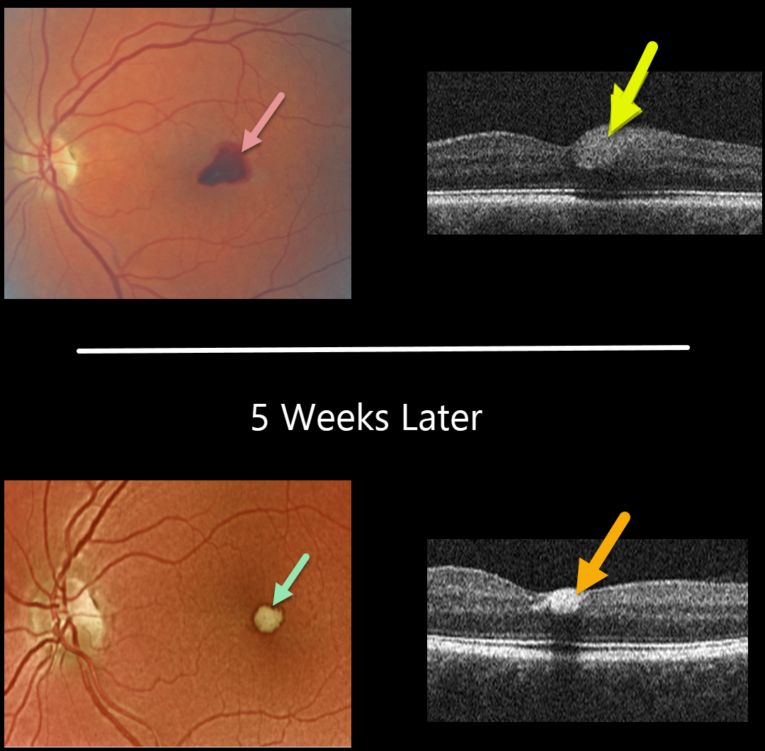
VALSALVA RETINOPATHY
Originally posted on @retina.rocks March 24, 2022
This 56YO female presented with acute vision loss in her left eye from Valsalva retinopathy. Vision was 20/100. A temporal foveal retinal hemorrhage is seen, located within the inner retina on OCT B-scan.
Five weeks later, the devitalized blood is contracting and turning yellow. The blood also became more hyperreflective on the OCT B-scan. All blood subsequently resolved with the return of normal vision.
Learning Points:
Valsalva hemorrhages are usually in the sub-internal limiting membrane (ILM) space, although this case was in the inner retina. These hemorrhages virtually always resolve without sequelae, although the sub-ILM blood can be drained into the inferior vitreous by creating a small opening in the ILM using either a thermal or Nd:YAG laser.
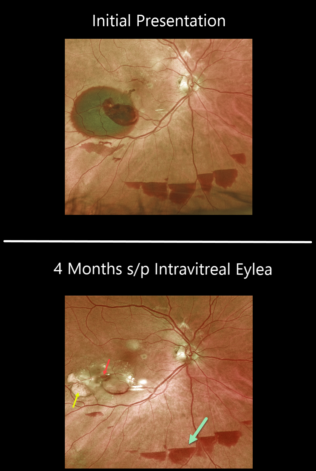
RETINAL ARTERIAL MACROANEURYSM (RAM)
Originally posted on @retina.rocks February 14, 2022
This 85 YO female with a retinal arterial macroaneurysm (RAM) continues to improve with 20/30 vision following a single Eylea injection four months earlier.
The macroaneurysm still appears patent. The old subretinal devitalized blood is devitalized and yellow. There are also multiple patches of resolving inferior subhyaloid blood.
Learning Points:
A RAM is thought to occur in a weakened arterial wall from arteriosclerosis and appears as a yellow-white dilated lesion along a retinal arteriole within the first 3 bifurcations from the optic nerve.
There are 2 distinct presentations: hemorrhagic or exudative. The hemorrhagic type often presents, as in this case, with pathognomonic preretinal, intraretinal, and subretinal blood. Acute macular hemorrhage can be treated with observation, anti-VEGF injections, or thermal laser.
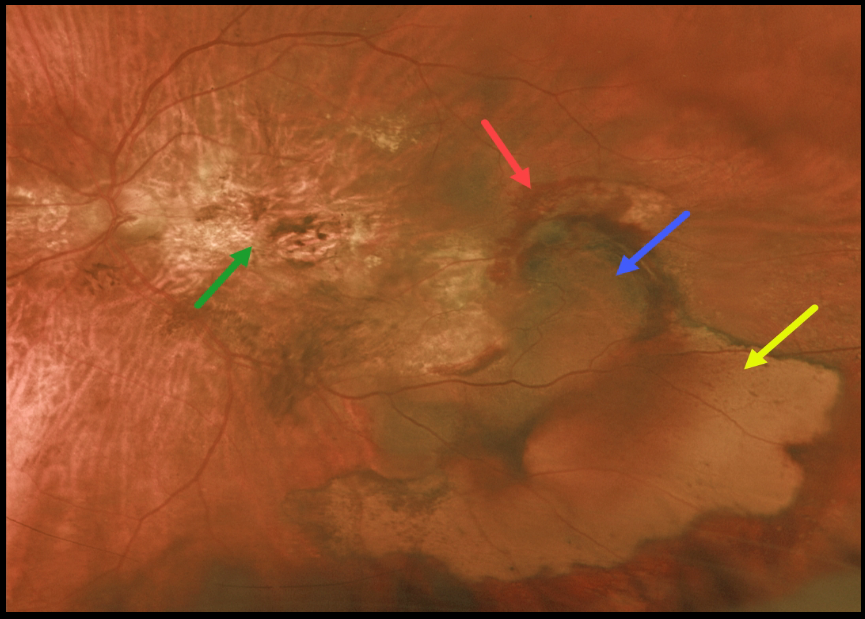
PERIPHERAL EXUDATIVE HEMORRHAGIC CHORIORETINOPATHY
Originally posted on @retina.rocks January 28, 2022
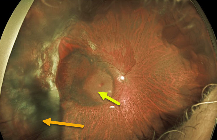
This 77YO female has been followed for years with vision of 20/200 OD and counting fingers OS from inactive age-related disciform scars. She then came in with sudden symptomatic vision loss and floaters in her left eye.
Vision was hand motion OS from a breakthrough vitreous hemorrhage from an inferotemporal ectopic choroidal neovascularization (CNV) that grew from the edge of the original macular scar. She was not taking anticoagulants or aspirin.
Following two monthly intravitreal Avastin injections, vision remained at hand motion. The vitreous hemorrhage had cleared, and the subretinal blood was resolving.
Optos imaging shows the original macular scarring, along with varying stages of resolving rub-RPE blood, subretinal blood, and devitalized, yellow subretinal blood.
Learning Points:
Ectopic CNV, also known as peripheral exudative hemorrhagic chorioretinopathy, is an exudative process similar to wet AMD but located in the retinal periphery.
Ectopic CNV can often masquerade as a choroidal mass or uveal melanoma. It can also cause extensive choroidal/suprachoroidal, subretinal, and vitreous hemorrhage, more commonly in patients who are on blood thinners.
The subretinal blood often spontaneously regresses without treatment, although anti-VEGF injections can be used for symptomatic or increasing exudation.
PERIPHERAL EXUDATIVE HEMORRHAGIC CHORIORETINOPATHY
Originally posted on @retina.rocks January 15, 2021
This 71YO male had a vitrectomy in 2018 for a breakthrough vitreous hemorrhage from ectopic choroidal neovascularization (CNV). He did well with 20/40 vision until a recurrent vitreous hemorrhage developed two years later.
The vitreous cleared with several monthly intravitreal Avastin injections, but vision remains at hand motion due to submacular blood, some of which is yellow and devitalized. We discussed repeat vitrectomy with subretinal TPA, but he elected for in-office anti-VEGF injections. He is not on any systemic blood thinners.
Learning Points:
Ectopic CNV, also known as peripheral exudative hemorrhagic chorioretinopathy, is a degenerative and exudative process similar to wet AMD but located in the retinal periphery.
Ectopic CNV can often masquerade as a choroidal mass or uveal melanoma. It can also cause extensive subretinal and vitreous hemorrhage, more commonly in patients who are on blood thinners.
The subretinal blood often spontaneously regresses without treatment, although anti-VEGF injections can be used for symptomatic or increasing exudation.
CHOROIDAL RUPTURE
Originally posted on @retina.rocks October 1, 2020
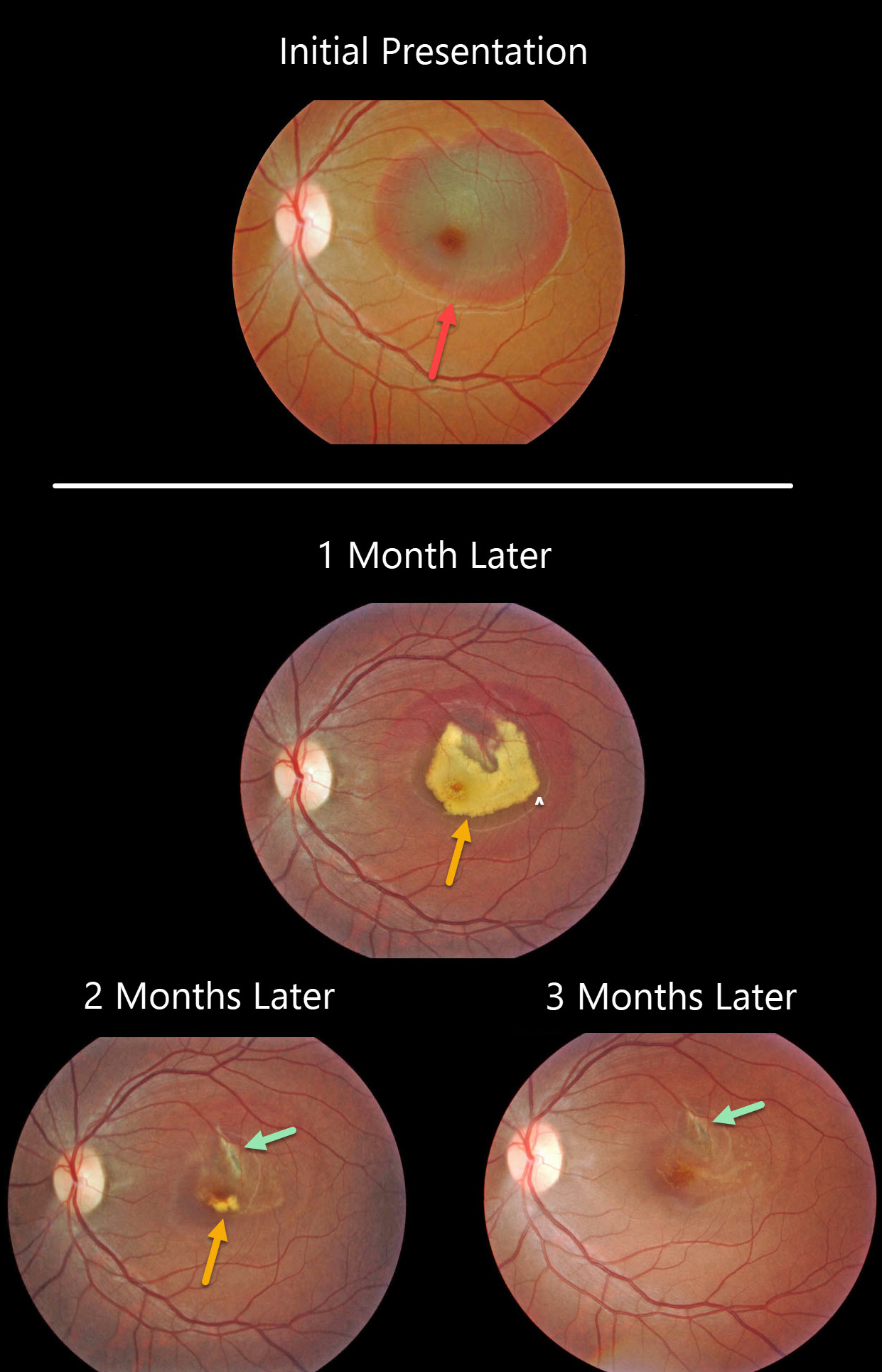
This patient presented with an acute submacular hemorrhage following a finger-to-eye injury.
The subretinal blood slowly resolved over the following three months. An underlying choroidal rupture is almost always the source of this traumatic blood.
Final vision was 20/400, either due to contusive foveal damage or possible toxicity from the subretinal blood.
Learning Point:
Choroidal ruptures are caused by blunt ocular trauma and are always circumferential with respect to the optic nerve.
These patients need to be followed since macular neovascularization can develop later in life.
MACULAR NEOVASCULARIZATION (MNV)
Originally posted on @retina.rocks July 8, 2020
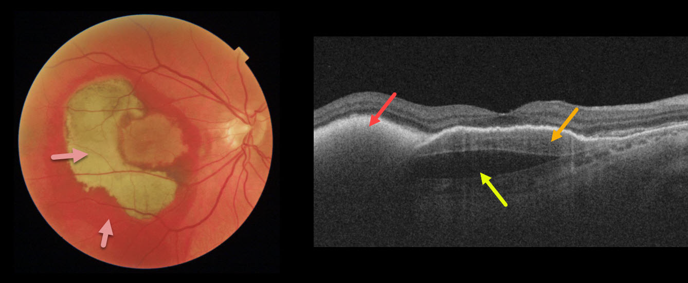
This patient’s vision remained 20/100 one month following vitrectomy with subretinal tissue plasminogen activator.
Surgery accomplished its goal of displacing most of the subfoveal blood, which is variably red and devitalized (yellow).
OCT shows the remaining subretinal hyperreflective blood, the causative sub-RPE type 1 macular neovascularization, and a subfoveal prechoroidal cleft.
Learning Points:
A prechoroidal cleft is a hyporeflective space between the RPE and Bruch’s membrane.
These are seen in up to about 20% of treated eyes with wet AMD, particularly with type 3 neovascularization and polypoidal choroidal vasculopathy.
They are usually associated with worse visual acuity due to potential complications, including RPE tear and subretinal hemorrhages.
For a more detailed discussion of prechoroidal clefts, see Kim et al Retina 2017;37;2047-2055.
PROLIFERATIVE DIABETIC RETINOPATHY (PDR)
Originally posted on @retina.rocks June 16, 2020
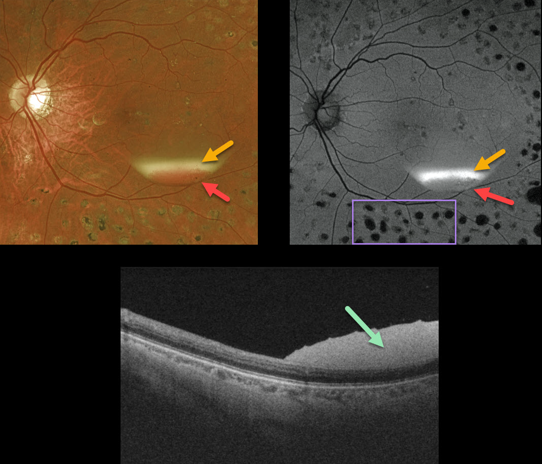
This patient, previously treated for proliferative diabetic retinopathy with panretinal photocoagulation (PRP), presented with an asymptomatic resolving two-toned subhyaloid hemorrhage. The devitalized yellow blood indicates chronicity, although some red blood remains.
The OCT confirms the subhyaloid location of the hyperreflective blood.
Red blood is dark on fundus autofluorescence (FAF), although yellow blood is extremely hyperautofluorescent. Lipofuscin within the RPE is the main fluorophore in FAF. The laser PRP scars are hypoautofluorescent due to loss of the RPE.
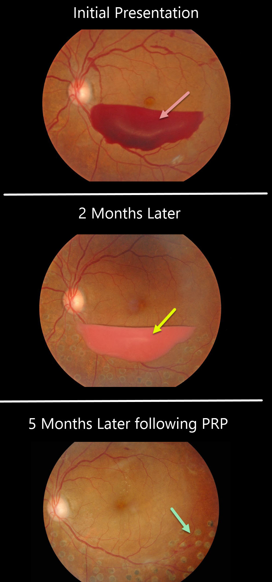
PROLIFERATIVE DIABETIC RETINOPATHY
Originally posted on @retina.rocks December 24, 2019
This patient presented with a sub-hyaloid hemorrhage due to proliferative diabetic retinopathy.
A subsequent photo two months later shows devitalized (yellowing) sub-hyaloid hemorrhage. He received panretinal photocoagulation (PRP) in the left eye and the sub-hyaloid hemorrhage completely resolved three months later. Anti-VEGF therapy was not used.
Learning Points:
PRP has been around since the 1960s and still remains the mainstay for treating proliferative disease. Often considered a “one and done,” patients can still develop break-through hemorrhages. Intravitreal anti-VEGF injections are often helpful, but some will require vitrectomy.


