

Diabetic Retinopathy Diabetic Macular Edema (DME)
<< Back to Cases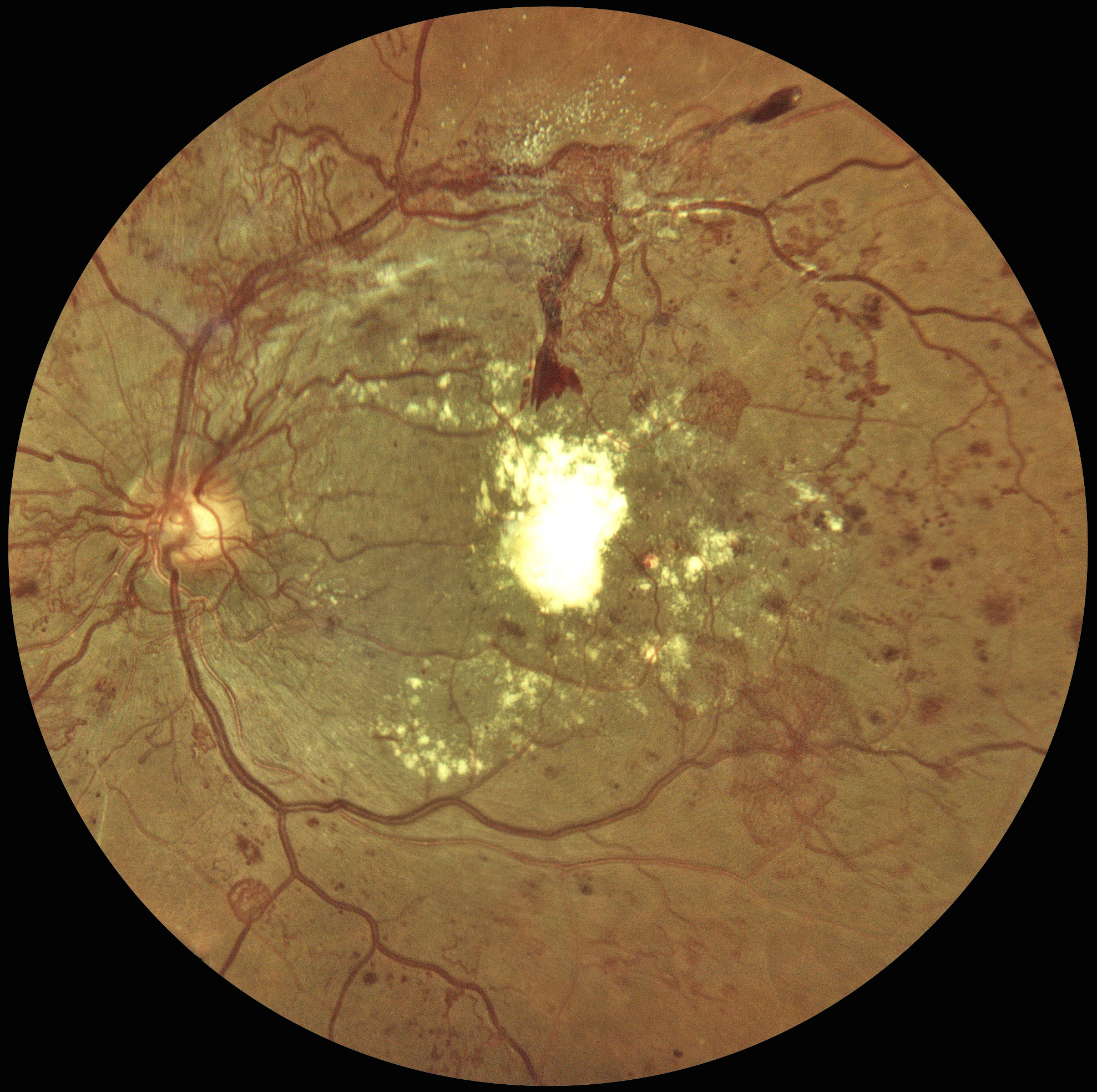
VERY SEVERE PDR AND DME FOLLOWING PATIENT LOST TO FU
Sowparnika Basavaraju
Originally posted on @retina.rocks June 16, 2026
This 48YO male with a history of poorly controlled type 2 DM was seen 4 years earlier for an eye examination elsewhere but was immediately lost to follow-up. He presented to us with hand-motion vision in his left eye.
Color fundus photography shows florid disc and peripheral neovascularization variably coating the posterior pole. A large plaque of subfoveal lipid is also noted.
Learning Points:
These findings would have been caught earlier if he had not been lost to follow-up, which, unfortunately, is quite common among diabetic patients (Obeid et al, Ophthalmology 2018;125:1386-1392; Gao et al, Ophthalmology Retina 2019;3:230-236; Green et al, AJO 2020;216:18-27; Ophthalmology Retina 2025;9:1167-1174, etc, etc).
At this stage, our patient will not recover central vision due to the subfoveal lesion, and our hope is to prevent further vision loss. Anti-VEGF therapy, panretinal photocoagulation, and macular photocoagulation were suggested. He is at significant risk for traction retinal detachment as the neovascularization involutes (crunch), and we will therefore follow him very closely.
HAPPY WORLD SMILE DAY
Pier Luigi Guerin
Originally posted on @retina.rocks October 4, 2024
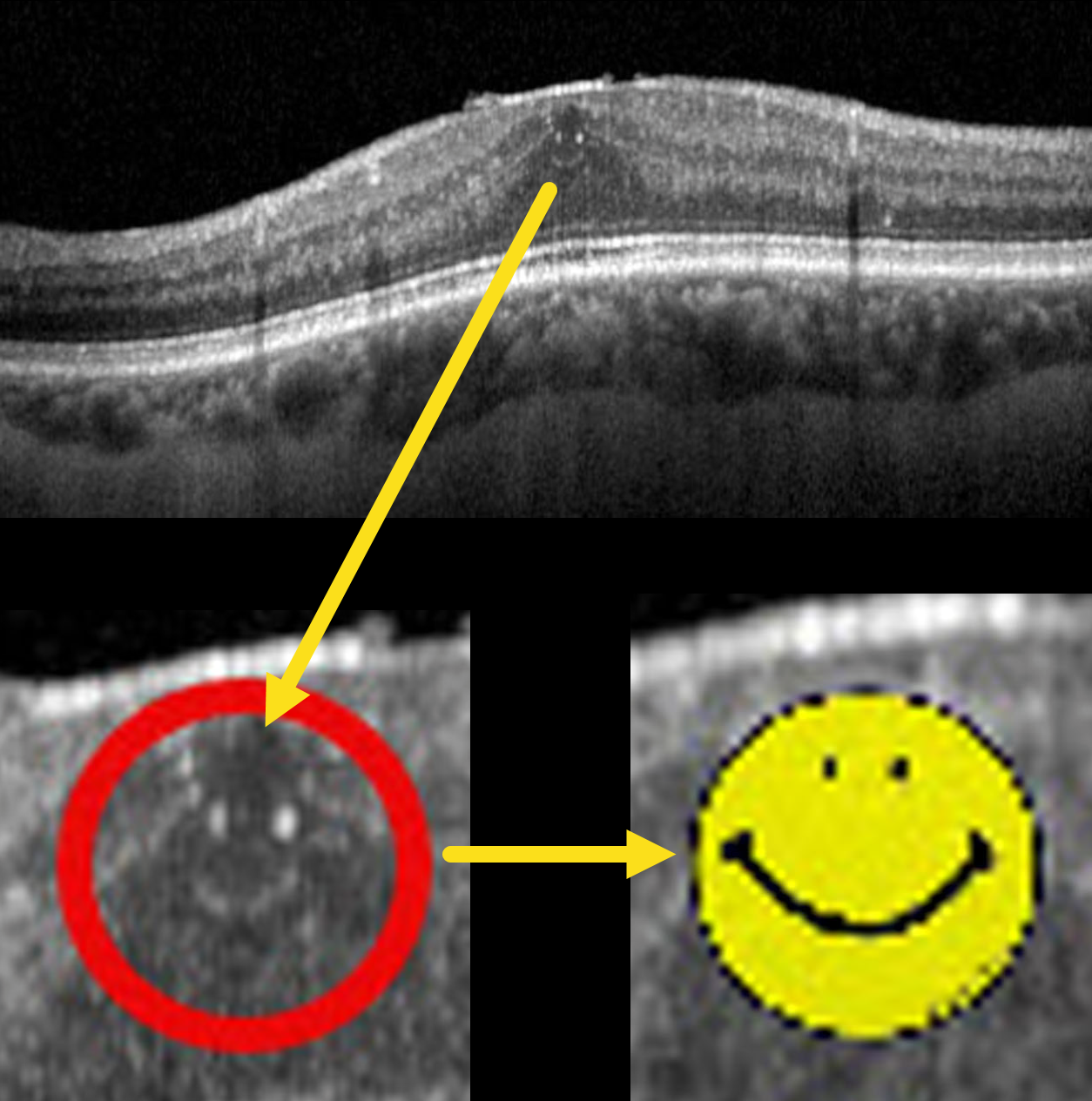
This 70YO male with type 2 diabetes presented with these asymptomatic OCT findings that made our day. Vision was 20/25.
World Smile Day is celebrated on the first Friday of October each year, a tradition that began in 1999. The day was created by Harvey Ball, the commercial artist who designed the iconic yellow smiley face. Ball was concerned that the over-commercialization of his symbol would dilute its original intent of spreading goodwill and cheer. To counteract this, he established World Smile Day to remind people of the power of a smile and to encourage acts of kindness around the world.
The day is marked by various activities that promote smiling and kindness. People are encouraged to perform acts of kindness and to smile more, spreading positivity and happiness. The Harvey Ball World Smile Foundation, established after Ball’s passing in 2001, continues to sponsor the day and organize events. These events range from community activities to global initiatives, all aimed at creating smiles and fostering a spirit of generosity and joy.
So, perform some random acts of kindness today and make the world a better place because you’re in it.
CHOROIDEREMIA CARRIER
Originally posted on @retina.rocks August 20, 2024
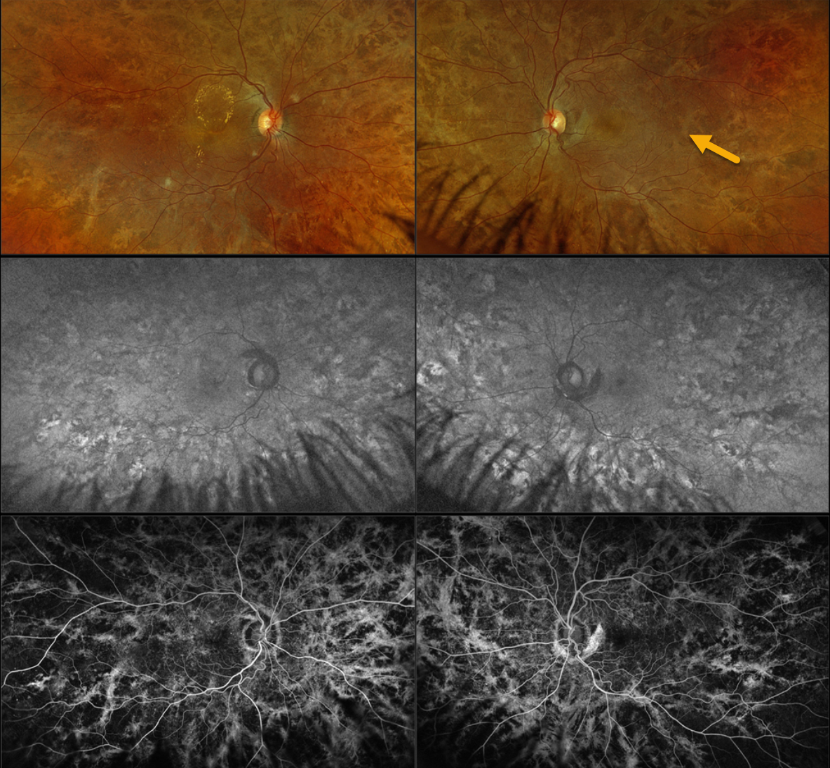
This 42YO female with type 2 diabetes was referred for asymptomatic diabetic macular edema (DME). There was no family history of ocular disease. Vision was 20/40 OD and 20/20 OS.
Optos color RGB imaging shows diffuse coarse subretinal hyperpigmentations throughout each posterior pole. Fundus autofluorescence (FAF) shows variable hyper- and hypo-FAF from these pigmentary changes, with fluorescein angiographic window defects. Genetic testing revealed a heterozygous pathogenic CHM mutation.
Learning Points:
Choroideremia is an X-linked recessive chorioretinal dystrophy caused by a mutation in the CHM gene located on the long arm of the X chromosome. Sons of female carriers have a 50% chance of developing choroideremia, and daughters have a 50% chance of becoming carriers. Some female carriers can develop asymptomatic pigmentary changes, as in our patient, or frank choroideremia due to irregular inactivation of the X chromosome (see Jauregui et al, AJO 2019;207:77-86).
FOCAL LASER FOR DIABETIC MACULAR EDEMA (DME) WITH SUBSEQUENT DEGENERATIVE LAMELLAR MACULAR HOLE
Originally posted on @retina.rocks August 15, 2024
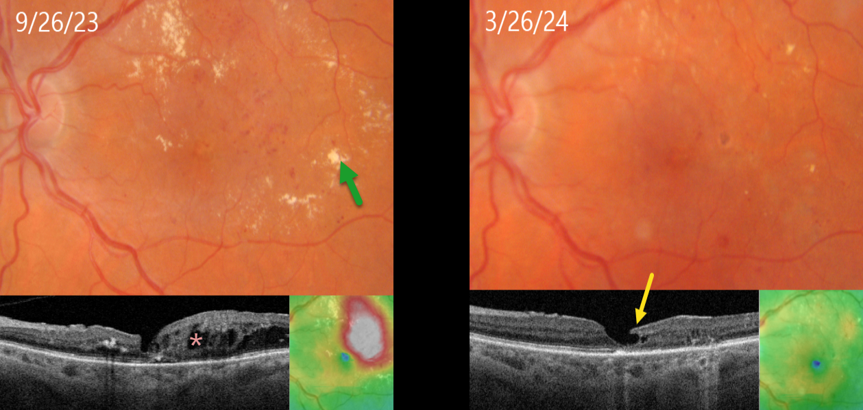
This 71YO female with type 2 diabetes presented diabetic macular edema (DME) and lipid exudates. Triton swept-source OCT shows central foveal thinning with surrounding cystic edema. Vision was 20/60. Thermal macular laser was applied.
Six months later, the edema completely resolved following this single treatment, with near total resolution of the macular lipid. A partial degenerative lamellar macular hole (LMH) is noted. Vision improved to 20/50.
Learning Points:
Our patient exemplifies why macular laser still has a place in any retinal surgeon’s toolbox and should be considered a viable option for treating non-central involved diabetic macular edema (NCI-DME) and even center-involved edema (CI-DME) when the leaking microaneurysms are outside the foveal avascular zone (FAZ). In our practice, macular laser remains the initial treatment for eyes with NCI-DME. When applied gently with small, low-power, and short-duration applications well outside the FAZ, patients rarely, if ever, experience scotoma. Both the treatment burden and the financial costs to society are dramatically less. And there is no risk for endophthalmitis.
Although the role of thermal laser for treating DME is currently a source of debate (see Eye 2022;36:485-486 and Eye 2022;36:483-484 for great pro-laser and anti-laser editorials) we are not sure what all the fuss is about. Old-fashioned lasers work and, in our opinion, are incredibly under-utilized.
LMH can be defined as tractional or degenerative (Govetto et al, AJO 2016;164:99-109). Our patient developed a partial degenerative LMH following likely degeneration or rupture of a foveal cyst following closure of the leaking microaneurysms. There is some debate regarding the benefits of surgery for symptomatic LMH, with some suggesting visual improvement (Morescalchi et al, Retina, 2020;40:1087-1093) and others noting little visual benefit (Mohammed and Thompson, J Vitreoretinal Diseases 2024;8:125-130). Since our patient was visually happy, observation was recommended.
DIABETIC MACULAR EDEMA (DME)
Originally posted on @retina.rocks August 29, 2023
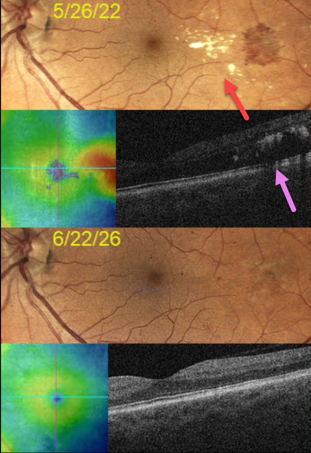
This 57YO man presented on 5/26/22 with asymptomatic diabetic macular edema (DME) in his left eye. Vision was 20/20.
Extensive lipid exudates with associated macular thickening were noted in the temporal macula. A gentle thermal laser was applied to the leaking vessels.
One year later, vision remains at 20/20, with total resolution of all lipid and DME.
Learning Points:
Beginning in the 1970s, macular laser was the gold standard for treating DME. In the early to mid-2000s, a perfect storm brewed with the advent of OCT technology and intravitreal injections. Unlike lasers, which take many months to work and rarely significantly improve vision, intravitreal injections cause a rapid and dramatic improvement in vision, making them the mainstay for treating DME ever since.
Clinically significant macular edema (CSME) is now an outdated term, as we are more concerned with whether the edema is central (central involved, CI-DME) or non-central (NCI-DME). Eyes with CI-DME are usually best treated with injections for superior visual outcomes.
However, our patient exemplifies why macular laser still has a place in any retinal surgeon’s toolbox, and should be considered a viable option for treating NCI-DME and even CI-DME when the leaking microaneurysms are outside the foveal avascular zone (FAZ).
In our practice, macular laser remains the initial treatment for eyes with NCI-DME. When applied gently with small, low-power, and short-duration applications well outside the FAZ, patients rarely, if ever, experience scotoma. Both the treatment burden and the financial costs to society are dramatically less. And there is no risk for endophthalmitis.
Although the role of thermal laser for treating DME is currently a source of debate (see Eye 2022;36:485-486 and Eye 2022;36:483-484 for great pro-laser and anti- laser editorials) we are not sure what all the fuss is about. Old-fashioned lasers work and, in our opinion, are incredibly under-utilized.
VALENTINE MACULAR EDEMA
Originally posted on @retina.rocks February 14, 2023
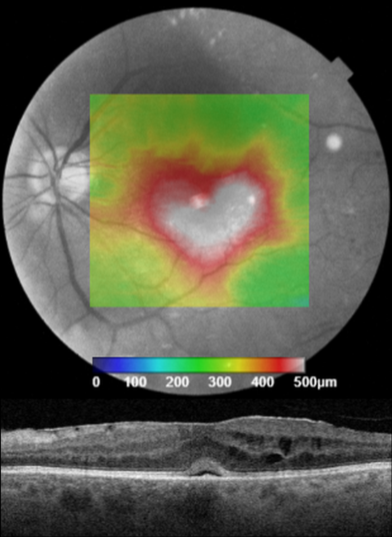
This patient presented with 20/200 vision from center-involved diabetic macular edema.
Unfortunately, we don’t have any further clinical information for this patient, but we still wish everyone a ‘heart-felt’ Happy Valentine’s Day!
DIABETIC MACULAR EDEMA (DME)
Originally posted on @retina.rocks January 26, 2023
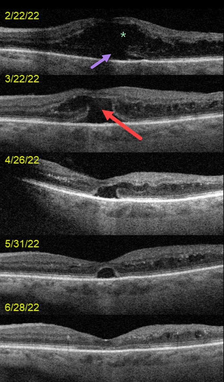
This 72YO female presented on 2/22/22 with counting-finger vision in her left eye.
OCT scanning at the initial visit shows severe center-involved diabetic macular edema (CI-DME) with shallow subretinal fluid. Anti-VEGF therapy was started.
One month later, vision remained at counting fingers. The retinal thickness had improved, but a new outer macular hole was noted.
Following 3 additional monthly injections, the edema continued to resolve with spontaneous closure of the outer macular hole. Vision was 20/70 at the 6/28/22 visit.
Learning Points:
Traditional primary full-thickness macular holes are caused by vitreomacular traction. Outer macular holes, however, have a much broader range of etiologies.
In our practice, we most commonly see these lesions in optic pit maculopathy and paraproteinemia (see Mansour et al, Ophthalmology 2014;121:1925-1932).
Our patient’s outer macular hole was likely caused by inner retinal exudation that extended through the outer retina. The return of central vision and closure of the defect, with relatively intact outer macular architecture, likely indicates that this was an outer macular dehiscence rather than a true loss of tissue. See Kumawat et al for a great review of atypical macular holes (Retina 2019;39:1236-1264).
FOCAL LASER FOR DIABETIC MACULAR EDEMA (DME)
Originally posted on @retina.rocks December 29, 2022
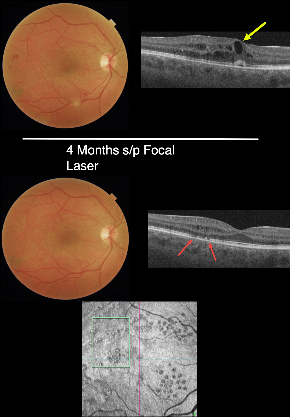
This 32YO female with type 1 diabetes was examined with no visual symptoms, and vision was 20/30 OD and 20/20 OS. Macular photography shows a foveal cyst and mild peripheral macular hemorrhages, and OCT scanning shows central and temporal cystic edema with trace subfoveal fluid.
Due to the lack of symptoms and good vision, we were hesitant to start anti-VEGF therapy, but due to concern for progressive, increased edema and vision loss, we decided to treat with macular laser.
Four months later, the edema virtually completely resolved. The laser scars appear as temporal defects in the ellipsoid zone, although they are barely visible funduscopically. The laser scars are more visible with outer retinal en face imaging.
Learning Points:
In their landmark 1985 paper (Arch Ophthalmology 1985;103:1796-1806), the Early Treatment Diabetic Retinopathy Study (ETDRS) introduced the term clinically significant diabetic macular edema (CSME). Broadly speaking, eyes with CSME, as determined by retinal thickening found on contact lens examination, have the threshold amount of macular edema that benefits from laser photocoagulation.
Macular laser remained the gold standard for diabetic macular edema (DME) for about 15 to 20 years. In the early to mid-2000s, a perfect storm brewed with the advent of OCT technology and intravitreal injections. Unlike lasers, which take many months to work and rarely significantly improve vision, intravitreal injections show a rapid and dramatic visual improvement, making them the mainstay for treating DME ever since.
CSME has become an antiquated term since we are now more concerned with whether the edema is central (central involved, CI-DME) or non-central (NCI-DME). Eyes with CI-DME are best treated with injections due to superior visual results.
However, our patient exemplifies why macular laser still has a place in any retinal surgeon’s toolbox, and should be considered a viable option for treating NCI-DME and even CI-DME when the leaking microaneurysms are outside the foveal avascular zone (FAZ). In our practice, macular laser remains the initial treatment for eyes with NCI-DME. When applied gently with small, low-power and short-duration applications well outside the FAZ, patients rarely, if ever, experience scotoma. Both the treatment burden and the financial costs to society are dramatically less. And there is no risk for endophthalmitis.
Although the role of thermal laser for treating DME is currently a source of debate (see Eye 2022;36:485-486 and Eye 2022;36:483-484 for great pro-laser and anti- laser editorials) we are not sure what all the fuss is about. Old-fashioned laser works and, in our opinion, is incredibly under-utilized.
OZURDEX GHOST
Originally posted on @retina.rocks November 26, 2021
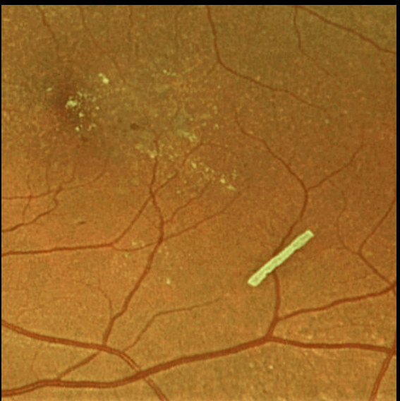
This 71YO female is getting Ozurdex intravitreal injections every 2 months to control her macular edema.
Learning Points:
The Ozurdex implant slowly releases the steroid and eventually totally absorbs, with the clinical effect usually lasting about 2 to 4 months.
The ghost-like remnants can persist for many months (see Kim et al, Retina 2020;40;2226-2231).
DIABETIC MACULAR EDEMA (DME)
Originally posted on @retina.rocks October 14, 2021
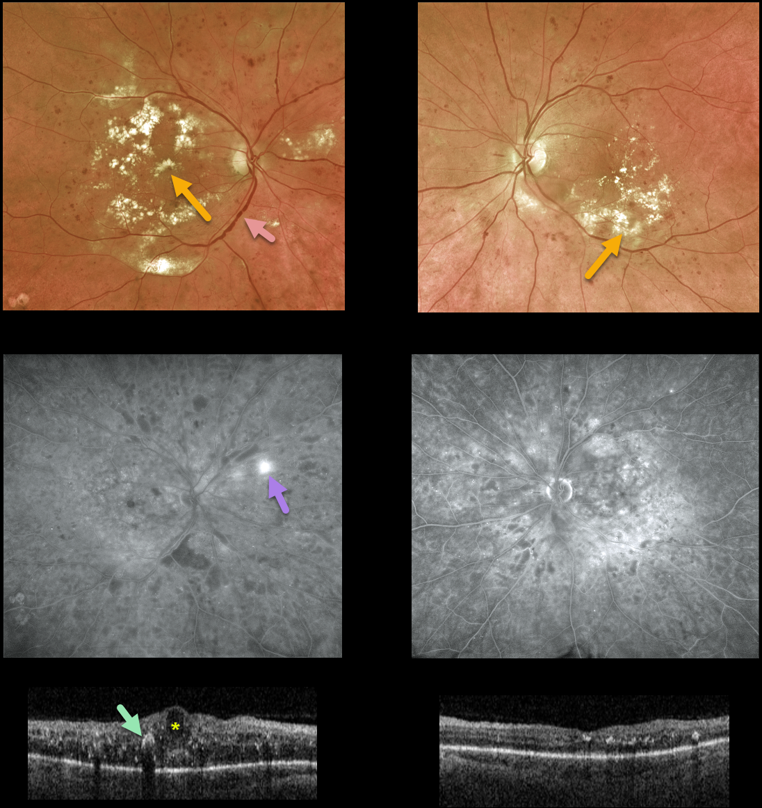
This 59YO male with a history of type 2 diabetes presented with bilateral diabetic macular edema. Vision was counting fingers OD and 20/30 OS.
There was extensive bilateral macular lipid, particularly in his right eye. Venous beading and sausaging were noted OD, indicating severe retinal ischemia.
Fluorescein angiography confirmed severe ischemia with peripheral temporal retinal neovascularization.
Triton swept-source OCT scanning shows severe central-involved diabetic macular edema (CI-DME) with scattered hyperreflective lipid flecks OD, non-central-involved diabetic macular edema (NCI-DME), and milder lipid OS.
Macular laser for the NCI-DME was recommended for his left eye. Monthly anti-VEGF injections were recommended for the CI-DME in his right eye. Remarkably, vision improved to 20/80 so far after a single injection.
Learning Points:
Anti-VEGF injections are the mainstay of treatment for CI-DME. It is expected that these injections will also stabilize, or possibly reverse, this patient’s proliferative disease.
We also plan to begin panretinal photocoagulation once the central edema resolves. There are multiple ways we could have approached his treatment plan. What would you do and why?
MIKE WAZOWSKI MACULOPATHY
Originally posted on @retina.rocks August 28, 2020
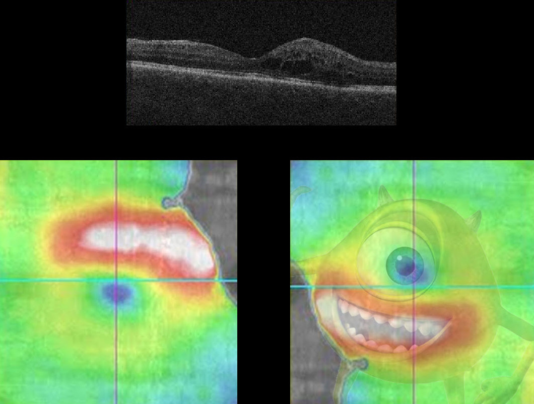
Here we see a patient with the ever-elusive Mike Wazowski Maculopathy (for you Baby Boomers, he stars in Monsters, Inc.).
The OCT image, rotated 180 degrees, details a classic Mike Wazowski pattern of edema.
In reality, the patient has some non-central diabetic macular edema.


