

Diabetic Retinopathy Proliferative (PDR) Panretinal Photocoagulation (PRP)
<< Back to Cases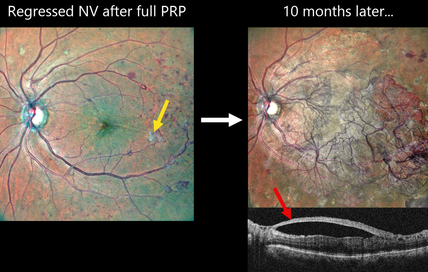
PROLIFERATIVE DIABETIC RETINOPATHY (PDR) WITH PROGRESSIVE NV DESPITE FULL PRP
Shishir Verghese
Originally posted on @retina.rocks March 4, 2026
This 60YO female with a history of type 2 diabetes underwent full panretinal photocoagulation (PRP) in her left eye. Following laser, MultiColor SLO imaging shows small residual areas of variably fibrosed retinal neovascularization. Vision was 20/30.
Ten months later, she presented complaining of 2 months of decreased vision in this eye. Vision was 20/60. MultiColor SLO imaging shows retinal neovascularization covering the posterior pole. On OCT, the fovea is remarkably attached with the hyperreflective neovascularization elevated along the partially detached posterior hyaloid.
Learning Points:
Although PRP is often considered a ‘one and done’ procedure, this is not always the case (Gonzalez et al, Ophthalmology 2021;128:1448-1457). Our patient is a perfect example of why these patients require lifelong monitoring even if their disease initially appears quiescent.
Given the aggressive neovascular proliferation, intravitreal anti-VEGF was given, followed by vitrectomy 5 days later. Although PRP is often considered a ‘one and done’ procedure, this is not always the case (Gonzalez et al, Ophthalmology 2021;128:1448-1457). Our patient is a perfect example of why these patients require lifelong monitoring even if their disease initially appears quiescent.
Given the aggressive neovascular proliferation, intravitreal anti-VEGF was given, followed by vitrectomy 5 days later.
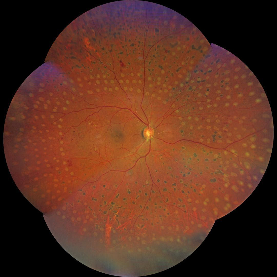
FILL-IN PANRETINAL PHOTOCOAGULATION
Giacomo De Rosa
Originally posted on @retina.rocks February 23, 2026
This 63YO male with type 2 diabetes presented to us with a history of recent panretinal photocoagulation (PRP) OD elsewhere. Vision was 20/20 OU.
Color photography shows irregularly sized and variably confluent laser scars mostly in the right inferior, nasal, and inferior midperiphery. Fill-in laser was applied at the slit lamp to complete the PRP.
Learning Points:
PRP should begin at least 1 disc diameter from the optic nerve and outside the major temporal arcades. Treatment is performed with a wide-angle lens or an indirect ophthalmoscope and should be applied from the posterior retina towards the periphery to prevent accidental macular burns. The laser spots should be 200-500 microns in size at the retina, be spaced about one burn-width apart, and should extend as far anteriorly as possible.
We generally do not recommend titrating the PRP based on involution of neovascularization, primarily due to the significant risk of the patient being lost to follow-up (Obeid et al, Ophthalmology 2018;125:1386-1392). Once the decision is made for PRP, we complete treatment in several sessions within a few months. Postoperative ultra-widefield imaging is helpful for assessing the adequacy of laser coverage, as areas of retinal ischemia are often undertreated (Sowmya L et al., Retina 2023;43:1922-1927).
We aren’t certain whether the initially treating laser surgeon planned further laser treatment, but the treatment was not performed in a controlled enough manner to control for burn size and spacing.
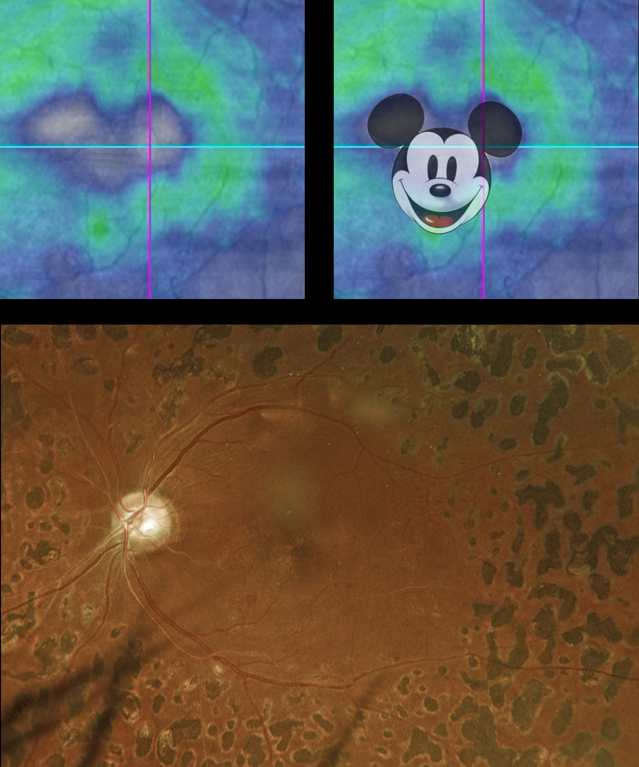
MICKEY MOUSE MACULOPATHY
Originally posted on @retina.rocks November 13, 2024
This 52YO diabetic male rodent has a history of proliferative diabetic retinopathy and full panretinal photocoagulation (PRP). Vision was stable at 20/30.
Optos color RGB imaging shows full PRP scarring. The macula is clinically ischemic with some vascular sheathing. Triton swept-source OCT shows nasal and central atrophy consistent with Mickey Mouse Maculopathy (MMM).
Created by Walt Disney and Ub Iwerks, Mickey Mouse celebrates his birthday on November 18th. This date commemorates his first appearance in the animated short film “Steamboat Willie” in 1928, famous for Mickey’s debut and for its synchronized sound.
On January 1, 2024, the famous mouse entered the public domain, allowing anyone to use this version of the character in their creative works without needing Disney’s permission or payment. However, Disney still retains the copyright on any subsequent variations of Mickey until they reach the 95-year mark.
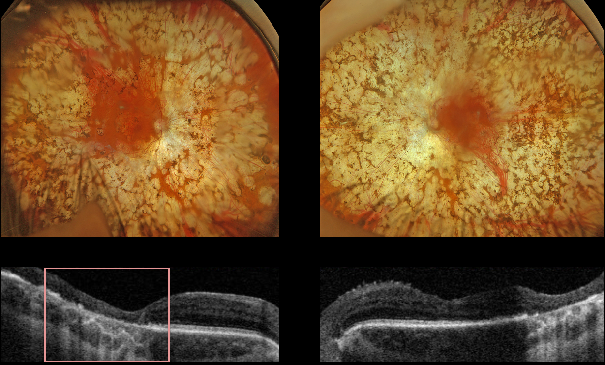
PANRETINAL PHOTOCOAGULATION
Originally posted on @retina.rocks February 18, 2024
This 53YO female with type 1 diabetes received bilateral panretinal photocoagulation (PRP) for proliferative diabetic retinopathy (PDR) about 30 years previously from an outside practice. She presented for her yearly retinopathy examination, and vision was stable at 20/200 OD and 20/30 OS.
Optos color RGB imaging shows bilateral confluent PRP scarring extending from within the arcades into the peripheries. Triton swept-source OCT of the right eye shows outer retinal atrophy extending through the foveal center from confluent macular laser scarring, with more temporal outer retinal atrophy in the left eye from confluent posterior PRP scarring.
Learning Points:
PRP should begin at least 1 disc diameter from the optic nerve and outside the major temporal arcades. Treatment is performed with a wide-angle lens or an indirect ophthalmoscope, and should be applied from the posterior retina towards the periphery to avoid accidental macular burns. The laser spots should be 200-500 microns in size at the retina, be spaced about one burn-width apart, and should extend as far anteriorly as possible.
The laser performed on this patient is wrong on so many levels, including spots extending into the macula, touching the nerve, and confluent elsewhere. The laser surgeon essentially gave this patient severe iatrogenic retinitis pigmentosa.
Although possibly appropriate before the advent of modern vitrectomy surgery and anti-VEGF therapy, this degree of laser should never be seen in today’s patients.
PANRETINAL PHOTOCOAGULATION
Originally posted on @retina.rocks July 7, 2022
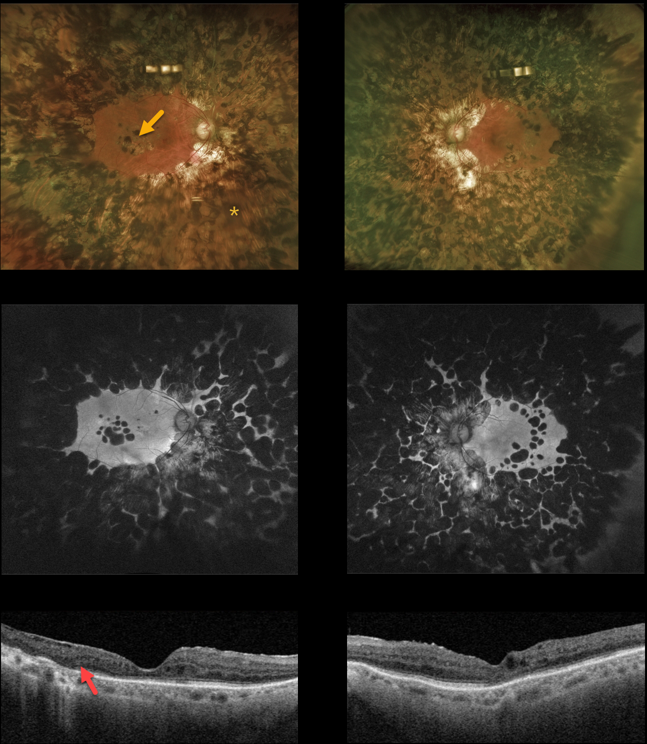
This 80YO diabetic female gave a history 0f prior retinal lasers elsewhere about 20 years earlier. Vision was 20/30 OD and 20/80 OS.
Optos color and fundus autofluorescent imaging show heavy bilateral panretinal photocoagulation (PRP) and macular laser scarring. Swept source OCT shows variable disorganization of the retinal layers and outer retinal atrophy. Visual fields (not shown) are severely constricted due to the ‘iatrogenic’ retinitis pigmentosa caused by the posterior and confluent laser scarring.
Learning Points:
Although possibly appropriate before the advent of modern vitrectomy surgery and anti-VEGF therapy, this degree of laser should never be seen in today’s patients. Laser should begin well outside the arcades and away from the optic nerve, extending as anteriorly as possible with the 200 to 500 micron-sized retinal burns spaced about one burn-width apart.
PANRETINAL PHOTOCOAGULATION
Originally posted on @retina.rocks November 11, 2021
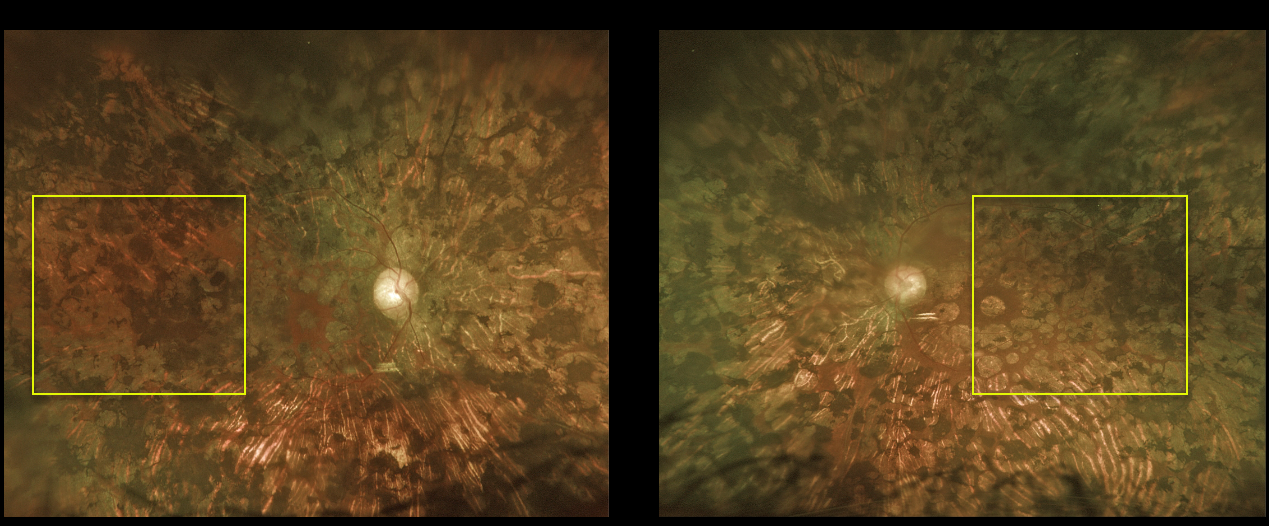
This 63YO female underwent incredibly intense, confluent, and posterior panretinal photocoagulation (PRP) and macular laser elsewhere before transferring her care to our office. Vision was hand motion OD and 20/400 OS.
The laser performed on this patient is wrong on so many levels, including spots extending to the macula, touching the nerve, and confluent elsewhere. The laser surgeon essentially gave this patient severe iatrogenic retinitis pigmentosa.
Learning Points:
PRP should begin at least 1 disc diameter from the optic nerve and outside the major temporal arcades. Treatment is performed with a wide-angle lens or an indirect ophthalmoscope and should be applied from the posterior retina towards the periphery to prevent accidental macular burns.
The laser spots should be 200-500 microns in size at the retina, spaced about one burn width apart, and extend as far anteriorly as possible.
PROLIFERATIVE DIABETIC RETINOPATHY (PDR)
Originally posted on @retina.rocks November 4, 2021
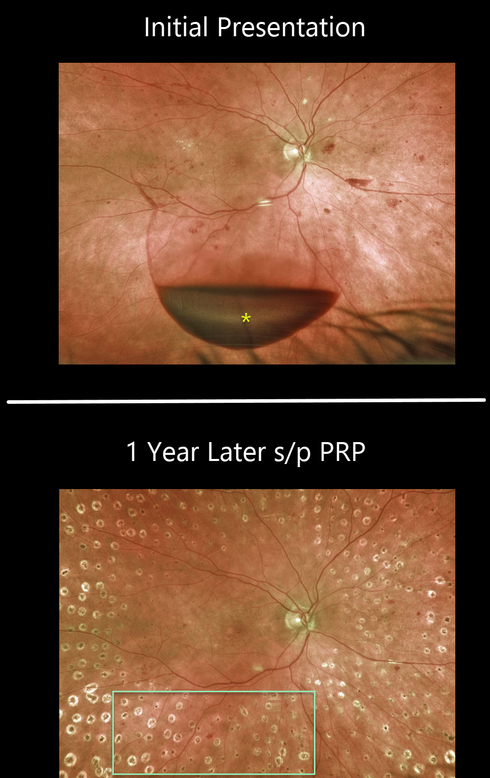
This 65YO female presented with 20/400 vision in her right eye. An acute inferior subhyaloid hemorrhage was noted along with non-central diabetic macular edema. Over the coming months, macular laser followed by panretinal photocoagulation (PRP) was performed.
One year later, vision was 20/50, with complete resolution of the preretinal blood and proliferative disease.
Learning Points:
Before PRP, patients with proliferative diabetic retinopathy (PDR) routinely went blind, and one of the treatments actually included pituitary ablation (for an amazing historical perspective on diabetic laser photocoagulation, see Goldberg and Jampol, Ophthalmology 1987;94;741-746).
PRP originally extended fairly posteriorly, and patients often needed fill-in treatment before vitrectomy and anti-VEGF treatments became available. Today, most laser surgeons will begin treatment well away from the nerve and major arcades to minimize the risks of central and peripheral vision loss.
Once considered a “one and done” treatment, more recent studies show that patients may still need additional laser, anti-VEGF injections, or vitrectomy (see Gonzalez et al, Ophthalmology 2021;128:1448-1457).
Some also advocate anti-VEGF injections as the primary treatment for PDR. However, most still prefer to add PRP at some point due to the risk of patients being lost to follow-up (see Gross et al, DRCR Protocol S, JAMA Ophthalmology 2018;136:1138-1148).
PANRETINAL PHOTOCOAGULATION
Originally posted on @retina.rocks March 5, 2021
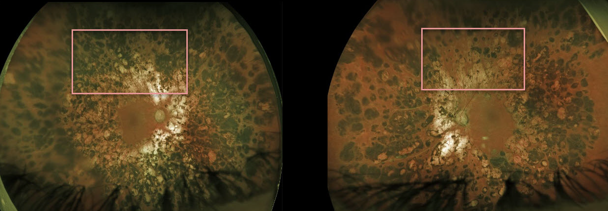
This 51YO male has heavy bilateral panretinal photocoagulation (PRP) scarring for proliferative diabetic retinopathy (PDR). All laser treatments were done years earlier at an outside practice.
The burns are confluent and extend into the arcades. Thankfully, the treating surgeon did not quite burn the optic nerves. However, the visual fields are severely constricted with essentially iatrogenic retinitis pigmentosa.
Learning Points:
Although possibly appropriate before the advent of modern vitrectomy surgery and anti-VEGF therapy, this degree of laser should never be seen in today’s patients.
Laser should begin well outside the arcades and away from the optic nerve, extending as anteriorly as possible, using 200- to 500-micron retinal burns spaced about one burn width apart.
PROLIFERATIVE DIABETIC RETINOPATHY (PDR)
Originally posted on @retina.rocks February 25, 2021
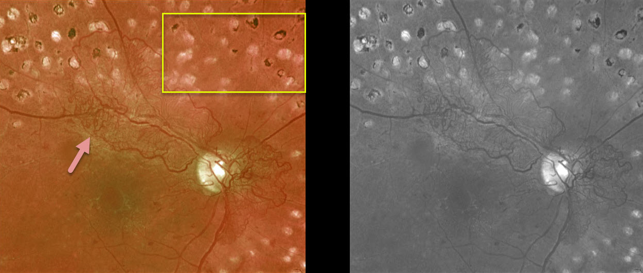
This is a 55YO diabetic patient who had been treated with complete panretinal photocoagulation (PRP). Vision was 20/40. Despite PRP, the patient still had significant residual posterior neovascularization.
Since it is not uncommon for neovascularization to persist following full laser, we are observing for now.
Learning Points:
Panretinal photocoagulation has been around since the 1960s and still remains the mainstay for treating proliferative disease. Often considered a “one and done,” patients can still develop breakthrough hemorrhages. Intravitreal anti-VEGF injections are often helpful, but some will require vitrectomy.
PROLIFERATIVE DIABETIC RETINOPATHY
Originally posted on @retina.rocks December 24, 2019
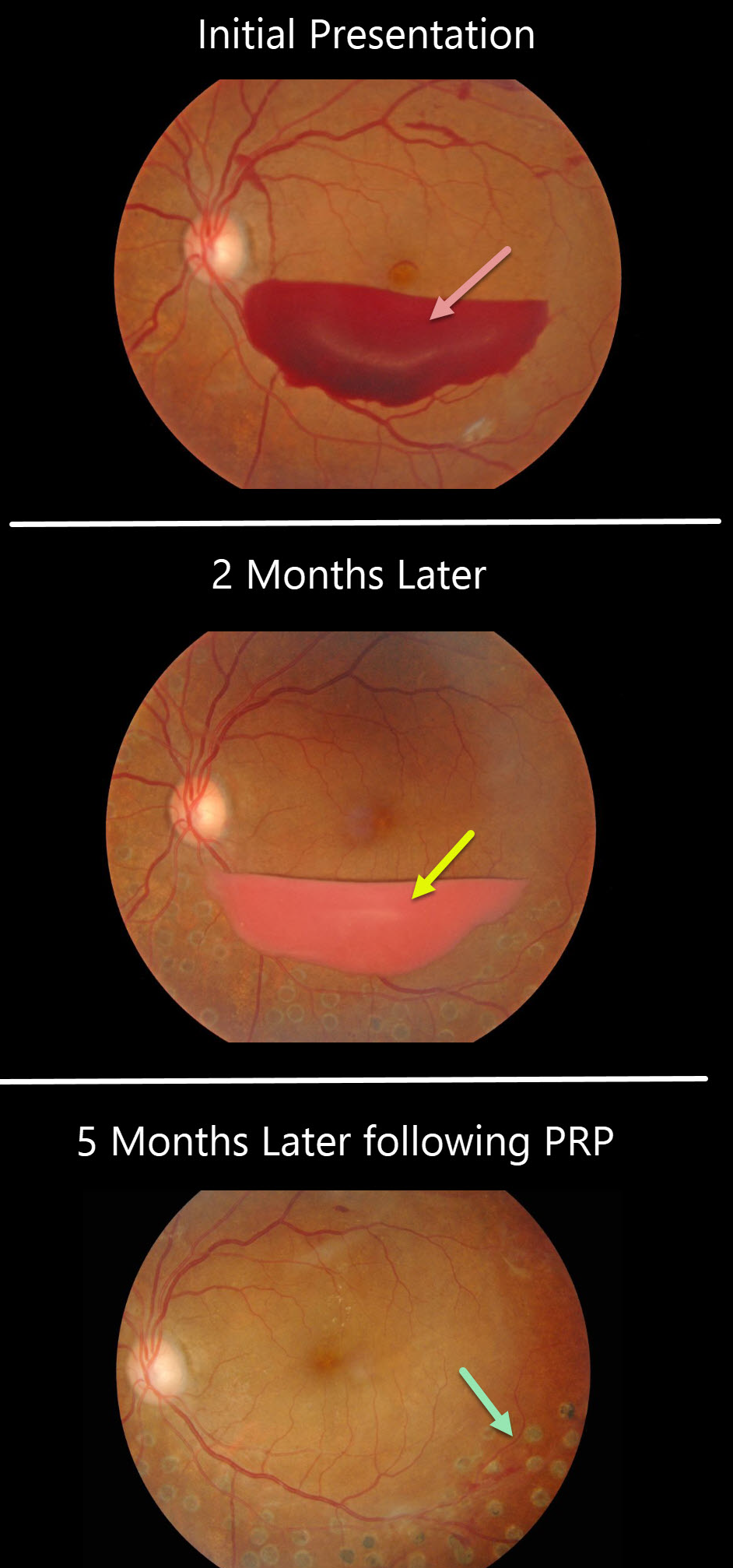
This patient presented with a sub-hyaloid hemorrhage due to proliferative diabetic retinopathy.
A subsequent photo two months later shows devitalized (yellowing) sub-hyaloid hemorrhage. He received panretinal photocoagulation (PRP) in the left eye and the sub-hyaloid hemorrhage completely resolved three months later. Anti-VEGF therapy was not used.
Learning Points:
PRP has been around since the 1960s and still remains the mainstay for treating proliferative disease. Often considered a “one and done,” patients can still develop break-through hemorrhages. Intravitreal anti-VEGF injections are often helpful, but some will require vitrectomy.


