

Diabetic Retinopathy Proliferative (PDR) Preretinal/Vitreous Hemorrhage
<< Back to Cases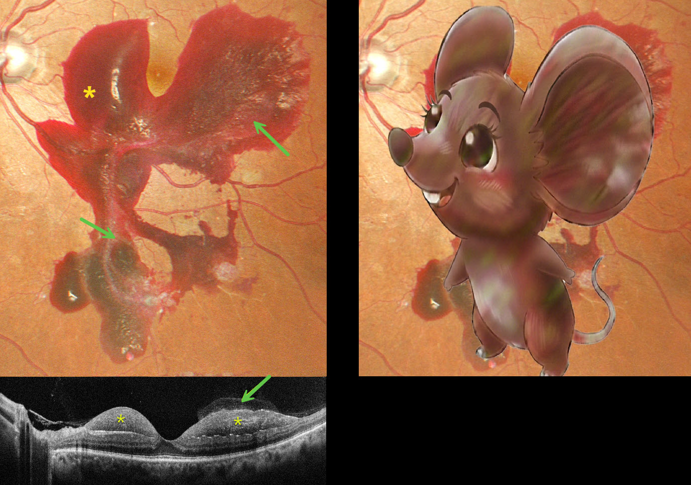
MOUSE MACULOPATHY AKA PROLIFERATIVE DIABETIC RETINOPATHY (PDR) WITH SUBHYALOID HEMORRHAGE
Originally posted on @retina.rocks February 24, 2026
This 48YO male with a 15-year history of poorly controlled type 2 diabetes presented with one day of blurred vision in his left eye. Vision was 20/20 bilaterally.
Pseudocolor SLO imaging shows a large macular hemorrhage, sparing the foveal center, which extends into the midperiphery. The hemorrhage is mostly subhyaloid with some breakthrough blood into the vitreous. The blood compartments are well visualized on OCT.
Learning Points:
The normal vitreous undergoes a decades-long process of orchestrated degeneration, including liquefaction and gradual separation from the retinal surface. The posterior vitreous detachment (PVD) begins in the perifoveal retina early in life, followed by a foveal PVD, then a peripheral PVD, and then finally a complete PVD when the vitreous separates from the optic nerve (Johnson AJO 2010;149:371-382). Although a diabetic subhyaloid hemorrhage can extend throughout the macula, our patient’s findings illustrate the normal increase in vitreofoveal adhesion.
PROLIFERATIVE DIABETIC RETINOPATHY (PDR)
Originally posted on @retina.rocks September 25, 2024
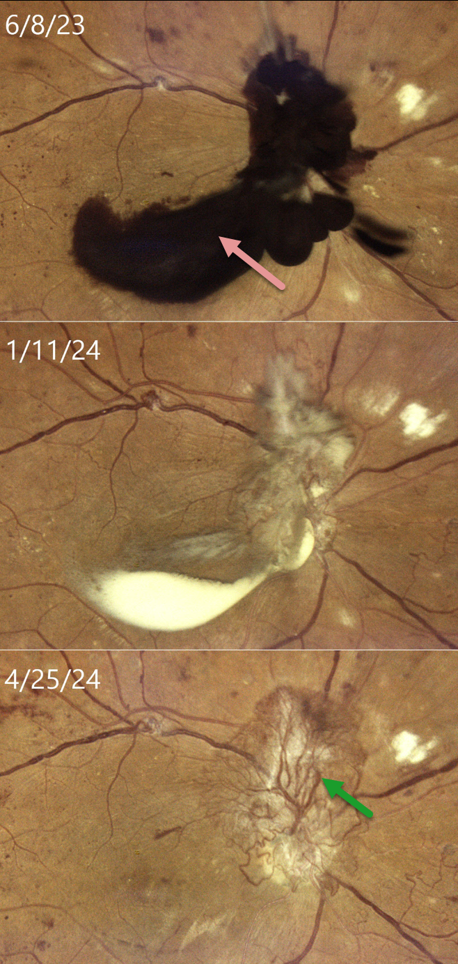
This 33YO female with type 2 diabetes was in the process of being treated with intravitreal Eylea, macular laser, and panretinal photocoagulation (PRP) for bilateral center-involved diabetic macular edema and proliferative diabetic retinopathy (PDR). She presented on 6/8/23 with recent vision loss OD and was overdue for this visit because of numerous medical issues. Vision had decreased to 20/400 OD.
Color imaging shows dark red preretinal blood obscuring the optic nerve and central macula. After several monthly Eylea injections and completion of the PRP, the preretinal blood became yellow and devitalized on 1/11/24, before finally completely absorbing, as noted at last examination on 4/25/24, with stable 20/400 vision. Despite full PRP, diffuse variably fibrosed neovascularization persists, along with venous beading. OCT scanning revealed no edema with central atrophy (not shown).
Our patient was initially in no rush for vitrectomy surgery, as vision in her fellow eye was 20/50. Conservative management with intravitreal injections and PRP was thankfully successful. This approach was also found to be safe and effective by Brar et al in a retrospective series of 22 eyes with foveal preretinal blood from PDR (Ind J Ophthalmol 2024;72:687-691).
DIABETIC SUBHYALOID HEMORHAGE
Originally posted on @retina.rocks January 9, 2024
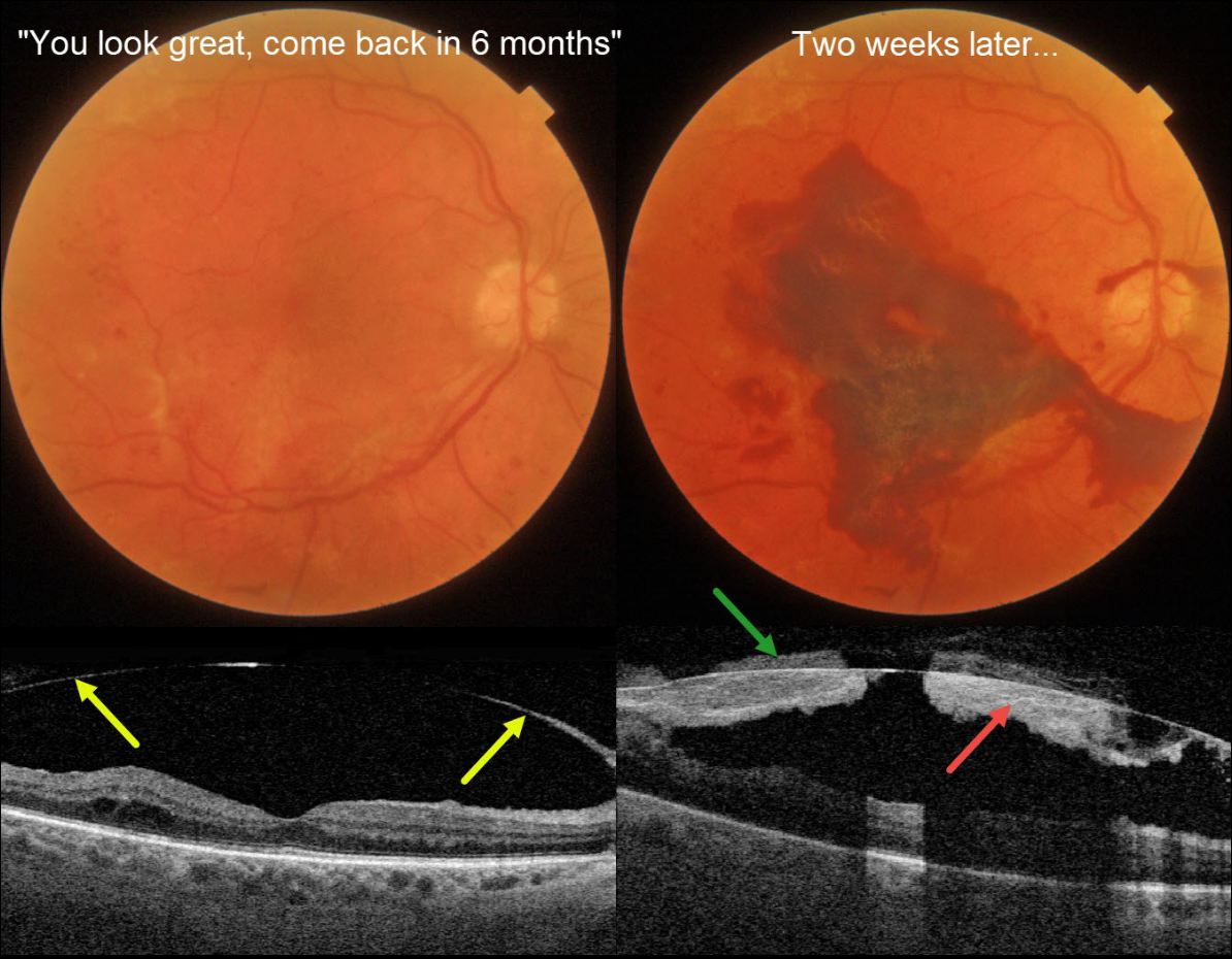
This 71YO diabetic male was examined over a year following completion of panretinal photocoagulation (PRP) for proliferative diabetic retinopathy (PDR). Vision was 20/50.
Triton color photography shows no macular edema, and swept-source OCT reveals a detached hyaloid elevated over the macula. He was told that everything looked great and to come back in 6 months. Famous last words…
Two weeks later, he presented with acute vision loss in this eye. Vision was counting fingers from a fresh macular subhyaloid hemorrhage. Both subhyaloid and prehyaloid blood are noted on Triton swept-source OCT.
An intravitreal Avastin injection was given, and vitrectomy will be recommended if the blood fails to clear.
Learning Points:
Although PRP is often a ‘one and done’ procedure, this is not always the case (Gonzalez et al, Ophthalmology 2021;128:1448-1457), and patients therefore need continued monitoring. Although our patient appeared perfect 2 weeks earlier, this case reminds us to remain humble in the presence of the retina and that even an involuted PDR requires lifelong follow-up.
PROLIFERATIVE DIABETIC RETINOPATHY (PDR)
Originally posted on @retina.rocks June 30, 2022
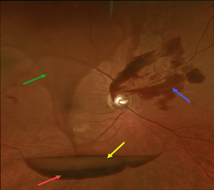
The preretinal blood in this 70YO male with proliferative diabetic retinopathy (PDR) has multiple layers.
The red arrow highlights subhyaloid blood, while the yellow arrow shows the yellowing, devitalized subhyaloid blood just above. The blue arrow shows subhyaloid blood emanating from disc neovascularization. Last but not least, the twister of blood within the vitreous gives the appearance of a tornado literally shaking up all of this blood.
Panretinal photocoagulation was started.
SEPHIROTH RETINOPATHY
Originally posted on @retina.rocks March 25, 2022
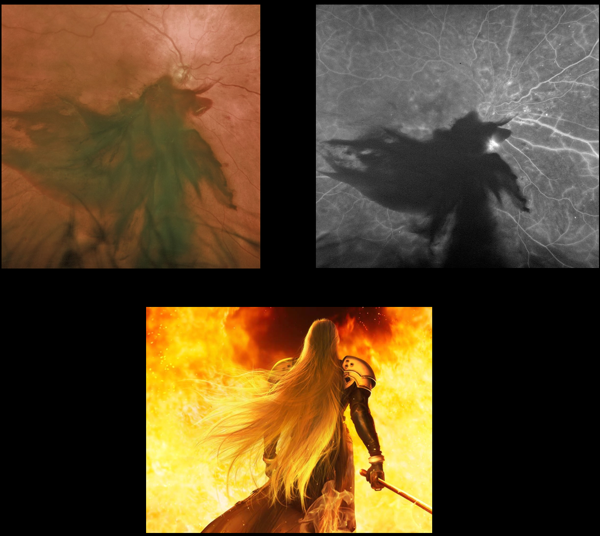
We all know it’s a large subhyaloid hemorrhage from proliferative diabetic retinopathy, but check your imagination and tell us what you see.
We see Sephiroth from Final Fantasy!
PROLIFERATIVE DIABETIC RETINOPATHY (PDR)
Originally posted on @retina.rocks February 1, 2022
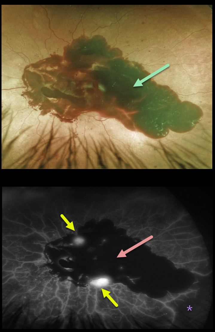
This 69YO male with type 2 diabetes presented with severe acute vision loss in his left eye from a large posterior pole subhyaloid hemorrhage.
The hemorrhage blocks on fluorescein angiography, with a few areas of leaking neovascularization and severe peripheral ischemia.
He was initially treated with intravitreal Avastin, and over the coming months, panretinal photocoagulation (PRP) was performed.
However, he later developed a large vitreous hemorrhage and will likely need vitrectomy surgery.
PROLIFERATIVE DIABETIC RETINOPATHY (PDR)
Originally posted on @retina.rocks November 4, 2021
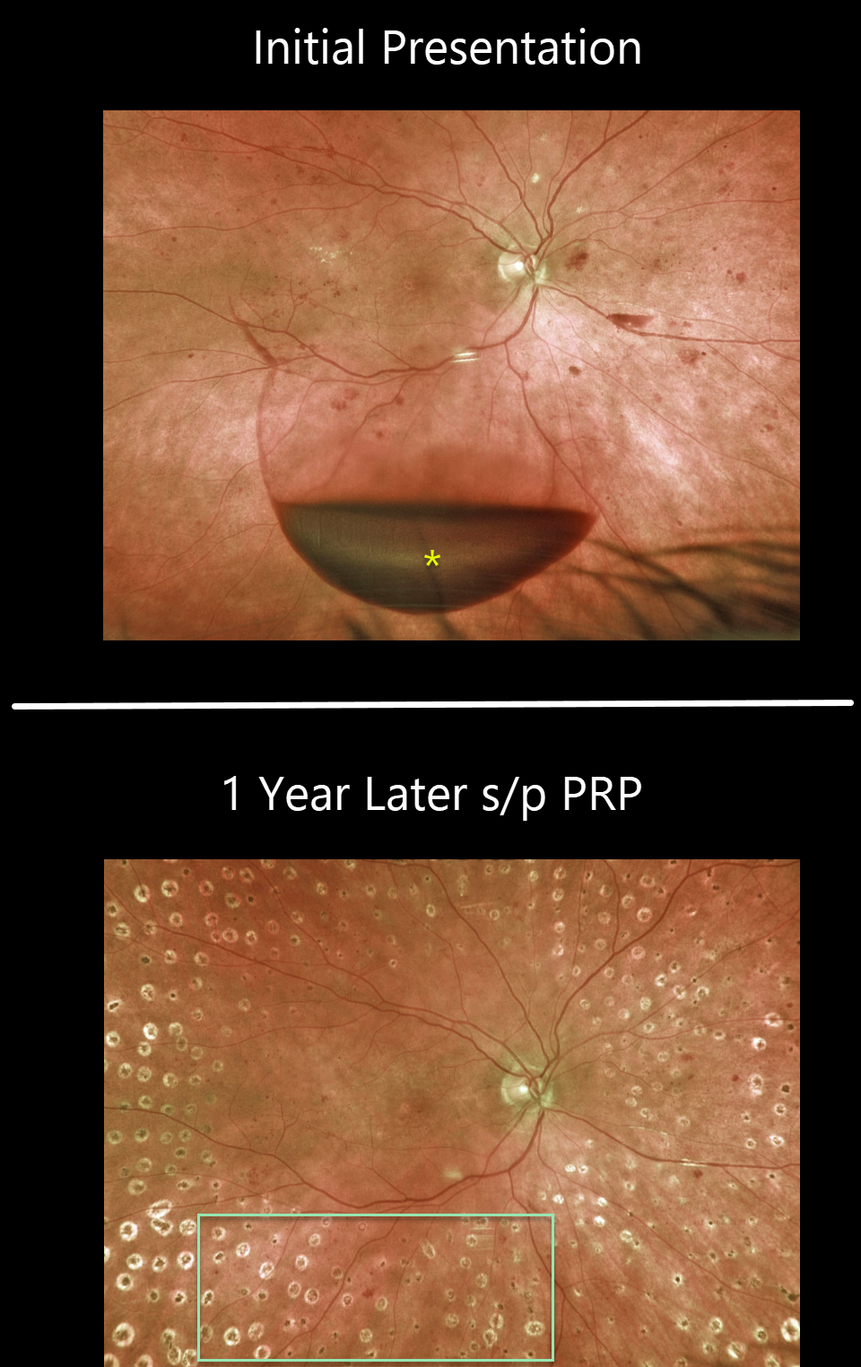
This 65YO female presented with 20/400 vision in her right eye. An acute inferior subhyaloid hemorrhage was noted along with non-central diabetic macular edema. Over the coming months, macular laser followed by panretinal photocoagulation (PRP) was performed.
One year later, vision was 20/50, with complete resolution of the preretinal blood and proliferative disease.
Learning Points:
Before PRP, patients with proliferative diabetic retinopathy (PDR) routinely went blind, and one of the treatments actually included pituitary ablation (for an amazing historical perspective on diabetic laser photocoagulation, see Goldberg and Jampol, Ophthalmology 1987;94;741-746).
PRP originally extended fairly posteriorly, and patients often needed fill-in treatment before vitrectomy and anti-VEGF treatments became available. Today, most laser surgeons will begin treatment well away from the nerve and major arcades to minimize the risks of central and peripheral vision loss.
Once considered a “one and done” treatment, more recent studies show that patients may still need additional laser, anti-VEGF injections, or vitrectomy (see Gonzalez et al, Ophthalmology 2021;128:1448-1457).
Some also advocate anti-VEGF injections as the primary treatment for PDR. However, most still prefer to add PRP at some point due to the risk of patients being lost to follow-up (see Gross et al, DRCR Protocol S, JAMA Ophthalmology 2018;136:1138-1148).
PROLIFERATIVE DIABETIC RETINOPATHY (PDR)
Originally posted on @retina.rocks August 23, 2021
This 53YO female with type 2 diabetes presented with acute vision loss from previously undiagnosed proliferative diabetic retinopathy (PDR).
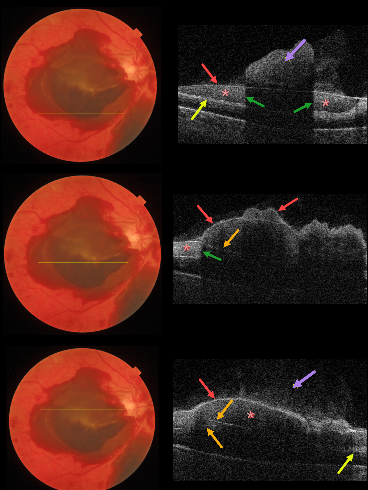
Color imaging shows multiple layers of preretinal blood. The blood is located above the neurosensory retina but is separated into several pockets due to posterior hyaloid splitting (vitreoschisis), rather than cleanly separating into a single plane.
Using the Triton swept-source OCT, we’ve taken four B-scans through different portions of the macula to highlight the complex anatomy of the preretinal blood.
Vitreous blood variably floats above the posterior hyaloid. The red arrows show the anterior edge of the posterior hyaloid. In some sections, the posterior hyaloid is split, separating the preretinal blood into several compartments.
Blood reflects and absorbs the OCT laser, making it difficult to see underlying retinal details (shadowing). The retinal nerve fiber layer is variably visible posterior to the blood.
PROLIFERATIVE DIABETIC RETINOPATHY (PDR)
Originally posted on @retina.rocks April 24, 2020
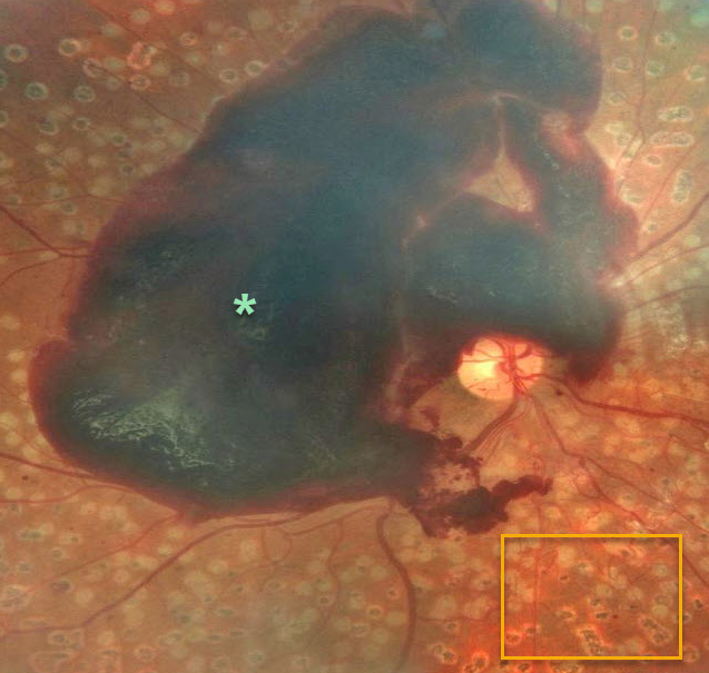
This patient still developed a large foveal subhyaloid hemorrhage despite very full prior panretinal photocoagulation (PRP).
Learning Points:
PRP has been around since the 1960s and still remains the mainstay for treating proliferative disease. Often considered a “one and done,” patients can still develop break-through hemorrhages. Intravitreal anti-VEGF injections are often helpful, but some will require vitrectomy.
PROLIFERATIVE DIABETIC RETINOPATHY (PDR)
Originally posted on @retina.rocks February 6, 2020
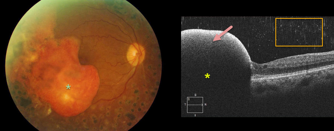
This patient with proliferative diabetic retinopathy has a dense subhyaloid hemorrhage. The yellowing subhyaloid blood indicates that this has been present for at least several months.
The OCT highlights the subhyaloid hemorrhage that blocks all underlying retinal detail and the hyper-reflective vitreous red blood cells gives a “snowy” appearance.
PROLIFERATIVE DIABETIC RETINOPATHY (PDR)
Originally posted on @retina.rocks January 31, 2020
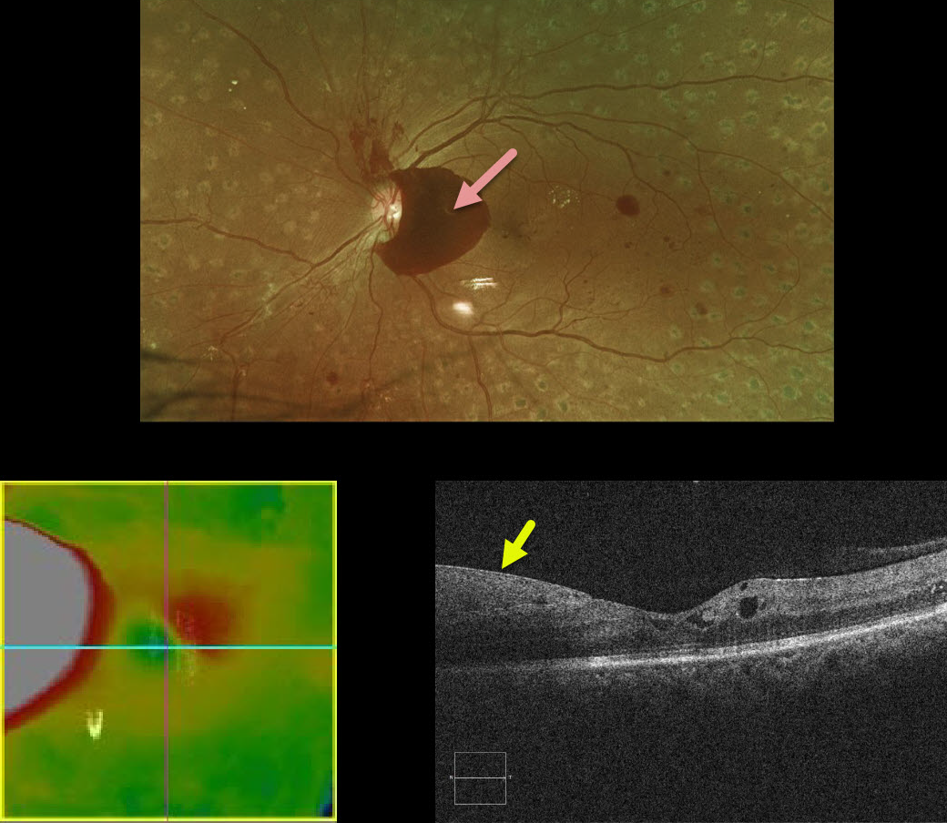
This patient with a history of involuted proliferative diabetic retinopathy following complete panretinal photocoagulation presented with what appeared on OCT to be new diabetic macular edema.
A closer inspection of the B-scan shows the retina to be normal with the pseudo-edema representing subhyaloid blood.
PROLIFERATIVE DIABETIC RETINOPATHY
Originally posted on @retina.rocks December 24, 2019
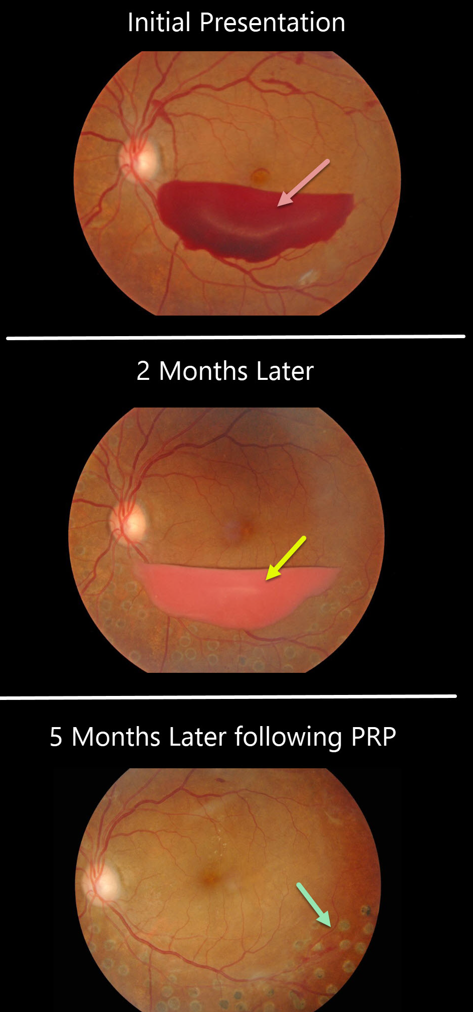
This patient presented with a sub-hyaloid hemorrhage due to proliferative diabetic retinopathy.
A subsequent photo two months later shows devitalized (yellowing) sub-hyaloid hemorrhage. He received panretinal photocoagulation (PRP) in the left eye and the sub-hyaloid hemorrhage completely resolved three months later. Anti-VEGF therapy was not used.
Learning Points:
PRP has been around since the 1960s and still remains the mainstay for treating proliferative disease. Often considered a “one and done,” patients can still develop break-through hemorrhages. Intravitreal anti-VEGF injections are often helpful, but some will require vitrectomy.


