

01 Disorders Causing Exudative and Hemorrhagic Detachment
<< Back to Cases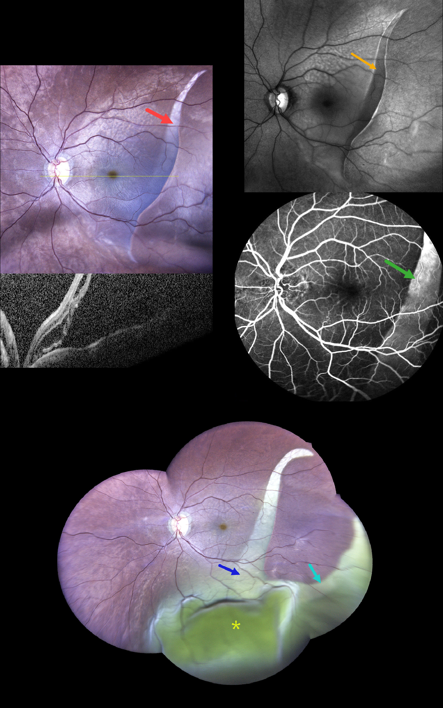
SEQUENTIAL GIANT RPE TEAR WITH SECONDARY EXUDATIVE RETINAL DETACHMENT
Rohan Suresh Ninan
Originally posted on @retina.rocks August 21, 2026
This 68YO male presented with sudden, painless decreased vision in his left eye 2 days ago. He gave a history of type 2 diabetes. Vision was 20/30 (6/9) in his normal OD and hand motion OS.
Widefield photography shows a large curvilinear area of pigment loss (giant RPE tear) extending from the superotemporal into the inferior midperiphery. OCT scanning shows marked macular subretinal fluid. On fundus autofluorescence (FAF), the lesion is hypo-FAF with a corresponding window defect on fluorescein angiography.
He returned 6 days later without new symptoms and stable vision. The prior RPE tear is enlarged in width, extends more inferiorly, and has developed a new branch extending into the inferotemporal midperiphery. A large inferior exudative retinal detachment is noted. Unfortunately, he was immediately lost to follow-up.
Learning Points:
A tear of the RPE is most often seen in RPE detachments associated with neovascular AMD, but can also occur in numerous other etiologies, including idiopathic central serous chorioretinopathy (ICSC) and polypoidal choroidal vasculopathy. The tear leaves a depigmented region where the RPE was and is now absent, along with a pigmented, relatively straight subretinal lesion from the scrolled RPE edge. Our patient’s RPE tear is atypical due to the lack of this hyperpigmented edge, indicating it is more of an RPE dehiscence than a scrolled tear, as well as the multibranched extension that developed 1 week later.
Giant retinal pigment epithelial (RPE) tears occurring outside the macula, defined as tears exceeding 3 clock hours, are most common following trauma or in bullous ICSC and polypoidal choroidal vasculopathy. We have seen these giant tears in previously clinically normal eyes, which later showed pachychoroid-related findings on ICG angiography and en face OCT.
POLYPOIDAL CHOROIDAL VASCULOPATHY
Originally posted on @retina.rocks July 8, 2026
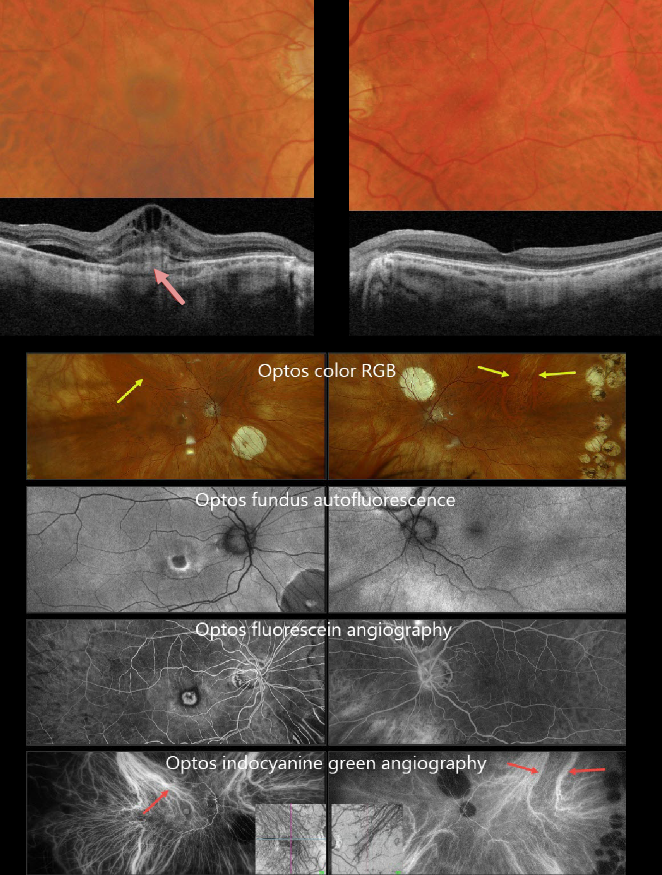
This 71YO white male underwent successful bilateral vitrectomies for rhegmatogenous retinal detachments by us years earlier. He had since moved out of state and was recently diagnosed with neovascular AMD in his right eye, which was treated with an initial anti-VEGF injection one month earlier. After moving back to our area, vision was 20/400 OD and 20/20 in his normal OS.
Triton color photography shows a poor foveal reflex with a surrounding subretinal pigment ring. Barely visible, smaller drusen are noted more peripherally. Swept-source OCT shows a probable type 1 (below the RPE) macular neovascularization (MNV) with overlying subretinal hyperreflective material, intraretinal and subretinal fluid. There are multiple smaller drusen in his left macula. Although the choroid appears thinned bilaterally, the vessels that are present mostly span the entire choroidal thickness.
Optos fundus autofluorescence (FAF) shows the MNV to be hypo-FAF with a rim of surrounding hyper-FAF (image 2). The MNV is hyperfluorescent on fluorescein angiography, surrounded by a rim of blockage from the subretinal pigment. The choroid is markedly abnormal on indocyanine green angiography, revealing that each macula is drained by the superotemporal choroidal vortex. These dilated choroidal vessels are also visible on color RGB imaging.
Learning Points:
Polypoidal choroidal vasculopathy (PCV) is a variant of type 1 macular neovascularization. PCV is likely distinct from age-related macular degeneration, occurring in a different population (more common in African Americans and Asians) and characterized by a relative lack of drusen and a thicker choroid. PCV presents differently in Caucasians, with multiple phenotypes that can include coexisting drusen, the presence or absence of a branching choroidal neovascular network, or background central serous chorioretinopathy findings (Sirks et al., Ophthalmology Retina 2025;9:994-1004).
PCV is part of the pachychoroid spectrum, more recently termed venous overload choroidopathy, which appears to be caused by relative vortex outflow resistance (Spaide et al., Progress Retinal Eye Research 2022;86;100973). The choroidal vortex system usually respects the horizontal meridian; anastomoses that cross it, as occurred in our patient, are characteristic of these eyes.
Historically, these lesions were believed to be somewhat resistant to anti-VEGF monotherapy and often required combined treatment with photodynamic therapy. More recent data from the PLANET study suggest that Eylea monotherapy is also effective (AJO 2019;204:80-89).
Given the lack of response to initial anti-VEGF therapy, intravitreal Eylea was given, followed by PDT 1 week later. Six weeks following PDT, vision remained stable, but the OCT showed marked improvement. We will continue to follow him closely.

FLATTENED DRUSENOID PED CAUSING SECONDARY MACULAR ATROPHY
Originally posted on @retina.rocks June 23, 2026
This 72YO female has been followed for years with intermediate AMD. On 3/26/25, she remained asymptomatic with 20/40 vision.
Color photography shows confluent central drusen creating a drusenoid PED with a radiating pigment figure, surrounded by large soft drusen. The drusenoid RPE detachment (PED) is confirmed on swept-source OCT. The RPE and outer retinal bands are obscured by subretinal hyperreflective material. She returned 6 months later with stable vision and no new symptoms. The PED is mostly flattened with some resolution of the hyperreflective material.
Learning Points:
Drusen commonly remodel over time and their collapse, as in our patient, can lead to drusenoid atrophy of the RPE (Filho et al, Retina 2013;33;1558-1566). Vision may or may not decrease following drusen regression. In our experience, drusenoid atrophy remains stable, unlike primary age-related macular atrophy, whose radius enlarges linearly over time (Shen et al, Ophthalmology Retina 2020;4;899-910). We therefore do not consider intravitreal complement inhibitors for these patients.
FIBRINOUS IDIOPATHIC CENTRAL SEROUS CHORIORETINOPATHY
Rohan Jain and Manish Nagpal
Originally posted on @retina.rocks June 22, 2026
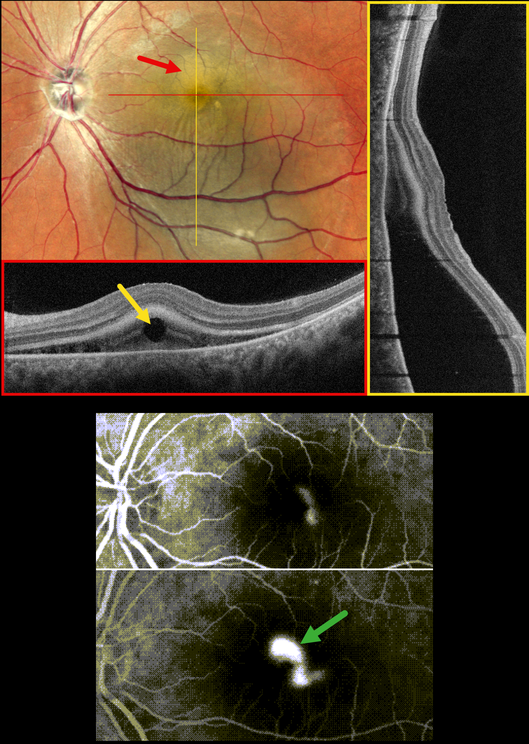
This 37YO male presented with one month of decreased vision in his left eye. There was no history of steroid or testosterone use. Vision was 20/20 in his normal OD and 20/40 OS.
MultiColor SLO imaging shows a large macular serous detachment extending into the inferior midperiphery, associated with fibrinous subretinal fluid. A horizontal OCT B-scan shows hyperreflective subretinal fibrin with a round hyporeflective lesion. A vertical B-scan shows the serous detachment extending inferiorly. Fluorescein angiography shows a smokestack leak.
Learning Points:
In idiopathic central serous chorioretinopathy (ICSC), fluorescein dye diffuses through a physiological break in the RPE and then pools within the subretinal space. The clearing within our patient’s inferotemporal subretinal fibrin (as evidenced by the hyporeflective round lesion on OCT) is likely caused by serous fluid entering the subretinal space and ‘washing away’ some of the fibrin.
Most patients with ICSC have a single, unilateral leak. Blot leaks are most common, with only 10% of cases exhibiting a smokestack. The subretinal fibrin noted in our patient is more commonly found in steroid-induced disease, although our patient denied steroid exposure.
PERIPHERAL EXUDATIVE HEMORRHAGIC CHORIORETINOPATHY
Originally posted on @retina.rocks June 19, 2026
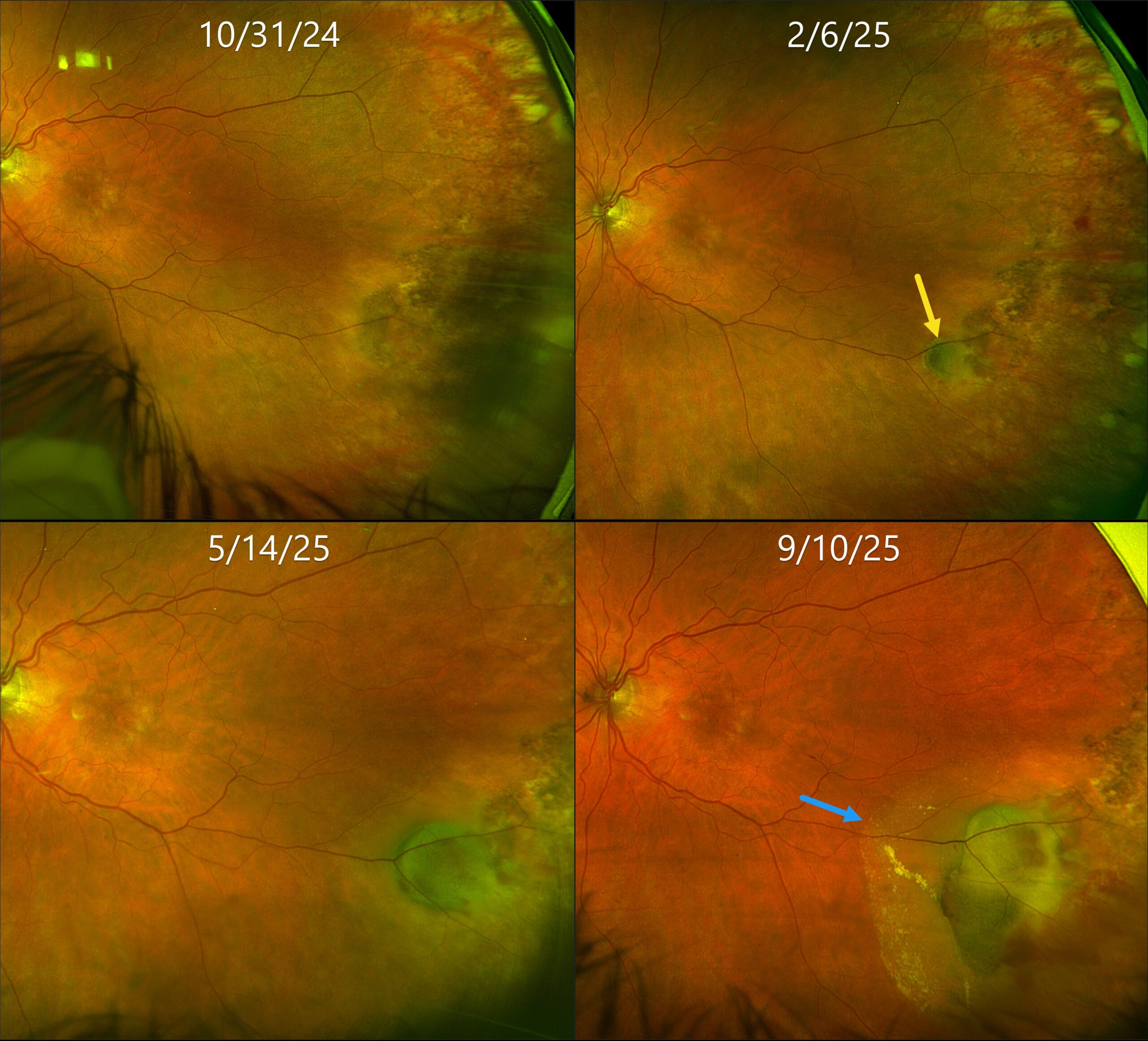
This 80YO female was initially referred for asymptomatic retinal findings in her left eye. Vision was 20/25.
Optos color RG imaging shows flat pigmented subretinal scarring in the left inferotemporal periphery. Three months later, a subretinal pigmented nodular lesion is noted along with a small dot of temporal blood. Over the following 7 months, this subretinal lesion enlarged with increasing blood and subretinal fluid approaching the macula. Although she remained asymptomatic, intravitreal anti-VEGF therapy was started.
Learning Points:
Ectopic choroidal neovascularization (CNV), commonly referred to as peripheral exudative hemorrhagic chorioretinopathy (PEHCR), is an exudative process similar to wet AMD but located in the retinal periphery. Ectopic CNV can often masquerade as a choroidal mass or uveal melanoma. It can also cause extensive choroidal/suprachoroidal, subretinal, and vitreous hemorrhage, more commonly in patients who are on blood thinners. The subretinal blood often spontaneously regresses without treatment, although anti-VEGF injections can be used for symptomatic or increasing exudation.
TOXEMIA
The European VitreoRetinal Society (EVRS), Berk Onaral and Ece Özdemir Zeydanlı
Originally posted on @retina.rocks May 1, 2026
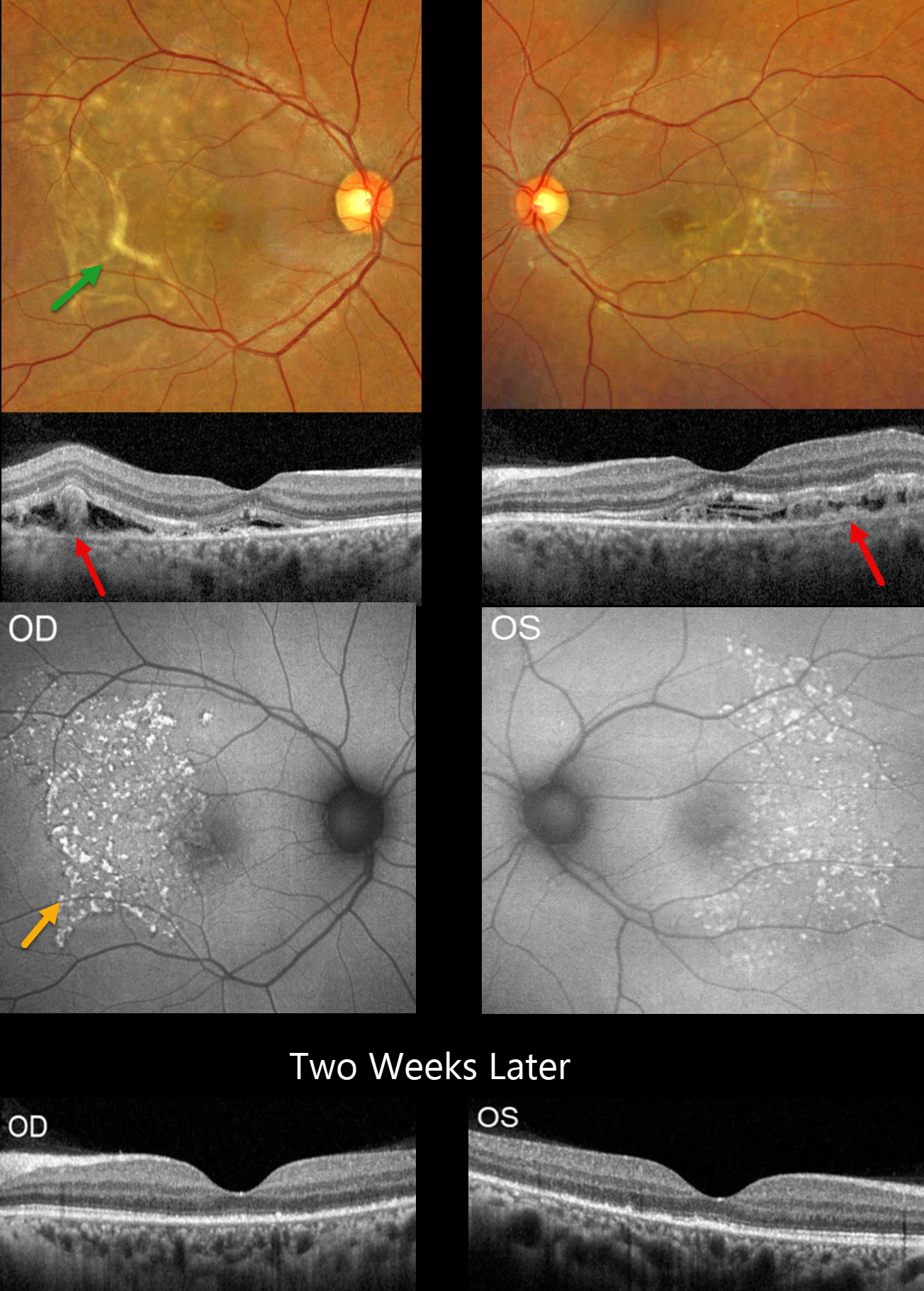
This 31YO postpartum female presented with sudden vision loss 3 days following delivery, which was complicated by severe preeclampsia (blood pressure was up to systolic 215 mmHg). Vision was 20/50 bilaterally.
Optos color RG imaging shows bilateral subretinal yellow bands and drusen-like changes. OCT shows disruption of the outer retinal bands with variable amorphous hyperreflective material and fluid. The RPE is variably thinned/absent as indicated by a visible Bruch’s membrane band. Fundus autofluorescence (FAF) shows extensive hyperreflective deposits in the temporal maculae.
Two weeks later, OCT shows resolved fluid with some restoration of the outer photoreceptor architecture.
Learning Points:
Posterior segment findings in toxemia of pregnancy, which develop in up to one-third of patients, are primarily due to choroidal ischemia with secondary RPE dysfunction and exudative retinal detachments. Outer retinal ischemia can cause bizarre and pathognomonic OCT changes as seen in our patient (Song et al, Graefes Arch Clin Exp Ophthalmol 2013;251:2647-2650).
The clinical and OCT findings recover remarkably within weeks following normalization of blood pressure. Upon resolution, the fundi can appear normal or reveal telltale subretinal pigment clumps (Elschnig spots), streaks (Siegrist streaks), or broad areas of RPE pigmentary changes with intraretinal migration that can mimic prior blunt trauma or rhegmatogenous retinal detachment.
PACHYCHOROID SPECTRUM PREVIOUSLY MISDIAGNOSED AS OCULAR HISTOPLASMOSIS
Steven Bloom
Originally posted on @retina.rocks April 22, 2026
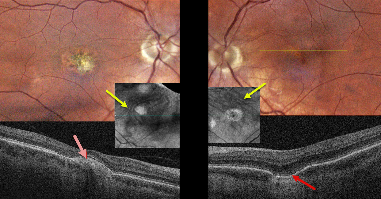
This healthy 39YO female gave a history 18 years earlier of prior anti-VEGF therapy OD for ocular histoplasmosis. She was visually asymptomatic, and vision was 20/40 OD and 20/30 OS.
Color photography of her left eye shows a patchy area of pigment loss in the superonasal fovea. OCT scanning through this area shows a choroidal excavation. The right macula has an overall mostly depigmented area of scarring, which appears as a mound of subretinal hyperreflectivity on OCT. Choroidal en face OCT shows dilated pachyvessels OU that cross the horizontal meridian (vortex anastomoses). There were no areas of peripheral chorioretinal scarring in either eye. Observation was recommended.
Learning Points:
Pachychoroid, meaning “thick” choroid, is secondary to large, dilated outer choroidal vessels. Increased oncotic pressure from these dilated vessels can cause RPE dysfunction, serous detachment, and macular neovascularization (MNV). Pachychoroid disease spectrum is a growing list of findings, including idiopathic central serous chorioretinopathy (ICSC), pachychoroid neovascularization, polypoidal choroidal vasculopathy, pachychoroid pigment epitheliopathy, pachydrusen, peripapillary pachychoroid syndrome, choroidal excavation, and pachyvitelliform maculopathy.
Our guess is that our patient had active ICSC when she was treated for her ‘ocular histoplasmosis.’ The lesion in the right macula is atypical for ICSC. But the other findings, including dilated choroidal vessels with vortex anastomoses and a choroidal excavation in her left eye, argue for pachychoroid as her unifying diagnosis.
PACHYVITELLIFORM MACULOPATHY
Originally posted on @retina.rocks April 7, 2026
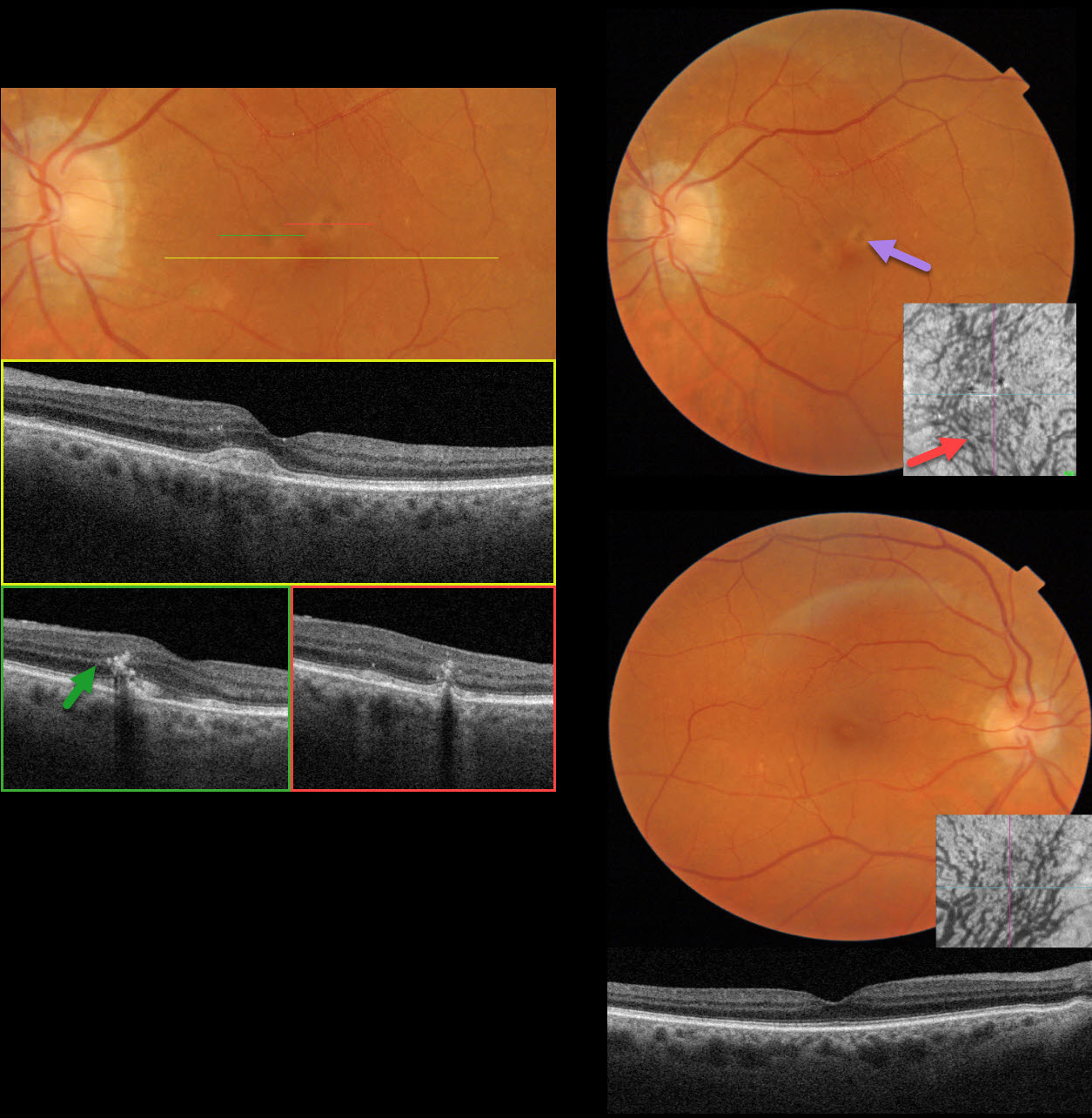
This 81YO female was referred for asymptomatic retinal findings. Vision was 20/30 OD and 20/30 OS.
Triton color imaging OS shows several yellowish subretinal pigment clumps in the superior fovea. Swept-source OCT through the central macula shows an acquired vitelliform lesion (AVL). Scans through each of the pigment clumps show coarse intraretinal hyperreflective foci. Some ectopic pachydrusen are noted in the inferotemporal right macula. OCT shows large outer choroidal vessels, and en face OCT shows the entire macula to be drained by the inferior vortex system with dilated vessels. The choroidal vessels are also dilated OS.
Learning Points:
Pachychoroid, meaning “thick” choroid, is secondary to large, dilated outer choroidal vessels. Increased oncotic or mechanical pressure from these dilated vessels can cause RPE dysfunction, serous detachment, and macular neovascularization (MNV). Our patient has pachychoroid findings despite having a ‘normal’ choroidal thickness on B-scan OCT. The choroidal thickness depends on numerous factors, including the patient’s age and refractive error. A high myope’s choroid should be quite thin, but if it appears as a ‘normal’ thickness, it is thick for that eye. We also often see pachychoroid disease in elderly patients whose choroid is abnormally thickened for their age, but appears ‘normally’ thick when not accounting for this.
Pachychoroid disease spectrum includes central serous chorioretinopathy, pachychoroid neovascularization, polypoidal choroidal vasculopathy, pachychoroid pigment epitheliopathy, pachydrusen, peripapillary pachychoroid syndrome, and focal choroidal excavation (Cheung et al, Eye 2025;39:819-834 ). Hilely et al recently described an additional entity, pachyvitelliform maculopathy (Br J Ophthalmol 2024;108:753-759), defined by AVL associated with pachychoroid features. The authors hypothesize that the thickened choroid causes RPE dysfunction, resulting in the accumulation of undigested photoreceptor outer segments with AVL formation.
We believe a better term for these disorders is venous overload choroidopathy, described by Spaide et al (Progress in Retinal and Eye Research 2022;86:100972). This hypothesis suggests scleral venous outflow as a unifying cause for this ever-expanding spectrum.
CHOROIDAL DETACHMENT
Originally posted on @retina.rocks March 19, 2026
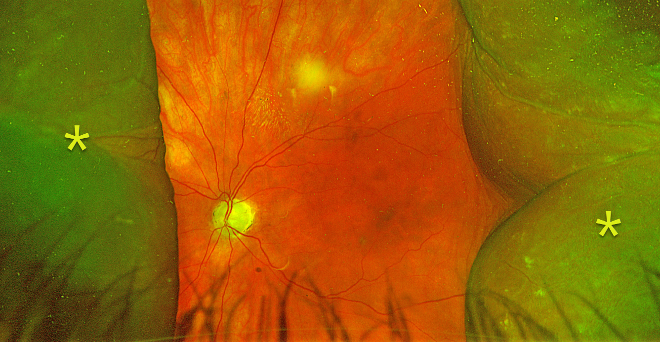
This 81YO female presented with counting fingers vision in her right eye 3 weeks following XEN Gel stent glaucoma surgery. Intraocular pressure was 11 mmHg.
Optos color RG imaging shows nasal and temporal choroidal detachments extending toward, but not into, the macula. Observation was recommended, and we expect the choroidals to resolve as she extends postoperatively.
Learning Points:
A choroidal detachment is defined by the abnormal presence of fluid or blood in the suprachoroidal space. Serous choroidal detachments, also known as choroidal effusions, are a frequent complication of glaucoma surgery. Several causative mechanisms are believed to be at play: hypotony allows fluid to accumulate in the suprachoroidal space, and inflammation increases the choroidal permeability. The detached ciliary body may also produce less aqueous humor, thus creating a self-perpetuating cycle. Often, observation is sufficient, but topical steroids and cycloplegics can help expedite resolution.
POLYPOIDAL CHOROIDAL VASCULOPATHY
Originally posted on @retina.rocks March 18, 2026
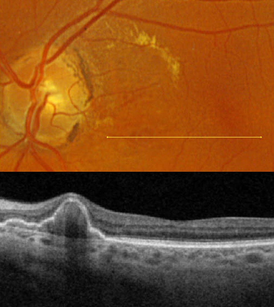
This 67YO female presented for a second opinion regarding possible age-related macular degeneration (AMD). She was without acute visual complaints. Vision was 20/25 in her normal OD and 20/30 OS.
Optos color RGB imaging shows macula lipid in the superonasal macula. Several red subretinal nodules are present inferior to the lipid. Triton swept-source OCT through these lesions shows a nodular and somewhat multi-lobulated RPE detachment (PED). We will follow her closely for any increase in exudation.
Learning Points:
At the Macula Society meeting in 1983 and at the Academy of Ophthalmology meeting in 1984, Brucker et al presented 7 patients with multiple recurrent sub-RPE and subretinal hemorrhages, which they termed the posterior uveal bleeding syndrome. This was followed by multiple authors reporting similar findings (Stern et al AJO 1985;100:560-569, Kleiner et al Retina 1990;10:9-17, and Yannuzzi et al Retina 1990;10:1-8). Dr. Gass, who was assigned as the reviewer for Yannuzzi’s manuscript titled “Idiopathic Polypoidal Choroidal Vasculopathy,” took issue with calling these lesions ‘polyps’ (Alexander J Brucker, personal communication 2/12/26), which are defined by Stedman’s Medical Dictionary as growths that protrude from a mucous membrane. When Yannuzzi challenged Dr. Gass for a better term, he could not come up with one; Yannuzzi’s terminology won out, and the rest is history. In our patient’s case, the clinical red ‘polyps’ corresponded to peaked, multilobulated RPE detachments on OCT.
PCV is likely distinct from age-related macular degeneration, occurring in a different population (more common in African Americans and Asians), with a relative lack of drusen and a thicker choroid. In our experience, many patients with ‘wet AMD’ actually have PCV or pachychoroid neovascularization.
SHOULD THIS LESION BE EMERGENTLY LASERED???
Originally posted on @retina.rocks March 11, 2026
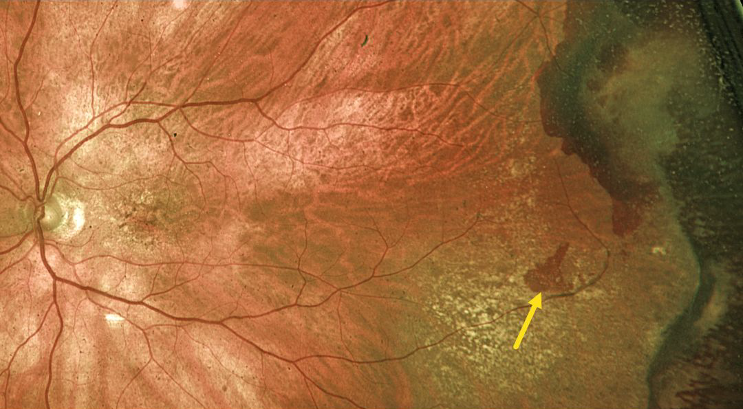
This 89YO male was referred for an asymptomatic retinal tear with possible retinal detachment in his left eye. Vision was 20/60.
Optos color RG imaging shows a red horseshoe-shaped lesion in the temporal midperiphery. Would you recommend immediate thermal laser demarcation of this retinal tear?
We hope not! This patient has asymptomatic and chronic ectopic choroidal neovascularization (CNV), which is evidenced by the more peripheral subretinal scarring and superotemporal dark subretinal blood. Ectopic CNV can often masquerade as a choroidal mass or uveal melanoma. It can also cause extensive choroidal/suprachoroidal, subretinal and vitreous hemorrhage, more commonly in patients who are on blood thinners. The subretinal blood often spontaneously regresses without treatment, although anti-VEGF injections can be used for symptomatic or increasing exudation.
We recommended observation and expect this lesion to remain relatively stable.
POLYPOIDAL CHOROIDAL VASCULOPATHY WITH EXCISION OF SUBRETINAL BLOOD
The European VitreoRetinal Society (EVRS), Anjana Mirajkar, Akansha Sharma, Vaidehi Sathaye, Iam Pramodsuman and Manish Nagpal
Originally posted on @retina.rocks February 27, 2026
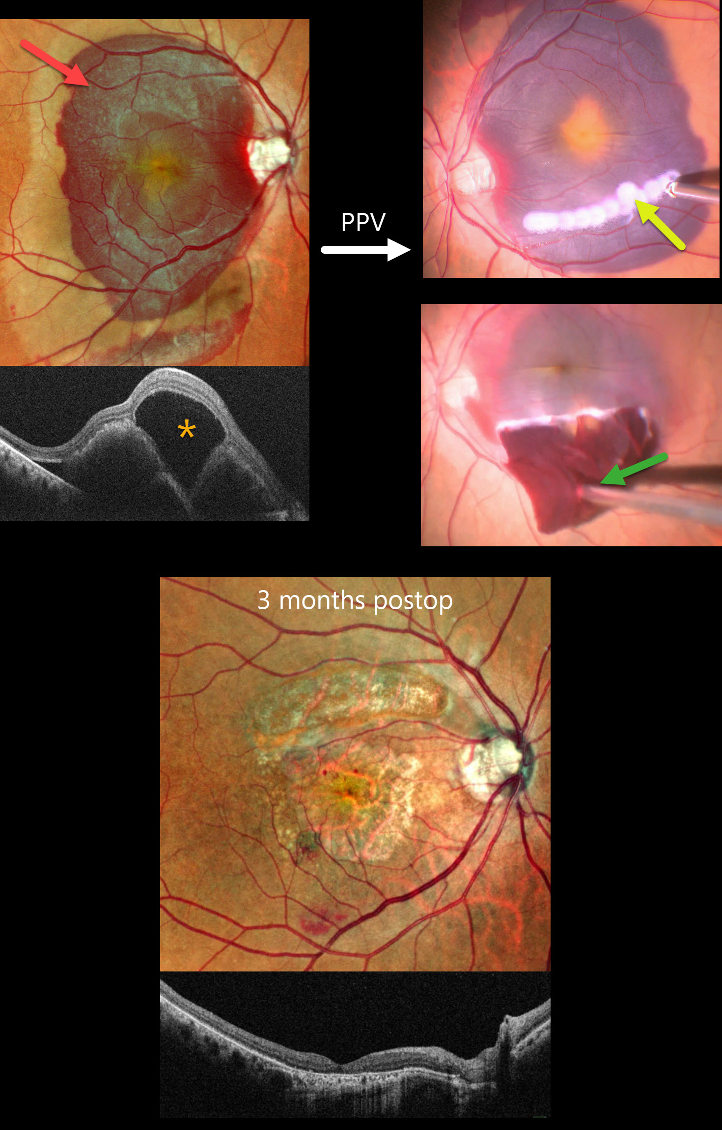
This 64YO female presented with 2 months of vision loss in her right eye. Vision was counting fingers OD and 20/30 in her normal OS.
Pseudocolor SLO imaging shows a large submacular hemorrhage. OCT scanning shows a central large bacillary layer detachment vs subretinal fluid overlying the elevated subretinal blood. Preoperative intravitreal tissue plasminogen activator (tPA) was injected, followed 2 days later by vitrectomy, a smile incision with excision of the subretinal clot, and gas tamponade.
Three months postoperatively, dry macular scarring is noted, and vision improved to 20/200.
Learning Points:
Common causes of submacular hemorrhage include neovascular age-related macular degeneration, polypoidal choroidal vasculopathy (PCV, our patient), trauma, and retinal arterial macroaneurysms. Multiple treatment options are available, including observation, anti-VEGF therapy, pneumatic displacement with positioning, and pars plana vitrectomy.
Surgery for thick submacular hemorrhage is often recommended due to the fear of possible toxicity from heme byproducts or fibrin formation (Toth et al, Arch Ophthalmol 1991;109:723-729). However, it is uncertain whether surgical results are better than anti-VEGF monotherapy (Gabrielle et all, Ophthalmology 2023;130:947-957). The value of adding TPA to any of these surgical options is also uncertain (Chotikkakamthorn et al, Retina 2025;45:394-401).
RPE TEAR
Originally posted on @retina.rocks February 2, 2026
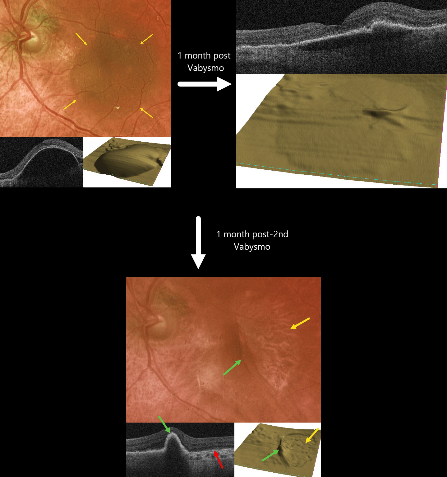
This 77YO female presented with recent vision loss in her left eye from new neovascular AMD. Vision was 20/400.
Optos color RG imaging shows a large pigmented retinal pigment epithelial detachment (PED) occupying most of the left macula. OCT scanning, B-scan, and RPE map confirm this large PED. Intravitreal Vabysmo was injected, and one month later, vision improved to 20/40. The PED nearly flattened.
Another Vabysmo injection was given, and one month later, she complained of some new vision loss. Vision was 20/80. A new RPE tear is noted, with bare RPE temporally and the retracted RPE nasally. Bare Bruch membrane is seen within the bed of the torn RPE. Vabysmo was injected, and treat-and-extend was begun.
Learning Points:
A tear of the RPE is a fairly common finding in RPE detachment (PED) associated with type 1 macular neovascularization (MNV, located below the RPE). Neovascularization can spontaneously contract or involute following treatment. It then puts traction on the overlying RPE, which can then rip at the opposite edge of the PED. Like a cheap window blind, the torn RPE retracts and scrolls towards the MNV. This leaves a depigmented region where the RPE was and is now absent, along with a pigmented, relatively straight subretinal lesion from the scrolled RPE edge. Anti-VEGF therapy can safely be continued following an RPE tear, with long-term visual outcome more closely related to the patient’s response to therapy than to the tear itself (Ophthalmology 2018;125:236-244), provided the tear is outside the foveal center.
TOXEMIA
Nilesh Kumar
Originally posted on @retina.rocks January 21, 2026
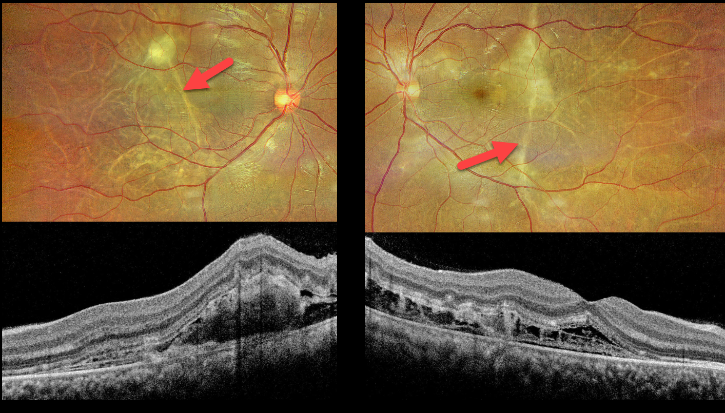
This 25YO female presented 10 days postpartum, complaining of bilateral blurred vision that began shortly following delivery, which was complicated by toxemia (preeclampsia). Blood pressure in our office was 180/90 mmHg. Vision was 20/30 bilaterally.
Optos color RG imaging shows multiple depigmented, wormlike subretinal tracks throughout each posterior pole. A larger contracting triangular-shaped lesion is noted in the superior left macula. Exudative fluid is present in the nasal maculas. OCT scanning shows outer retinal disorganization, an increased number of outer retinal bands, and pockets of variably hyperreflective fibrinous outer retinal fluid.
Learning Points:
Posterior segment findings in toxemia of pregnancy, which develop in up to one-third of patients, are primarily due to choroidal ischemia with secondary RPE dysfunction and exudative retinal detachments. Outer retinal ischemia can cause bizarre and pathognomonic OCT changes as seen in our patient (Song et al, Graefes Arch Clin Exp Ophthalmol 2013;251:2647-2650).
The clinical and OCT findings recover remarkably within weeks following normalization of blood pressure. Upon resolution, the fundi can appear normal or reveal telltale subretinal pigment clumps (Elschnig spots), streaks (Siegrist streaks), or broad areas of RPE pigmentary changes with intraretinal migration that can mimic prior blunt trauma or rhegmatogenous retinal detachment.
INTERMEDIATE AMD WITH DRUSENOID PEDS
Originally posted on @retina.rocks January 8, 2026
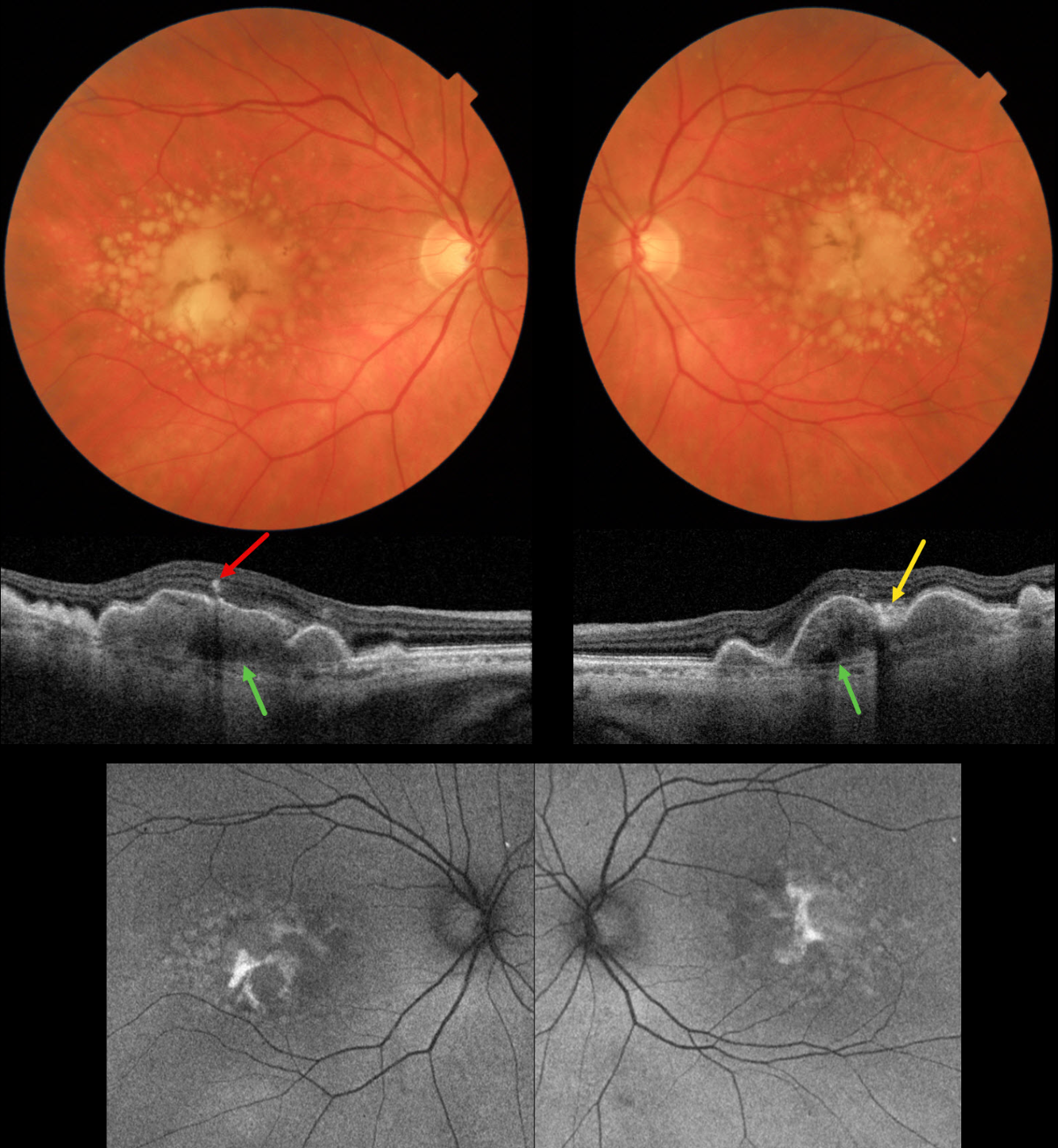
This 72YO female presented for her 6-month AMD examination. She was visually asymptomatic, and vision was 20/40 OU.
Triton color imaging shows bilateral chronic foveal RPE detachments (PEDs) with surrounding soft drusen. A radiating pigment figure overlies each PED. Swept-source OCT shows hyperreflective material beneath each PED, with some overlying hyperreflective foci and hyperreflective material likely representing lipofuscin. Bruch membrane is visible beneath the PED and surrounding soft drusen. Fundus autofluorescence (FAF) shows variable hyper-FAF from the pigment figures and lipofuscin.
Learning Points:
RPE detachments are most commonly associated with AMD and the central serous-pachychoroid spectrum. In cases of AMD, lesions can be avascular (as in our patient) or vascularized as part of a type 1 neovascular complex (below the RPE). AMD-related PEDs can form primarily or as a confluence of soft drusen. When they collapse, they may lead to secondary macular atrophy (Yu et al, Ophthalmology 2019;126:261-273).
The radiating pigment figure, described originally by Dr. Gass, is pathognomonic for a chronic PED and persists even after a PED flattens. Avascular PEDs are typically not treated. In contrast, anti-VEGF therapy is both safe and effective for vascularized PEDs, despite the risk of a RPE tear (Khanani et al, Retina 2018;38;2103-2117).
ANGIOID STREAKS WITH MNV AND PATTERN DYSTROPHY PHENOTYPE
Shraddha Raj Shrivastava and Manish Nagpal
Originally posted on @retina.rocks January 6, 2026
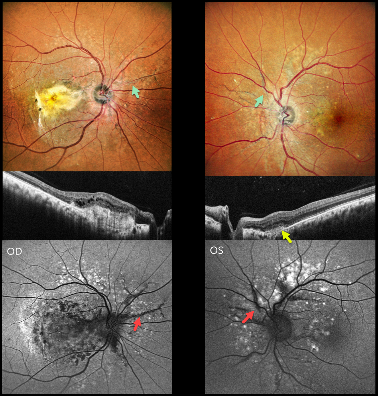
This healthy 42YO male presented with 10 months of vision loss in his right eye and 1 month of mild blurred vision in his left eye. Vision was counting fingers OD and 20/20 OS.
Pseudocolor SLO imaging shows angioid streaks extending around and radiating outwards from each optic nerve. There are drusen-like changes around each posterior pole, along with clumps of subretinal pigment temporally.
OCT scanning shows hyperreflective subretinal fibrosis with mild overlying cystic retinal edema OD. A much smaller area of subretinal hyperreflectivity (indicating a type 2 macular neovascularization, MNV) with overlying subretinal hyperreflective material is noted OS. The angioid streaks on fundus autofluorescence (FAF) are hypo-FAF due to RPE atrophy. The drusen-like changes are hyper-FAF.
Learning Points:
Retinal findings in pseudoxanthoma elasticum (PXE) most commonly include angioid streaks and a subretinal peaux d’orange appearance, most pronounced in each temporal macula. However, pattern dystrophy-like changes, as in our patient, can sometimes be the predominant phenotype (Murro et al, Graefe’s 2020;258:1881-1892). As with typical pattern dystrophies, the findings are dynamic and best captured with multimodal imaging.
PXE is caused by a mutation in the ABCA6 protein and is virtually always transmitted as an autosomal recessive disorder, although autosomal dominant transmission may rarely occur (Plomp et al, Am J Med Genet A 2004;126:403-412).
Our patient had no clinical signs of PXE and denied any family history of this disease. He was referred to his primary care doctor for further evaluation. Intravitreal anti-VEGF therapy was started OS.
PSEUDO-UVEAL MELANOMA FROM CHOROIDAL DETACHMENT
Originally posted on @retina.rocks December 25, 2025
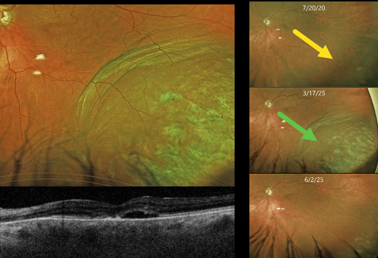
This 85YO female presented with an elevated pigmented choroidal mass in her inferotemporal periphery, which extended into her distal macula (Optos color RG). This mass was absent 5 years earlier. Circumferential choroidal folds extend along the posterior edge of the lesion. Triton swept-source OCT shows a blister of subretinal fluid and a thickened choroid.
She has a history of uncontrolled glaucoma and underwent a XEN Gel Stent procedure 8 days earlier. Intraocular pressure (IOP) was 8 mmHg.
Learning Points:
A choroidal detachment is defined by the abnormal presence of fluid or blood in the suprachoroidal space. Serous choroidal detachments, also known as choroidal effusions, are a frequent complication of glaucoma surgery. Several causative mechanisms are believed to be at play: hypotony allows fluid to accumulate in the suprachoroidal space, and inflammation increases the choroidal permeability. The detached ciliary body may also produce less aqueous, thus creating a self-perpetuating cycle. Often, observation is sufficient, but topical steroids and cycloplegics can be helpful in promoting resolution.
Other than the obvious appearance of this lesion immediately following glaucoma surgery, clues to this not being a uveal MM include the choroidal folds along the posterior extent of the choroidal detachment and an OCT consistent with hypotony maculopathy. About 10 weeks later, the IOP was 9 mmHg, and the choroidal effusion had completely resolved.
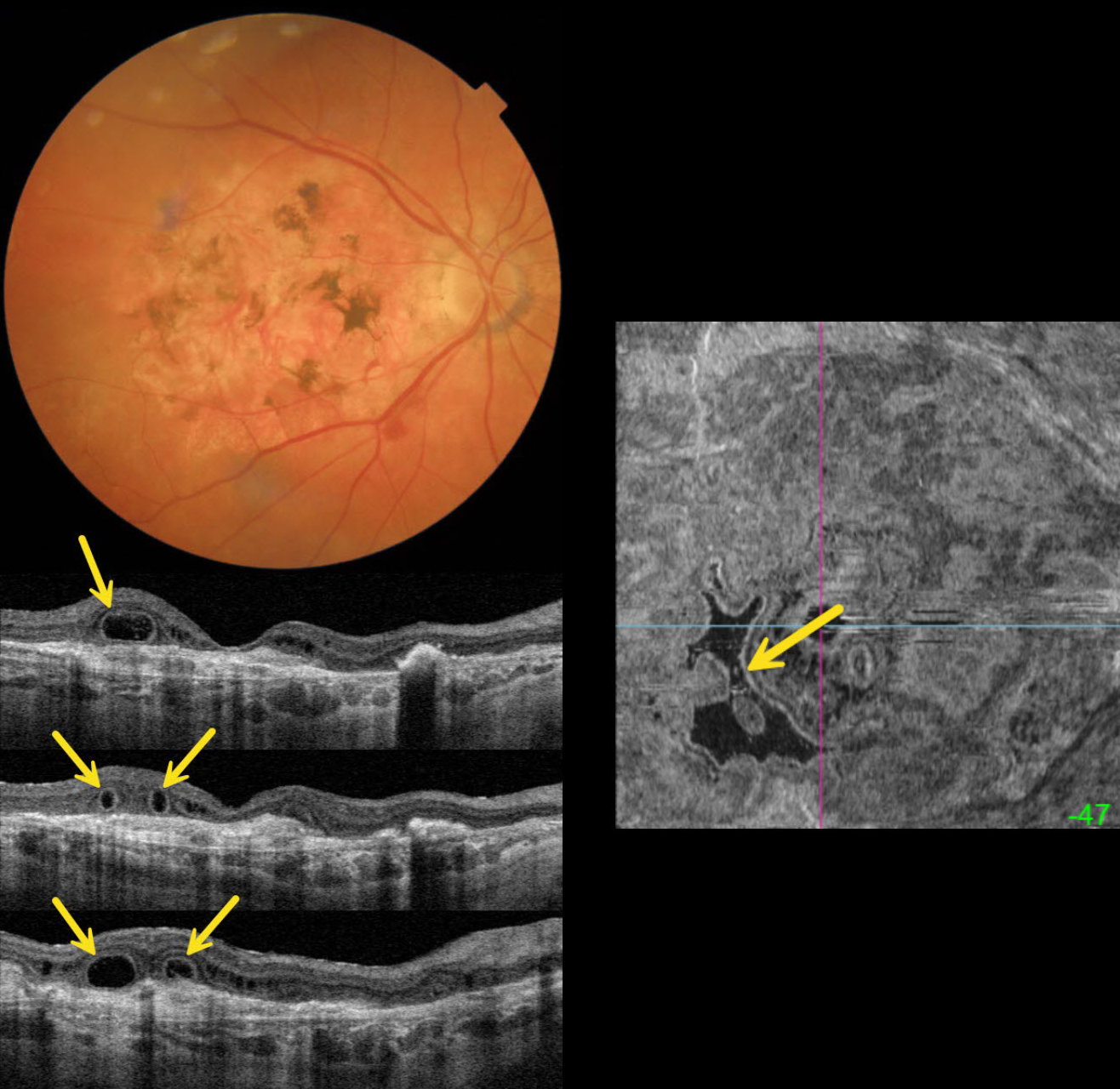
OUTER RETINAL TUBULATION IN NEOVASCULAR AMD
Originally posted on @retina.rocks November 18, 2025
This 89YO female is receiving ongoing intravitreal Eylea in her 20/40 left eye for neovascular AMD. She has a stable 20/400 atrophic disciform scar in her right eye.
Triton color imaging shows a variably pigmented flat disciform scar. Swept-source OCT shows variable outer retinal atrophy overlying a hyperreflective inactive macular neovascularization (MNV). A complex pattern of outer retinal tubulation (ORT) is seen on numerous B-scans. En face OCT shows another view of these meandering tubular structures.
Learning Points:
ORT is often noted overlying inactive MNV and should not be confused with exudative fluid or cysts, which lack a hyperreflective border. The outer hyperreflective band likely represents inner segment mitochondria undergoing fission and translocation toward the nucleus (Litts et al, Retina 2018;38:445-461). ORT, initially described by Zweifel et al (Arch Ophthalmol 2009;127:1596-1602), is a neurodegenerative condition of the photoreceptors and Muller cells associated with atrophy affecting the outer retina and retinal pigment epithelium, including advanced AMD and inherited retinal diseases.
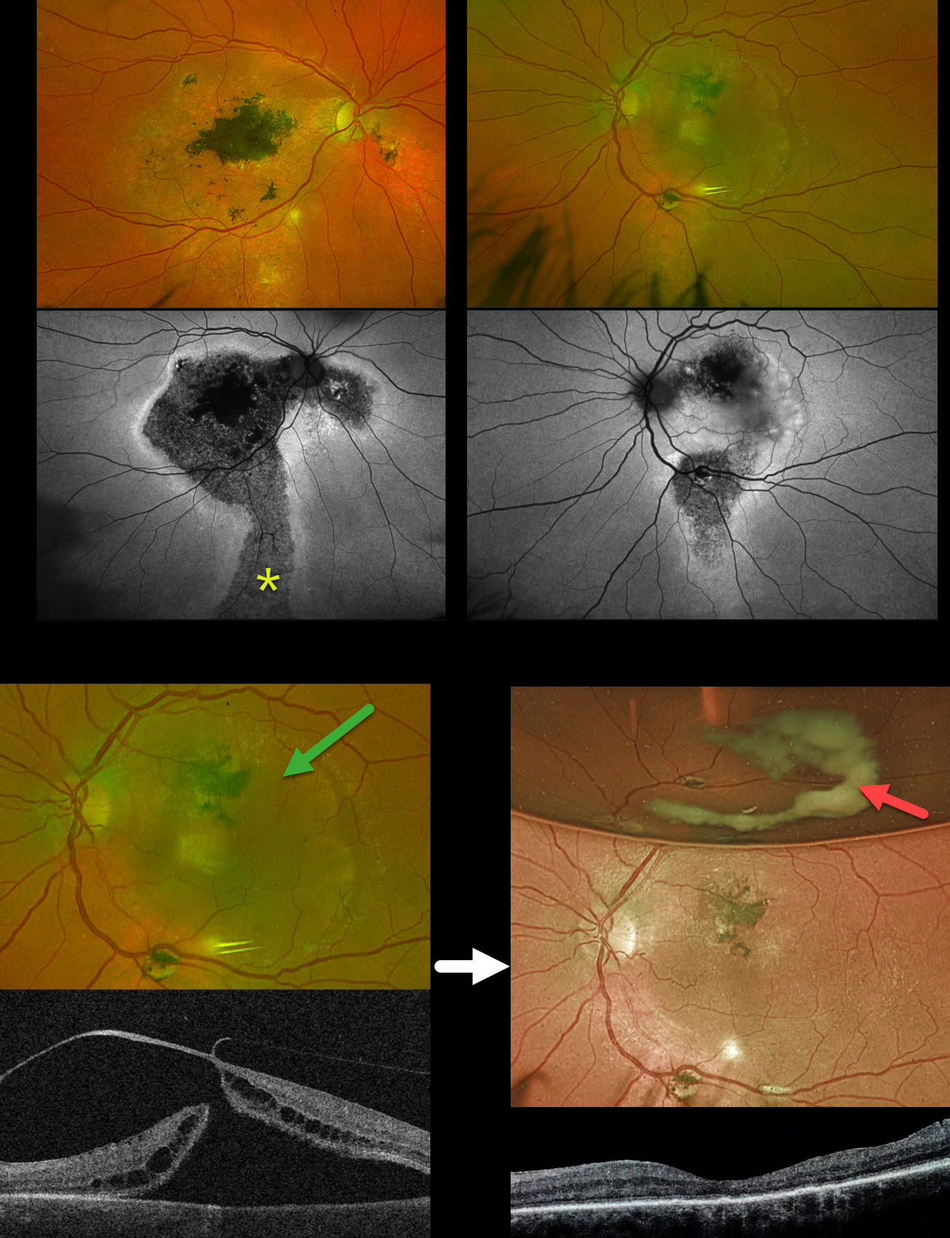
PRIOR CHRONIC ICSC WITH NEW TRACTIONAL MACULAR HOLE-RRD WITH POSTOPERATIVE MENISCUS MICROPYON
Originally posted on @retina.rocks October 29, 2025
This 58YO male was referred for a retinal detachment in his left eye. He has a long history of idiopathic central serous chorioretinopathy (ICSC). He was previously treated elsewhere for neovascular AMD OD. For many years, he has been receiving intravitreal Avastin OS every 1-2 months. Vision was counting fingers OD and 20/400 OS.
Optos color RG imaging shows bilateral macular scarring with confluent hyperpigmented foveal changes and intraretinal pigment migration, especially in his right eye. Inferior hypopigmented gutters are most obvious on fundus autofluorescence (FAF) as hypo-FAF with surrounding hyper-FAF margins. A rhegmatogenous retinal detachment (RRD) occupies the entire left macula. Triton swept-source OCT shows a full-thickness macula hole, active vitreoretinal traction on the temporal edge of the hole, and a macular detachment.
The tractional macular hole with secondary RRD in our patient’s left eye is completely unrelated to his poorly treated ICSC. One month following vitrectomy, vision improved to 20/100. The retina was completely attached, and the macular hole was closed. A meniscus micropyon is adherent to the inferior meniscus of the gas bubble.
Learning Points:
Photodynamic therapy (PDT) is currently the standard of care for ICSC with symptomatic acute or chronic subretinal fluid. Unfortunately, Visudyne is rarely available outside the US, which has led to the adoption of unproven therapies, including mineralocorticoid receptor antagonists (eplerenone), beta blockers, and intravitreal anti-VEGF agents. Recent meta-analyses and systematic reviews confirm that these alternative therapies are ineffective (Felipe et al Int J Retina Vitreous 2022;8;34 and Kim et al Ophthalmology 2025;132:343-353). Despite the clear lack of efficacy in the literature, specialists both within and outside the US continue to use these drugs (Vankatesh et al Int J Retina Vitreous 2023;9;61). Our patient’s macular findings are a testament to what can happen when proven therapies are ignored.
Russell et al recently reported a unique ophthalmoscopic sign, a meniscus micropyon, that follows PPV with gas tamponade for all causes (Retina 2024;44:1766-1776). Features they noted that distinguish this sterile micropyon from post-PPV fibrin include a delayed appearance, hyperautofluorescence, and absence of anterior chamber or vitreous fibrinous strands/sheets. Vitreous biopsy in two of their cases revealed predominantly white blood cells. The micropyon appearance was associated with epiretinal proliferation (80%). Recurrent RRD from proliferative vitreoretinopathy (PVR) developed 33% of eyes, 12% re-detached without frank PVR, and 18% developed worsening epiretinal membranes.
Our patient’s meniscus micropyon disappeared as the bubble resolved. Three months postoperatively, vision was 20/60, and the retina remained completely reattached without PVR.
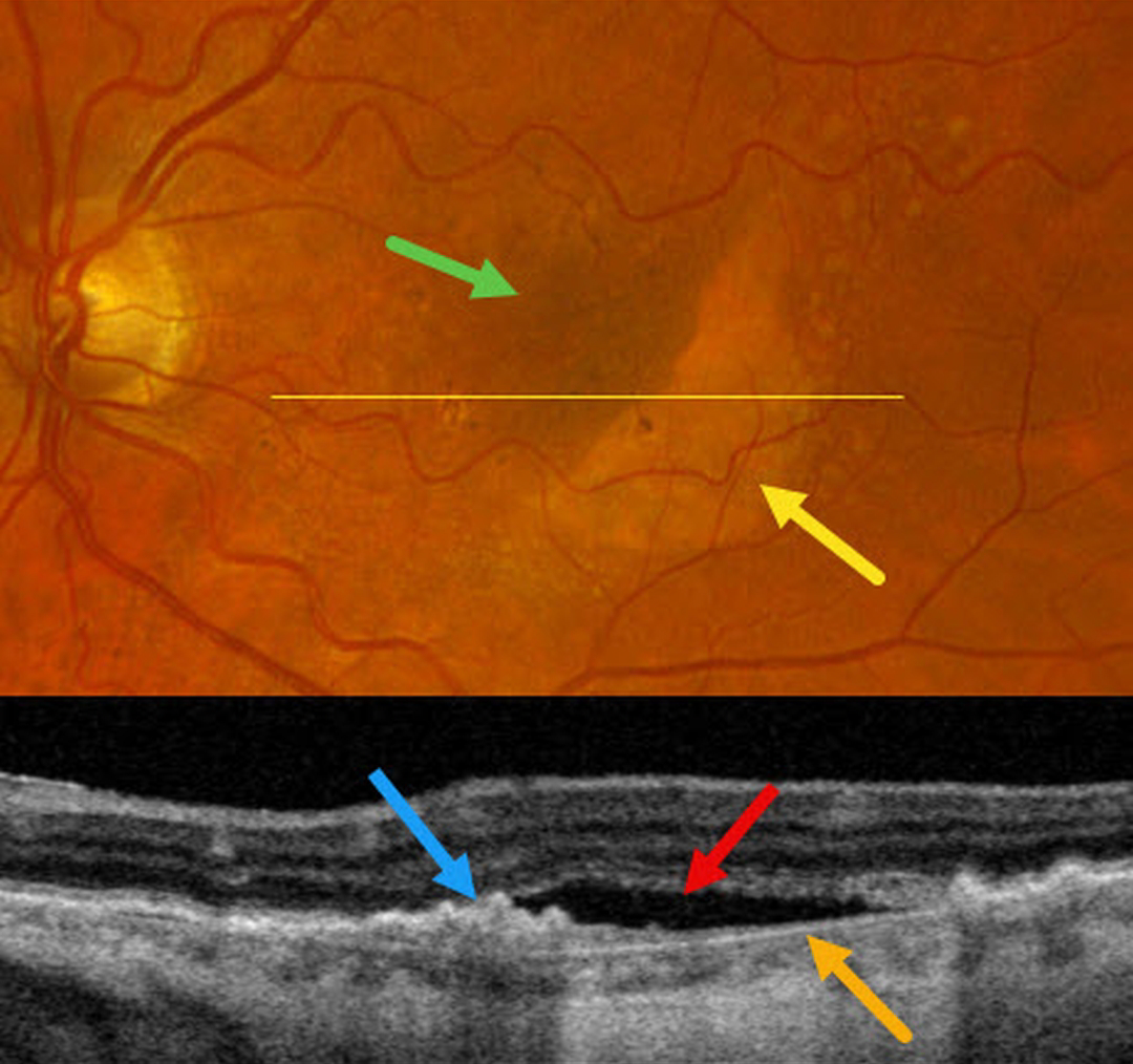
RPE TEAR
Originally posted on @retina.rocks October 14, 2025
This 71YO female complained of one month of decreased vision in her left eye. Vision was 20/30 OD and 20/200 OS.
Moderate larger drusen with pigment clumps were noted in her right eye (not shown). Optos color RGB imaging of her left eye shows a crescent of subretinal pigment loss in the inferotemporal macula with increased subretinal pigment superonasally. OCT scanning within the area of pigment loss shows bare Bruch membrane with overlying subretinal fluid. The RPE nasally is retracted and bunched onto itself.
Learning Points:
A tear of the RPE is most seen in RPE detachments (PEDs) associated with neovascular AMD, but can also occur in numerous other etiologies, including central serous chorioretinopathy and polypoidal choroidal vasculopathy. The tear leaves a depigmented region where the RPE was and is now absent, along with a pigmented, relatively straight subretinal lesion from the scrolled RPE edge.
Our patient may harbor a macular neovascularization beneath the retracted RPE. A therapeutic trial of monthly intravitreal Avastin was thus started.
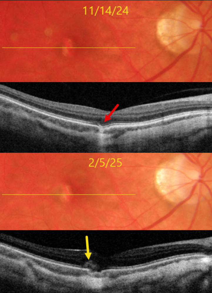
CHOROIDAL EXCAVATION WITH MACULAR NEOVASCULARIZATION
Originally posted on @retina.rocks September 30, 2025
This 39YO myopic female was referred on 11/14/24 for an asymptomatic lesion in her right macula. Vision was 20/25 OD and 20/25 in her normal OS.
Triton color imaging shows a small vertically oval area of subretinal pigment loss temporal to the right fovea. Swept-source OCT shows a focal choroidal excavation (FCE) within this region.
She returned on 2/5/25 with recent blurred vision in this eye. Vision was 20/30. A new type 2 (above the RPE) macular neovascularization (MNV) is noted along the temporal edge of the FCE.
Learning Points:
FCE, originally described by Jampol et al. in 2006, is likely part of the pachychoroid spectrum, as it is usually associated with a thickened choroid and pachyvessels (not noted in our myopic patient). The FCE can be observed, although symptomatic manifestations, including active central serous retinopathy or MNV (as in our patient), often require treatment. An excellent review is by Verma et al, Br J Ophthalmol 2021;105:1043-1048.
One month after a single intravitreal Avastin injection, vision was 20/30, her symptoms had resolved, and the lesion was completely dry on funduscopy and OCT. We are following her closely with PRN therapy.
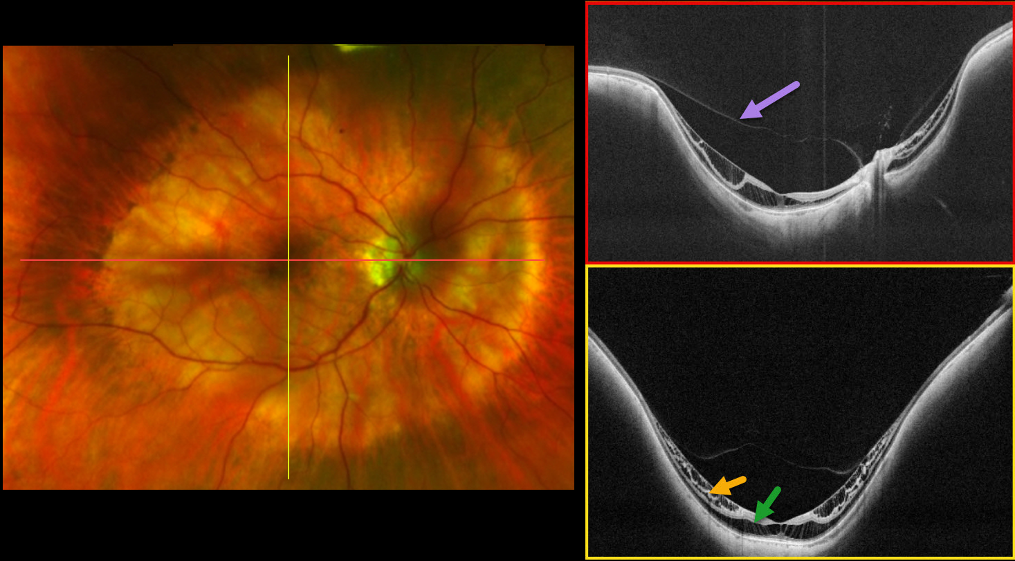
MYOPIC TRACTION MACULOPATHY
Barbara Parolini and Tommaso Pellegrino
Originally posted on @retina.rocks September 26, 2025
This 64YO female has a history of high myopia with posterior staphyloma. The axial length is 24.8 mm. Vision in the right eye has been deteriorating to 20/40.
Optos color RG imaging shows a posterior staphyloma, Type I (wide macula), in the right eye, which encircles the macula and disc. Ultra-widefield OCT (DreamOCT Intalight, 26 mm scan) shows a partial posterior vitreous detachment over the macula into the staphyloma with inner and outer retinal schisis within the staphyloma. There is no retinal detachment.
Learning Points:
Pathologic or high myopia is usually defined as a refractive error of at least -6 diopters with an axial length of at least 26.5 mm, so our patient technically is not a ‘high’ myope. 3D MRI imaging allows for a more nuanced definition and has shown that the ocular deformations can affect the entire eye, the equatorial, or just the posterior globe (Luo et al, JAMA Ophthalmology 2023;141:768-774). As defined by Luo et al, our patient has a posterior bulb-shaped globe. The ultra-widefield OCT is designed to image the entire posterior profile of the eye, which shows the geometry of our patient’s staphyloma.
The image also shows that myopic traction maculopathy (MTM) underlies a partial vitreous detachment. This confirms that the main cause of MTM is not the vitreous but the posterior staphyloma. Furthermore, when looking at the horizontal scan, the thickness of the retina (or, in other words, the severity of the schisis) is not evenly distributed into the staphyloma. It is more pronounced in the temporal side of the macula. The optic nerve pushes the sclera anteriorly, buckling the retina in that area and releasing the traction.
In the vertical scan, the schisis is uniformly distributed in the whole staphyloma. According to the ATN (atrophic, tractional, and neovascular) classification system for myopic maculopathy (Ruiz-Medrano et al, Progress Retinal Eye Research 2019;69:80-115), our patient has A1 (tessellated fundus only), T2 (inner + outer foveoschisis), and N0 (no myopic CNV). According to the myopic traction maculopathy staging system (MSS, Parolini et al, Ophthalmic Surg Lasers Imaging Retina 2023;54:153-157), our patient is affected by MTM in Stage 2a (predominantly outer macular schisis with no macular hole).
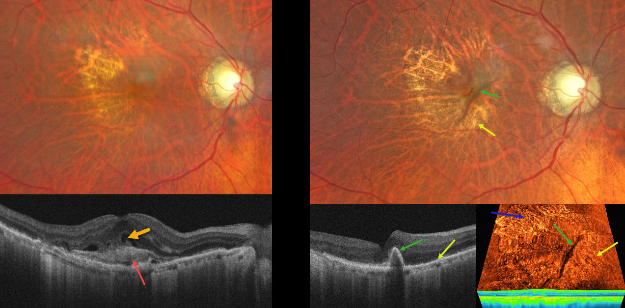
NEOVASCULAR AMD WITH RAPIDLY RESOLVED SHRM FOLLOWING RPE TEAR
Ayushi Gupta and Vishal Agrawal
Originally posted on @retina.rocks August 18, 2025
This 82YO male presented with recent vision loss in his right eye. Vision was 20/200.
Color photography shows a poor foveal reflex with superotemporal geographic atrophy. OCT scanning shows subretinal hyperreflective material (SHRM) with overlying retinal fluid. A possible bacillary layer detachment is also noted. About 6 weeks following a single intravitreal anti-VEGF injection, vision improved to 20/50. A new RPE tear is noted, with the bed of the tear inferonasal to the retracted hyperpigmented RPE. Despite persistent flow signals on OCT angiography, there is no fluid on structural OCT. The SHRM has completely resolved. A treat-and-extend anti-VEGF regimen was begun.
Learning Points:
A tear in the RPE is a fairly common finding in RPE detachment (PED) associated with type 1 macular neovascularization (MNV) located below the RPE. The neovascularization can spontaneously contract or involute following treatment. It then puts traction on the overlying RPE, which can then rip at the opposite edge of the PED. Like a cheap window blind, the torn RPE retracts and scrolls towards the MNV. This leaves a depigmented region where the RPE was and is now absent, along with a pigmented, relatively straight subretinal lesion from the scrolled RPE edge.
SHRM is an OCT finding that is invisible clinically. This material, located between the neurosensory retina and RPE, is thought to consist of fluid, fibrin, blood, and other fibrovascular tissues. SHRM is a biomarker of active disease that decreases with successful anti-VEGF therapy (Willoughby et al, Ophthalmology 2015;122:1846-1853). Persistent SHRM is associated with an increased incidence of scar formation and worse vision.
It is impossible to determine whether the anti-VEGF injection alone or in combination with the RPE tear caused such a rapid resolution of the SHRM. Fortunately, our patient’s vision remained quite good since the RPE tear just spared the foveal center.
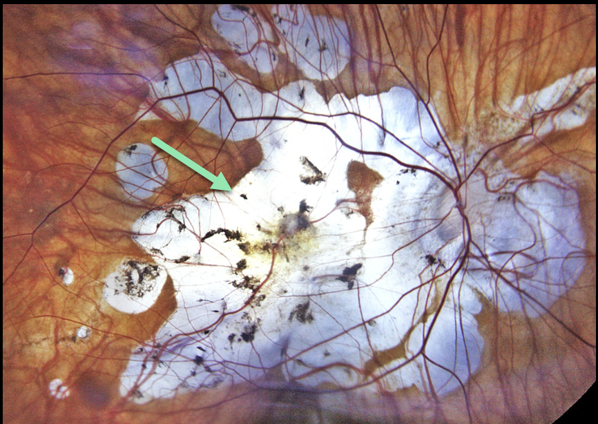
AMBLYOPIC MYOPIC STAPHYLOMA WITH MATURE CATARACT
Liubov Sitaruk
Originally posted on @retina.rocks August 11, 2025
This 64YO female has a lifetime history of anisometropic amblyopia with a myopic posterior staphyloma OD and presented with a mature cataract in this eye. Vision was light perception. She was clearly informed that surgery would likely not restore much vision. The goal was to remove the dense opacity and maximize any remaining visual potential.
Postoperative color imaging of the posterior pole shows severe peripapillary and myopic atrophy. Vision was 20/250.
Our patient reminds us that amblyopia is not an absolute contraindication for cataract surgery. Preoperative counseling is critical so that the patient understands the surgical goals. In general, the adage “under promise and over deliver” is a good rule for any healthcare provider to live by.
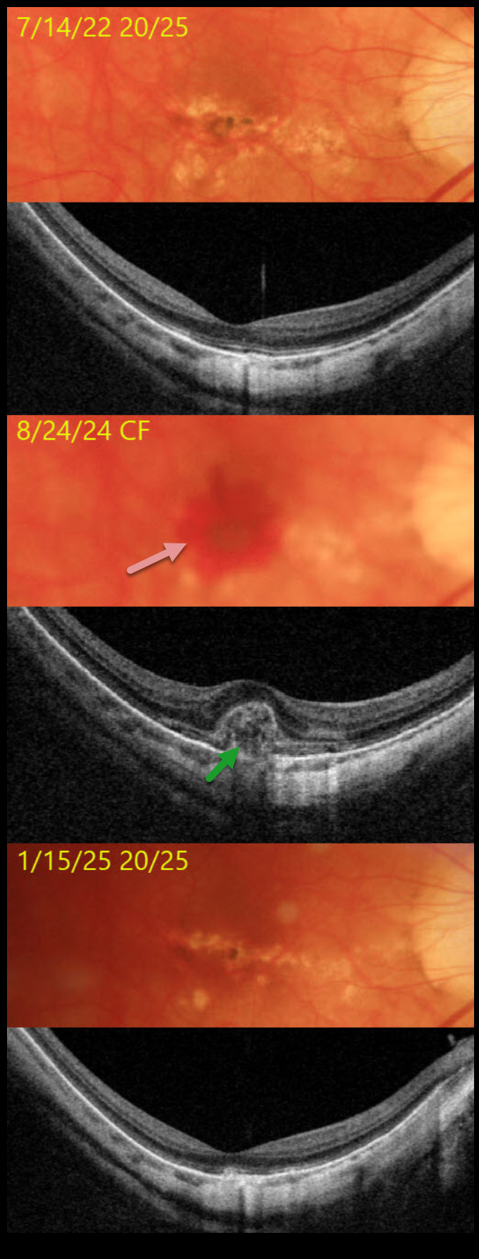
MYOPIC MACULAR NEOVASCULARIZATION
Originally posted on @retina.rocks August 6, 2025
This 53YO highly myopic male was seen on 7/14/22 for a routine exam with 20/25 vision. Triton color imaging shows dry myopic degeneration, macular pigment loss, and clumping. Triton swept-source OCT shows variable loss of the outer retinal bands and diffuse choroidal thinning.
He returned on 8/24/24 with vision loss from a counting-fingers submacular hemorrhage. OCT shows hyperreflective subretinal blood elevating and extending into the outer retina. Following monthly intravitreal Avastin injections, vision improved to 20/25 on 1/15/25. All blood resolved, and the OCT mostly returned to its pre-blood appearance.
Learning Points:
In our experience, myopic macular neovascularization (MNV) presents quite differently from neovascular AMD. Significant subretinal blood (as in our patient), subretinal fluid, or intraretinal fluid are rarely seen. Often, an active MNV will present as a new dot of subretinal blood or pigment. Occasionally, there will just be symptomatic vision loss or metamorphopsia without clinical or OCT findings. Anti-VEGF therapy is effective, and these lesions can often be managed with PRN therapy, which is what we recommended for our patient (Sakata et al, Retina 2023;43:1863-1871).
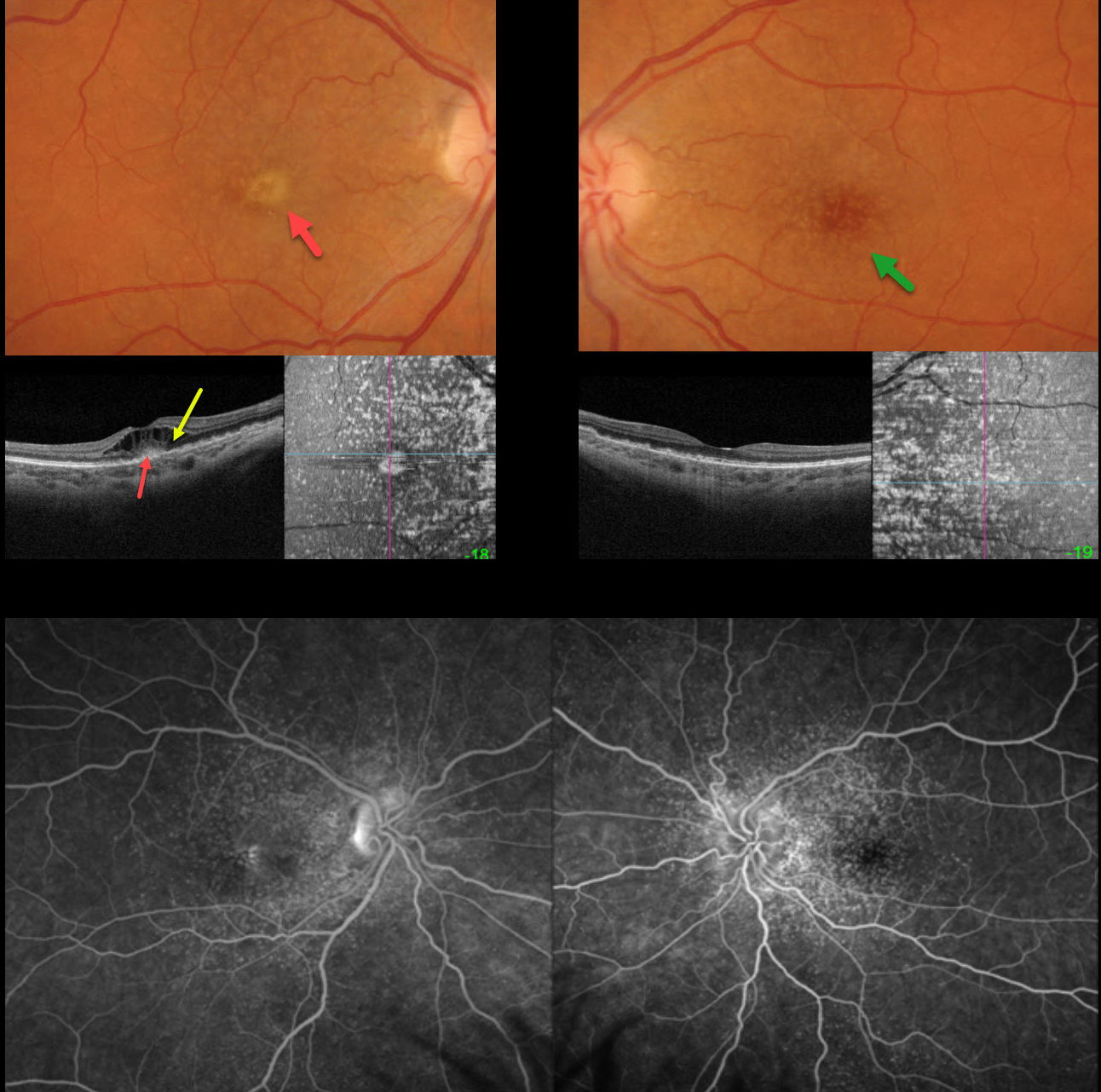
CUTICULAR DRUSEN WITH MACULAR NEOVASCULARIZATION
Originally posted on @retina.rocks August 5, 2025
This 47YO female presented with one week of vision loss in her right eye. Vision was counting fingers OD and 20/25 OS.
Optos color RGB imaging shows innumerable cuticular macular drusen, which appear on swept-source OCT B-scanning as arrowhead-like RPE elevations and on en face imaging as numerous hyperreflective spots. A yellow subfoveal lesion OD appears on OCT as hyperreflectivity in the outer retina. Marked overlying cystoid edema is seen. Fluorescein angiography shows late pooling in the foveal cysts OD, with hyperfluorescent window defects from the drusen.
One month following an initial intravitreal Avastin injection, vision improved to 20/80, with complete resolution of the vitelliform lesion and cystoid edema. Treat-and-extend treatment was started.
Learning Points:
Originally described by Gass as basal laminar drusen (AJO 1985;99:445-459), cuticular drusen were initially thought to represent nodular thickening of the RPE basement membrane. They appear clinically as numerous, round, yellow-white ‘hard drusen’, and on OCT as small, arrowhead-shaped drusen located above Bruch’s membrane and below the RPE (Spaide and Curcio, Retina 2010;30:1441-1454).
Our patient’s angiographic appearance echoes Dr. Gass’ original description of “a beautiful stars-in-the-sky or Milky-Way pattern” of hyperfluorescence. Cuticular drusen are classically associated with avascular vitelliform lesions (Finger et al, Retina 2010;30:1455-1464).
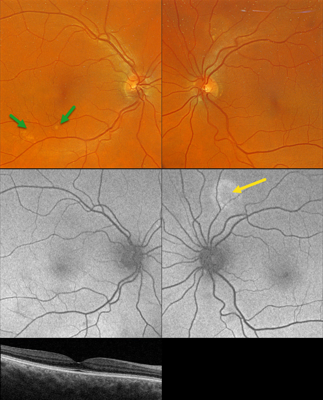
PACHYCHOROID SPECTRUM PREVIOUSLY MISDIAGNOSED AS AMD
Originally posted on @retina.rocks July 30, 2025
This healthy 65YO founder of Retina Rocks presented for his once-per-decade ‘eye examination’ which included an undilated slit lamp examination by his partner, Triton swept-source OCT, and Optos color RGB imaging. His mother had a history of neovascular AMD, and he has a known history of drusen. Given his family history, he has been taking AREDS2 supplementation for many years. There were no ocular symptoms, and vision was 20/20 bilaterally.
Triton color imaging shows some pachydrusen in the inferotemporal right macula. Swept-source OCT is normal except for a thickened choroid. Optos fundus autofluorescence (FAF) shows a round area of hyper-FAF superior to the left nerve.
Learning Points:
Pachydrusen were described by Spaide as a distinct pachychoroid phenotype (Retina 2018;38:708-716). Unlike typical AMD, where cone-based soft drusen are symmetrically localized to the foveal region, pachydrusen are eccentrically located, far less numerous, and have an irregular contour. Since choroidal thickness is influenced by numerous factors, including age and refractive status (Xie et al, IOVS 2022;63;2;34), these must always be considered when reviewing an individual patient’s scans. Eyes with AMD typically have age-related choroidal thinning, but eyes with pachydrusen, as in this case, are associated with a relatively thickened choroid.
Our patient became aware of his macular drusen well before pachychoroid was described by Freund in 2013 (Warrow et al, Retina 2013;33:1659-1672), and he attributed his early macular drusen to AMD. In 2025, he understands that his findings are consistent with the pachychoroid spectrum, as evidenced by the bilaterally thickened choroid, pachydrusen OD, and FAF findings suggestive of prior extramacular central serous OS. He does not have intermediate AMD, is at minimal risk for developing AMD, and thus doesn’t need AREDS2 supplementation. However, like the rest of us, he is only human and finds emotional comfort in continuing to take these supplements.
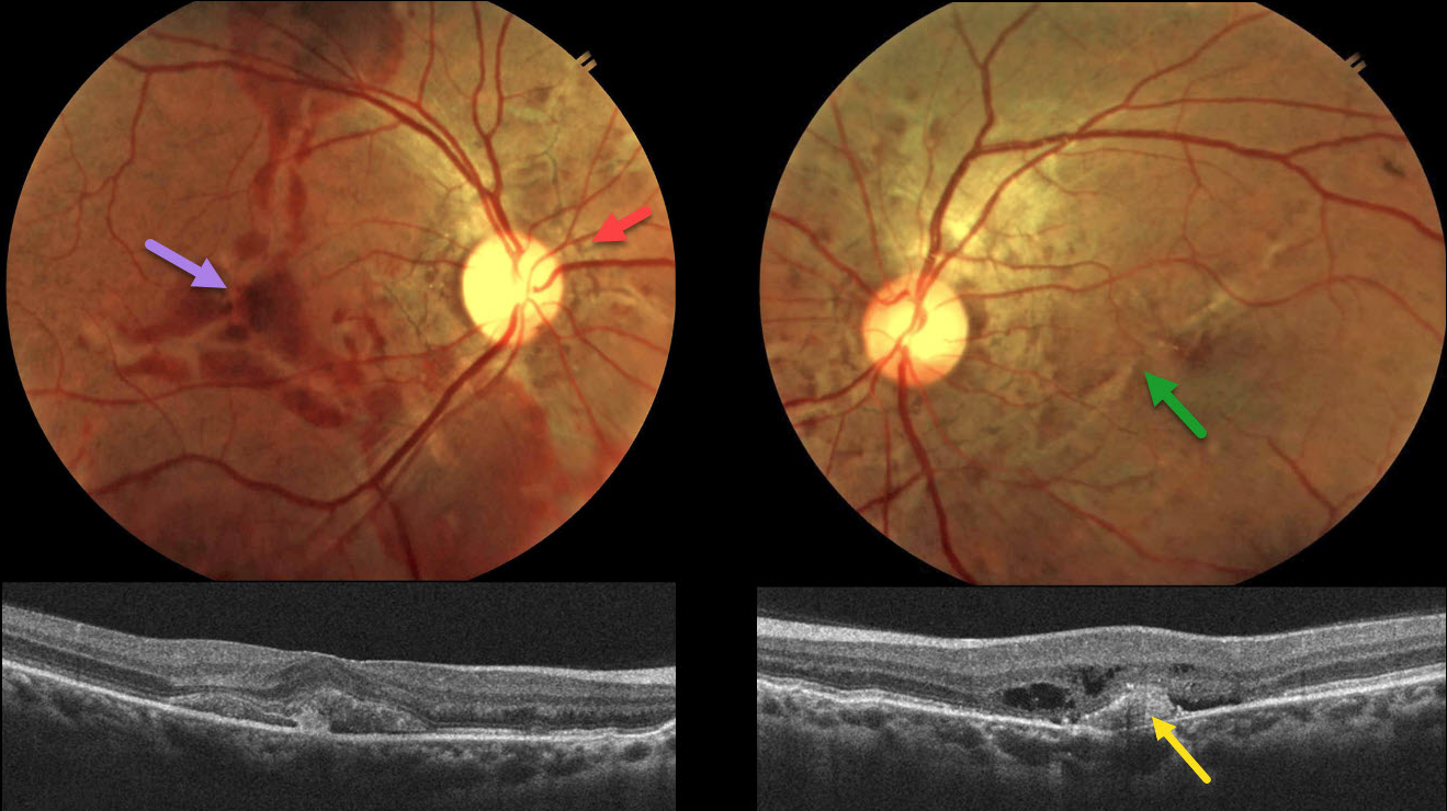
ANGOID STREAKS WITH TRAUMATIC CHOROIDAL RUPTURE AND SUBRETINAL BLOOD
Neelam Khatwani
Originally posted on @retina.rocks July 2, 2025
This 38YO male presented with 10 days of bilateral vision loss after being hit in the right eye with a fist. He denied any past medical history, and family history was negative for eye disease. Vision was 20/200 OD and 20/80 OS.
Color fundus photography shows irregular orange-gray lines radiating outwards from each nerve. Variable areas of subretinal blood are noted in the right macula, extending into the superior and inferior midperipheries. There is milder subretinal blood OS along with an irregular oblique macular angioid streak vs choroidal rupture. OCT OD shows subretinal hyperreflective material, likely blood. A possible type 1 (below the RPE) MNV with some outer retinal fluid is seen OS.
An anti-VEGF injection was given bilaterally, but unfortunately, the patient was immediately lost to follow-up.
Learning Points:
Initially described by Doyne in 1889, angioid streaks are orange-yellow, irregular, crack-like dehiscences in Bruch’s membrane associated with atrophic degeneration of the overlying RPE. The term “angioid” derives from its resemblance to blood vessels. Although they are historically mistakenly associated with numerous conditions, the only real association is with pseudoxanthoma elasticum (PXE, Nadelmann et al, Eye 2023;37:1596-1601). Patients are at significant risk for vision loss from MNV or pattern dystrophy-like changes (Murro et al, Graefe’s 2020;258:1881-1892).
Even minor trauma can cause choroidal ruptures with subretinal blood (Agrawal et al, JAMA Ophthalmology 2017;135(3):e165466). Unlike typical choroidal ruptures that are usually curvilinear with the nerve, the lesions from the brittle Bruch’s membrane in PXE appear as numerous irregular ruptures usually radiating from the nerve. Even though our patient sustained a direct injury to his right globe, the trauma was significant enough to cause choroidal ruptures in his left eye. The bilateral subretinal hemorrhages may be purely avascular, as their multifocality suggests.
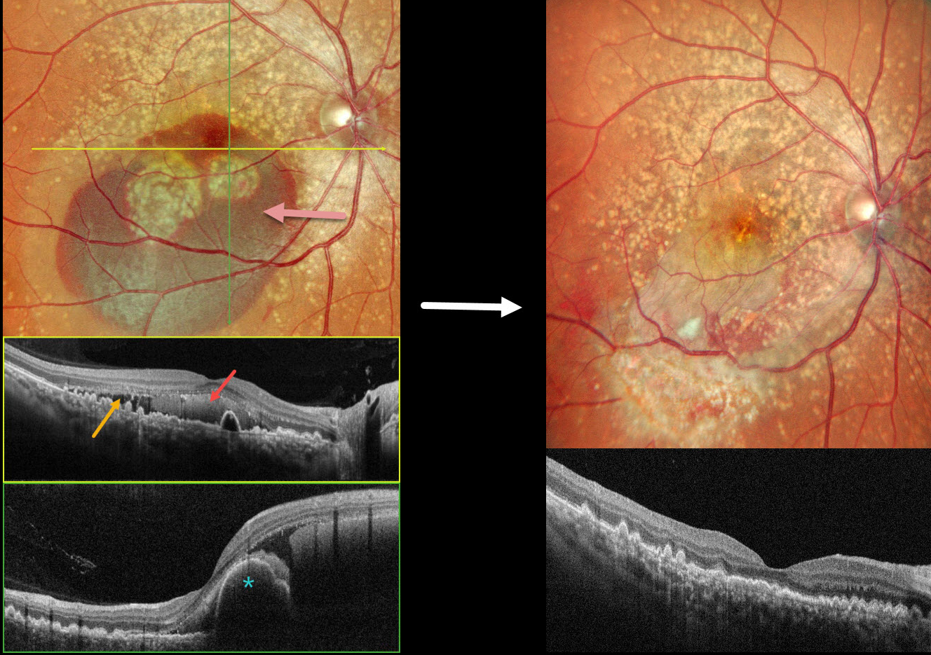
DOMINANTLY INHERITED RADIAL BASAL LAMINAR DRUSEN WITH POSSIBLE POLYPOIDAL CHOROIDAL VASCULOPATHY
Malvika Singh and Manish Nagpal
Originally posted on @retina.rocks June 25, 2025
This 49YO female presented with 3 days of decreased vision in her OD. Vision was 20/70 OD and 20/15 OS. There was no family history of eye disease.
Pseudocolor SLO imaging shows extensive mixed and confluent drusen extending through each macula into the midperipheries. Inferior submacular blood extends into the right foveal center. OCT scanning shows extensive conically shaped drusen. The subretinal blood in her right eye is hyperreflective, with additional temporal subretinal fluid. A bilobed nodular RPE detachment is present within the nasal blood.
She underwent pars plana vitrectomy, fluid-air exchange, and intravitreal Avastin. One month postoperatively, vision improved to 20/15 OD with resolved fluid and trace residual blood.
Learning Points:
Doyne’s honeycomb macular dystrophy, also known as dominantly inherited radial basal laminar drusen or Malattia Leventinese, is a rare macular disorder caused by a mutation in the EFEMP1 gene on chromosome 2p16. The EFEMP1 protein is a member of the fibulin family of extracellular matrix glycoproteins. The defective protein creates an abnormally thickened RPE basement membrane.
Centrally large, nodular, and confluent drusen are noted, along with a temporal radiating pattern of smaller cuticular drusen. Later, there may be variable amounts of RPE hyperplasia and fibrous metaplasia. Macular neovascularization, as in this case, may also develop. Our patient’s pre-op right OCT was suggestive of polypoidal choroidal vasculopathy, although this is not a known association with Doyne’s.
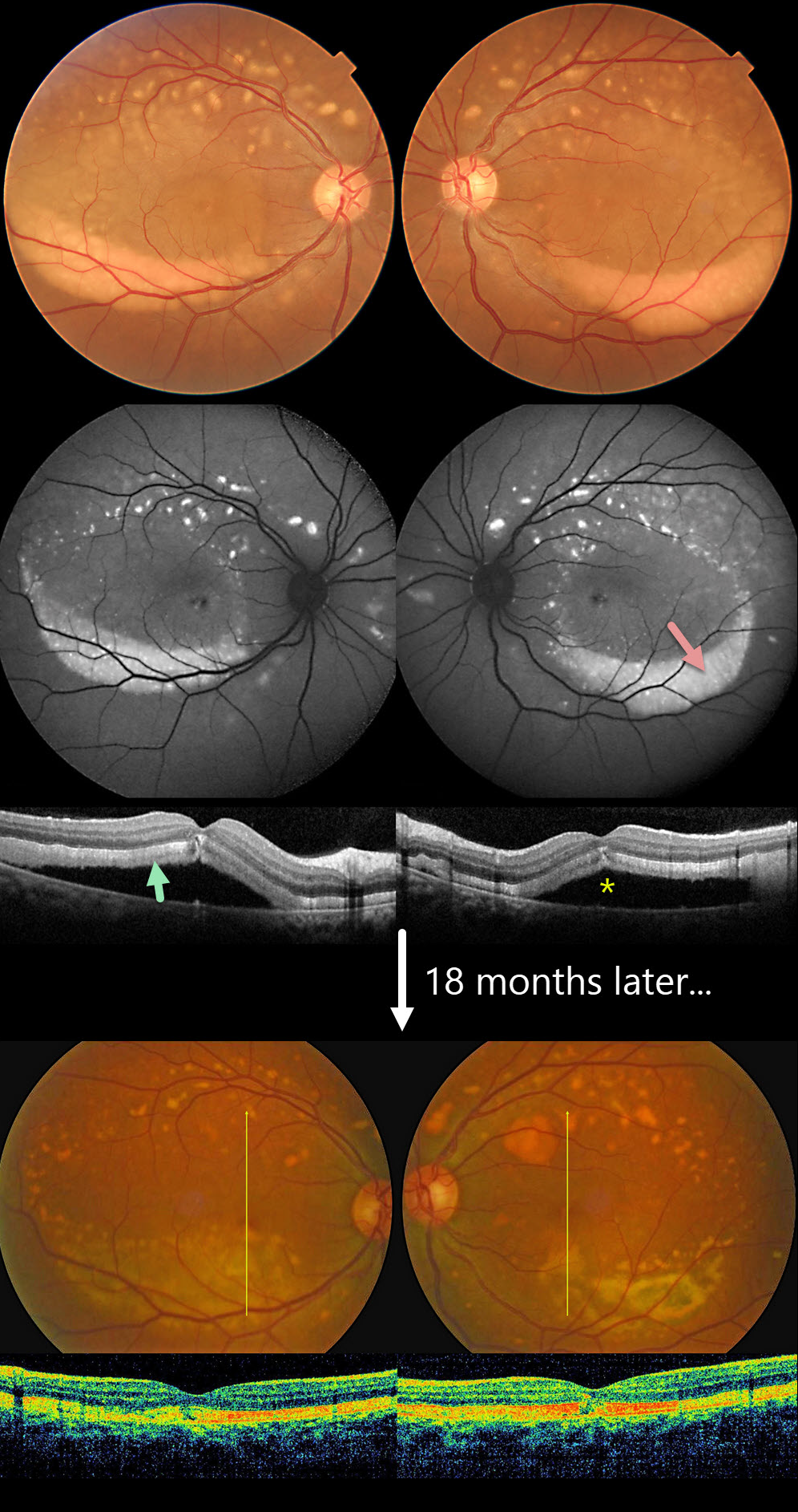
ACUTE EXUDATIVE POLYMORPHOUS VITELLIFORM MACULOPATHY
Krishnendu Nandi
Originally posted on @retina.rocks June 24, 2025
This 38YO male presented with 6 months of bilateral vision loss. Vision was 20/80 OU.
Color imaging shows multiple symmetrical yellowish subretinal variably round lesions centered in each superior macula and extending superiorly. Confluent subretinal vitelliform material is settled in the inferior maculas. This material is markedly hyper-autofluorescent. On OCT, the outer retina is diffusely infiltrated by this hyperreflective material, with associated serous macular detachments.
Oncologic workup was completely negative. When last examined 18 months later, vision was 20/30 OD and 20/30 OS. Color imaging shows remodeling of both the superior macular lesions and the inferiorly layered vitelliform material. The subretinal fluid is resolved with variable outer retinal band defects.
Learning Points:
Acute exudative polymorphous vitelliform maculopathy (AEPVM) is a rare condition characterized by acute vision loss from multifocal serous retinal detachments that later progress to subretinal vitelliform lesions. Initially, serous detachments can mimic central serous retinopathy, including choroidal thickening. The condition is often self-limiting, and vision usually recovers within several months. The yellow vitelliform lesions may take 1-2 years to completely resolve. These patients need a systemic workup for metastatic disease, since paraneoplastic retinopathy, including metastatic melanoma, can mimic idiopathic AEPVM.
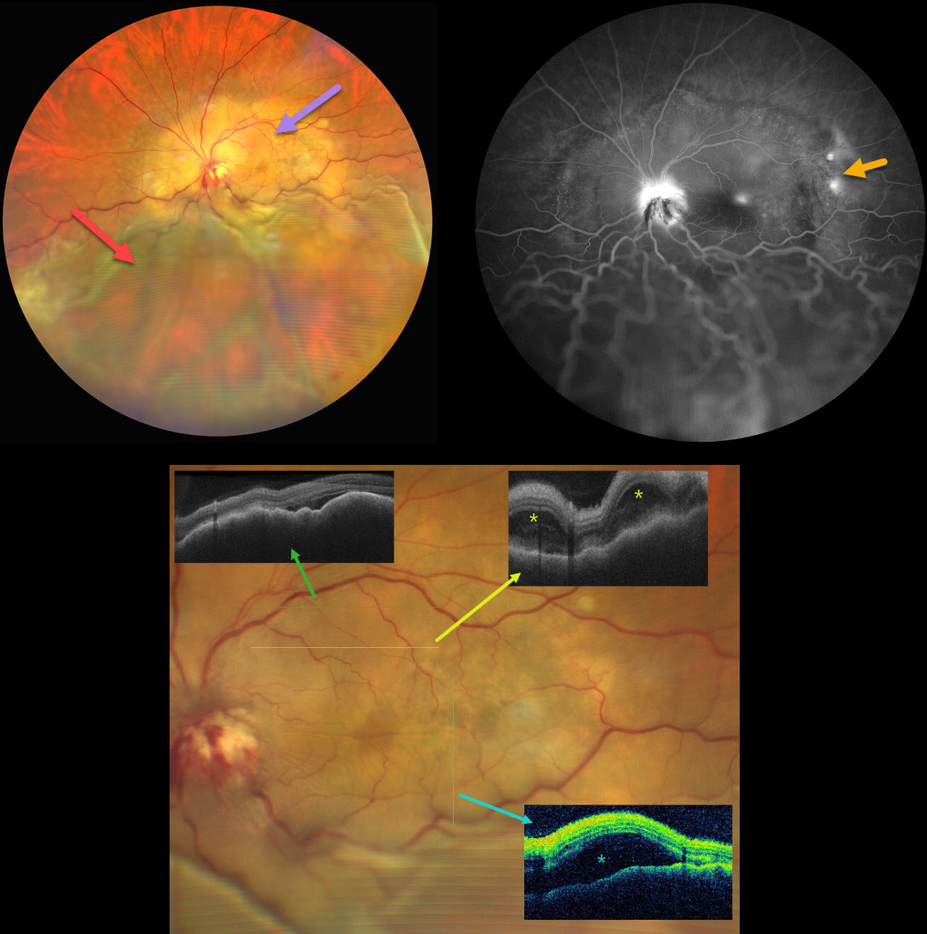
CHOROIDAL METASTASES WITH EXUDATIVE RETINAL DETACHMENT
Ayushi Gupta and Vishal Agrawal
Originally posted on @retina.rocks June 18, 2025
This 47YO female with a known history of metastatic breast carcinoma presented with several weeks of decreased vision in her left eye. She was on active chemotherapy and hormonal therapy. Vision was no light perception OD following trauma 30 years earlier, and hand motion OS.
Ultrawidefield color photography shows a large, amelanotic yellow choroidal lesion involving the entire macula and peripapillary choroid, which extends into the midperiphery. A large secondary inferior exudative retinal detachment involves the entire inferior hemisphere. This fluid shifted when laid supine. The optic disc is hyperemic with indistinct margins and associated blood.
OCT scanning shows a thickened, undulating choroid with variable overlying subretinal fluid and bacillary layer detachments. Fluorescein angiography shows multifocal subretinal leaks, choroidal lesion staining with possible overlying pooling, and intense nerve leakage.
Learning Points:
Although uveal melanoma is the most common primary intraocular malignancy, metastatic choroidal tumors are the most common intraocular malignancies. Lung and breast cancer are the most common sources.
Our patient had a known history of metastatic disease when she presented with this large lesion occupying her entire posterior pole. The extent of the metastasis and the overlying exudation are atypical in our experience. She was immediately referred to her oncologist for external-beam radiation, but was lost to follow-up.
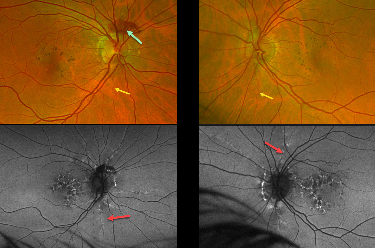
PSEUDOXANTHUM ELASTICUM WITH ANGIOID STREAKS AND PATTERN DYSTROPHY PHENOTYPE
Mattie Adams
Originally posted on @retina.rocks June 4, 2025
This 53YO female was referred for asymptomatic macular findings. Her mother has a history of pseudoxanthoma elasticum (PXE). Vision was 20/25 OD and 20/40 OS.
Optos color RG imaging shows coarse subretinal pigment clumps bilaterally. Subretinal blood is noted along the superior aspect of the right nerve from a peripapillary choroidal neovascularization. A few barely visible angioid streaks radiate from each nerve.
Fundus autofluorescence (FAF) shows variable hyper- and hypo-FAF flecks. The angioid streaks appear as hyper-FAF lesions radiating outwards from each nerve.
Learning Points:
Retinal findings in PXE most commonly include angioid streaks and a subretinal peaux d’orange appearance, most pronounced in each temporal macula. However, pattern dystrophy-like changes, as in our patient, can sometimes be the predominant phenotype (Murro et al, Graefe’s 2020;258:1881-1892). As with typical pattern dystrophies, the findings are dynamic and best captured with multimodal imaging.
PXE is caused by mutations in the ABCA6 gene, and our patient tested positive for homozygous pathogenic ABCA6 mutations. PXE is virtually always transmitted as an autosomal recessive disorder, although autosomal dominant transmission may rarely occur (Plomp et al, Am J Med Genet A 2004;126A:403-412).
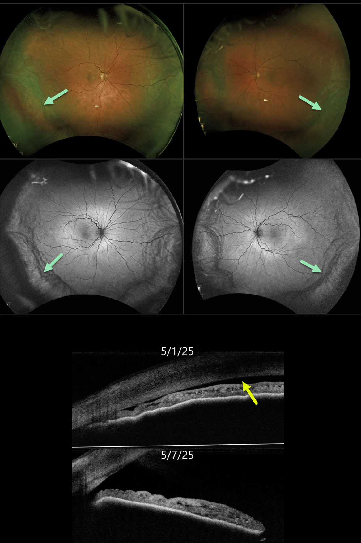
ESCITALOPRAM-INDUCED UVEAL EFFUSIONS WITH ANGLE CLOSURE GLAUCOMA
Nikolaos Zagorianos, Mattie Adams, Jessica Haycraft and Brett Abney
Originally posted on @retina.rocks June 3, 2025
This 67YO female woke with bilateral blurred vision and severe eye pain. She was started on Lexapro (escitalopram) six months earlier. Vision was 20/400 OD and 20/100 OS.
Slit lamp examinations showed bilateral shallow anterior chambers, with no angle structures visible in either eye on gonioscopy and anterior segment OCT. Intraocular pressures (IOP) were 50 mmHg OD and 44 mmHg OS.
Optos color RG and green channel imaging shows bilateral peripheral encircling choroidal detachments. Anterior segment OCT confirmed a flat anterior chamber. Rocklatan, Simbrinza, Alphagan, Atropine, and Diamox were administered in the office, with the IOP decreasing to 38 OD and 36 OS. We contacted her primary care doctor to discontinue the Lexapro. One week later, vision improved to 20/30 OD and 20/25 OS. IOP was 16 OD and 13 OS, the anterior chamber angles were open clinically and on OCT, and the choroidal detachments were decreasing.
Learning Points:
Drug-induced ciliochoroidal effusions with secondary angle closure glaucoma (ACG) are a known complication of topiramate therapy, but have also rarely been reported with selective serotonin reuptake inhibitors, including escitalopram (Zelefsky et al, AJO 2006;141:1144-1147). Unlike typical ACG, which is cured with a peripheral iridotomy, these drug-induced ACG attacks are treated with dilation. Atropine paralyzes the ciliary body, which allows the lens/Iris diaphragm to return to its natural state, rotating backward and increasing the anterior chamber depth. Topical steroids help decrease uveal vascular permeability, thereby reducing choroidal effusion.
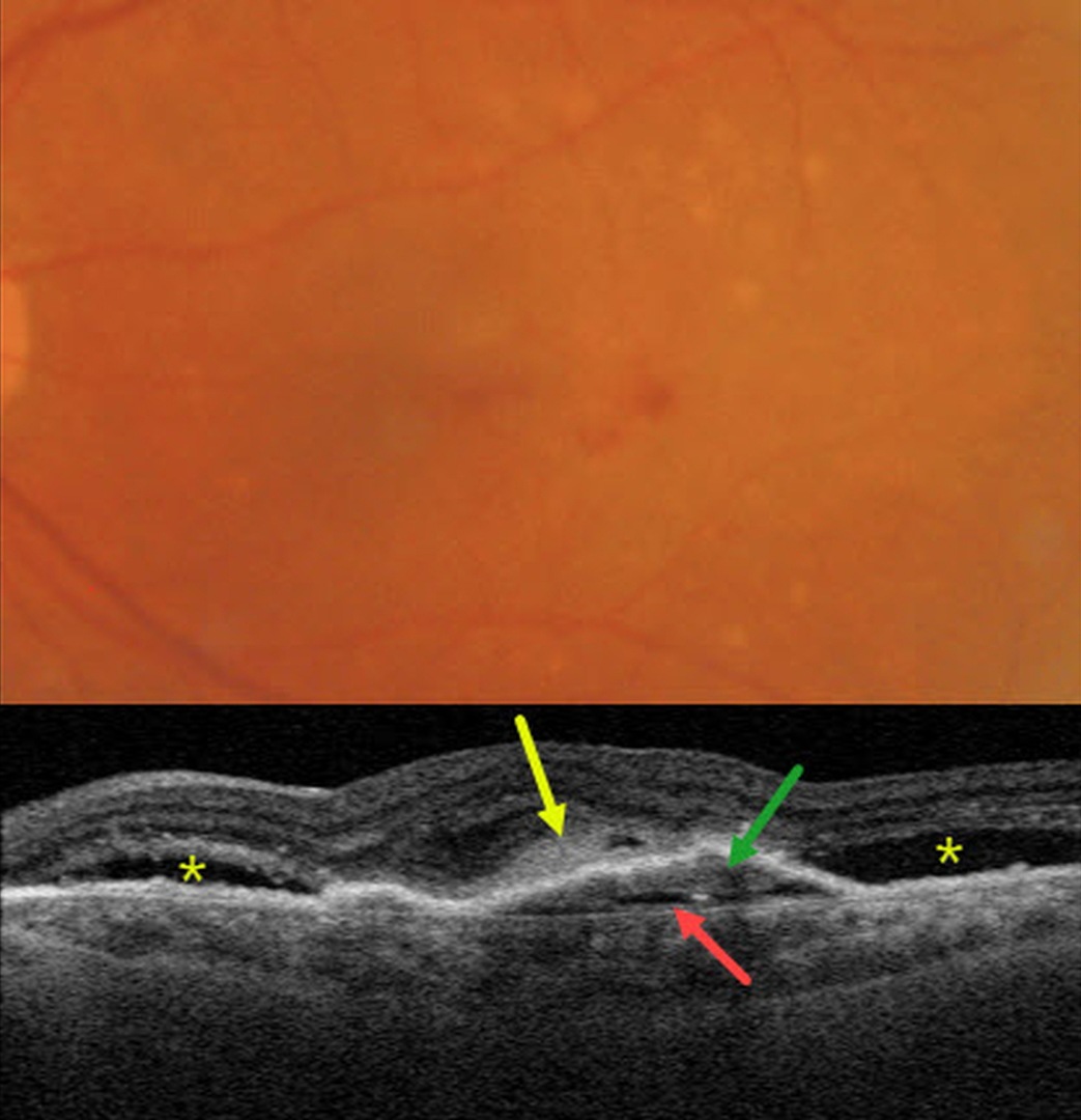
NEOVASCULAR AMD WITH SUBRETINAL HYPERREFLECTIVE MATERIAL (SHRM) AND PRECHOROIDAL CLEFT
Originally posted on @retina.rocks May 28, 2025
This 80YO female presented with counting-fingers vision in her left eye from treatment-naive neovascular AMD (nAMD).
Triton color photography shows a few retinal hemorrhages and more peripheral large drusen. Swept-source OCT shows a shallow irregular RPE detachment with underlying type 1 macular neovascularization (MNV). A hyporeflective prechoroidal cleft is noted below the MNV and subretinal hyperreflective material (SRHM) above. There is shallow outer retinal fluid above the SRHM. Subretinal fluid is seen on either side of the MNV. Intravitreal Avastin therapy was begun.
Learning Points:
Our patient’s OCT includes numerous findings of active MNV, including SRHM, prechoroidal cleft, and subretinal and intraretinal fluid.
SHRM is an OCT finding that is invisible clinically. This material, located between the neurosensory retina and retinal pigment epithelium (RPE), is thought to consist of fluid, fibrin, blood, and other fibrovascular tissues. SHRM is a biomarker of active disease that decreases with successful anti-VEGF therapy (Willoughby et al, Ophthalmology 2015;122:1846-1853). Persistent SHRM is associated with an increased incidence of scar formation and worse vision.
A prechoroidal cleft is a hyporeflective space between the RPE and Bruch’s membrane. It is seen in up to 20% of treated eyes with nAMD, particularly with type 3 neovascularization (intraretinal MNV) and polypoidal choroidal vasculopathy. They are usually associated with worse visual acuity due to potential complications, including RPE tears and subretinal hemorrhages (Kim et al, Retina 2017;37:2047-2055).
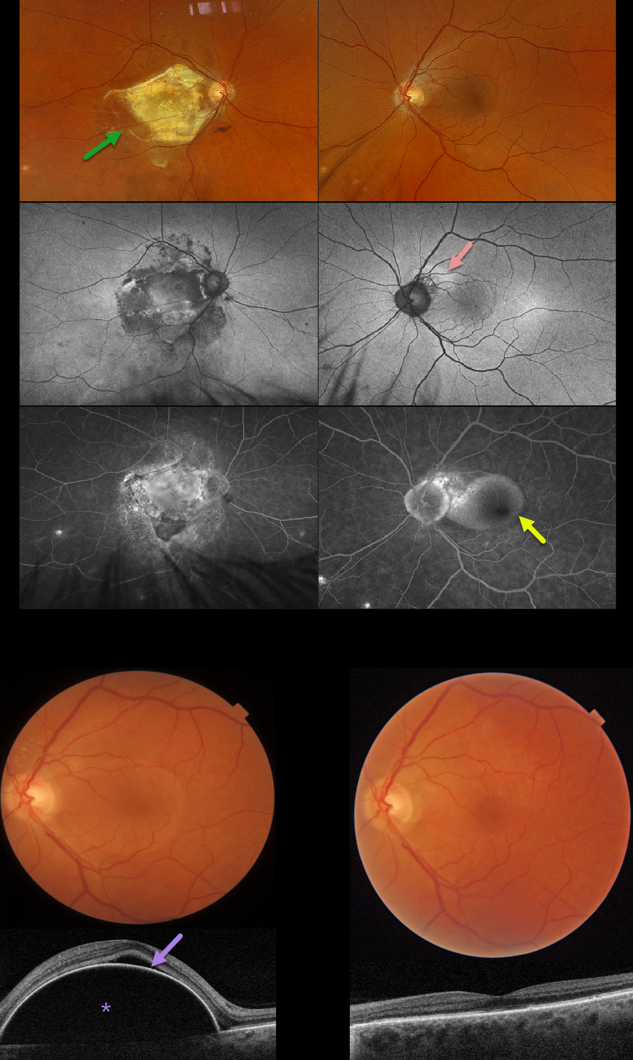
POLYPOIDAL CHOROIDAL VASCULOPATHY
Originally posted on @retina.rocks May 14, 2025
This 70YO male presented with 1 week of vision loss in his left eye. He has a long history of polypoidal choroidal vasculopathy (PCV). He received prior anti-VEGF injections OD, but his vision subsequently decreased to counting fingers from a fibrotic disciform scar due to poor compliance with office visits. Several years earlier, he developed a peripapillary polyp in his left eye that was resistant to Eylea monotherapy but responded well to combination treatment with photodynamic therapy (PDT).
Optos color RGB imaging shows a fibrotic disciform scar OD and a retinal pigment epithelial detachment (PED) OS. Fundus autofluorescence (FAF) shows variable hyper-FAF OS in the region of the prior polyp. Ill-defined angiographic leakage is noted superotemporal to the left nerve, with an uneven-filling PED. Triton swept-source OCT confirms a large and highly elevated PED with some overlying subretinal fluid.
Given the prior excellent response to combination treatment, intravitreal Eylea followed by PDT to the peripapillary notch was applied. Six weeks later, vision improved to 20/25, and the PED completely resolved with an almost normal OCT.
Learning Points:
Polypoidal choroidal vasculopathy (PCV) is a variant of type 1 macular neovascularization. PCV is likely distinct from age-related macular degeneration, occurring in a different population (more common in African Americans and Asians) and characterized by a thicker choroid and a relative lack of drusen. Historically, these lesions were believed to be somewhat resistant to anti-VEGF monotherapy, often requiring combined treatment with photodynamic therapy. More recent data from the PLANET study suggest that Eylea monotherapy is also effective (AJO 2019;204:80-89). However, our patient was resistant to monotherapy and responded remarkably well to combination therapy.
Originally described by Gass (Retina 1984;4:205-220), a notched PED is a sign of an occult MNV. The notch represents a type 1 MNV, which tethers the overlying RPE from the surrounding more highly elevated PED. Hydrostatic leakage from the MNV may contribute to the larger PED.
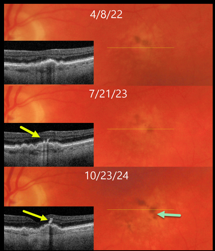
DRY AMD WITH PIGMENT CLUMPING REMODELING
Originally posted on @retina.rocks April 10, 2025
This 71YO male has been followed for several years with intermediate AMD.
On 4/8/22, vision was 20/40 in his asymptomatic left eye. There is a shallow RPE detachment (PED) with faint surrounding drusen and pigment loss on both Triton color imaging and swept-source OCT. Clumping of subretinal pigment is noted along the superior edge of the PED on color imaging.
He returned for his 1-year examination on 7/21/23 with stable 20/40 vision. A new pigment clump is noted along the temporal edge of the PED, and this is seen as a hyperreflective plume on OCT.
On 10/23/24, vision decreased to 20/100, with extension of the plume into the inner retina. There is continued remodeling of the pigment clumps on color imaging.
Learning Points:
The widely used Beckman AMD clinical classification defines intermediate AMD as including large drusen > 125 microns and/or any AMD pigmentary abnormalities (Ferris et al, Ophthalmology 2013;120:844-851). This classification was modified in 2024 to include a third risk factor, subretinal drusenoid deposits, which significantly increases the risk for progression to late AMD (Agron et al, Ophthalmology 2024;131:1164-1174).
These pigment clumps, which are visualized on OCT as hyperreflective foci (HRF), are found in over 50% of eyes with AMD (Ho et al, Ophthalmology 2011;118:687-693), and are thought to represent thickened RPE or intraretinal migrating RPE cells (Laiginhas et al, Ophthalmology Science 2022;2:100116). The migrating RPE cells often track along Muller cells creating a hyperreflective plume (Cao et al, IOVS 1021;62(10):34). These RPE cells transdifferentiate into macrophage-like cells. Macular atrophy will often develop underlying the HRF.
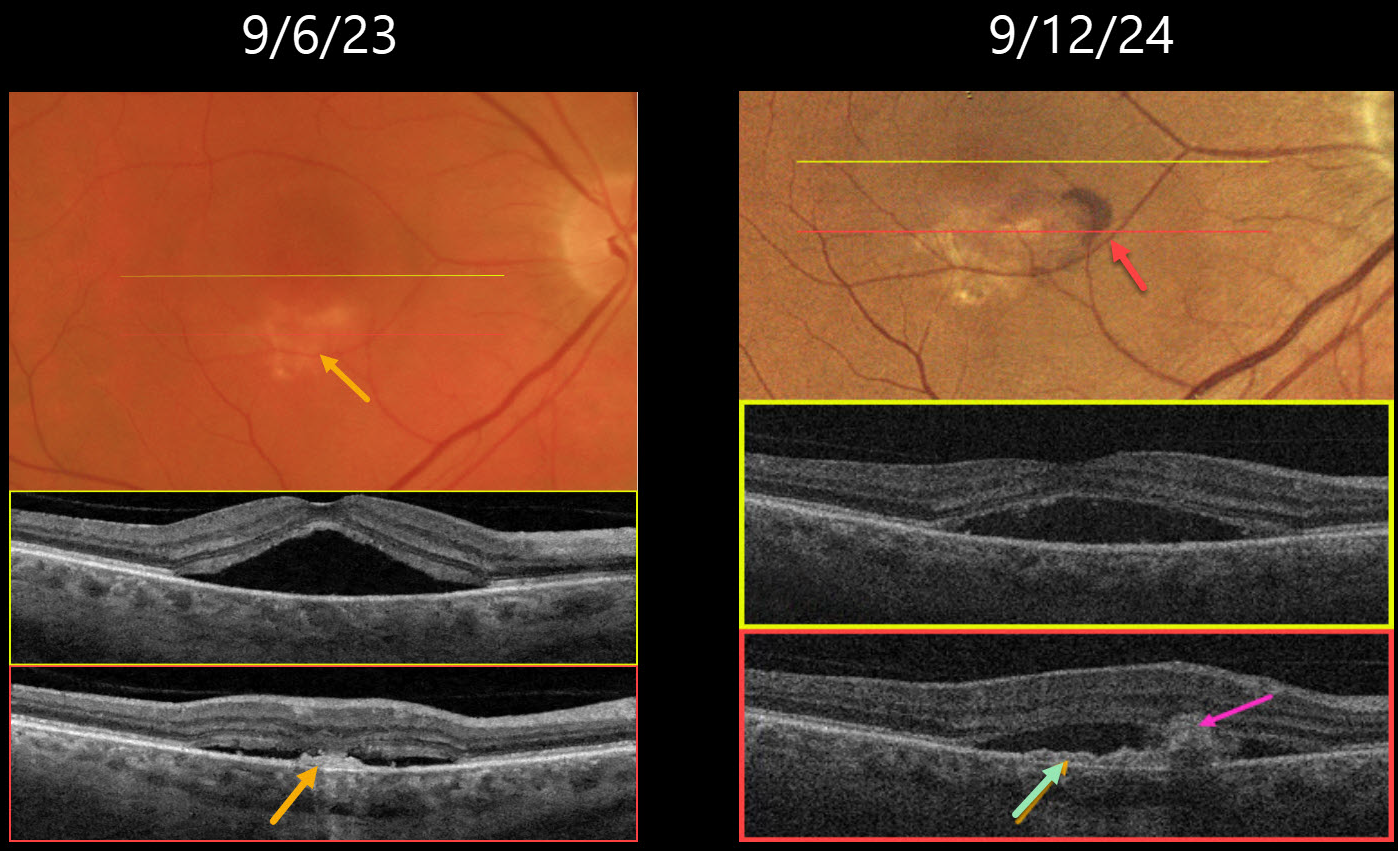
PACHYCHOROID NEVOASCULARIZATION
Originally posted on @retina.rocks March 31, 2025
This 64YO male presented on 9/6/23 with several weeks of vision loss in his right eye. Vision was 20/40 OD.
Triton color imaging shows a faint round foveal serous detachment. Swept-source OCT confirms a classic idiopathic central serous chorioretinopathy (ICSC) foveal detachment. There is a small hyperreflective placoid elevation of the RPE along the inferior edge of the detachment corresponding to a yellow-white area of subretinal pigmentation. Due to significant symptoms, he elected for early photodynamic therapy (PDT), which successfully resolved the serous fluid two months later with 20/30 vision (not shown).
He did well until he returned on 9/12/24 with recurrent blurred vision OD, which had decreased slightly to 20/40. There is new subretinal blood within the region of the persistent placoid RPE elevation inferiorly. OCT shows recurrent serous foveal fluid centrally. There is an enlargement of the shallow inferior RPE elevation adjacent to the subretinal blood. Monthly intravitreal Avastin therapy was started.
Learning Points:
Initially described by Pang and Freund, pachychoroid neovasculopathy is a variant of type 1 (below the RPE) macular neovascularization (MNV) found as part of the pachychoroid spectrum, including ICSC and pachychoroid pigment epitheliopathy (Retina 2015;35:1-9). About one-third of chronic ICSC eyes with flat irregular RPE detachments will harbor an MNV on OCT angiography (Bousquet et al, Retina 2018;38:629-639), which is probably a form of pachychoroid neovascularization (Hwang et al, Retina 2020;40:1724-1733).
In retrospect, our patient most likely had a pachychoroid neovascularization, which initially responded to PDT. When he returned one year later with a definite active MNV, we elected to start anti-VEGF monotherapy. He was unfortunately immediately lost to follow-up.
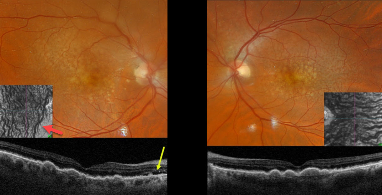
PERIPAPILLARY PACHYCHOROID SYNDROME
Originally posted on @retina.rocks March 20, 2025
This 77YO male was referred for asymptomatic neovascular AMD in his right eye. Vision was 20/50 bilaterally.
Optos color RGB imaging shows extensive bilateral larger drusen and subretinal drusenoid deposits, which are evident on Triton swept-source OCT. Localized subretinal fluid is noted just temporal to the right nerve.
Although the choroid is not particularly thickened, it is relatively thick for a 77YO, and large vessels span the full-thickness choroid. En face choroidal OCT shows markedly abnormal dilated vessels.
Learning Points:
Originally described by Phasukkijwatana et al in 2018 (Retina 2018;38:1652-1667), peripapillary pachychoroid syndrome presents with nasal macular intraretinal and subretinal fluid. These eyes can look very similar to those of patients with optic pit maculopathy, but without the pit. Additional findings include a thicker nasal than temporal macular choroid, choroidal folds (75% of cases), shorter axial lengths (<23mm), and hyperopia. There is no angiographic leakage despite the presence of OCT fluid, which often spontaneously waxes and wanes. Symptomatic fluid can successfully be treated with photodynamic therapy.
We assume our patient’s fluid is related to his pachychoroid disease rather than nAMD. Observation was recommended.
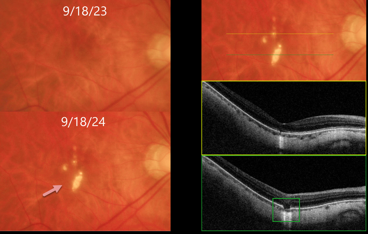
LACQUER CRACKS
Originally posted on @retina.rocks March 17, 2025
This 54YO highly myopic female is being followed with PRN Avastin injections in her left eye for myopic macular neovascularization. She was asymptomatic, and vision was stable at 20/50 OD and 20/60 OS.
Optos color RGB imaging of her right macula shows numerous lacquer cracks. Triton swept-source OCT shows outer retinal and RPE loss with increased choroidal transmission. Remarkably, her macula was normal one year earlier. Continued observation was recommended.
Learning Points:
Lacquer cracks are a common finding in patients with high myopia. They consist of breaks in Bruch’s membrane due to the outpouching/stretching of the pathologically myopic eye and increase the risk for macular neovascularization. Acutely, they are often associated with avascular subretinal blood, which resolves with observation.
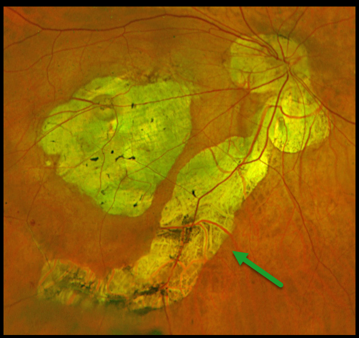
MYOPIC PERIVASCULAR CHORIORETINAL ATROPHY
Yuenpang Cheung
Originally posted on @retina.rocks February 25, 2025
Optos color RG imaging of this highly myopic patient shows severe myopic degeneration extending from the central macula inferiorly, along with peripapillary atrophy that extends beneath the entire inferotemporal arcade.
Learning Points:
Perivascular chorioretinal atrophy is a newly described finding in highly myopic eyes (Benahmed et al, AJO 2025;271:498-506). The atrophy occurs along the first few bifurcations of the major retinal vessels and always develops in eyes with a posterior staphyloma. An anterior scleral protrusion along the retinal vessel is seen in 74% of eyes, suggesting that anomalous scleral curvature may cause this unusual atrophy.
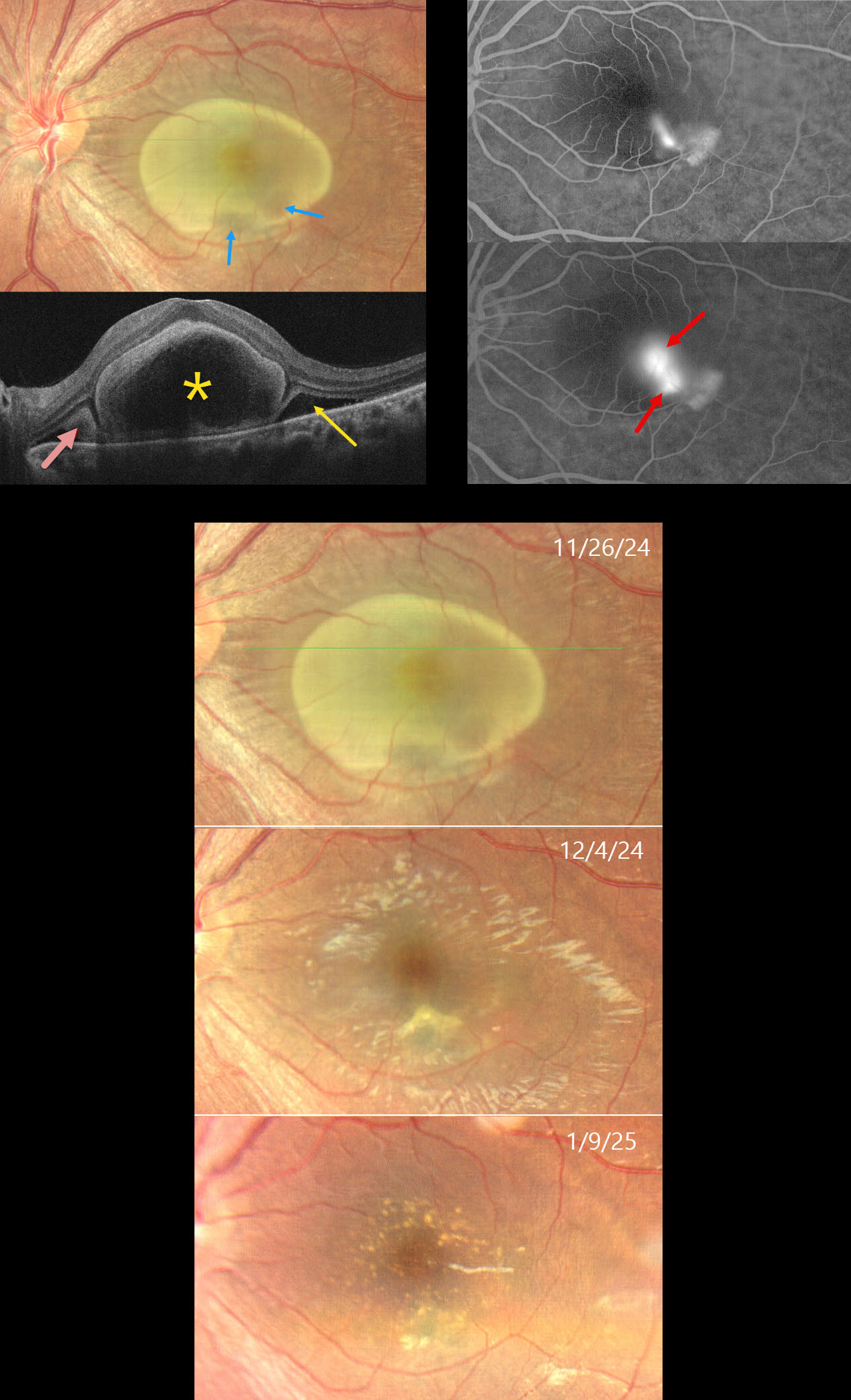
FIBRINOUS IDIOPATHIC CENTRAL SEROUS CHORIORETINOPATHY
Ayushi Gupta and Vishal Agrawal
Originally posted on @retina.rocks February 24, 2025
This 31YO female complained of decreased vision in her left eye for 6 weeks. She was diagnosed elsewhere with cysticercosis and referred to us for further evaluation. Vision was 20/20 in her normal OD and 20/200 OS.
Color photography shows a yellow oval lesion occupying nearly the entire macula. OCT scanning shows a large subfoveal bacillary layer detachment (BALAD). Hyperreflective fibrin likely lines the inner border of the BALAD. Subretinal fibrin and subretinal fluid (SRF) are noted on either edge of the BALAD. Fluorescein angiography shows either a smokestack or two independent blot leaks with staining inferotemporally. The yellow foveal lesion is relatively depigmented within the region of the angiographic leakage. The lesion and fluid spontaneously resolved over about 6 weeks, with residual drusen-like changes. Vision at the final visit was 20/20.
Learning Points:
In idiopathic central serous chorioretinopathy (ICSC), fluorescein dye diffuses through a physiological break in the RPE and then pools within the subretinal space. The clearing within our patient’s inferotemporal subretinal fibrin (as evidenced by the hyporeflective temporal SRF on OCT) is likely caused by serous fluid entering the subretinal space and ‘washing away’ some of the fibrin.
Most patients with ICSC have a single, unilateral leak. Blot leaks are most common, with only 10% of cases exhibiting a smokestack. The multifocal leaks and subretinal fibrin noted in our patient are more commonly found in steroid-induced disease, although our patient denied steroid exposure. Bacillary layer (cone and rod inner and outer segments) detachment appears as a unique dome-shaped collection of intraretinal fluid from photoreceptor splitting. These detachments can be observed in an increasing number of conditions, most classically Vogt-Koyanagi-Harada disease (Cicinelli et al, Ophthalmology Retina 2020;4:454-456). BALAD is also a biomarker for severe ICSC with steroid use (Casella et al, Int J Retina Vitreous 2024;10;96).
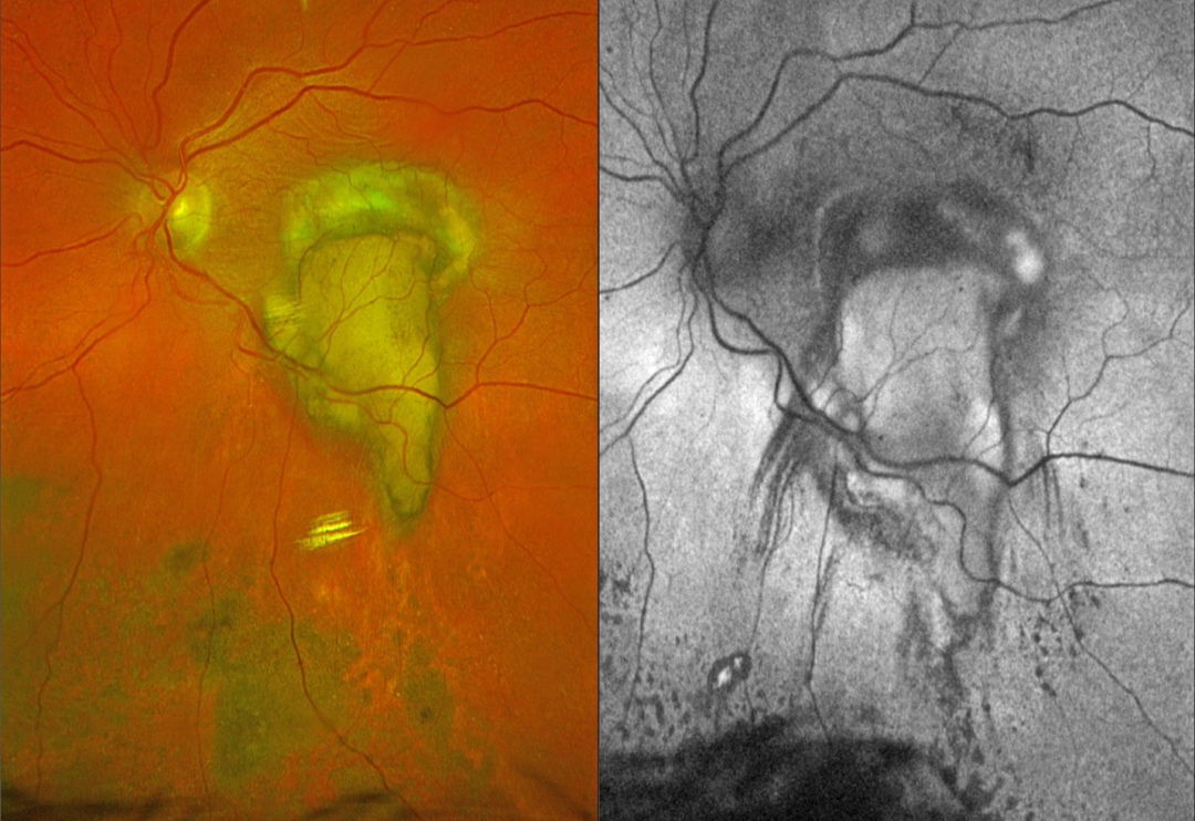
NEOVASCULAR AMD
Originally posted on @retina.rocks February 5, 2025
This 79YO female underwent vitrectomy for a breakthrough vitreous hemorrhage from neovascular AMD. Vision was 20/400.
Postoperative Optos color RGB imaging shows an oval fibrotic macular scar with a tongue-like projection of organized devitalized yellow blood extending inferiorly. Coarse subretinal pigment extends into the inferior midperiphery. On fundus autofluorescence (FAF), the fibrotic portion of the scar and inferior subretinal pigment are hypo-FAF, and the devitalized blood is hyper-FAF. Steaks of inferiorly radiating hypo-FAF also extend from the tongue-like yellow blood.
Learning Points:
This complex healing neovascular lesion has many signs of chronicity. There is no fresh, red blood. The remaining blood is yellow and devitalized, developing as heme products degrade. The coarse peripheral subretinal pigment is a marker of prior resolved subretinal blood and appears on OCT as subretinal hyperreflective material. Finally, the subretinal fibrosis where the breakthrough blood originated represents the usual endpoint of untreated neovascular disease, which is thankfully much less common since the advent of anti-VEGF therapy in the mid-2000s.
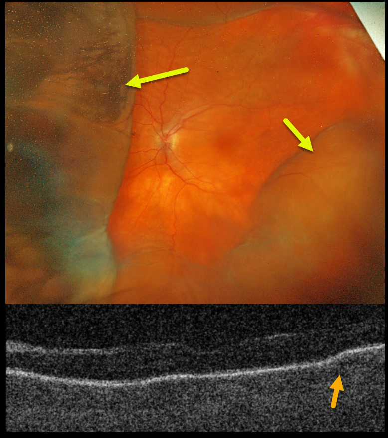
CHOROIDAL DETACHMENT
Mattie Adams
Originally posted on @retina.rocks January 27, 2025
This 78YO female presented with acute vision loss in her left eye. Penetrating keratoplasty (PKP) was performed 9 months earlier. When seen on 8/26/24, vision was counting fingers in part due to irregular astigmatism from the residual PKP sutures, which were then removed. The cornea was Seidel negative after removal. She returned one week later complaining of a curtain in her visual field. Her anterior chamber was flat, and her corneal graft was dehisced for 3 clock hours.
Optos color RGB imaging shows a variably elevated choroidal detachment. Triton swept-source OCT shows chorioretinal folds.
Learning Points:
A choroidal detachment is defined by the abnormal presence of fluid or blood in the suprachoroidal space. Serous choroidal detachments, also known as choroidal effusions, are a frequent complication of glaucoma surgery. Several causative mechanisms are believed to be at play: hypotony allows fluid to accumulate in the suprachoroidal space and inflammation increases the choroidal permeability. The detached ciliary body may also produce less aqueous, thus creating a self-perpetuating cycle. Often, observation is sufficient but topical steroids and cycloplegics can be helpful to aid in resolution.
Hypotony maculopathy is characterized by low IOP and chorioretinal folds, most commonly following glaucoma filtration surgery. Optic nerve edema can also be seen due to decreased axoplasmic transport.
Our patient was immediately taken to surgery for suturing of the dehisced PKP. The choroidals and chorioretinal folds immediately improved and completely resolved two months later (not shown).
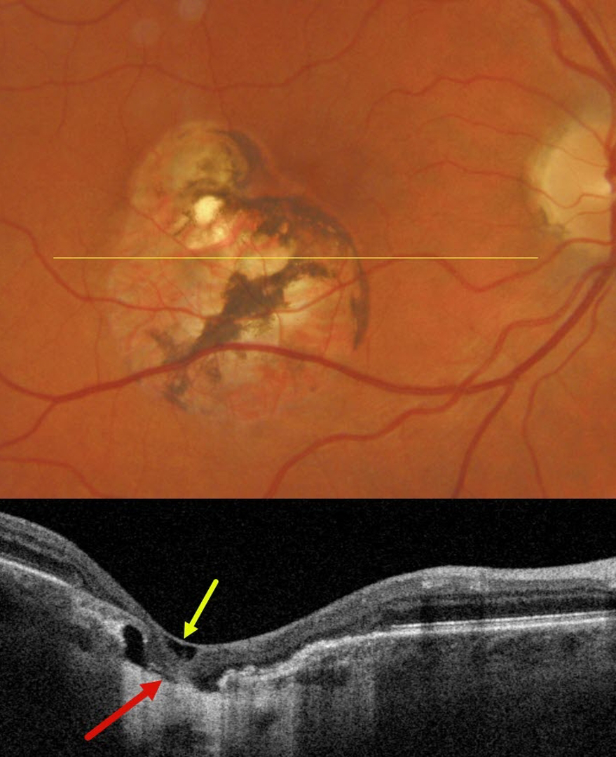
OCULAR HISTOPLASMOSIS LASER SCAR
Originally posted on @retina.rocks January 8, 2025
This 52YO male has been followed by us for many years for ocular histoplasmosis (OHS). Decades earlier, he received thermal laser from an outside practice for macular neovascularization (MNV) in his right eye. When recently seen for his yearly examination, vision was 20/20 OD, and he remained completely visually asymptomatic.
Triton color imaging shows a variably pigmented inferotemporal macular laser scar just sparing the foveal center. Swept-source OCT through this scar shows internal limiting membrane draping, variable outer- and mid-retinal atrophy, variable RPE loss, and more centrally a complete foveal cavitation with draping of the featureless neurosensory retina onto bare sclera.
Learning Points:
Thermal laser photocoagulation was the only treatment for MNV prior to the introduction of photodynamic therapy (PDT) in 2000 and anti-VEGF therapy in 2005. Unlike age-related MNV, which had an extremely high failure rate due to persistent/recurrent lesions, treating OHS-related lesions was often a one-and-done. The Macular Photocoagulation Study found that 74% of extrafoveal OHS MNV were without recurrences at year 5 (Arch Ophthalmology 1991;109:1109-1114). It is always amazing to us how few patients, including this patient, ever complained of a laser-induced scotoma, even when treatment extended close to the macular center.
We currently rarely perform thermal laser for extrafoveal MNV and prefer either PRN anti-VEGF monotherapy or combined anti-VEGF with PDT.
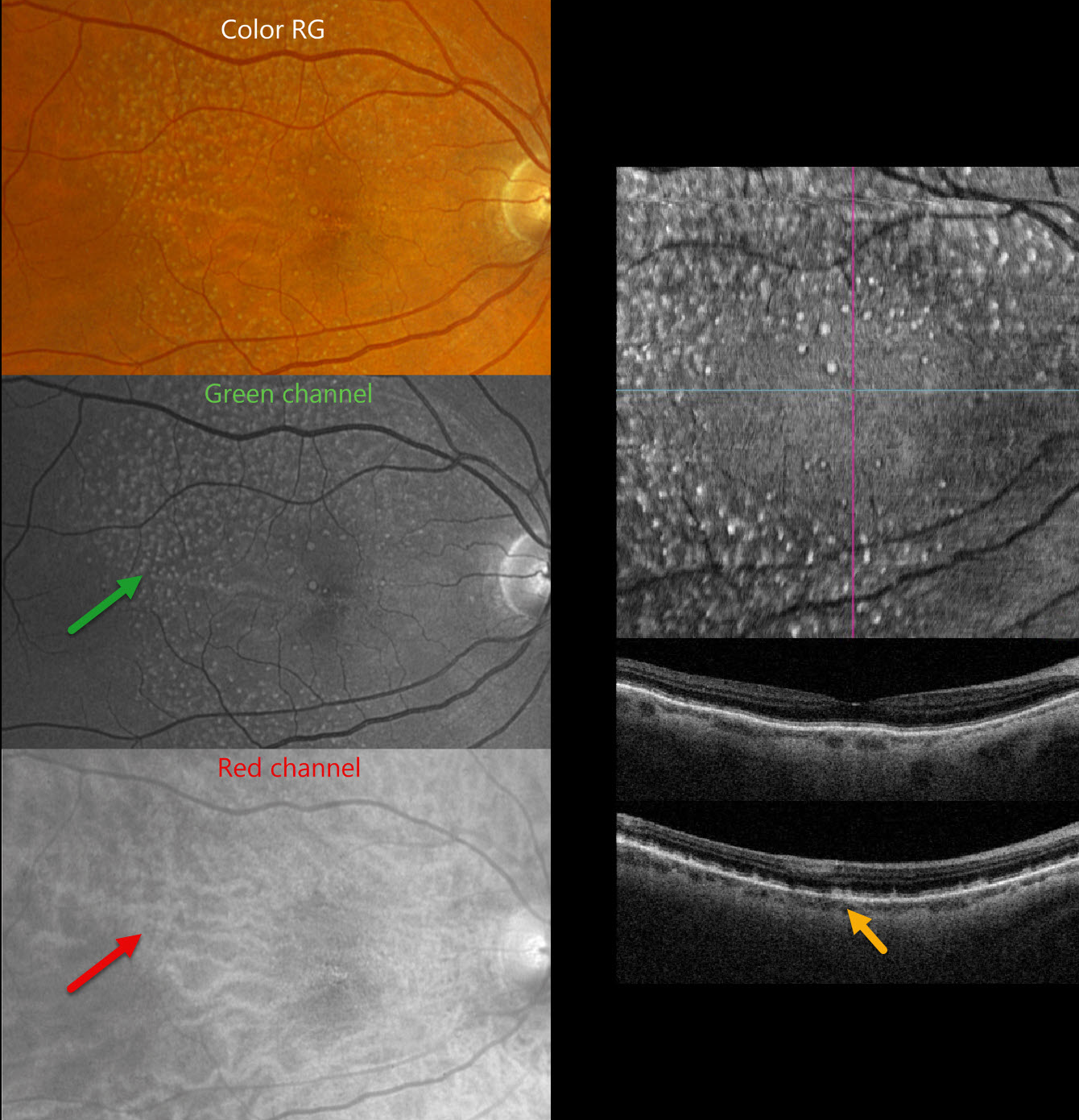
SUBRETINAL DRUSENOID DEPOSITS (SDD)
Originally posted on @retina.rocks January 6, 2025
This 70YO male was referred for age-related macular degeneration (AMD). He was visually asymptomatic, and vision was 20/25 OD and 20/50 OS.
Optos color RG imaging shows extensive, mostly small drusen centered outside the fovea. These lesions are well visualized on the green channel but disappear on the red channel. Triton swept-source OCT shows these ‘drusen’ to represent subretinal drusenoid deposits (SDD), with numerous hyperreflective deposits scattered above the RPE. En face imaging through the outer retina most dramatically shows these SDD.
Learning Points:
SDD are polymorphous, light-gray, interconnected accumulations located above the RPE and are thus best visualized with blue or green illumination, as in our patient. SDD are now considered the third clinical feature for progression to late AMD (Agron et al, Ophthalmology 2022;129:1107-1119), in addition to large drusen and pigment abnormalities as defined in the Beckman classification (Ferris et al, Ophthalmology 2013;120:844-851).
They are present in about one-quarter of older adults with healthy maculas and in more than half of those with early-to-intermediate dry AMD. Clinically, they are often mistaken for conventional drusen and consist of lipid-rich material similar yet distinct from that found in soft drusen.
SDD are localized to rods, unlike typical soft drusen, which are more central and cone-based. As with drusen, SDD are dynamic and can grow, shrink, resolve, or even recur. There is an increased risk for delayed dark adaptation, macular atrophy, and types 2 and 3 MNV. In fact, type 2 neovascularization in the setting of AMD occurs almost solely in eyes with SDD.
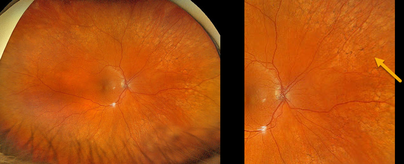
PERIPHERAL RETICULAR DEGENERATION OF THE RPE
Originally posted on @retina.rocks January 2, 2025
This 67YO female shows prominent bilateral peripheral reticular degeneration of the RPE (PRDRPE) with Optos ultra-widefield color RGB imaging.
Learning Points:
The reticular changes at first glance may appear like the ‘bone spicules’ found with retinitis pigmentosa, which are caused by disinhibited RPE cells migrating along retinal capillaries following photoreceptor degeneration. The findings in PRDRPE are part of the age-related macular degeneration (AMD) spectrum, are subretinal and located in the periphery.
A recent meta-analysis by Forshaw et al found PRDRPE to be present in 68-83% of eyes with AMD (Ophthalmology Retina 2019;3:734-743). It is associated with decreased dark adaptation, which may help explain why many patients with AMD have trouble with night vision (Lains et al, AJO 2018;190:113-124).
ANGIOID STREAKS
Anand Temkar and Manish Nagpal
Originally posted on @retina.rocks November 21, 2024
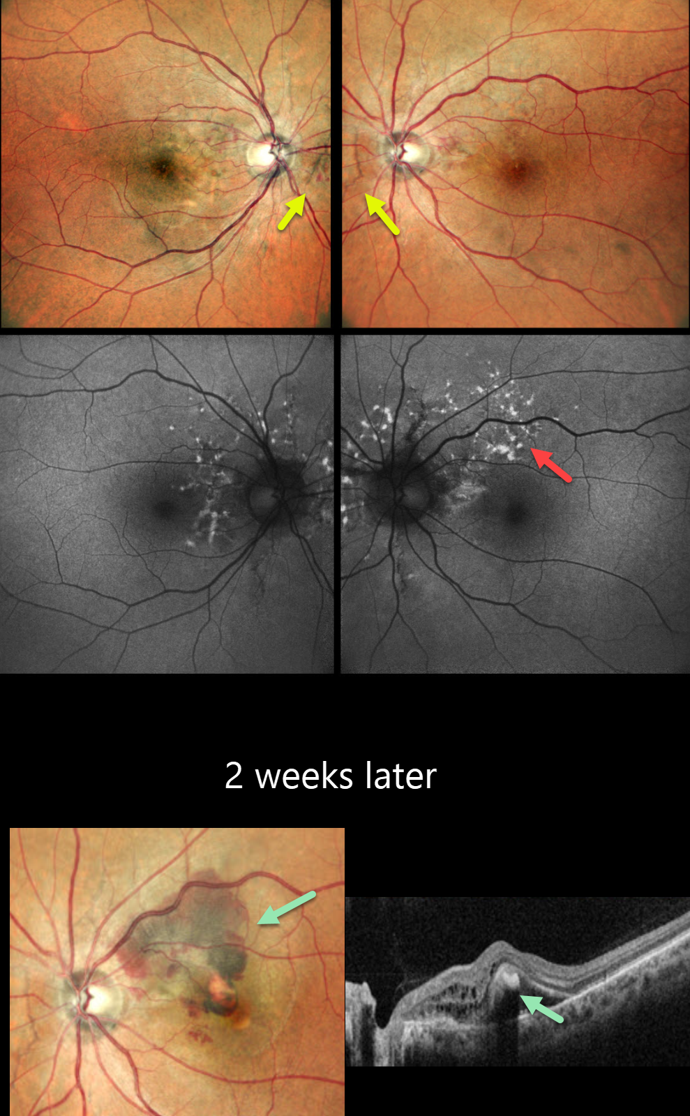
This 45YO male presented for a second opinion for bilateral wavy vision for 2 months. He was diagnosed elsewhere with macular neovascularization (MNV) and received two prior anti-VEGF injections (last 10 days earlier). Vision was 20/20 OU.
Pseudocolor SLO imaging shows subtle bilateral peripapillary angioid streaks associated with a few retinal hemorrhages. Fundus autofluorescence (FAF) shows numerous mostly peripapillary hyper-FAF flecks. Initial observation was recommended.
He returned two weeks later with new symptoms OS, and vision decreased to 20/120. New submacular fluid and blood are noted clinically and on OCT, and monthly anti-VEGF therapy was restarted. Unfortunately, he was lost to follow-up after 3 injections.
Learning Points:
Angioid streaks are orange-yellow, irregular, crack-like dehiscences in Bruch’s membrane associated with atrophic degeneration of the overlying RPE. These patients, especially those with pseudoxanthoma elasticum (PXE), are at significant risk for MNV as occurred in each of our patient’s eyes. MNV responds best to anti-VEGF therapy, and treatment is usually ongoing as with the typical neovascular AMD protocols. However, long-term vision is often poor due to RPE and choroidal atrophy (Mori et al, Graefe’s 2020;258:1863-1869).
Our patient had no family history of PXE or suggestive skin lesions, so skin biopsy or genetic testing was not performed. However, the funduscopic changes and macular dystrophy-like FAF findings are fairly pathognomonic for PXE.
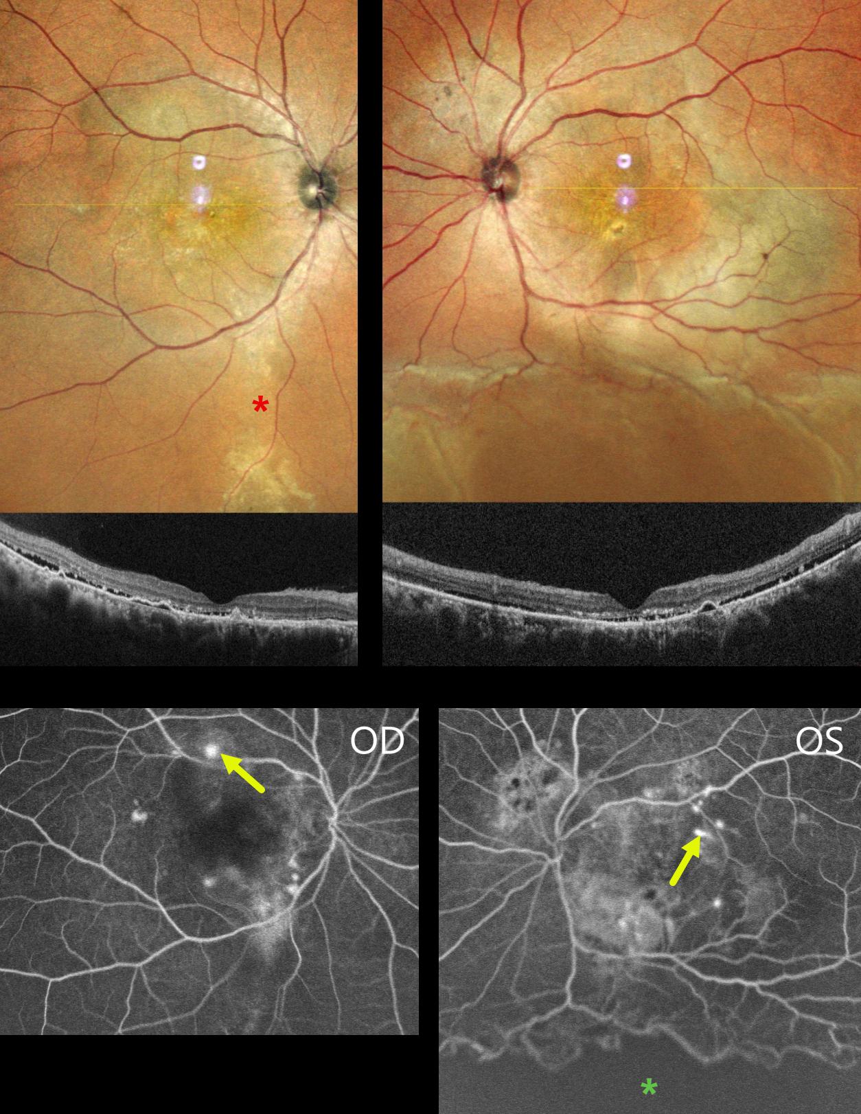
CENTRAL SEROUS CHORIORETINOPATHY WITH EXUDATIVE RD AND COMCOMITANT TB
Anand Temkar, Navneet Mehrotra, and Manish Nagpal
Originally posted on @retina.rocks November 7, 2024
This 39YO male presented with 4 months of bilateral vision loss. He was diagnosed elsewhere with chronic idiopathic central serous chorioretinopathy (ICSC) and was placed on azathioprine and spironolactone 3 months earlier. There was no history of steroid use or injections. Vision was 20/120 OD and 20/200 OS.
MultiColor imaging shows macular pigmentary changes that extend in a gutter inferiorly OD. Diffuse macular pigmentary changes are noted in his left eye that communicate with an inferior exudative retinal detachment. OCT scanning shows shallow subretinal fluid with hyperreflective shed outer segments, outer retinal hyperreflective foci, and RPE detachments. Fluorescein angiography shows bilateral multifocal subretinal blot leakage. A broad area of ischemia is present overlying the inferior detachment of the OS.
Mantoux skin testing (15x18mm) and QuantiFERON-TB Gold testing were positive. Chest CT showed fibrotic strands in the right lung. He was referred for systemic antituberculosis treatment. A week later, his local ophthalmologist reported that his vision and retinal findings improved, but he was then lost to follow-up.
Learning Points:
Our patient presented with severe bilateral idiopathic central serous chorioretinopathy (ISCS) with multifocal leaks in each eye and a secondary exudative detachment in his left eye. There are rare case reports of TB occurring in patients with ICSC, and these are likely coincidental findings (Khan et al, BMJ Case Rep 2017:bcr-2016-216471).
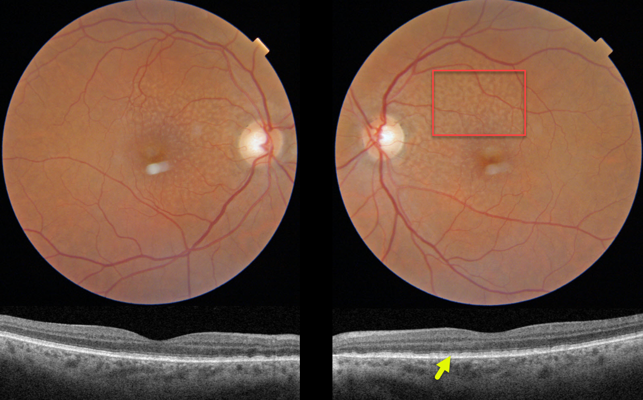
SUBRETINAL DRUSENOID DEPOSITS
Originally posted on @retina.rocks November 6, 2024
This 70YO female was referred for asymptomatic age-related macular degeneration. Vision was 20/30 OD and 20/25 OS.
Triton color imaging of both eyes shows symmetrical ribbon subretinal drusenoid deposits. On swept-source OCT, these lesions appear as hyperreflective material mostly between the RPE and ellipsoid zone (EZ).
Learning Points:
Subretinal drusenoid deposits (SDD) are polymorphous light-gray interconnected accumulations located above the RPE. They are present in about one-quarter of older adults with healthy maculas and in more than half of those with early-to-intermediate dry AMD. They can appear as an array of dots, interconnected reticular bands (as in our patient), or confluent lesions (Suzuki and Spaide, AJO 2014;157:1005-1012). The dot variant is associated with macular neovascularization (MNV) and the confluent variant with macular atrophy (Zhou et al, Ophthalmology 2016;123:1530-1540).
Clinically, they are often mistaken for conventional drusen and consist of similar yet distinct lipid-rich material found in soft drusen. They are localized to rods, unlike typical soft drusen, which are more central and cone-based. As with drusen, SDD are dynamic and can grow, shrink, resolve, or even recur.
Since they are located above the RPE, they are best visualized with blue or green illumination. On OCT, they appear in 3 stages (Zweifel et al, Ophthalmology 2010;117:303-312) growing upwards from the RPE towards the external limiting membrane: stage 1, diffuse deposition of granular hyperreflective material between the RPE and the EZ, stage 2 material alters the EZ contour, and stage 3 conical appearance breaking through the EZ. Rod function seems to be preferentially affected, and there is an associated increased risk of delayed dark adaptation, macular atrophy and types 2 and 3 MNV. In fact, type 2 neovascularization in the setting of AMD occurs almost solely in eyes with subretinal drusenoid deposits.
In addition to large drusen and pigmentary abnormalities, SDD is the 3rd risk factor for progression to late AMD (Agron et al, Ophthalmology 2022;129:1107-1119). For a recent review, see Wu et al, Progress Retinal Eye Research 2022;88;101017.
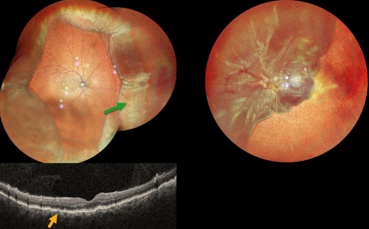
CHOROIDAL DETACHMENT
Anand Temkar and Manish Nagpal
Originally posted on @retina.rocks October 31, 2024
This 52YO male presented with 2 weeks of vision loss OD. He had a complicated ocular history of multiple prior surgeries elsewhere, including cataract surgery OD 2 months earlier and multiple retinal detachment surgeries OS. Vision was 20/30 OD and LP OS. Pressure was 8mmHg with no wound leaks on slit lamp OD.
MultiColor imaging shows encircling peripheral choroidal detachments OD, and OCT shows shallow macular chorioretinal folds. A total retinal detachment is noted OS with bare choroid nasally and inferiorly.
Learning Points:
A choroidal detachment is defined by the abnormal presence of fluid or blood in the suprachoroidal space. Serous choroidal detachments, also known as choroidal effusions, are a frequent complication of glaucoma surgery or following any intraocular procedure. Frank wound leaks usually require surgical repair. Otherwise, conservative management, often with topical/systemic steroids, is recommended.
Since our patient’s surgical wound was secure and the anterior chamber was formed, topical and oral steroids were prescribed.
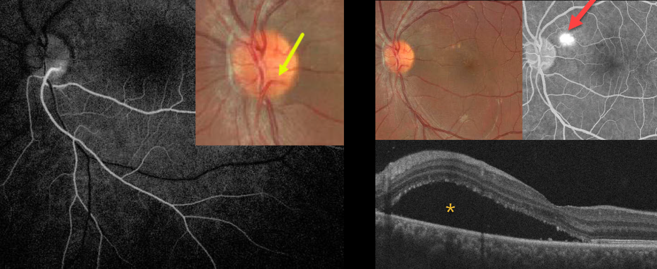
CILIORETINAL ARTERY + CENTRAL SEROUS CHORIORETINOPATHY
Mayelinne García, Luis Montero and Alberto Ruiz
Originally posted on @retina.rocks September 24, 2024
This healthy 25YO male presented with 3 weeks of vision loss in his left eye from active idiopathic central serous chorioretinopathy (ICSC). Vision was 20/20 in his normal OD and 20/110 OS.
A cilioretinal vessel originates within the inferotemporal aspect of the nerve and arches inferiorly. Fluorescein angiography reveals that this cilioretinal artery supplies the entire inferotemporal retina. Color imaging of the macula shows a large serous detachment with some drusen-like changes. This detachment is confirmed on OCT scanning. A blot leak is seen angiographically. Due to the patient’s symptoms, thermal laser was applied to the angiographic leak.
Learning Points:
The inner two-thirds of the neurosensory retina are supplied by the central retinal artery, with the choroid supplying the RPE and photoreceptor layer. Cilioretinal arteries, which arise from the posterior choroidal circulation, are present in up to 50% of individuals and are bilateral in about 25% (Schneider et al, Acta Ophthalmologica 2021;99:e310-e318).
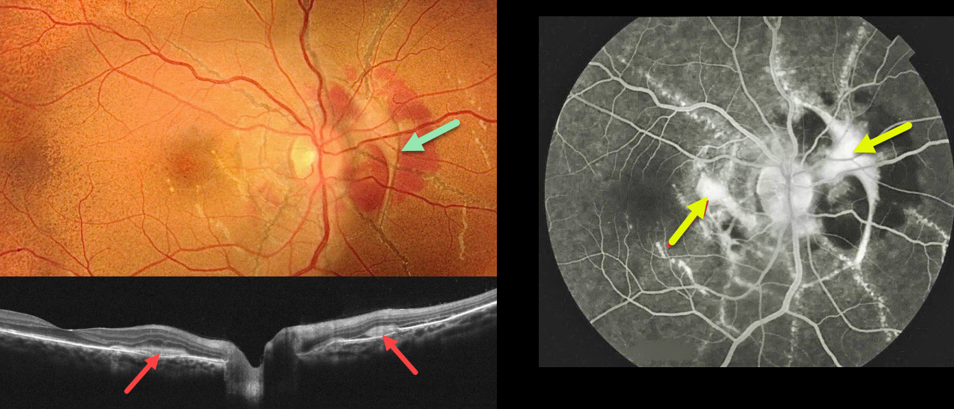
ANGIOID STREAKS
Seif Anwar
Originally posted on @retina.rocks September 19, 2024
This healthy 18YO female presented with recent vision loss in her right eye. Vision was 20/40 OD and 20/30 OS.
Fundus photography shows angioid streaks extending around and radiating outwards from each optic nerve. The posterior pole has a diffuse orange skin (peaux d’orange) appearance. A fibrosed macular neovascularization (MNV) encircles the right nerve and extends into the nasal macula.
On OCT scanning, the hyperreflective MNV is located above the RPE (type 2 lesion) and is associated with minimal exudation. The MNV stains on fluorescein angiography. Intravitreal Lucentis therapy was started in her right eye.
Learning Points:
Angioid streaks are orange-yellow, irregular, crack-like dehiscences in Bruch’s membrane associated with atrophic degeneration of the overlying RPE. The orange skin (peaux d’orange) appearance is caused by a calcified Bruch’s membrane giving a yellow mottled ‘coquille d’oeuf’ (eggshell) appearance that contrasts with the normal orange RPE and choroidal pigmentation (Spaide, Retina 2015;35:392-397). These patients are at significant risk for MNV, as occurred in our patient’s right eye.
Angioid streaks are often missed clinically because their color and orientation blend with the overlying retinal vessels. Individuals with the extensive streaks and peaux d’orange, as seen in our patient, are pathognomonic for PXE, although less extensive streaks can also be seen in sickle cell and age-related macular degeneration (Eye 2023;37:1596-1601).
Our patient gave no family history of PXE. Genetic testing was not possible due to financial reasons, but we will refer her to dermatology for a skin biopsy to definitively establish this diagnosis.
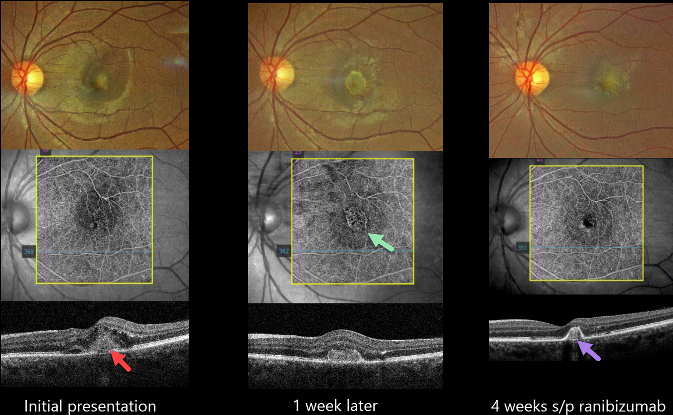
IDIOPATHIC MACULAR NEOVASCULARIZATION (MNV)
Nilesh Kumar
Originally posted on @retina.rocks August 7, 2024
This 20YO female presented with 2 weeks of decreased vision in her left eye. Vision was 20/25 in her normal right eye and 20/60 in her left eye.
Color imaging shows foveal fluid with an amelanotic subfoveal lesion. OCT angiography (OCTA) shows a small subfoveal macular neovascularisation (MNV). An OCT B-scan shows a type 2 MNV (above the RPE) with intraretinal fluid and numerous hyperreflective foci.
Although we recommended an intravitreal ranibizumab biosimilar that day, she refused treatment and returned one week later with 20/200 vision and marked enlargement of the MNV on OCTA.
The injection was given, and 4 weeks later, vision improved to 20/40, with a dramatic treatment response characterized by decreased size and exudation.
Learning Points:
Idiopathic MNV in younger patients was first described by Cleasby in 1976 (AJO 1976;81:590-596). They tend to be type 2 lesions and have a better natural history than typical neovascular AMD (Ophthalmology 1998;105:1816-1820). There is usually an excellent response to anti-VEGF therapy (Kodjikian et al, Retina 2022;42:290-297), as in our patient who initially had both a dramatic rapid enlargement of the untreated lesion, followed by an equally rapid treatment response.
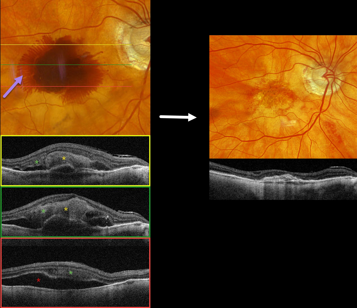
NEOVASCULAR AMD WITH HENLE LAYER HEMORRHAGE
Originally posted on @retina.rocks July 29, 2024
This 97YO male has a history of counting-finger vision in his left eye due to foveal macular atrophy. On 2/29/24, vision was 20/40 in his non-exudative right eye. He presented on 3/12/24 with sudden vision loss OD and new hand motions vision.
Optos color RGB imaging shows fairly dense central subretinal blood, with surrounding radiating blood in the deeper Henle’s layer (outer plexiform layer). Triton swept-source OCT shows variable thick subretinal blood and more inferior subretinal fluid. Henle’s blood is seen as a diffuse hyperreflective obscuration of the outer retinal layers. Vitrectomy with subretinal TPA was performed two days later. Two months later, vision was counting fingers, and the macula was completely free of fluid and blood.
Learning Points:
Radiating deep macular hemorrhages are seen in multiple conditions, including Valsalva and trauma. We have not previously seen these hemorrhages in neovascular AMD. Their unique radiating appearance results from blood accumulating in Henle’s layer. Kon Graversen et al described hemorrhagic unilateral retinopathy, an idiopathic disorder with a similar appearance (Retina 2014;34:483-489).
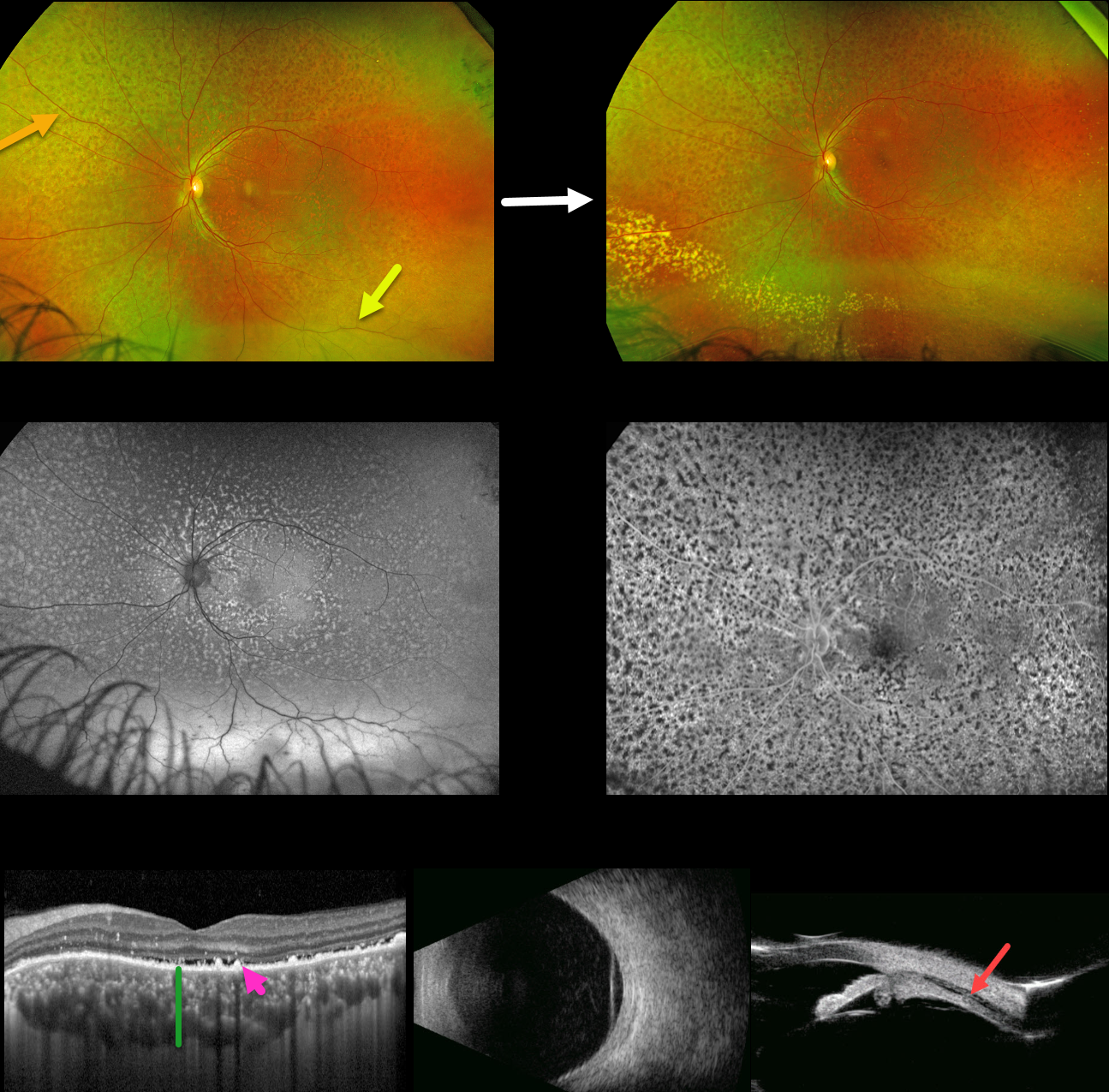
IDIOPATHIC UVEAL EFFUSION SYNDROME vs UNILATERAL BDUMP
Carl-Joe Mehanna
Originally posted on @retina.rocks July 17, 2024
This 48YO healthy male presented with two months of progressive loss of vision in his left eye. Vision was 20/50 OD due to a subcapsular cataract and 20/100 OS with a clear lens. Anterior segments were normal.
Optos color RG imaging of the left eye shows a diffuse orange-red leopard spot appearance to the fundus with an inferior exudative retinal detachment. Fundus autofluorescence (FAF) shows these lesions to be markedly hyper-FAF and block on fluorescein angiography.
Macular OCT shows a thickened choroid, shallow subretinal fluid, and multiple hyperreflective lipofuscin deposits on top of the RPE. B-scan ultrasonography shows a diffusely thickened choroid with overlying retinal detachment, and ultrasound biomicroscopy shows a ciliary body effusion.
Extensive medical workup, including bloodwork and brain MRI, was negative for malignancy or infectious processes (including syphilis, HIV, and TB). He was lost to follow-up for a year, when he presented with resolved subretinal but with new peripheral lipid. He has since been lost to follow-up.
Learning Points:
The idiopathic uveal effusion syndrome (non-nanophthalmic) is a rare entity presenting with idiopathic exudative choroidal, ciliary body, and shifting retinal detachment due to impaired transscleral suprachoroidal drainage caused by an abnormal thickened sclera (Elagouz et al, Surv Ophthalmology 2010;55:134-145). Chronic detachment can cause leopard-spot changes and vision loss.
The differential diagnosis includes leukemic infiltration, central nervous system lymphoma, and bilateral diffuse uveal melanocytic proliferation (BDUMP). The most likely cause of these red subretinal lesions is RPE-engorged, subretinal lipofuscin, which was noted on our patient’s OCT.
The five cardinal signs of idiopathic uveal effusion syndrome include 1) multifocal round/oval red patches at the level of the RPE, 2) early hypofluorescence of these patches, 3) multiple uveal melanocytic tumors with diffuse uveal thickening, 4) exudative retinal detachment, and 5) rapidly progressive cataract (Gass et al, Arch Ophthalmol 1990;108:427-533).
In our patient’s case, we feel the most likely diagnosis is idiopathic uveal effusion syndrome. However, we have seen similar findings in BDUMP, which can present unilaterally (Spaide, Retinal Cases & Brief Reports 2018;12:263-165). However, BDUMP is usually a paraneoplastic process, and our patient’s metastatic workup was negative.
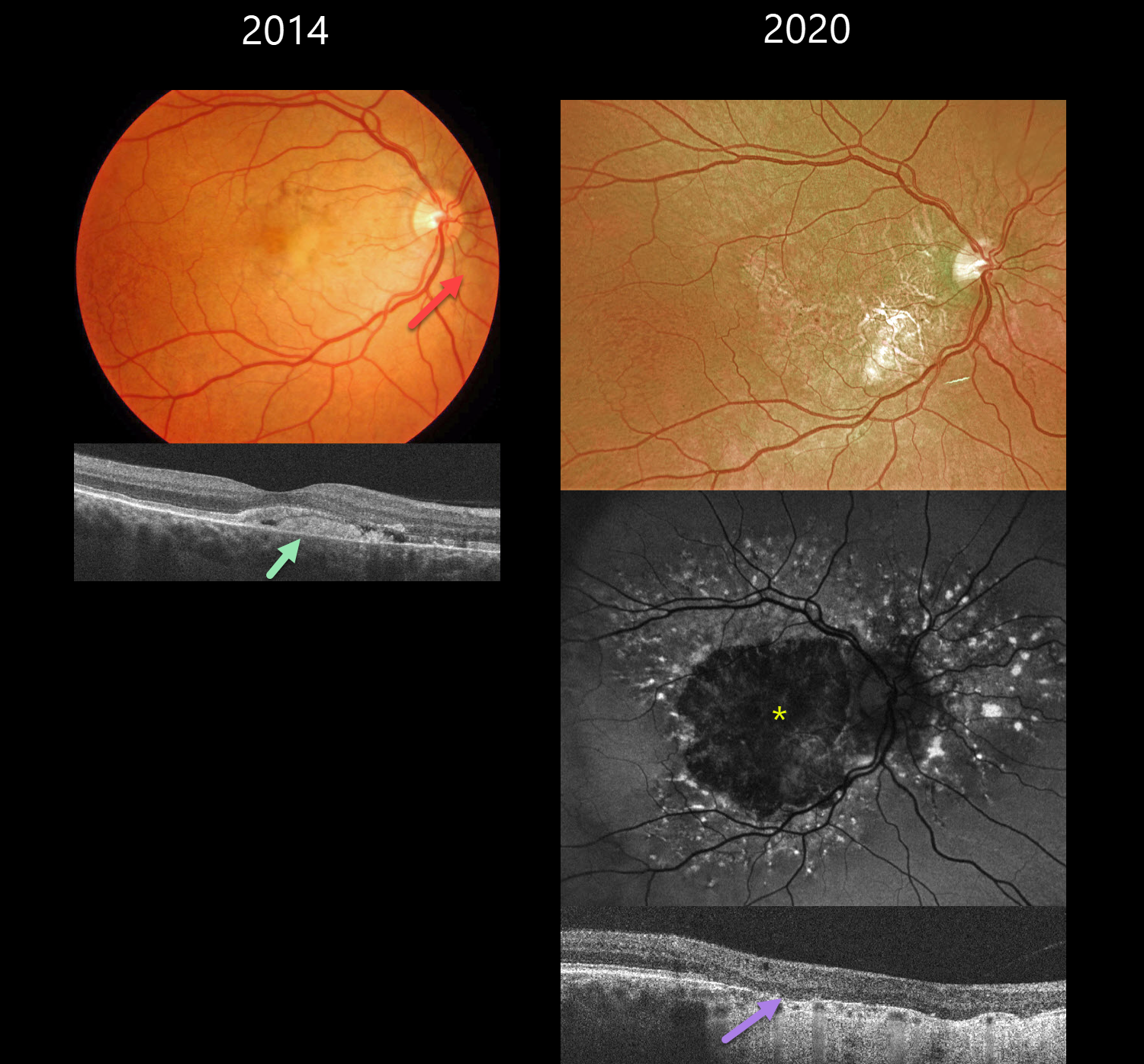
PSEUDOXANTHUM ELASTICUM WITH ANGIOID STREAKS AND PATTERN DYSTROPHY PHENOTYPE
Originally posted on @retina.rocks July 11, 2024
This 39YO male with a known history of pseudoxanthoma elasticum presented on 2/19/14. Findings were bilateral at this, and subsequent visits, so only his right eye images will be shown and discussed.
Color photography shows nasal yellowish subfoveal pigmentary changes with a peaux d’orange appearance temporally. Some peripapillary angioid streaks are noted. OCT shows hyperreflective vitelliform material and fluid below the retina, with some of this material also infiltrating the outer retina.
He has been followed without treatment, and when examined on 7/17/20, vision was 20/200 OD and 20/80 OS. Optos color RG imaging shows a variably atrophic macula. The atrophy on fundus autofluorescence is hypo-FAF. There is a fairly dramatic pattern of hyper-FAF spots radiating out from this atrophy. OCT shows outer retinal atrophy and no fluid. When last examined on 7/6/23, vision was 20/200 OU.
Learning Points:
Retinal findings in PXE most commonly include angioid streaks and a subretinal orange skin (peaux d’orange) appearance, most pronounced in each temporal macula. However, pattern dystrophy-like changes, as in our patient, can sometimes be the predominant phenotype (Murro et al, Graefe’s 2020;258:1881-1892). As with typical pattern dystrophies, the findings are dynamic and best captured with multimodal imaging.
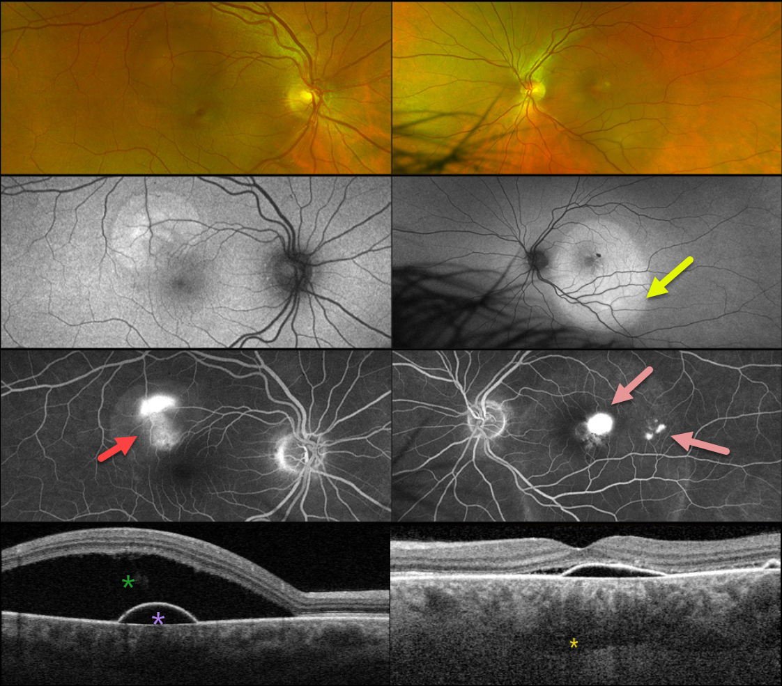
IDIOPATHIC CENTRAL SEROUS CHORIORETINOPATHY WITH LOCULATED OUTER CHOROIDAL FLUID
Originally posted on @retina.rocks June 25, 2024
This healthy 48YO male presented with 1-2 weeks of decreased vision. There was no past ocular history. Vision was 20/25 OD and 20/70 OS.
Optos color RG imaging shows a superotemporal serous detachment OD with a larger serous detachment throughout the left macula. Triton swept-source OCT shows a shallow RPE detachment with some nasal subretinal fluid. Loculated outer choroidal fluid is seen. A more dramatic serous detachment with an underlying RPE detachment is noted OD.
On fundus autofluorescence, these serous detachments show hyper-FAF. Fluorescein angiography shows a smokestack leak OD with multifocal blot leaks OS.
We will initially observe our patient and recommend photodynamic therapy if the fluid fails to resolve over the coming months.
Learning Points:
Initially described by Spaide and Ryan (AJO 2015;160:1211-1216), loculated outer choroidal fluid is found in about two-thirds of patients with idiopathic central serous chorioretinopathy (ICSC). It is best appreciated with swept-source or enhanced depth imaging OCT.
This fluid most commonly interdigitates with the larger outer choroidal vessels, as in our patient, but can rarely appear in the suprachoroidal space. It is more common with a thicker choroid and sclera (AJO 2022;235:197-203).
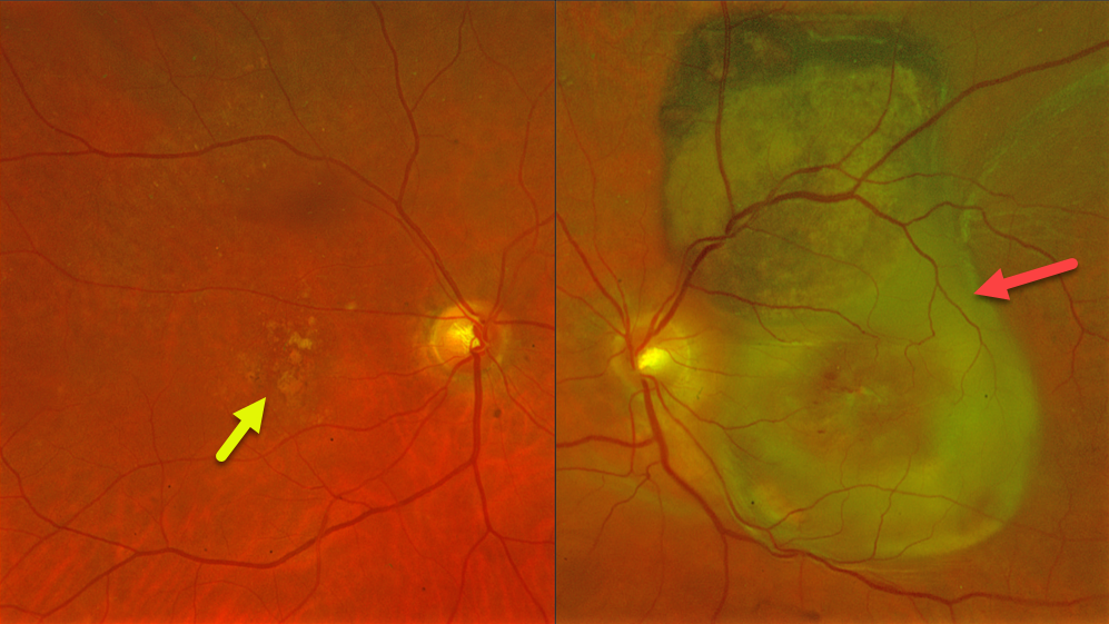
PSEUDO-UVEAL MELANOMA FROM NEOVASCULAR AMD SUBRETINAL FIBROSIS
Fraser McKay
Originally posted on @retina.rocks June 20, 2024
This 78YO female presented with at least several months of vision loss in her left eye. Vision was 20/40 OD and light perception OS. She was examined 2 years earlier with 20/400 vision OS from macular atrophy.
Optos color RG imaging OS shows a variably pigmented and slightly thickened subretinal lesion extending from the superior macula into the superior midperiphery. Subretinal fibrosis extends through the macula, along with retinal blood dots. Intermediate AMD with mixed drusen was noted OD.
Learning Points:
Suprachoroidal, subretinal, or sub-RPE blood can rarely simulate a uveal malignant melanoma. We suggested a trial of monthly anti-VEGF therapy for this treatment naïve neovascular AMD lesion, although the visual prognosis remains quite guarded given the extent of submacular fibrosis.
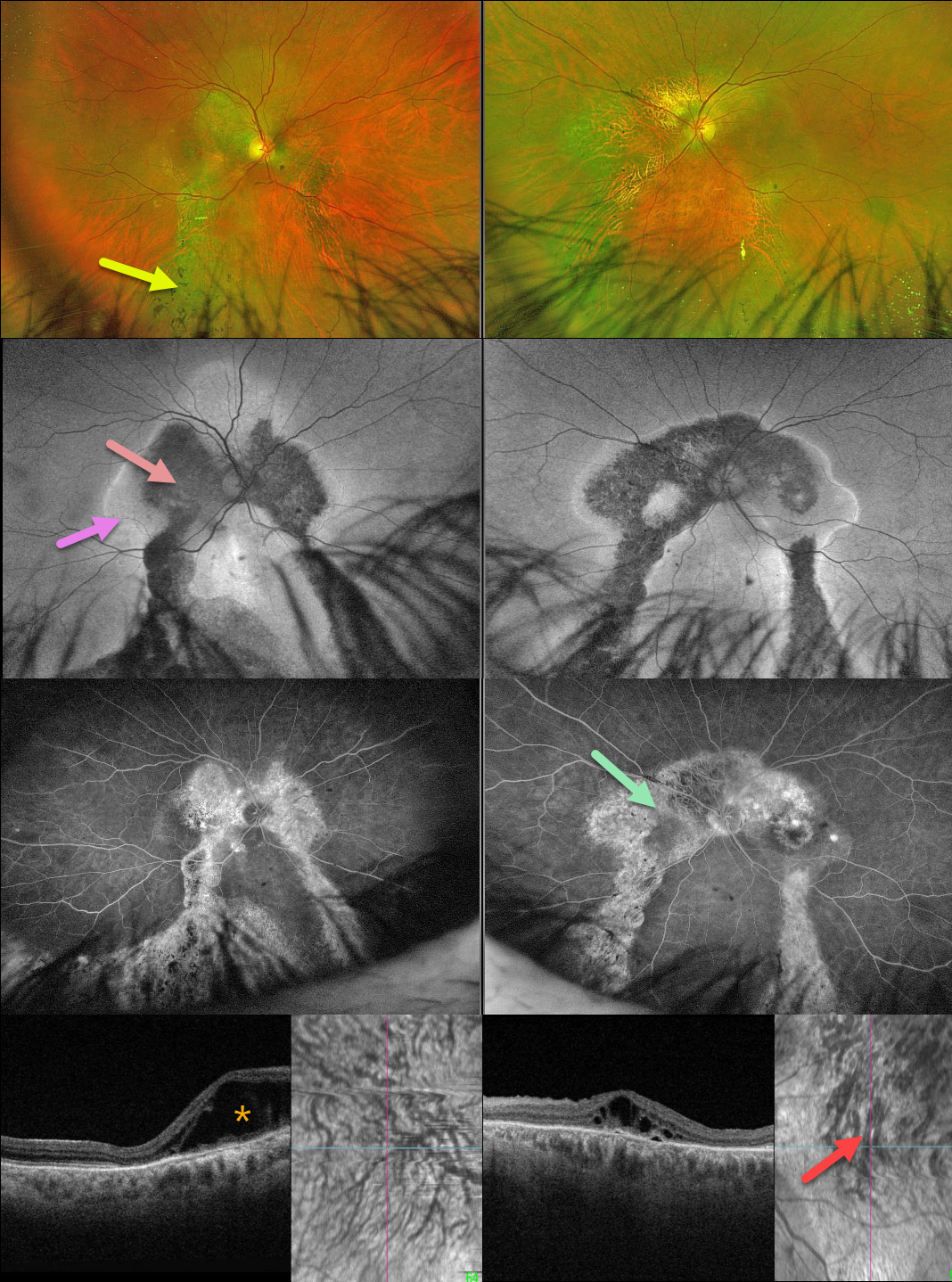
POSSIBLE PACHYCHOROID SPECTRUM
Originally posted on @retina.rocks June 11, 2024
This healthy 62YO male presented with a several-year history of bilateral vision loss. There was no family history of eye disease. Vision was 20/400 OU.
Optos color RG imaging shows bilateral, symmetrical peripapillary and macular pigmentary changes extending into the gutters towards the inferior peripheries. Intraretinal pigment migration (spicules) is noted inferiorly.
On fundus autofluorescence (FAF), these areas are hypo-FAF centrally with hyper-FAF along their margins, and exhibit window defects on fluorescein angiography.
Outer retinal fluid is noted nasally OD and centrally OS on Triton swept-source OCT. En face OCT shows bilateral choroidal pachyvessels with anastomoses. Genetic testing was completely negative.
Learning Points:
Gutters are commonly seen in pathology with chronic subretinal leakage, including central serous retinopathy, choroidal hemangioma, choroidal nevus, and uveal melanoma. These pigmentary changes are usually best imaged with FAF. Our patient’s OCT certainly shows pachychoroid features, but the symmetrical double gutters in each eye would be quite unusual for central serous retinopathy.
Our patient’s gutters are somewhat reminiscent of a patient we have previously posted with the Senior-Løken Syndrome, although this is associated with renal failure and an NPHP1 mutation, which our patient did not have.
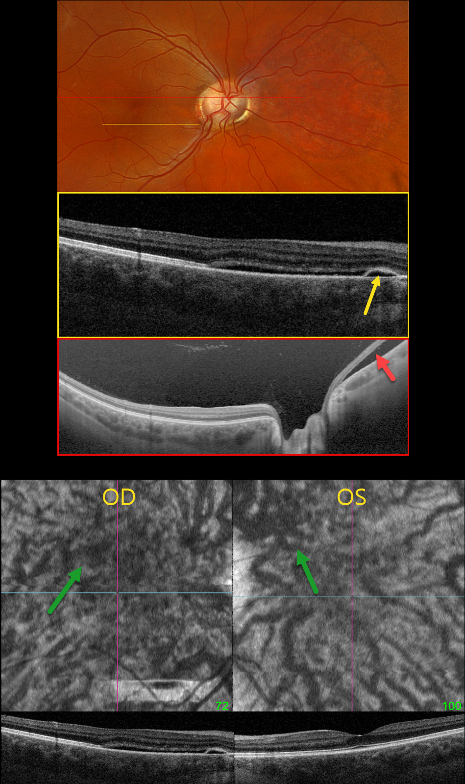
IDIOPATHIC CENTRAL SEROUS CHORIORETINOPATHY WITH INCIDENTAL OPTIC NERVE PIT
Originally posted on @retina.rocks May 30, 2024
This healthy pregnant 32YO female presented with recent mild central vision loss in her right eye. Vision was 20/40 OD and 20/30 OS.
Optos color RGB imaging shows an anomalous right optic disc with an optic pit both nasally and temporally. Triton swept-source OCT shows both pits and subretinal fluid extending from the nerve nasally. OCT through the macula shows shallow nasal subretinal fluid extending into the macular center, along with a tiny nasal RPE detachment. This fluid does not communicate with the nerve or temporal pit. Optos Silverstone swept-source OCT through the posterior pole shows the subretinal fluid nasal to the nerve. Macular en face and B-scans show a thickened choroid with anomalous, thickened, and tortuous vessels.
Our patient’s symptoms are from idiopathic central serous retinopathy (ICSC), most likely related to her pregnancy. The macular fluid does not communicate with the temporal disc pit, and her macular OCT findings are classic for central serous. Fluorescein angiography was not performed due to her pregnancy. Although she has optic pit ‘maculopathy,’ this is in the nasal retina from her temporal disc pit. Observation was recommended.
Learning Points:
An optic nerve pit is a rare (about 0.1% prevalence) congenital anomaly thought to be caused by incomplete closure of the optic fissure during gestation. Pits are most commonly located within or along the inferior-temporal disc margin and usually appear as a gray depression. While most optic pits are asymptomatic, they can cause vision loss by forcing fluid into the inner and outer retina, and ultimately into the subretinal space (optic pit maculopathy). The source of the fluid remains a topic of ongoing debate, with the fluid originating either from the vitreous or the subarachnoid space.
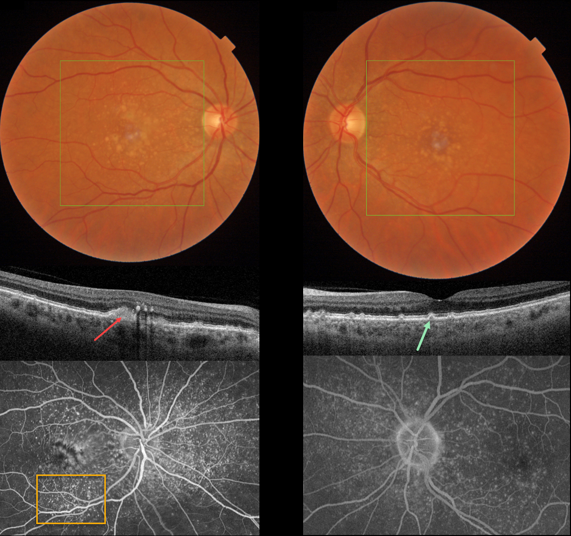
CUTICULAR DRUSEN WITH VITELLIFORM LESION
Originally posted on @retina.rocks May 28, 2024
This 57YO female was referred for asymptomatic macular changes. Vision was 20/20 bilaterally.
Triton color imaging shows bilateral confluent central soft drusen and smaller, more peripheral macular drusen. Swept-source OCT shows bilateral, small arrowhead-shaped cuticular drusen. A hyperreflective vitelliform lesion is noted OD, along with some hyperreflective deposits in the outer plexiform layer. Optos fluorescein angiography shows extensive bilateral hyperfluorescent cuticular drusen along with a staining vitelliform lesion OD.
Learning Points:
Originally described by Gass as basal laminar drusen (AJO 1985;99:445-459), cuticular drusen were initially thought to represent nodular thickening of the RPE basement membrane. They appear clinically as numerous, round, yellow-white ‘hard drusen’, and on OCT as small, arrowhead-shaped drusen located above Bruch’s membrane and below the RPE (Spaide and Curcio, Retina 2010;30:1441-1454).
Our patient’s angiographic appearance echoes Dr. Gass’ original description of “a beautiful stars-in-the-sky or Milky-Way pattern” of hyperfluorescence. Cuticular drusen are classically associated with avascular vitelliform lesions (Finger et al, Retina 2010;30:1455-1464), as in this case, but can also develop macular neovascularization.
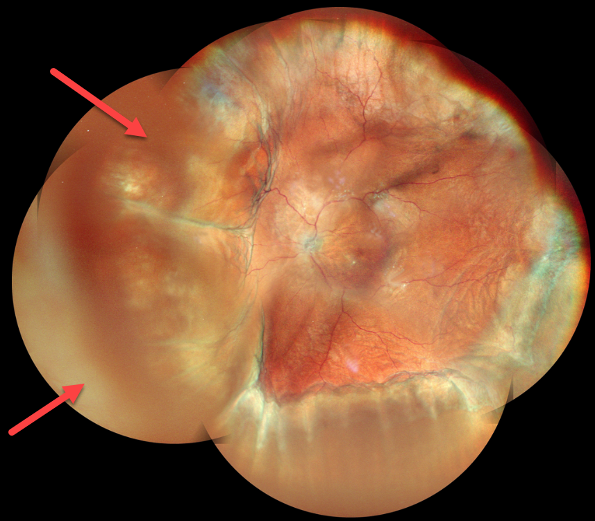
CHOROIDAL DETACHMENT
Anand Temkar and Manish Nagpal
Originally posted on @retina.rocks May 23, 2024
This 63YO male underwent glaucoma filtering surgery with mitomycin C one month earlier at an outside practice. Vision was 20/30, and intraocular pressure was 4. Montage color imaging shows extensive choroidal detachments. He was immediately lost to follow-up.
Learning Points:
A choroidal detachment is defined by the abnormal presence of fluid or blood in the suprachoroidal space. Serous choroidal detachments, also known as choroidal effusions, are a frequent complication of glaucoma surgery.
Several causative mechanisms are believed to be at play: hypotony allows fluid to accumulate in the suprachoroidal space, and inflammation increases the choroidal permeability. The detached ciliary body may also produce less aqueous humor, thus creating a self-perpetuating cycle. Often, observation is sufficient, but topical steroids and cycloplegics can be helpful in facilitating resolution.
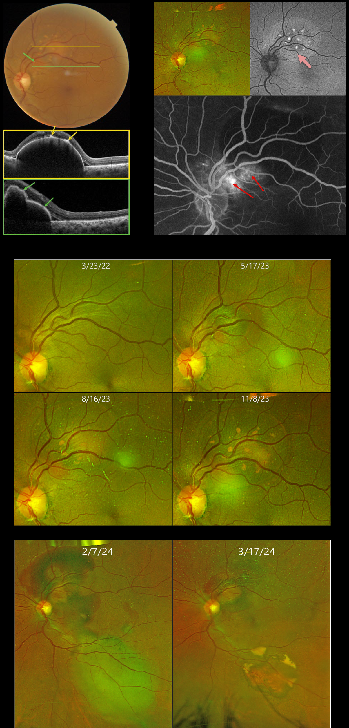
POLYPOIDAL CHOROIDAL VASCULOPATHY
Originally posted on @retina.rocks May 21, 2024
This 59YO female was initially referred to us on 3/23/22 with a 1-year history of vision loss in her right eye. Vision was counting fingers OD and 20/25 OS.
Over the past 10 months, a gradually enlarging RPE detachment developed in the left superonasal macula. On 11/8/23, Triton swept-source OCT confirms a highly elevated RPE detachment. The orange lipofuscin deposits seen on color imaging appear as hyperreflective lesions on OCT. Scanning through the reddish polyp shows a multi-lobulated elevation of the RPE. The lipofuscin deposits markedly hyper-autofluoresce. The large variably staining/leaking polyp creates a notch along the inferonasal edge of the PED.
Due to the patient’s history of a large macular hemorrhage resulting in counting fingers vision in her fellow eye, half-fluence photodynamic therapy (PDT) was applied to the polyp in her left eye. However, one month following PDT on 2/27/24, there was increased blood around the treated superior lesion, with new blood inferiorly.
Multiple full-fluence PDT applications were then applied to the old and new lesions. Six weeks later, the patient’s fundus was dramatically improved, with near-complete resolution of all blood and fluid. Vision was 20/20, and we will continue to follow her closely.
Learning Points:
Polypoidal choroidal vasculopathy (PCV) is a variant of type 1 macular neovascularization. PCV is likely distinct from age-related macular degeneration, occurring in a different population (more common among African Americans and Asians), with a relative lack of drusen and a thicker choroid. The sub-RPE vessels were classically described using indocyanine green angiography, although newer literature suggests that OCT is likely just as effective at diagnosing these vascular polyps (Permadi et al., Int J Retina Vitreous 2022;8;14).
Historically, these lesions were believed to be somewhat resistant to anti-VEGF monotherapy, often requiring combined treatment with photodynamic therapy. Newer data from the PLANET study suggest that Eylea monotherapy is also successful (AJO 2019;204:80-89).
Originally described by Gass (Retina 1984;4:205-220), a notched PED is a sign of an occult MNV. The notch represents a type 1 MNV, which tethers the overlying RPE from the surrounding more highly elevated PED. Hydrostatic leakage from the MNV may contribute to the larger PED.
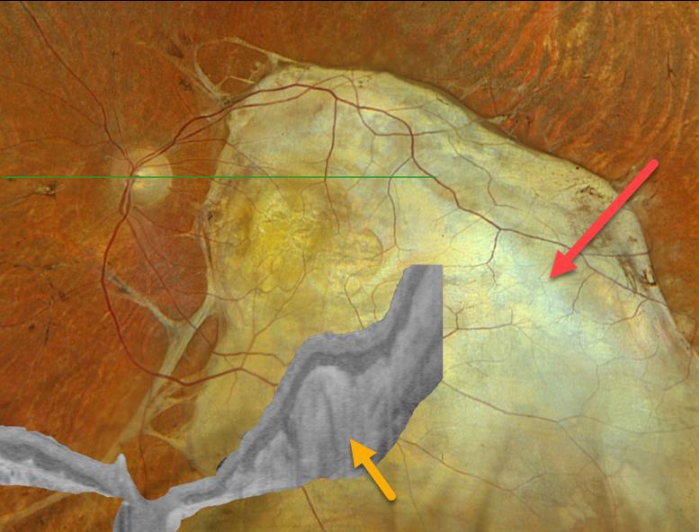
SUBRETINAL FIBROSIS
Originally posted on @retina.rocks April 30, 2024
This 69YO male has stable vision loss in his left eye from an age-related disciform scar.
Optos color RGB imaging shows massive subretinal fibrosis occupying virtually the entire macula, which extends into the inferior midperiphery. Optos Silverstone OCT shows markedly thickened hyperreflective subretinal fibrosis. Vision was remarkably 20/400. Continued observation was recommended.
Learning Points:
Disciform scars were the norm for untreated neovascular AMD (nAMD) prior to the anti-VEGF era that began in the mid-2000s. New-onset nAMD diagnosed and treated with timely anti-VEGF dosing virtually never goes on to develop these lesions. There may be progressive macular atrophy following years of therapy, but not this type of fibrotic scarring.
For an extensive discussion on the SD-OCT findings of these age-related fibrotic lesions, see the AOS thesis by Souied et al (AJO 2020;214:151-171).
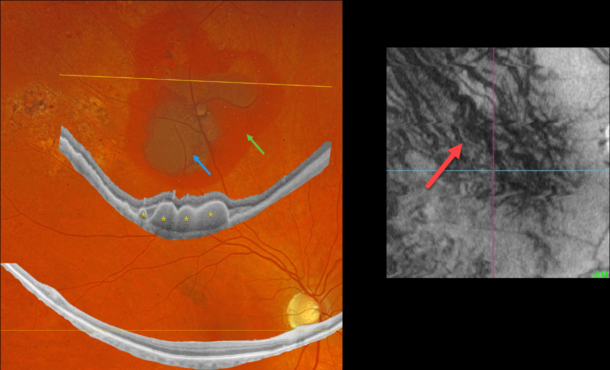
PERIPHERAL EXUDATIVE HEMORRHAGIC CHORIORETINOPATHY
Originally posted on @retina.rocks April 17, 2024
This 64YO diabetic male presented with asymptomatic retinal findings in his left eye. Vision was 20/40 OU.
Optos color RGB imaging shows superior subretinal and sub-RPE blood. Optos Silverstone widefield swept-source OCT through this lesion shows prominent multiple choroidal polyps. Subretinal pigmentary clumping temporal to the blood likely represents findings from absorbed subretinal blood. Moderate non-proliferative retinopathy is noted.
En face imaging is quite abnormal with vortex anastomoses and dilated choroidal vessels.
Observation was recommended.
Learning Points:
Pachychoroid, meaning “thick” choroid, is secondary to large, dilated Haller vessels. Increased oncotic/mechanical pressure from these dilated vessels can cause RPE dysfunction, serous detachments, and macular neovascularization.
What defines a ‘pachy’ choroid remains quite unclear (Spaide, Retina 2021;41:231-237), since pachychoroid-related findings can be present despite a ‘normal’ macular choroidal thickness as in our patient.
Spaide recently described venous overload choroidopathy, a unifying concept that better explains the underlying pathogenesis (Progress Retinal Eye Research 2022;86:100973).
The pachychoroid spectrum also includes ectopic neovascularization (aka peripheral exudative hemorrhagic chorioretinopathy), with the choroid showing progressive thickening towards the temporal periphery (Shroff et al, Retina 2021;41:1518-1525).
Our patient’s widefield OCT beautifully shows the choroidal polyps responsible for the peripheral subretinal bleeding.
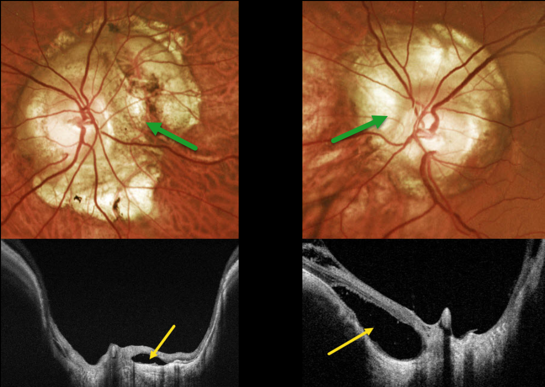
OPTIC NERVE CONUS + PERIPAPILLARY RETINAL DETACHMENT
Originally posted on @retina.rocks March 13, 2024
This 42YO male with high myopia was referred for asymptomatic optic nerve changes. Vision was 20/30 OD and 20/25 OS.
Optos color RG imaging shows bilateral peripapillary atrophy. Clinically, this scarring is markedly excavated from the surrounding retinal plane. Triton swept-source OCT confirms peripapillary staphyloma. Localized retinal detachments are noted bilaterally, particularly within the nasal aspect of atrophy around the left nerve. Within these detachments, there is variable loss of RPE, absence of chorioid, and variable retinal disorganization.
Learning Points:
An optic nerve conus, most commonly found in high myopes, is a staphylomatous-like depression of the optic nerve and surrounding tissue.
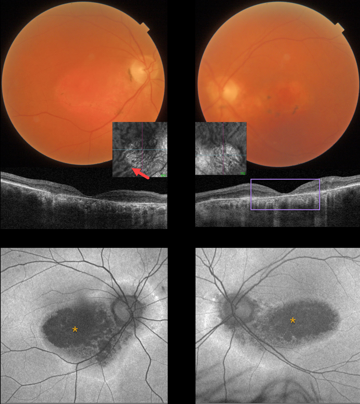
HYDROXYCHLOROQUINE TOXICITY + PACHYCHOROID
Originally posted on @retina.rocks March 11, 2024
This 67YO female has a known history of Plaquenil toxicity. Her bilateral 20/200 vision and macular findings have been stable since discontinuing this medication 30 years ago.
Triton color imaging and swept-source OCT show classic findings for severe hydroxychloroquine toxicity, including an oval area of foveal pigment loss and outer retinal atrophy.
Most patients will have preserved central foveal pigment giving a bulls-eye appearance, although our patient has loss of outer retina and RPE throughout. Interestingly, choroidal en face imaging shows bilateral dilated choroidal pachyvessels.
On fundus autofluorescence (FAF), the central maculas are hypo-FAF, although an atypical hyper-FAF ring extends around each optic nerve.
Learning Points:
Current screening guidelines should make Plaquenil toxicity, as found in our patient, a thing of the past. Last revised in 2016, the American Academy of Ophthalmology (AAO) recommends that a baseline exam be performed before starting the medication, with annual screenings beginning at least 5 years after initiating treatment, unless major risk factors are present. However, in practice, patients are usually screened yearly once they are placed on this medication.
Optical coherence tomography (OCT) is performed annually to assess outer retinal findings, including ellipsoid zone loss. Humphrey 10-2 visual field testing (24-2 for Asians, since their macular involvement is usually more peripheral) is also needed annually, as about 10% of patients will have field loss despite normal examinations and OCT testing.
Plaquenil dosing should also be based on real, not ideal, weight to better predict the optimal dose. See Marmor et al Ophthalmology 2016;123:1386-1394 for the full screening guidelines.
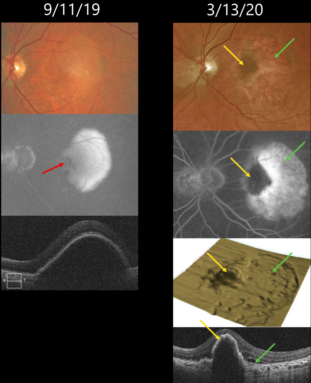
RPE TEAR
Originally posted on @retina.rocks February 26, 2024
This 72YO male was followed for many years with stable intermediate AMD. On 9/11/19, he presented with a new symptomatic retinal pigment epithelial detachment (PED) with 20/30 vision. Fluorescein angiography shows an uneven-filling PED with a nasal hypofluorescent notch. A large PED is confirmed on OCT scanning.
He initially did well with monthly Eylea injections. While on treat-and-extend therapy, he presented on 3/13/20 without new visual symptoms, although his vision had decreased to 20/200.
Optos color RG imaging shows a new, large temporal RPE tear. On multimodal imaging, the bed of the tear is depigmented on color imaging, hyperfluorescent on fluorescein angiography (window defect), and shows a crater on the 3D OCT RPE map, and shows bare Bruch’s membrane on B-scan.
The retracted edge of the tear is hyperpigmented on color imaging, hypofluorescent on fluorescein angiography (blockage), an elevated mound on the 3D OCT RPE map, and a rippled, elevated PED on B-scan.
We subsequently decided on PRN therapy, with the last injection given on 3/12/21. When last examined, vision was stable at 20/400 with a stable dry lesion.
Learning Points:
A tear in the RPE is a fairly common occurrence in PED associated with type 1 macular neovascularization (MNV) located below the RPE. The neovascularization can spontaneously contract or involute following treatment. It then puts traction on the overlying RPE, which can then rip at the opposite edge of the PED.
Like a cheap window blind, the torn RPE retracts and scrolls towards the MNV. This leaves a depigmented region where the RPE was and is now absent, along with a pigmented, relatively straight subretinal lesion from the scrolled RPE edge.
Originally described by Gass (Retina 1984;4:205-220), a notched PED is a sign of an occult MNV. The notch represents a type 1 MNV, which tethers the overlying RPE from the surrounding more highly elevated PED. Hydrostatic leakage from the MNV may contribute to the larger PED.
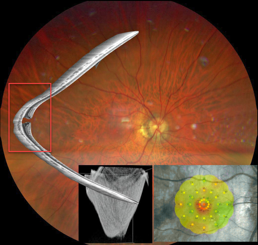
STAPHYLOMA + MACULAR HOLE
Barbara Parolini and Veronika Matello
Originally posted on @retina.rocks February 13, 2024
This Eyecare Clinic (in Brescia, Italy) case was submitted by Barbara Parolini, Veronika Matello, Giulia Freschi, and Roberta Penzani.
This 48YO highly myopic male presented for a sudden drop in vision. Vision was 20/100 OD and 20/20 in OS.
Color photography shows a blunted macular reflex. Widefield OCT shows a conical-shaped staphyloma of the entire posterior pole with a full-thickness macular hole. Microperimetry shows a decreased but not absent foveal sensitivity.
According to the Myopic Traction Maculopathy Staging System (Parolini et al, Eur J Ophthalmol 2021;31:1299-1312), this eye is in Stage 1c. The left eye showed a similar staphyloma but without a macular hole (not shown). The patient declined surgery.
Learning Points:
A macular staphyloma consists of a concave posterior bowing of the eye wall in patients with high myopia. These myopic patients also have a markedly thinned choroid throughout the posterior pole.
Due to stretching of the posterior retinal structures, these eyes are at increased risk of numerous findings, including macular schisis/traction and macular neovascularization.
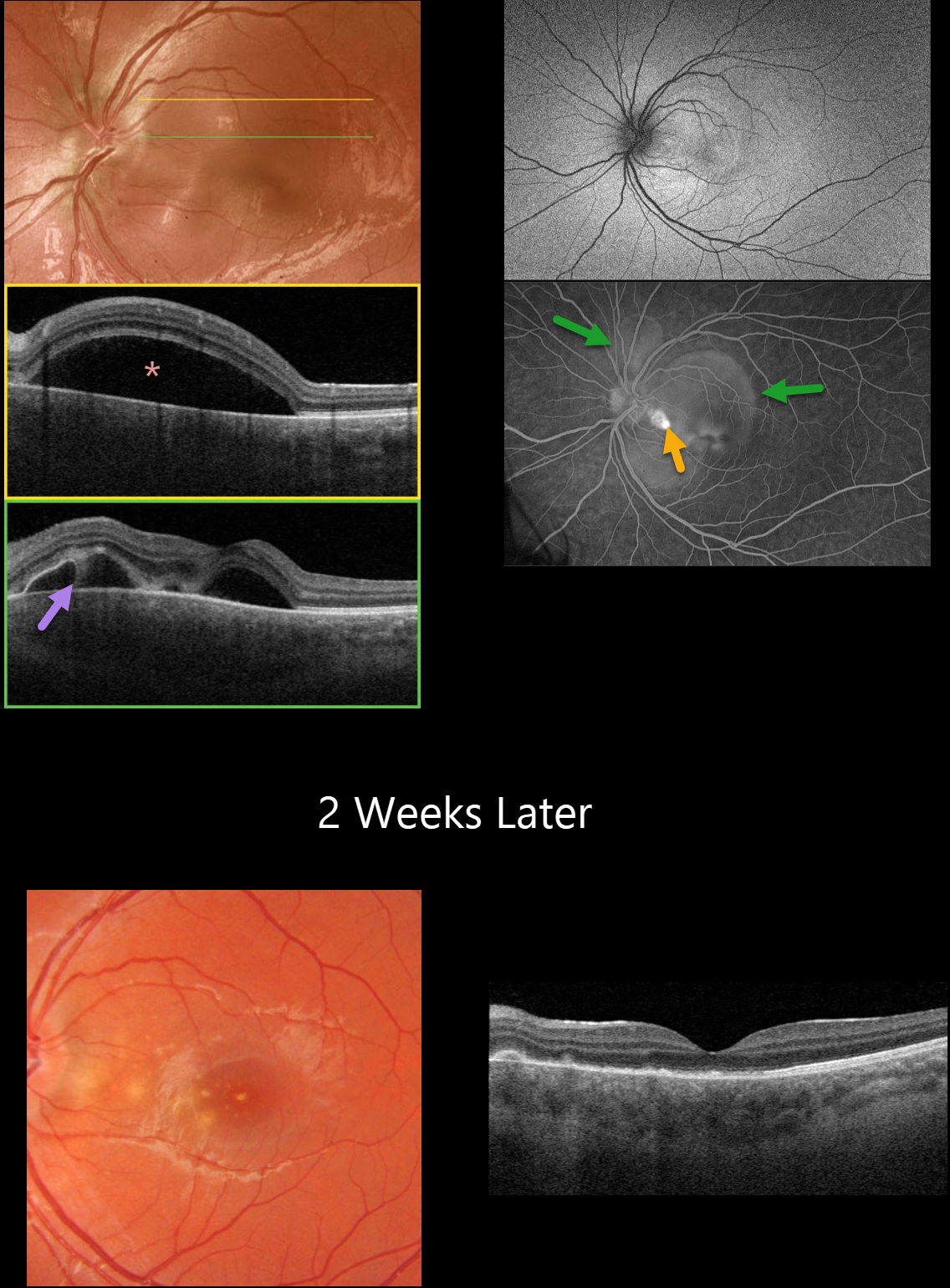
POST-COVID FIBRINOUS CENTRAL SEROUS CHORIORETINOPATHY
Originally posted on @retina.rocks January 31, 2024
This 12YO boy presented with a few days of painless vision loss in his left eye. He had COVID one week earlier. There was no prior medical history. He was on no systemic medications. Vision was 20/30 in his normal right eye and 20/50 in his left eye.
Optos color RG imaging of his left eye shows a large macular serous detachment. Triton swept-source OCT confirms the serous detachment along with subretinal fibrin.
Fundus autofluorescence (FAF) shows faint peripapillary and macular hyper-FAF, and fluorescein angiography shows pooling beneath a nasal macular RPE detachment and larger areas of pooling within the multifocal serous detachments.
When he returned following 2 weeks of observation, the fluid and BALAD completely regressed with secondary drusen-like changes. Variable outer retinal atrophy is seen on OCT.
Learning Points:
Prior case reports of idiopathic central serous chorioretinopathy (ICSC) following COVID have been reported, although these have developed in patients who were also treated with systemic corticosteroids.
Fuganti et al reported 2 patients, both treated with systemic steroids, with post-COVID ICSC with BALAD (AJO Case Reports 2022;28;101690). We suspect our patient’s findings may have been COVID-related, given the atypical presentation and rapid resolution of the BALAD and serous fluid.
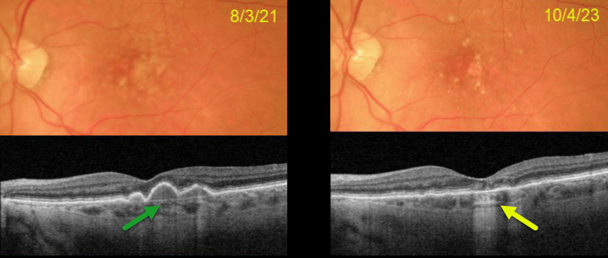
FLATTENED DRUSEN CAUSING SECONDARY MACULAR ATROPHY
Originally posted on @retina.rocks January 8, 2024
This 77YO female presented on 8/3/21 with 20/30 vision in her left eye. Optos color RG imaging shows confluent central soft drusen, which are confirmed on Triton swept-source OCT.
On 10/4/23, vision was 20/25. The central drusen flattened with secondary drusenoid atrophy. Continued observation was recommended.
Learning Points:
Drusen commonly remodel over time and their collapse, as in our patient, can lead to drusenoid atrophy of the RPE (Filho et al, Retina 2013;33;1558-1566). Vision may or may not decrease following regression of drusen.
In our experience, drusenoid atrophy remains stable, unlike primary age-related macular atrophy, whose radius enlarges linearly over time (Shen et al, Ophthalmology Retina 2020;4;899-910). We therefore do not consider intravitreal complement inhibitors for these patients.
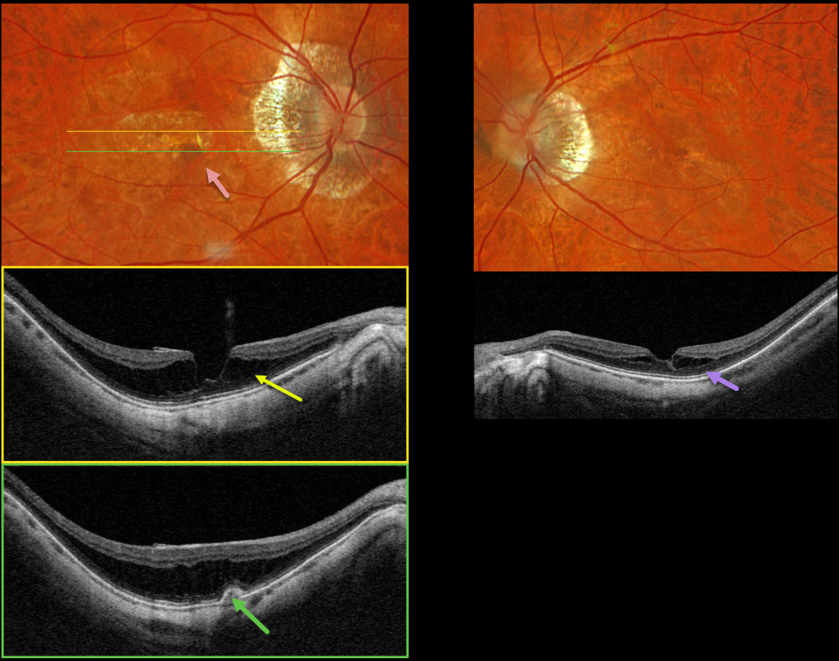
MYOPIC MACULAR NEOVASCULARIZATION
Originally posted on @retina.rocks January 1, 2024
This 72YO female recently moved to our area and self-referred herself to establish new retinal care. One year earlier, she received a series of five monthly anti-VEGF injections OD for myopic macular neovascularization (MNV), and felt that her vision was stable since the last injection about 6 months earlier. Vision was 20/60 OD and 20/40 OS.
Optos color RGB imaging OD shows myopic peripapillary atrophy, central macular pigment loss, and a pigmented subretinal lesion.
Triton swept-source OCT through the macular center shows a shallow staphylomatous posterior bowing of the sclera with overlying outer macular schisis. The choroid is diffusely thinned. Scanning the pigmented lesion reveals a small type 1 MNV (below the RPE) without fluid. Imaging of her left eye shows much milder, central schisis in the outer plexiform layer. Observation was recommended.
Learning Points:
In our experience, myopic MNV presents quite differently from those with neovascular AMD. Significant subretinal blood, subretinal fluid or intraretinal fluid are rarely seen.
Often, an active MNV will present as a new dot of subretinal blood or pigment. Occasionally, there will just be symptomatic vision loss or metamorphopsia without clinical or OCT findings.
Anti-VEGF therapy is effective, and these lesions can often be managed with PRN therapy (Sakata et al, Retina 2023;43:1863-1871) as in our case.
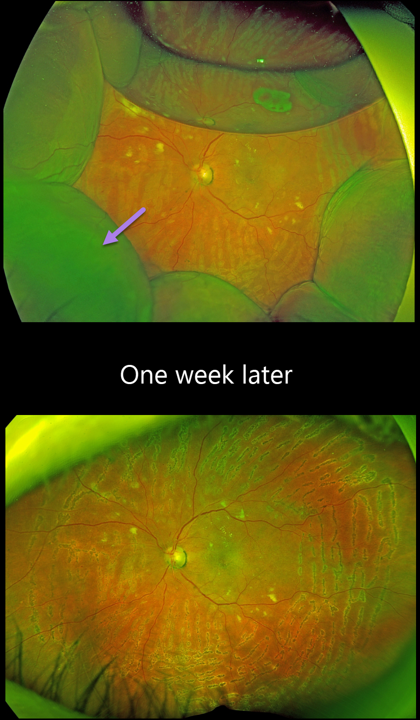
CHOROIDAL DETACHMENT
Originally posted on @retina.rocks December 11, 2023
This 66YO female presented for her postoperative exam following an uneventful pars plana vitrectomy one day earlier for a diabetic traction retinal detachment.
Optos RG imaging shows 360 degrees of peripheral choroidal detachments with a 30% air bubble. Fresh panretinal photocoagulation burns are noted, and the retina is attached. All wounds were secure, and the intraocular pressure was 20mmHG.
We are not sure why our patient presented with these choroidals 1 day postoperatively, since her pressure was normal and there were no wound leaks.
Uveal effusion is a rare transient complication following heavy panretinal photocoagulation (Gentile et al, Ophthalmology 1996;103:827-832), although our patient’s endolaser did not appear overly intense.
When examined one week later, vision was 20/400, and the IOP was 17. The choroidals had spontaneously and completely resolved.
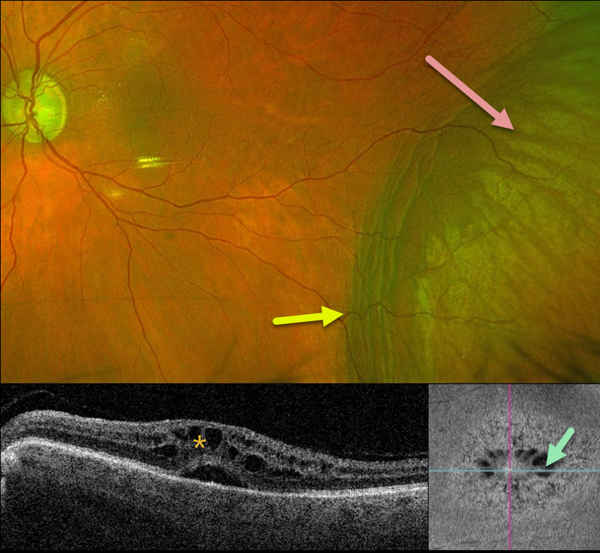
CHOROIDAL DETACHMENT
Originally posted on @retina.rocks November 30, 2023
This 84YO male has a long history of advanced chronic open-angle glaucoma with numerous surgical procedures. For several years, his left eye has had stable hypotony, with an intraocular pressure (IOP) of about 3-6 mmHg. Vision is 20/60.
Optos color RG imaging shows an inferotemporal choroidal detachment with chorioretinal folds along its posterior margin. Triton swept-source OCT shows cystoid edema with subretinal fluid, and en face imaging shows radiating mid-retina foveal cysts.
Since his symptoms, vision, IOP, and retinal findings remain stable, we are continuing to follow him without further intervention.
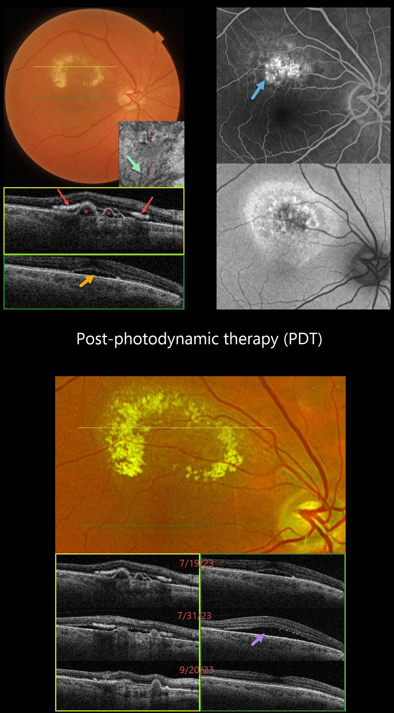
POLYPOIDAL CHOROIDAL VASCULOPATHY
Originally posted on @retina.rocks November 27, 2023
This 66YO female complained of 2 weeks of vision loss in her right eye. Vision was 20/40 OD and 20/20 in her normal OS.
Color imaging shows a circinate area of lipid in the superior macula. Triton swept-source OCT through the superior macula shows several sub-RPE polyps with hyper-reflective subretinal lipid. Scanning through the central macula shows shallow foveal subretinal fluid. Dilated inferior choroidal vessels are noted on en face OCT imaging.
Optos fluorescein angiography shows superior macular staining and leakage, with variable hyper-autofluorescence surrounding this region. Intravitreal Avastin was injected on 7/19/23, followed by photodynamic therapy (PDT) on 7/26/23.
She returned 5 days following PDT on 7/31/23 with increased blurriness. Vision remained at 20/40, although increased SRF was noted on OCT. Observation was recommended.
On 9/20/23, vision improved to 20/30, all SRF resolved, and the polyps were flattening.
Learning Points:
Polypoidal choroidal vasculopathy (PCV) is a variant of type 1 (below the RPE) macular neovascularization. It can be treated with either anti-VEGF monotherapy or combined with PDT to achieve better polyp regression and reduced treatment burden.
Transient PDT-induced increased exudative SRF sometimes develops immediately following treatment (Manayath et al, Retina 2020;40:135-144) but resolves spontaneously, as in our patient.
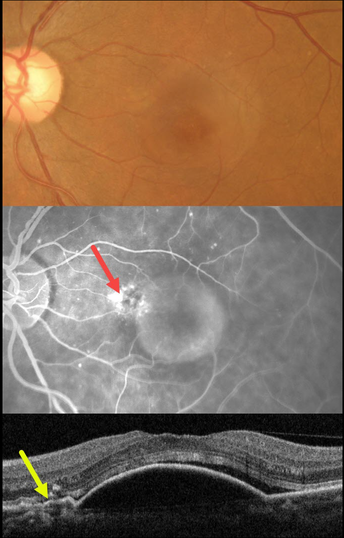
OCCULT MNV
Asma Samsudeen and Ashish Sharma
Originally posted on @retina.rocks November 23, 2023
This 62YO female presented with 3 weeks of vision loss in her left eye. Vision was 20/30.
Color imaging shows a large central macular retinal pigment epithelial detachment (PED). Fluorescein angiography shows a somewhat unevenly filling PED, with staining vs leakage along its superonasal edge. OCT scanning shows a large PED, shallow overlying temporal subretinal fluid, and shallow irregular elevation of the RPE nasally indicative of a type 1 macular neovascularization (MNV).
The uneven angiographic pooling is also suggestive of an occult MNV. Anti-VEGF therapy was advised.
This case was submitted by Asma Samsudeen and Ashish Sharma.
Learning Points:
Originally described by Gass (Retina 1984;4:205-220), a notched PED is a sign of an occult MNV. The notch represents a type 1 MNV, which tethers the overlying RPE from the surrounding more highly elevated PED. Hydrostatic leakage from the MNV may contribute to the larger PED.
Although now considered a classic clinical and angiographic finding, in 1984 this was evidently not the case, as the reviewers for this article advised against publication. Based solely on who the contributing author was, the journal’s editor overruled his reviewers, published Dr. Gass’ manuscript, and the rest was literally history!
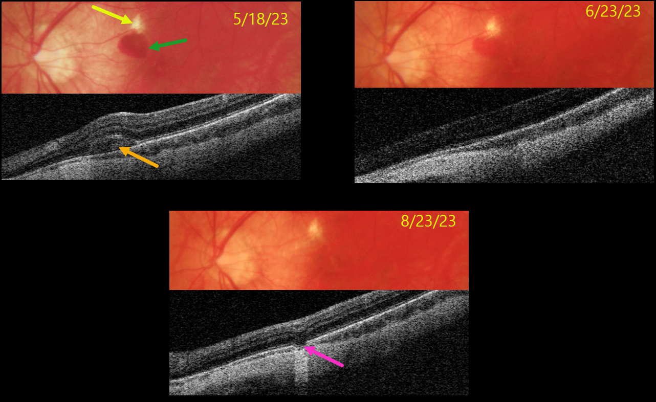
LACQUER CRACK
Originally posted on @retina.rocks November 13, 2023
This 33YO highly myopic male presented on 5/18/23 with vision loss in his left eye. Vision was 20/40.
Color photography shows a superior macular lacquer crack just superior to a round subretinal hemorrhage. The blood is hyperreflective on Triton swept-source OCT. The blood gradually absorbed with observation.
When last examined on 8/23/23, the blood had completely resolved, and vision remained at 20/40, but with resolved symptoms. OCT scanning through the lacquer crack shows a defect in the outer retinal bands and RPE.
Learning Points:
Lacquer cracks are a common finding in patients with high myopia. They consist of breaks in Bruch’s membrane due to the outpouching/stretching of the pathologically myopic eye and increase the risk for macular neovascularization. In some cases, similar to this patient, self-limiting small subretinal avascular hemorrhages occur that do not require anti-VEGF therapy.
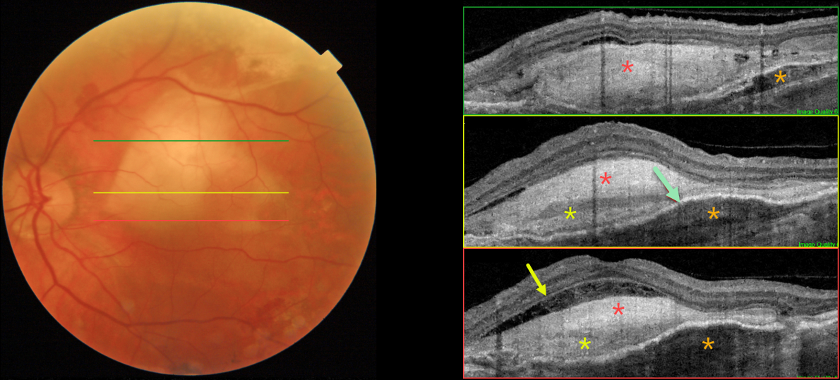
DISCIFORM SCAR
Originally posted on @retina.rocks October 24, 2023
This 81YO male has a history of counting-finger vision in his left eye due to a stable age-related fibrosed disciform scar.
Color imaging shows a fibrosed disciform scar in the central and superior macula. Triton swept-source OCT through different regions of the scar shows a constellation of interesting findings. The scar is variably hyperreflective, with areas of increased and relatively decreased reflectivity. Most inferiorly, the outer plexiform layer is clearly visible overlying an area of fluid and disorganized outer retina. Superiorly, the fibrosis indents and compresses the underlying RPE and choroid. Finally, the fibrosed neovascular lesion is both above (type 2) and below (type 1) the RPE, with the RPE variably elevated.
Learning Points:
Disciform scars were the norm for untreated wet AMD prior to the anti-VEGF era that began in the mid-2000s. This type of scarring is virtually no longer seen in eyes treated early in the course of their neovascular disease, although some subretinal fibrosis remains in 40% of eyes after 10 years of anti-VEGF treatment (Chong et al, Retina 2020;40:2285-2295).
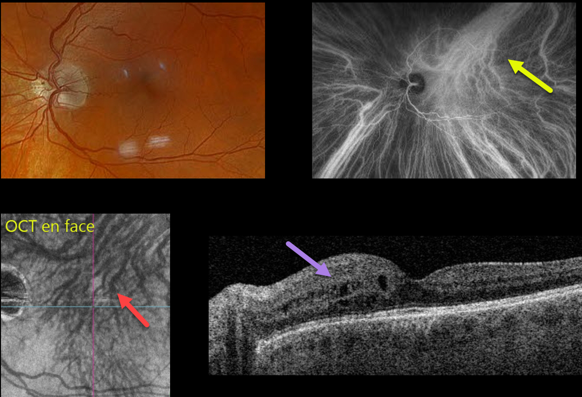
PERIPAPILLARY PACHYCHOROID SYNDROME
Originally posted on @retina.rocks October 16, 2023
This 59YO male has been followed for the past year with asymptomatic peripapillary pachychoroid syndrome (PPS) in his left eye. Vision is stable at 20/25 OU.
Optos color RGB imaging shows a normal left macula. Indocyanine green (ICG) angiography is normal OD (not shown), but reveals a dilated vortex system superotemporally that drains nearly the entire macula, along with a strikingly similar appearance from the OCT choroidal en face.
Triton swept-source OCT was normal OD (not shown), but shows mild nasal thickening with cysts in the inner nuclear layer OS.
Learning Points:
Originally described by Phasukkijwatana et al in 2018 (Retina 2018;38:1652-1667), PPS presents with nasal macular intraretinal and subretinal fluid. These eyes can look very similar to those of patients with optic pit maculopathy, but without the pit.
Additional findings include a thicker nasal than temporal macular choroid, choroidal folds (75% of cases), shorter axial lengths (<23mm), and hyperopia. There is no angiographic leakage despite the OCT fluid. This fluid will often spontaneously wax and wane. Symptomatic fluid can successfully be treated with photodynamic therapy.
Intervortex venous anastomoses are common in pachychoroid-related disorders (Spaide et al Retina 2021;41:997-1004). In this case, the choroidal anastomosis and dilated superior vortex system were beautifully imaged on both ICG and en face OCT.
In our practice, we continue to find that en face imaging often provides a readily accessible, unique, and compelling perspective for a variety of pathologies, and we hope other clinicians will add this to their daily clinic routine.
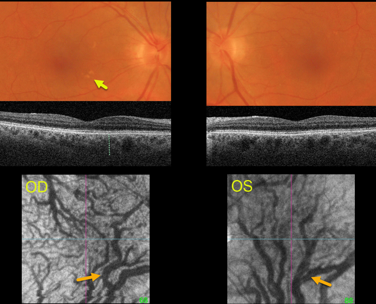
CHOROIDAL VENOUS ANASTAMOSIS
Originally posted on @retina.rocks October 5, 2023
This healthy 67YO female was examined for a symptomatic posterior vitreous detachment in her right eye. She was previously visually asymptomatic, and vision was 20/25 OU.
Optos color RGB imaging shows several large irregular drusen in the nasal right macula and faint drusen in the superotemporal left macula.
Triton swept source OCT shows a relatively thickened choroid for the patient’s age, along with large choroidal vessels. En face imaging through each choroid shows marked vortex dilation bilaterally. Each macula, particularly in her left eye, is drained by the inferior vortex system.
Learning Points:
Pachychoroid, meaning “thick” choroid, is secondary to large dilated Haller vessels. Increased oncotic pressure from these dilated vessels can cause RPE dysfunction, serous detachment, and macular neovascularization.
Another, more subtle finding is pachydrusen. Unlike typical age-related drusen that are pinpoint or round and centered in each macula, pachydrusen are fewer in number, more ectopic, and often have an irregular contour (Spaide, Retina 2018;38:708-716).
The choroid is typically thinned with AMD, but eyes with pachydrusen have thicker choroids. Although our patient’s macular clinical findings were quite subtle and the OCT B-scans were suggestive of pachychoroid, her en face images most dramatically show the choroidal pathology.
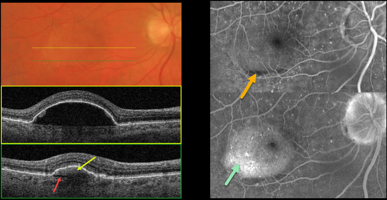
NOTCHED PED
Originally posted on @retina.rocks September 27, 2023
This 64YO male presented with recent central vision loss in his right eye. Vision was 20/40.
Color imaging shows a large central macular retinal pigment epithelial detachment (PED). Triton swept-source OCT through the central portion of the PED shows underlying hyporeflectivity. Scanning through the inferior portion shows a hyperreflective type 1 macular neovascularization (MNV) with a thin underlying prechoroidal cleft.
Optos fluorescein angiography shows an inferior linear filling defect within the MNV. The PED also shows late uneven pooling.
Learning Points:
Originally described by Gass (Retina 1984;4:205-220), a notched PED is a sign of an occult MNV. The notch represents a type 1 MNV, which tethers the overlying RPE from the surrounding more highly elevated PED. Hydrostatic leakage from the MNV may contribute to the larger PED.
Although now considered a classic clinical and angiographic finding, in 1984 this was evidently not the case, as the reviewers for this article advised against publication.
Based solely on who the contributing author was, the journal’s editor overruled his reviewers, published Dr. Gass’ manuscript, and the rest was literally history! The uneven angiographic pooling is also suggestive of an occult MNV.
A prechoroidal cleft is a hyporeflective space between the RPE and Bruch’s membrane. These are seen in up to about 20% of treated eyes with nAMD, particularly with type 3 neovascularization (intraretinal MNV) and polypoidal choroidal vasculopathy.
They are usually associated with worse visual acuity due to potential complications, including RPE tears and subretinal hemorrhage (Kim et al, Retina 2017;37:2047-2055).
Anti-VEGF therapy was recommended, and the patient was warned of the possibility of RPE development with or without treatment (Ersoz et al., Surv Ophthalmology 2017;62:493-505).
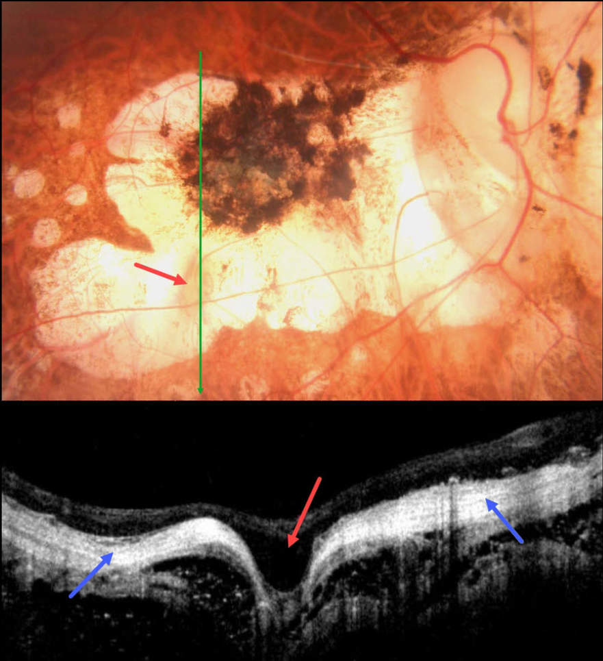
HIGH MYOPIA
Asma Samsudeen and Ashish Sharma
Originally posted on @retina.rocks September 14, 2023
This 58YO male with a history of degenerative myopia presented with 6 months of vision loss in his right eye. He was blind in his left eye following vitrectomy surgery 7 years earlier. Vision was counting fingers OD.
Color photography shows severe variably pigmented atrophic myopic degeneration. An area of orange coloration is seen within the larger area of atrophy inferiorly.
On OCT, this area appears as a focal staphyloma. The overlying retina is draped across the staphyloma. The choroid is so thin as to be invisible on OCT, with a hyperreflective underlying sclera. The orbital tissue is well seen posterior to the sclera.
Learning Points:
Degenerative myopia is a major cause of worldwide visual impairment. Due to the stretching of the posterior retinal structures, there is an increased risk for numerous findings, including atrophic retinal and choroidal thinning, posterior staphyloma, macular schisis/traction, and macular neovascularization (see Ohno-Matsui et al for a recent review, Invest Ophthalmol Vis Sci 2021;62(5);5)..
Initially described by Freund et al (Arch Ophthalmol 2003;121:197-204), intrachoroidal cavitations are most commonly found in high myopes as a peripapillary yellow-orange subretinal lesion. It is difficult to determine whether our patient has a cavitation above the staphyloma.
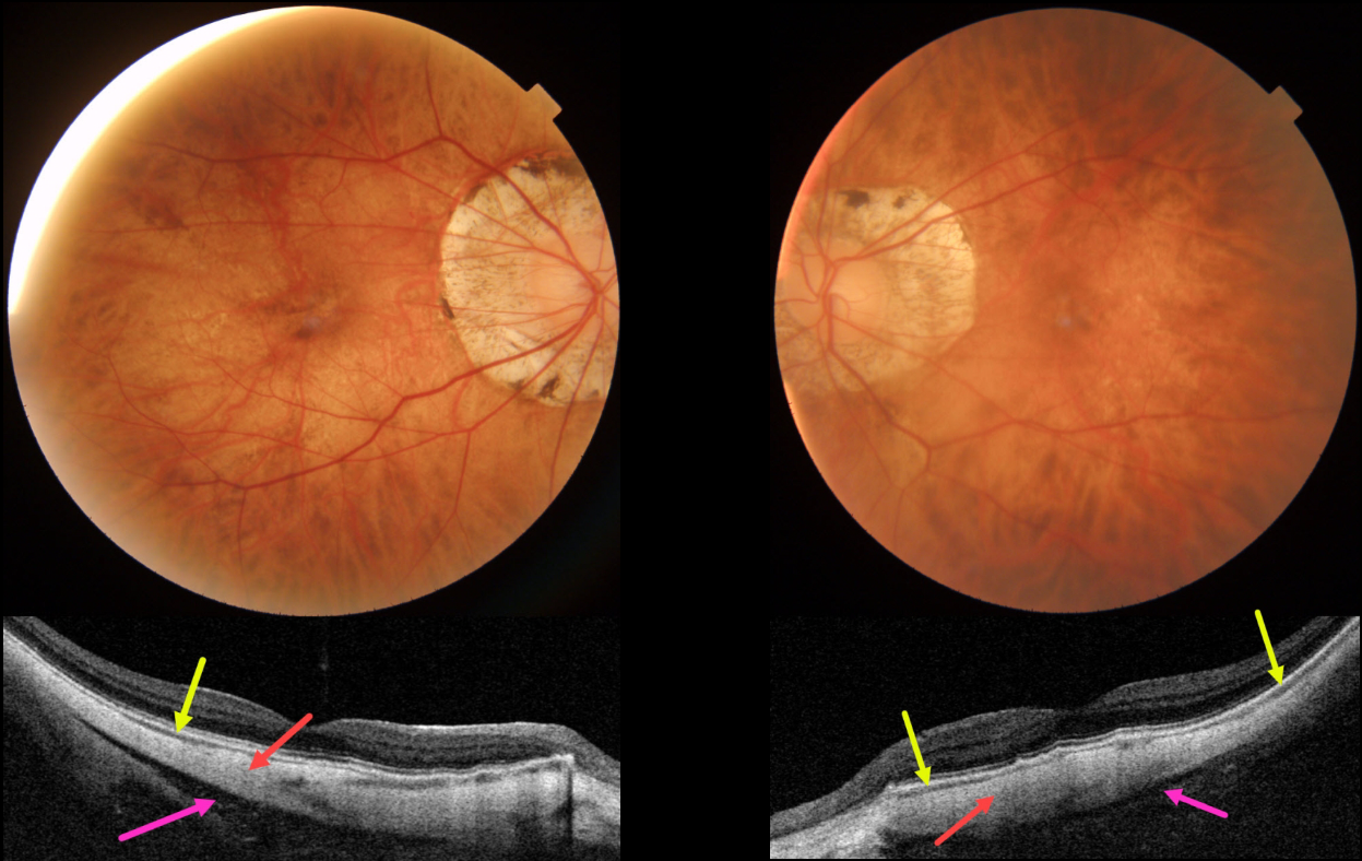
HIGH MYOPIA
Originally posted on @retina.rocks September 11, 2023
This 63YO female with severe high myopia underwent bilateral cataract surgery many years ago, along with successful vitrectomy for retinal detachment in her right eye. Vision was 20/50 OD and 20/30 OS.
Color imaging shows bilateral myopic peripapillary atrophy and macular pigmentary loss. Swept-source OCT shows a dramatically thinned or absent choroid and full-thickness, thinned sclera measuring about 300 microns bilaterally (normal posterior scleral thickness should be about 1mm). A hyporeflective region behind the sclera likely represents Tenon’s capsule, with the orbit visible more posteriorly.
Learning Points:
The era of ocular OCT began in 1991 with the initial report from Dr. Fujimoto’s lab at MIT (Huang et al, Science 1991;254:1178-1181). The technology was acquired by Zeiss in 1994, and after several failed commercial iterations, the Zeiss Stratus time-domain OCT in 2002 made it a must-have instrument for eye care.
Spectral domain units became available in 2008, followed by swept source units in 2015. Each subsequent generation can capture more information at greater retinal/choroidal depth. The swept source technology, in particular, is best able to visualize choroidal detail.
Our patient’s axial myopia with thinned choroid and sclera, along with the swept-source technology, provides a unique ability to visualize Tenon’s capsule and the anterior orbit.
For a wonderful historical review of the development, commercialization, and impact of OCT, see Fujimoto and Swanson, Invest Ophthalmol Vis Sci 2016;57;OCT1-OCT13.
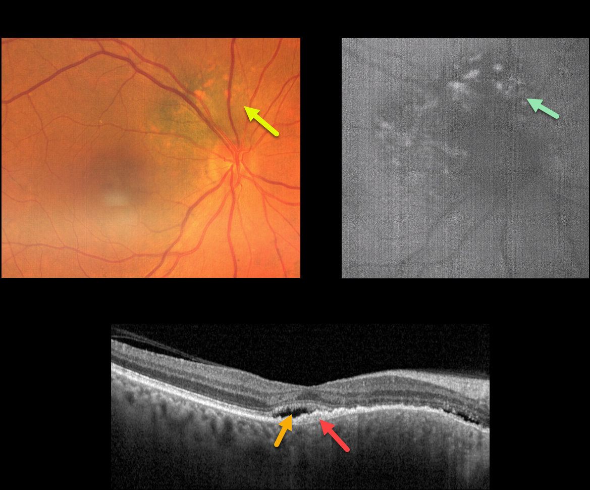
CHOROIDAL NEVUS MISDIAGNOSED AS CENTRAL SEROUS
Emma Oreskovic and Natasa Draca
Originally posted on @retina.rocks September 7, 2023
This 71YO female was referred for further treatment of chronic idiopathic central serous chorioretinopathy (ICSC) of her right eye, for which she had previously received unsuccessful anti-VEGF therapy at another institution. Vision was 20/100 OD and 20/20 in her normal left eye.
Fundus photography shows a variably pigmented, minimally thickened peripapillary choroidal nevus that extends from about 7-1 o’clock around the optic nerve margin. Overlying orange pigmentation is noted, which variably hyper-autofluoresces.
OCT scanning shows shallow foveal subretinal fluid with a subretinal layer of thickened hyperreflective material extending nasally overlying the thickened nevus. Beneath this is a very shallow, irregular elevation of the RPE.
Learning Points:
Clinical characteristics of choroidal nevi at risk of progressing to uveal malignant melanoma (MM) were initially described by Shields et al. (Ophthalmology 1995;102:1351-1361).
The most recent version of this famous acronym (Shields et al, Retina 2019;39:1840-1851) is To Find Small Ocular Melanoma Doing Imaging (TFSOM-DIM), which stands for To (Thickness >2mm) Find (subretinal Fluid) Small (Symptoms, vision <= 20/50) Ocular (Orange pigment) Melanoma (Melanoma hollow on ultrasonography) DIM (DIaMeter > 5mm).
ICSC and pachychoroid disorders, similar to syphilis, are one of the ‘great imitators,’ including uveal MM masquerading as ICSC (Negretti et al, Ophthalmology Retina 2023;7:171-177).
In our patient, on the central macular OCT B-scan, one sees a thickened choroid, subretinal fluid, and hyperreflective shed outer segments/lipofuscin, all of which are suggestive of ICSC.
This case emphasizes the importance of looking beyond the OCT, using both the clinical exam and multimodal imaging, for every patient, including those with chronic ICSC, to arrive at the correct diagnosis and treatment.
Since our patient has several high-risk characteristics (fluid, symptoms, and orange pigment), she was referred to an ocular oncologist for further management.
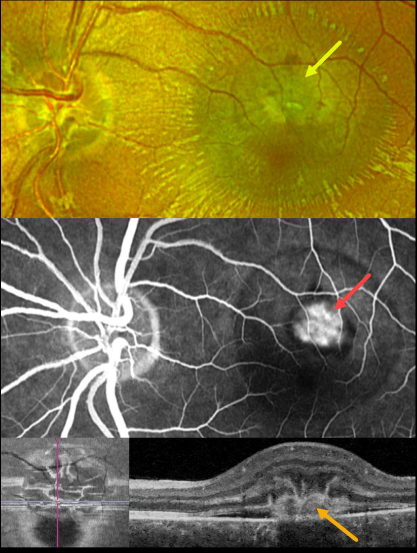
IDIOPATHIC MACULAR NEOVASCULARIZATION (MNV)
Originally posted on @retina.rocks September 6, 2023
This 23YO male presented with a recent loss of vision in his left eye. Vision was 20/60.
Optos color RG imaging shows a depigmented subretinal lesion in the superior fovea with overlying dots of blood and radiating retinal striae.
Fluorescein angiography shows a well-defined superior extrafoveal macular neovascularization (MNV). Swept-source B-scan OCT through this lesion shows a type 2 (above the RPE) MNV associated with shallow subretinal fluid and overlying radiating hyperreflective lines (pitchfork sign). These radiating lesions are uniquely visualized with en face OCT.
Intravitreal Avastin was begun.
Learning Points:
The pitchfork sign was originally described by Hoang et al (Retina 2013;33:1049-1055) as a unique OCT finding in inflammatory MNV. Falavarajani et al more recently noted that this finding is also associated with non-inflammatory MNV (Ophthalmic Surg Lasers Imaging Retina 2019;50:719-725).
The pathogenesis of the avascular pitchfork spikes is unknown, but may be related to outer retinal traction or Müller cell activation.
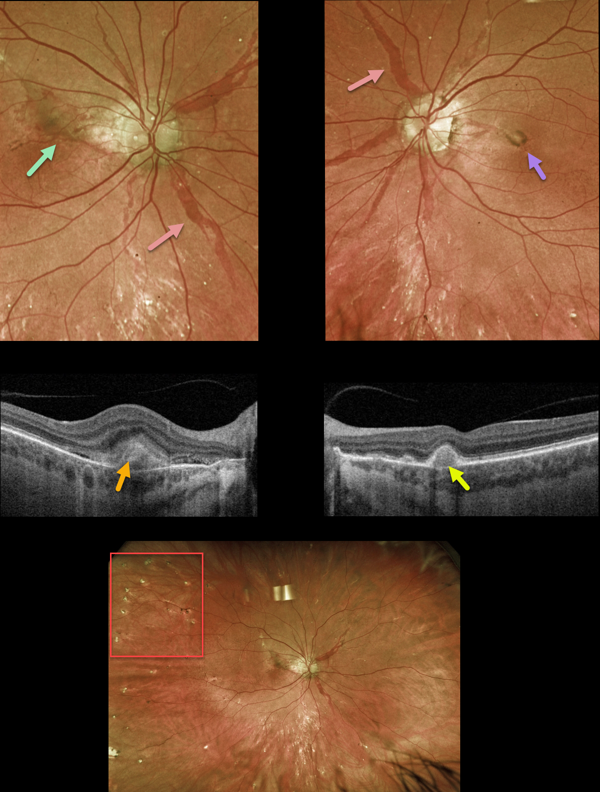
ANGIOID STREAKS
Originally posted on @retina.rocks September 5, 2023
This 42YO female with a known history of pseudoxanthoma elasticum (PXE) presented with new central flashes in her right eye. Vision was 20/25 OD and 20/30 OS.
Optos color RG imaging shows large, irregularly dilated’ angioid streaks’ radiating outwards from each optic nerve. A pigmented macular neovascularization (MNV) with some subretinal blood is noted in the superior right macula, and a small inactive MNV is noted in the left nasal fovea. Innumerable small subretinal comet-shaped lesions (‘comet rain’) are noted.
Swept-source OCT through the right MNV shows a hyperreflective, thickened type 2 lesion (above the RPE). OCT through the left MNV shows a small, nodular, hyperreflective type 1 lesion (below the RPE).
Learning Points:
Angioid streaks are orange-yellow, irregular, crack-like dehiscences in Bruch’s membrane associated with atrophic degeneration of the overlying RPE. They are often missed clinically because their color and orientation blend with the overlying retinal vessels.
Our patient’s lesions are about the most dramatic and red angioid streaks we’ve ever seen and are pathognomonic for PXE, but less extensive streaks can also be seen in sickle cell and age-related macular degeneration.
The comet lesions are also pathognomonic for PXE. These lesions develop from the outer retina as a neurodegenerative process and strongly resemble the hyporeflective core with a surrounding hyperreflective rim found in outer retinal tubulation (see Murro et al., Graefe’s 2018;256:1605-1614).
These patients are at significant risk for macular neovascularization (MNV), as occurred in each of our patients’ eyes. Intravitreal Avastin therapy was started in her right eye. The left MNV will be observed, as it involuted spontaneously without prior symptoms.
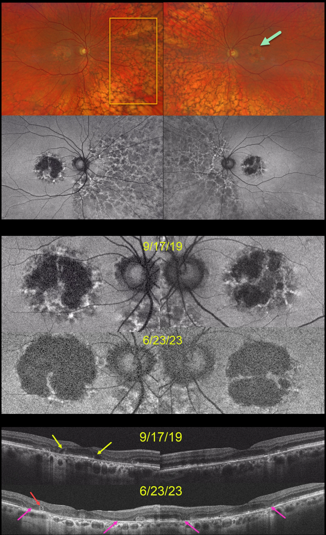
PENTOSAN POLYSULFATE SODIUM TOXICITY VS AMD MACULAR ATROPHY
Will Gibson
Originally posted on @retina.rocks September 4, 2023
This 59YO was examined on 9/17/19. She was taking Elmiron (pentosan polysulfate sodium, PPS) for 17 years for interstitial cystitis. Vision was 20/40 OD and 20/30 OS.
Optos color RG imaging shows bilateral central-sparing macular atrophy (MA) and extensive, posterior peripheral reticular degeneration of the RPE (PRDRPE).
Fundus autofluorescence shows hypo-FAF from the MA with surrounding punctate and linear areas of hyper-FAF. Based on these findings, she decided to stop taking Elmiron.
When last examined on 6/23/23, vision dropped to 20/800 OD and was relatively stable at 20/40 OS. The MA progressed bilaterally clinically and on FAF and OCT. Hyporeflective OPL degeneration (wedge defects) is noted on the initial 9/17/19 OCT. On the 6/23/23 OCTs, an area of outer retinal tubulation (ORT) is seen temporally OD, and progressive outer retinal and RPE atrophy are noted bilaterally.
Learning Points:
Elmiron was approved by the FDA in 1996 for the treatment of interstitial cystitis. Recently, a unique PPS retinopathy has been described. Toxicity seems to develop over many years and can mimic more common disorders, including age-related macular degeneration and macular dystrophies.
Peripapillary hypoautofluorescence, more densely packed macular autofluorescent changes, and earlier central macular involvement suggest PPS toxicity over other causes (see Barnes et al Ophthalmology Retina 2020;4:1196-1201), including maternally inherited diabetes and deafness (MIDD).
ORT is often noted overlying inactive macular neovascularization with ongoing anti-VEGF therapy and should not be confused with exudative fluid or cysts, which lack a hyperreflective border. The outer hyperreflective band likely represents inner segment mitochondria undergoing fission and translocation towards the nucleus (Litts et al, Retina 2018;38:445-461).
ORT, initially described by Zweifel et al (Arch Ophthalmol 2009;127:1596-1602), is a neurodegenerative condition of the photoreceptors and Muller cells associated with outer retinal and retinal pigment epithelium atrophy, including advanced AMD and inherited retinal diseases.
The hyper-FAF surrounding the initial MA is more typical for PPS toxicity, and progressive atrophy is not uncommon even with stopping therapy (Jung et al, JAMA Ophthalmol 2023;141:260-266). However, in our experience, PPS usually produces more widespread macular findings. Given the PRDRPE, the MA could also be age-related. Or maybe our patient had baseline age-related MA superimposed on Elmiron toxicity?
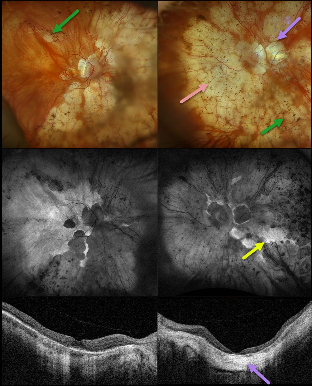
RETINITIS PIGMENTOSA + MYOPIC DEGENERATION
Originally posted on @retina.rocks August 30, 2023
This 78YO female has been followed by us for several years for retinitis pigmentosa (RP) and degenerative myopia. Vision is stable at 20/30 OD and hand motion OS. Multimodal imaging shows a fascinating fusion of findings for these two disorders.
Optos color RGB imaging shows typical RP findings of bone spicules (intraretinal pigment migration) extending from outside the arcades into the peripheries, superimposed on severe degenerative myopic changes.
Fundus autofluorescence (FAF) shows hypo-FAF within the areas of myopic atrophy, with hyper-FAF more peripherally from the RP-induced outer retinal atrophy.
A macular staphyloma is also seen in the left eye and confirmed on swept-source OCT. The degenerative myopic changes just spare the right macular center, which has no staphyloma.
Learning Points:
Genetic testing showed pathogenic heterozygous mutations for OCA2 and RPGR. Fortunately, the RP spares each macula. Her myopic degeneration has been stable, and we are hopeful that her central vision will be preserved in her right eye.
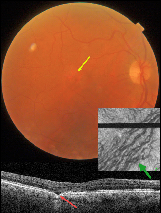
CHOROIDAL EXCAVATION
Originally posted on @retina.rocks August 15, 2023
This 74YO male was seen for a routine eye examination. He told us he had a laser procedure on his right eye 18 years ago. Based on this image, what type of laser was it?
Fundus photography shows a normal macula except for an area of depigmentation in the temporal fovea.
Swept-source OCT through this area shows a small focal choroidal excavation. Although the choroid appears to have normal thickness, large outer choroidal vessels spanning virtually the entire choroid are noted nasally.
Choroidal en face OCT is markedly abnormal with dilated vessels and vortex anastomoses.
Our patient shows several pachychoroid-related findings, including a thick choroid for their age, large choroidal vessels, FCE, and vortex anastomoses. Our patient currently has asymptomatic pachychoroid despite having a ‘normal’ choroidal thickness on OCT.
So what type of laser did our patient have? Photodynamic therapy for central serous retinopathy, which has remained in remission since treatment in 2005
Learning Points:
Pachychoroid, meaning “thick” choroid, is secondary to large dilated Haller vessels. Increased oncotic pressure from these dilated vessels can cause RPE dysfunction, serous detachment, and macular neovascularization.
The choroidal thickness depends on numerous factors, including refractive error and age. A high myope’s choroid should be quite thin, but if it appears as a ‘normal’ thickness, it is thick for that eye. We also often see pachychoroid disease in elderly patients whose choroid is abnormally thickened for their age, but appears ‘normally’ thick when not accounting for this.
FCE, originally described by Jampol et al in 2006, is likely part of the pachychoroid spectrum, as it is usually associated with a thickened choroid and pachyvessels. The FCE can be observed, although symptomatic manifestations, including active central serous retinopathy or macular neovascularization, often require treatment. An excellent review is by Verma et al, Br J Ophthalmol 2021;105:1043-1048.
The choroidal venous system, as with the neurosensory retinal vasculature, usually respects the horizontal meridian. With pachychoroid disorders, these anastomoses develop, and in this case, the inferior vortex system drains nearly the entire macula.
For a wonderful review highlighting the concept of venous overload choroidopathy, see Spaide et al, Progress Retinal Eye Research 2022;86:100973.
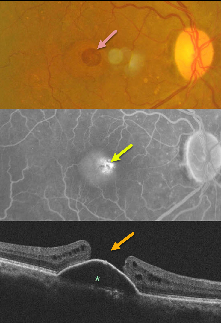
MACULAR HOLE
Shilpi Narnaware and Prashant Bawankule
Originally posted on @retina.rocks August 8, 2023
This 60YO female presented with 1 month of vision loss in her right eye. Vision was 20/100 OD and 20/40 OS.
Fundus photography of her right eye shows a large macular hole with some nasal pachydrusen. There was an old branch retinal vein occlusion without macular drusen in her left eye (image not shown).
Fluorescein angiography shows central pooling beneath a retinal pigment epithelial detachment (PED) with increased transmission through the macular hole. OCT scanning shows a PED with an overlying large macular hole.
This case presents a unique and interesting challenge in that macular hole surgery likely would not close the hole, given the underlying PED. She received 3 monthly Lucentis injections in her right eye, with no change in the PED.
Our thought would be to try a different anti-VEGF agent, such as Eylea, or consider adding photodynamic therapy for a possible pachychoroid-related avascular PED.
A similar case showed spontaneous closure of a macular hole following anti-VEGF therapy (see Chakraborty and Sheth, Indian J Ophthalmol Case Rep 2023;3:439-440).
If our patient’s macular hole remained open once the PED flattened, then macular hole surgery would be performed.
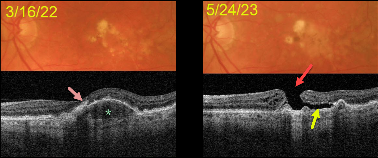
MACULAR HOLE AFTER COLLAPSE OF PED
Originally posted on @retina.rocks July 25, 2023
This 72YOM was examined with asymptomatic intermediate AMD bilaterally. Vision was 20/40 OD and 20/50 OS.
Color imaging of his left eye initially shows mostly large, soft drusen and pigment clumps. Swept source OCT shows a RPE detachment (PED) with overlying hyperreflective foci.
He returned over a year later without new symptoms, despite vision dropping to counting fingers. OCT shows a large macular hole overlying the collapsed PED. Shallow subretinal fluid lies below the temporal aspect of the hole.
The development of a macular hole following spontaneous collapse of a PED is quite unusual. Fortunately, vision in his right eye remains at 20/40 with intermediate dry AMD.
Since our patient was completely asymptomatic, he did not want macular hole surgery, and we will monitor him twice yearly.
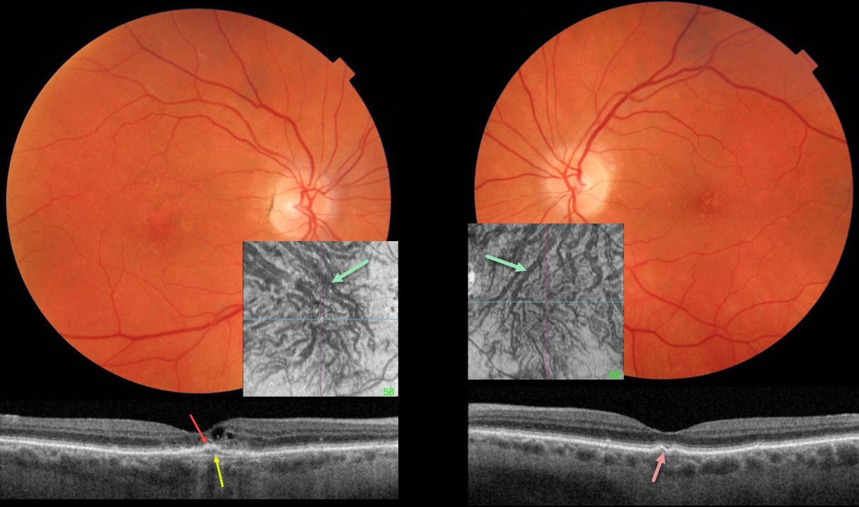
PACHYCHOROID NEOVASCULARIZATION
Originally posted on @retina.rocks July 18, 2023
This 85YO female presented with recent vision loss in her right eye. Vision was 20/200 OD and 20/40 OS.
Fundus photography of her right eye shows granular foveal pigmentary changes with a few faint, medium-sized drusen in her left eye.
Swept-source OCT OD shows a shallow, irregular elevation of the foveal RPE with visible Bruch’s membrane. Some cuticular drusen are noted bilaterally. Although the choroid itself is not thickened, large choroidal vessels are seen bilaterally, and choroidal en face imaging shows abnormal, dilated superior anastomotic vortex vessels that drain the entirety of each macula.
Learning Points:
Pachychoroid, meaning “thick” choroid, is secondary to large dilated Haller vessels. Increased oncotic pressure from these dilated vessels can cause RPE dysfunction, serous detachment, and macular neovascularization (MNV).
Our patient has pachychoroid despite having a ‘normal’ choroidal thickness on B-scan OCT. The choroidal thickness depends on numerous factors, including the patient’s age and refractive error. A high myope’s choroid should be quite thin, but if it appears as a ‘normal’ thickness, it is thick for that eye.
We also often see pachychoroid disease in elderly patients whose choroid is abnormally thickened for their age, but appears ‘normally’ thick when not accounting for this.
Originally described by Pang and Freund (Retina 2015;35:1-9), pachychoroid neovasculopathy is a variation of type 1 MNV (located below the RPE). About one-fifth of patients diagnosed with neovascular AMD actually have pachychoroid neovasculopathy (Farvardin et al, Int J Retina Vitreous 2022;8:78), and about one-third of eyes will progress to polypoidal choroidal vasculopathy by 5 years (Siedlecki et al, Ophthalmology Retina 20222;6:807-813).
Although our 85YO patient also has macular drusen, the summation of her findings most likely points to pachychoroid disease as the cause of her MNV. Anti-VEGF therapy was begun.
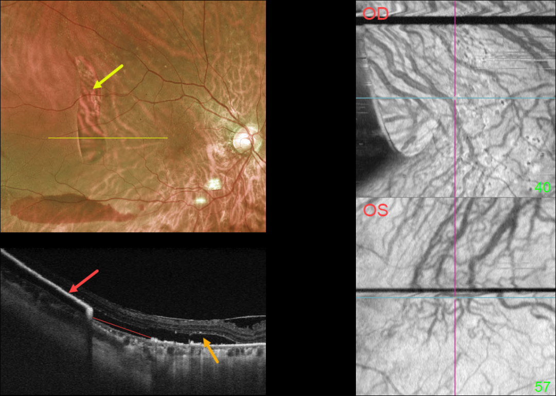
RPE TEAR
Originally posted on @retina.rocks June 14, 2023
This 73YO female presented with acute vision loss in her right eye. Vision was 20/40 OD and 20/30 OS. There was a several-year history of type 2 diabetes with mild bilateral nonproliferative diabetic retinopathy.
Optos imaging shows a large vertical area of depigmentation in the distal superotemporal macula. There is a serous detachment surrounding this region, along with a far inferior subretinal hemorrhage.
Swept-source OCT through this lesion shows bare Bruch’s membrane from an RPE tear. The retracted RPE is markedly hyperreflective more temporally. Subretinal fluid overlies this region.
Macular choroidal en face imaging shows bilateral anomalous vasculature with choroidal anastomoses.
Learning Points:
A tear of the RPE is most commonly seen in RPE detachments (PEDs) associated with neovascular AMD, but can also occur in numerous other etiologies, including central serous chorioretinopathy and polypoidal choroidal vasculopathy.
The tear leaves a depigmented region where the RPE was and is now absent, along with a pigmented, relatively straight subretinal lesion from the scrolled RPE edge.
The choroidal en face OCT findings are more consistent with the pachychoroid spectrum. RPE tears are found in polypoidal choroidal vasculopathy, another pachychoroid disorder (Zhao et al, Retina 2020;40:477-489).
See Ersoz et al for a great review on RPE tear classification, pathogenesis, predictors, and management (Surv Ophthalmology 2017;62:493-505).
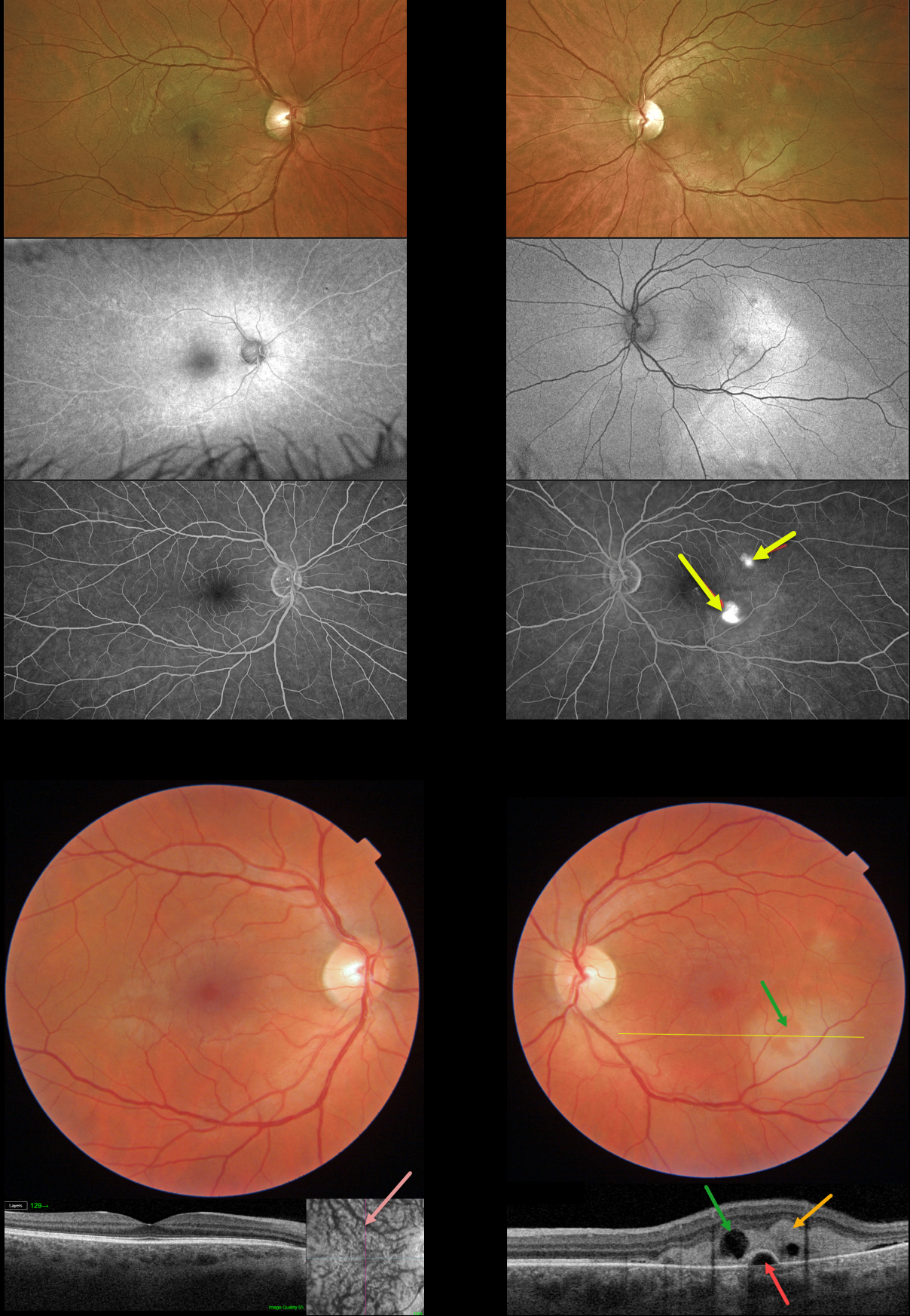
FIBRINOUS IDIOPATHIC CENTRAL SEROUS CHORIORETINOPATHY
Mattie Adams
Originally posted on @retina.rocks April 20, 2023
This 42YO male presented with mild symptoms of decreased color saturation and 20/40 vision in his left eye. The right eye was normal with 20/20 vision.
Color imaging shows creamy white subretinal fibrin in the inferotemporal left macula with a smaller area of fibrin superotemporally. Swept-source OCT through the larger inferior lesion shows hyperreflective, subretinal fibrin extending into the outer retinal layers. There is a small underlying retinal pigment epithelial detachment (PED), with a small overlying hyporeflective space corresponding funduscopically to an area devoid of fibrin.
Although the right macula appears normal funduscopically and on B-scan OCT, en face imaging shows somewhat dilated choroidal vessels.
Optos fundus autofluorescence (FAF) shows a hyper-FAF gutter extending from both fibrinous lesions. Fluorescein angiography shows two hyperfluorescent blot leaks.
Learning Points:
In idiopathic central serous chorioretinopathy (ICSC), fluorescein dye diffuses through a physiologic break in the RPE and then pools within the subretinal space. As in this case, the subretinal leak is almost always associated with a PED.
The clearing within the inferotemporal subretinal fibrin, which corresponds to the hyporeflective space within the fibrin on OCT, is caused by serous fluid entering the subretinal space and ‘washing away’ some of the fibrin.
Most patients with ICSC have a single, unilateral leak. Blot leaks are most common, with only 10% of cases exhibiting a smokestack leak. The multifocal leaks and subretinal fibrin noted in our patient are more commonly found in steroid-induced disease (our patient denied steroid exposure). Due to the complete lack of symptoms, initial observation was recommended.
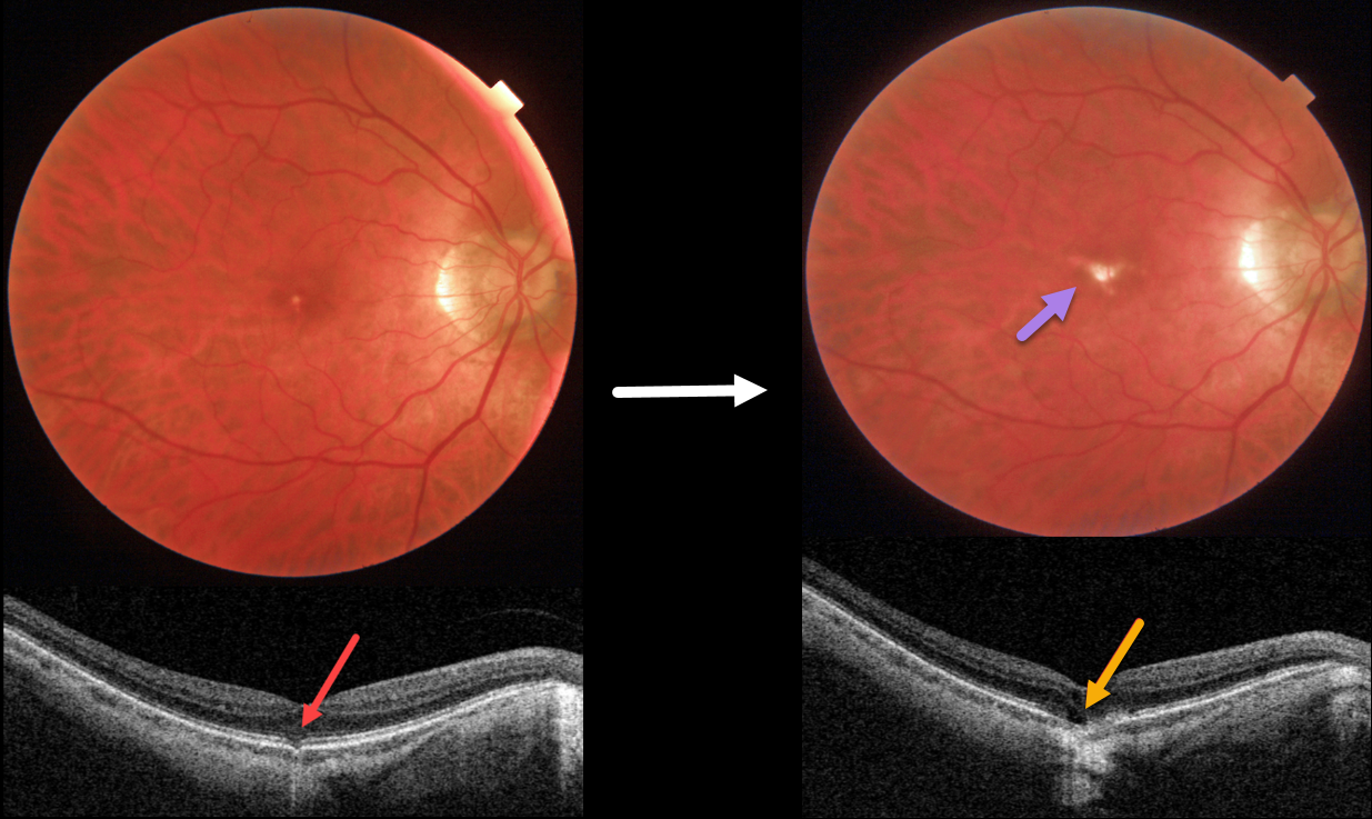
OUTER FOVEAL MICRODEFECT
Originally posted on @retina.rocks April 13, 2023
This 58YO male was referred for an asymptomatic foveal lesion in his right eye. There was a history of high myopia prior to LASIK surgery years earlier. Vision was 20/40 OD.
Color photography shows a small atrophic foveal scar. OCT shows a focal EZ defect with a possible tiny choroidal excavation. He returned about 2 years later for a routine follow-up visit.
Although he was binocularly without a new complaint, his vision had decreased to 20/200. There was now a definite, much enlarged foveal lacquer crack. OCT scanning shows a new outer foveal microdefect.
Learning Points:
Outer foveal microdefects (Cohen et al, Ophthalmology Retina 2021;5:553-561) have been described in numerous conditions, including macular telangiectasia, tamoxifen use, ABCA4 disorders (cone-rod dystrophies, Stargardt disease, and fundus flavimaculatus), phototoxicity, trauma, and vitreomacular traction disorders.
We are not aware of a prior report showing these lesions in association with myopic lacquer cracks. These defects, caused by focal loss of the outer retinal layers, leave an optically empty space that can be mistaken for subretinal fluid but is more of an optical gap within the outer retina.
Focal choroidal excavation (FCE), originally described by Jampol et al in 2006, is likely part of the pachychoroid spectrum since it is usually associated with a thickened choroid and pachyvessels. The FCE can be observed, although symptomatic manifestations, including active central serous retinopathy or macular neovascularization, often require treatment. An excellent review is by Verma et al, Br J Ophthalmol 2021;105:1043-1048.
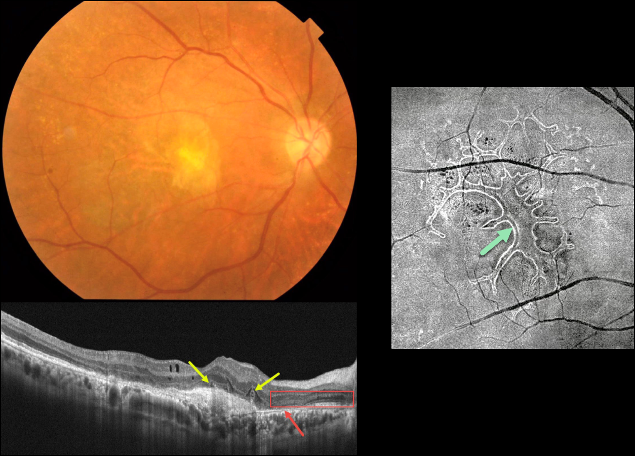
WET AMD WITH OUTER RETINAL TUBULATION
Originally posted on @retina.rocks April 12, 2023
This 63YO male received several intravitreal anti-VEGF injections for neovascular AMD in his right eye, the most recent being 6 months earlier. Vision is counting fingers.
Color photography shows an inactive atrophic foveal scar. OCT scanning shows a hyperreflective macular neovascularization (MNV). The outer retina overlying the MNV is disorganized with several overlying hyporeflective lesions with surrounding hyperreflective rims. More nasally, there is complete RPE loss with bare Bruch’s membrane. The overlying retina is anomalous with additional hypo- and hyperreflective bands.
En face OCT scanning through the outer retina provides a more dramatic view of the extensive tubulation-branching network.
Learning Points:
Outer retinal tubulation (ORT) is often noted overlying inactive MNV with ongoing anti-VEGF therapy and should not be confused with exudative fluid or cysts, which lack a hyperreflective border. The outer hyperreflective band likely represents inner segment mitochondria undergoing fission and translocation toward the nucleus (Litts et al, Retina 2018;38:445-461).
ORT, initially described by Zweifel et al (Arch Ophthalmol 2009;127:1596-1602), is a neurodegenerative condition of the photoreceptors and Muller cells associated with atrophy affecting the outer retina and retinal pigment epithelium, including advanced AMD and inherited retinal diseases.
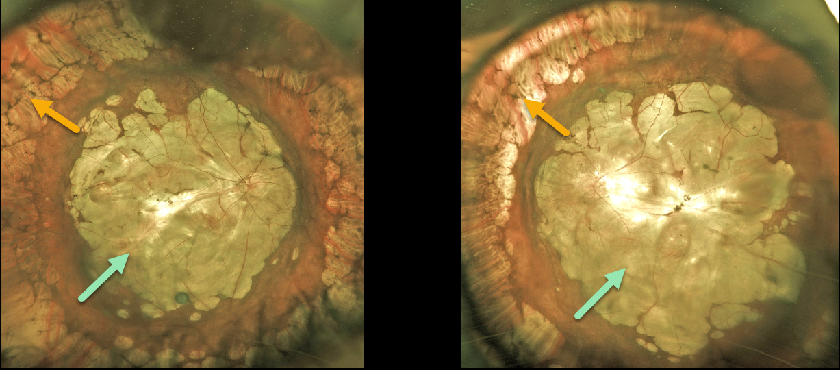
HIGH MYOPIA WITH STAPHYLOMA
Originally posted on @retina.rocks April 4, 2023
This 72YOM has a lifelong history of severe degenerative myopia. Vision was hand motion bilaterally.
Optos color imaging of both eyes shows confluent myopic posterior staphylomas and chorioretinal atrophy extending into each retinal midperiphery. Encircling cobblestone degeneration is seen more anteriorly.
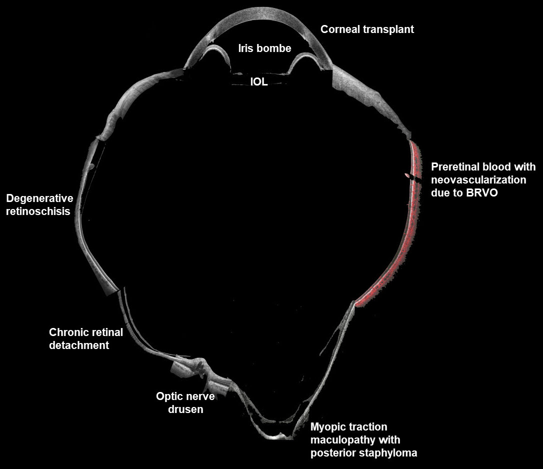
WIDEFIELD OCT
Veronika Matello and Barbara Parolini
Originally posted on @retina.rocks April 1, 2023
This composite widefield OCT image combines multiple pathologies to create a quite sick virtual eye. All the scans were taken with the Canon Xephilio S1.
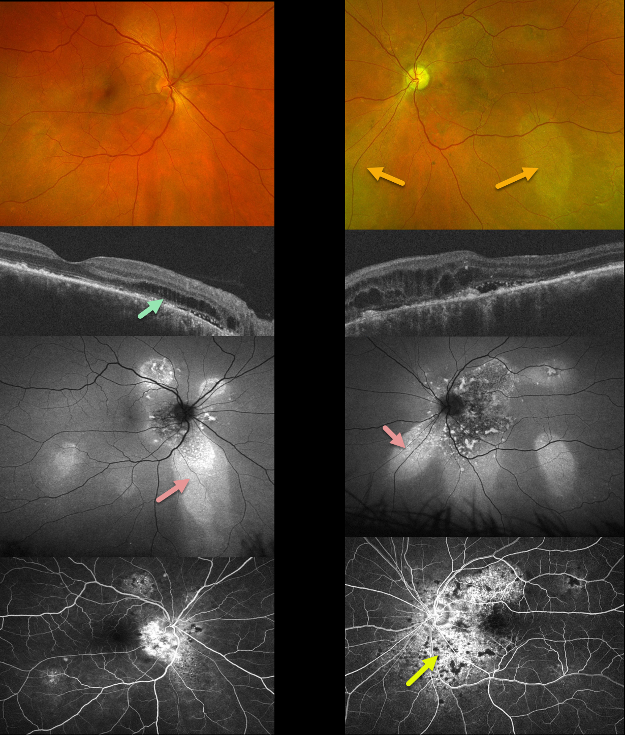
PACHYCHOROID SYNDROME
Will Gibson
Originally posted on @retina.rocks March 22, 2023
This 51YO male has been followed for years with self-limited episodes of idiopathic central serous retinopathy (ICSC).
Optos color imaging of both eyes shows regions of inferior pigmentary loss, most pronounced in the OS. These findings are much more dramatic on fundus autofluorescence. Swept-source OCT shows variable bilateral loss of the outer retinal bands. Outer retinal fluid with trace subretinal fluid is seen centrally and nasally OS. Fluorescein angiography shows variable staining.
Learning Points:
This patient has numerous features of the pachychoroid spectrum, including idiopathic central serous retinopathy (ICSC), pachychoroid neovasculopathy, polypoidal choroidal vasculopathy, pachychoroid pigment epitheliopathy, peripapillary pachychoroid syndrome, and choroidal excavation.
Our patient’s current findings are dominated by the peripapillary pachychoroid syndrome. Originally described by Phashukkuwatana et al (Retina 2018;1652-1667), findings include peripapillary choroidal thickening associated with nasal macular subretinal and/or intraretinal fluid. Most patients also have chorioretinal folds.
The fluid waxes and wanes, and is responsive to photodynamic therapy if treatment is needed for persistent symptomatic foveal fluid (Iovino et al, Eye 2022;36:716-723). Our patient is being observed for now.
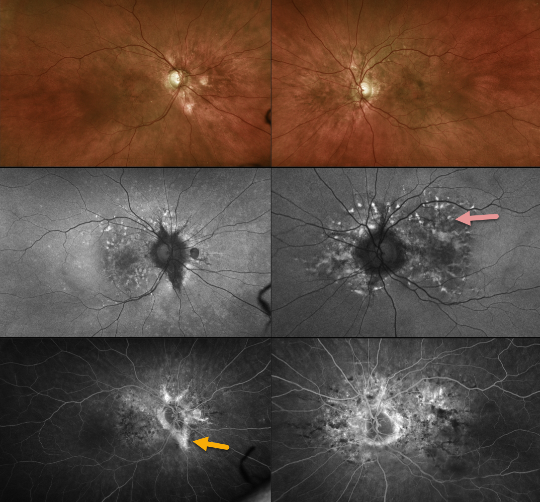
PSEUDOXANTHUM ELASTICUM WITH ANGIOID STREAKS AND PATTERN DYSTROPHY PHENOTYPE
Originally posted on @retina.rocks March 20, 2023
This 55YO female has been followed for years with macular pattern dystrophy-type changes due to pseudoxanthoma elasticum (PXD). Vision is 20/25 OU.
Color imaging shows variable mild pigmentary changes. Optos fundus autofluorescence imaging shows variable hyperautofluorescent flecks with variable angiographic staining. Some staining angioid streaks are noted in her right eye.
Learning Points:
Retinal findings in PXE most commonly include angioid streaks and a subretinal orange skin (peaux d’orange) appearance, most pronounced in each temporal macula.
However, pattern dystrophy-like changes, as in our patient, can sometimes be the predominant phenotype in some PXE patients (Murro et al, Graefe’s 2020;258:1881-1892). As with typical pattern dystrophies, the findings are dynamic and best captured with multimodal imaging.
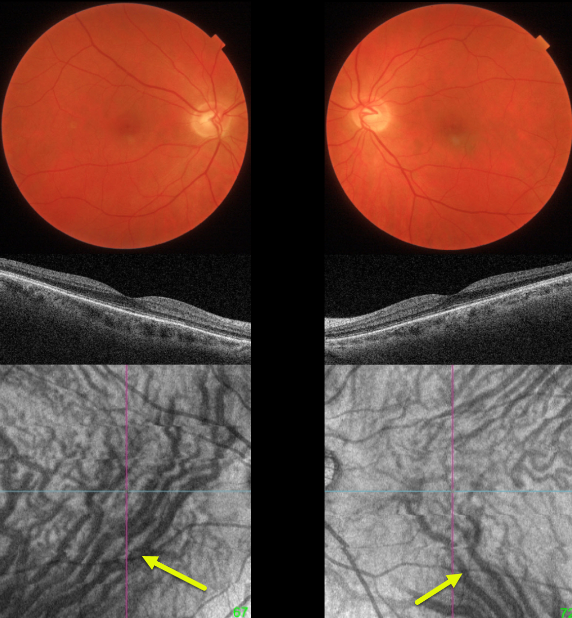
PACHYCHOROID PIGMENT EPITHELIOPATHY
Originally posted on @retina.rocks March 6, 2023
This 51YO minus 5 diopter myope presented without visual complaint for a routine yearly examination. Vision was 20/25 OU.
Both maculas are normal on fundus photo and B-scan OCT. However, en face OCT shows bilateral dilation of the inferior vortex system. The right inferior vortex also drains nearly the entire macula.
Learning Points:
The choroidal venous system, as with the neurosensory retinal vasculature, usually respects the horizontal meridian. With pachychoroid disorders, these anastomoses develop, and in this case, each inferior vortex system drains nearly the entire macula.
For a wonderful review highlighting the concept of venous overload choroidopathy, see Spaide et al, Progress Retinal Eye Research 2022;86:100973.
Pachychoroid, meaning “thick” choroid, is secondary to large dilated Haller vessels. Increased oncotic pressure from these dilated vessels can cause RPE dysfunction, serous detachments, and macular neovascularization.
Our patient has asymptomatic pachychoroid despite having a ‘normal’ choroidal thickness on B-scan OCT. The choroidal thickness depends on the refractive error. A high myope’s choroid should be quite thin, but if it appears as a ‘normal’ thickness, it is thick for that eye.
We also often see pachychoroid disease in elderly patients whose choroid is abnormally thickened for their age but appears ‘normally’ thick when not accounting for this. Although our patient’s retinal findings are completely normal clinically, these subclinical pachychoroid findings put her at risk for the various pachychoroid disorders
For an absolutely fascinating editorial discussing the ambiguity of how best to define what ‘pachychoroid’ actually means, see Spaide Retina 2021;41:231-237.
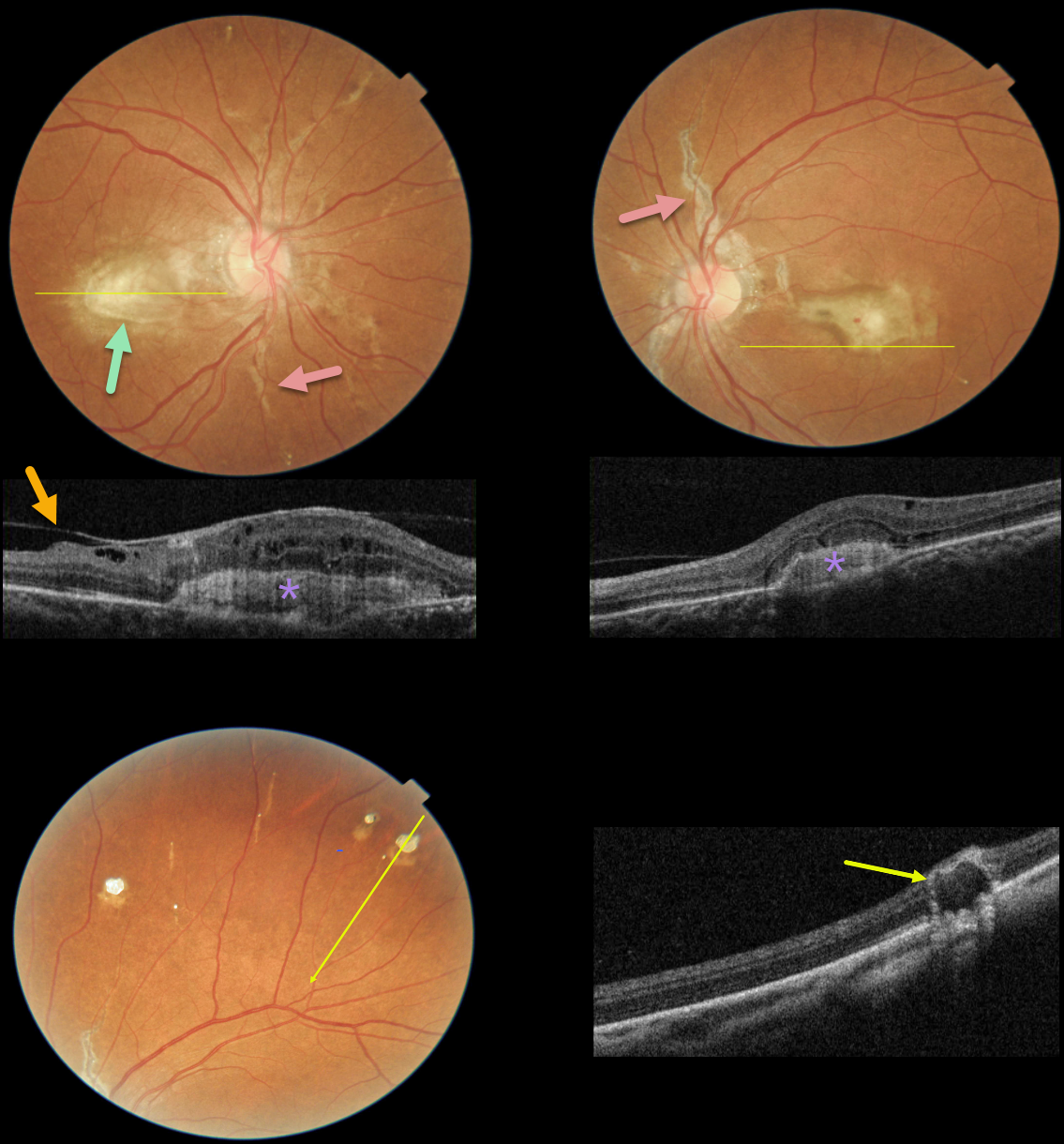
ANGIOID STREAKS
Asma Samsudeen and Ashish Sharma
Originally posted on @retina.rocks February 17, 2023
This 49YO female presented with one month of blurred vision. There was no past medical history except for a seizure disorder. Vision was 20/80 OD and 20/120 OS.
Fundus photographs show bilateral angioid streaks radiating from each optic nerve. Bilateral fibrosed subfoveal macular neovascularization (MNV) is present, and a small dot of blood is seen overlying the left macular lesion.
OCT scanning through the central right macula shows a hyperreflective, laminated MNV with overlying cystic retinal edema and a partially detached hyaloid vs macular pucker. OCT scanning through the inferior left macula also shows a hyperreflective MNV below the RPE associated with some overlying retinal thickening.
Numerous small subretinal comet-shaped lesions (‘comet rain’) are noted bilaterally. OCT scanning through one of these lesions shows a full-thickness hyporeflective lesion with a surrounding hyperreflective rim.
Learning Points:
Angioid streaks are orange-yellow, irregular, crack-like dehiscences in Bruch’s membrane associated with atrophic degeneration of the overlying RPE. They are often missed clinically since their color and orientation often blend in with the overlying retinal vessels.
Our patients’ lesions are easier to diagnose due to significant overlying RPE changes. The extent and prominence of the angioid streaks in our patient are pathognomonic for PXE, but less extensive streaks are seen in sickle cell disease and age-related macular degeneration.
These patients are at significant risk for macular neovascularization (MNV), which can be successfully treated with intravitreal anti-VEGF therapy.
The comet lesions are pathognomonic for PXE. These lesions develop from the outer retina as a neurodegenerative process and strongly resemble the hyporeflective core with a surrounding hyperreflective rim found in outer retinal tubulation (see Murro et al, Graefe’s 2018;256:1605-1614). Full-thickness lesions, as in our patient, were reported by Barteselli and Viola (Retina 2015;35:1051-1052).
Although our patient has no known personal or family history of PXE, her retinal findings indicate she has this disease. Her MNV appears chronic and fibrosed, and it is unclear why her symptoms were only present for one month. However, due to her recent symptoms and presence of overlying retinal fluid and blood, anti-VEGF therapy will be started.
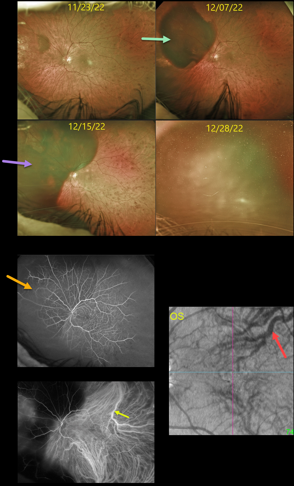
SUBRETINAL HEMORRHAGE
Originally posted on @retina.rocks January 31, 2023
This diabetic male had been followed for over 5 years in our practice, receiving PRN intravitreal injections for diabetic macular edema.
On 8/10/22, he presented with a large superotemporal subretinal hemorrhage in his right eye that progressed into the macula (not shown). Vitrectomy with evacuation of the subretinal blood was performed.
Three months later, an inferior retinal detachment with proliferative vitreoretinopathy was noted (not shown). Staged cataract surgery followed by vitrectomy with silicone oil was performed.
At the 12/7/22 visit, the patient complained of new inferotemporal peripheral field loss, noting that “the same thing is happening in my left eye.”
Unfortunately, a superonasal subretinal hemorrhage was indeed found, and intravitreal Avastin injection was given the following day. However, one week later, the subretinal blood spread posteriorly.
Fluorescein angiography shows nonproliferative diabetic retinopathy with blockage superonasally from the subretinal blood. ICG angiography shows a large dilated superotemporal choroidal vein. En face OCT shows abnormal vortex systems, including a large, dilated superotemporal vortex vein.
On 12/28/22, the subretinal blood spread further with breakthrough bleeding into the vitreous.
Cataract surgery was performed, followed by vitrectomy; however, intraoperatively, once the vitreous hemorrhage was removed, he had a massive subretinal hemorrhage with an inoperable retinal detachment with proliferative vitreoretinopathy.
We believe our patient has an extremely unusual and aggressive variant of pachychoroid disease, as evidenced by the abnormal choroidal vasculature noted on ICG and en face OCT.
We have no explanation for why both eyes developed these massive subretinal hemorrhages, or why they developed almost concurrently. We plan on injecting his right eye with ongoing intravitreal Eylea in hopes of preventing recurrent subretinal hemorrhages.
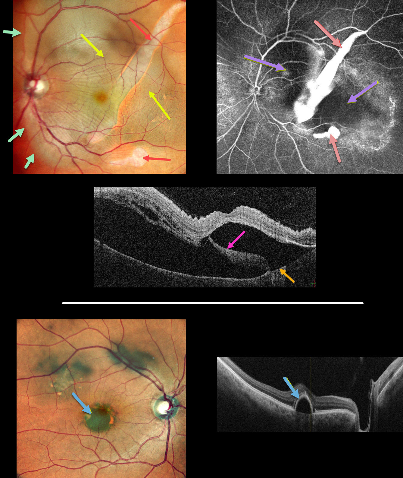
GIANT RPE TEAR
Anjana Mirajkar, Manish Nagpal, Navneet Mehrotr, and Akansha Sharma
Originally posted on @retina.rocks January 20, 2023
This 62YO male presented with 1 month of vision loss in his left eye. Vision was 20/30 OD and 20/60 OS.
Multicolor imaging of his left eye shows a giant RPE tear extending from the inferior macula into the superotemporal midperiphery, along with a much smaller tear just inferior to the macula. The beds of the RPE tears are depigmented. Increased subretinal pigmentation is noted on either edge of the giant tear. Subretinal fluid variably elevates the macula with inner retinal folds radiating from the optic nerve temporally. This subretinal fluid extends into the superior and temporal midperipheries.
B-scan OCT shows marked subretinal fluid. Bare Bruch’s membrane is noted temporally, and the RPE tear is lifted nasally and floating within the serous detachment.
Fluorescein angiography shows marked hyperfluorescent window defects from the absent RPE with blockage on either side of the giant tear from the redundant retracted RPE. He was placed on a 5-day tapering course of oral steroids but was subsequently lost to follow-up.
An asymptomatic retinal pigment epithelial detachment (PED) is noted in his right macula. The choroid does not appear thickened on OCT B-scan.
Learning Points:
A tear of the RPE is most commonly seen in RPE detachments (PEDs) associated with neovascular AMD, but can also occur in numerous other etiologies, including central serous chorioretinopathy and polypoidal choroidal vasculopathy. The tear leaves a depigmented region where the RPE was and is now absent, along with a pigmented, relatively straight subretinal lesion from the scrolled RPE edge.
Our patient’s presentation is unique in that RPE tears are usually much smaller and tend to rip in just one direction. The edges of our patient’s RPE retracted both nasally and temporally. RPE tears are usually singular, and ours had an additional, much smaller secondary tear more inferiorly.
Although our patient’s findings seem to be in the pachychoroid spectrum, the lack of a thickened choroid on OCT scanning makes the etiology less certain.
See Ersoz et al for a great review on RPE tear classification, pathogenesis, predictors and management (Surv Ophthalmology 2017;62:493-505).
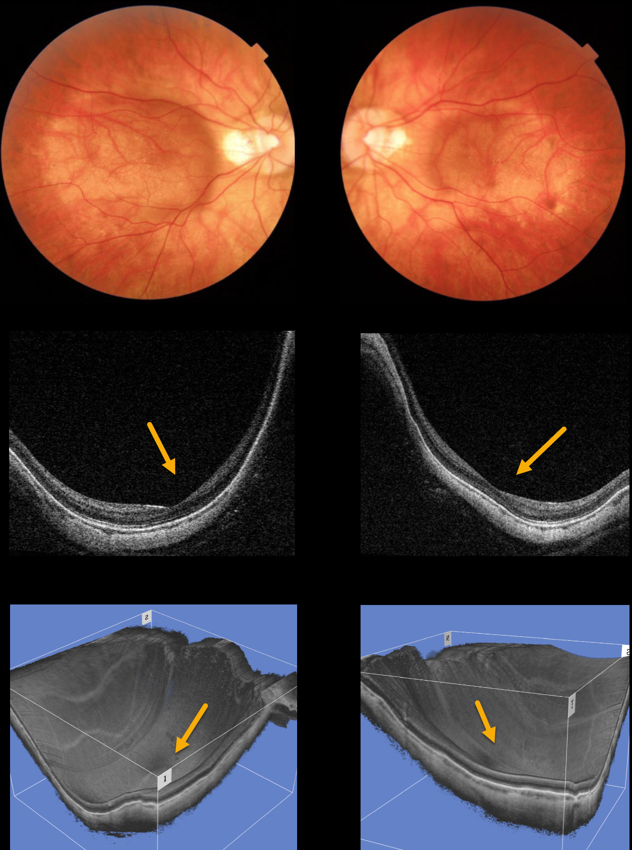
HIGH MYOPIA
Originally posted on @retina.rocks January 6, 2023
This 42YO male has a history of high myopia, of -14 diopters OD and -12.5 diopters OS. Vision is 20/60 OU.
Color photographs show bilateral macular staphyloma. Swept-source OCT B-scans and 3-D reconstruction more dramatically show the macular staphylomas.
Learning Points:
A macular staphyloma is a concave posterior bowing of the sclera in patients with high myopia. These myopic patients also have a markedly thinned choroid throughout the posterior pole.
Due to stretching of the posterior retinal structures, these patients are at increased risk of numerous findings, including macular schisis/traction and macular neovascularization (MNV).
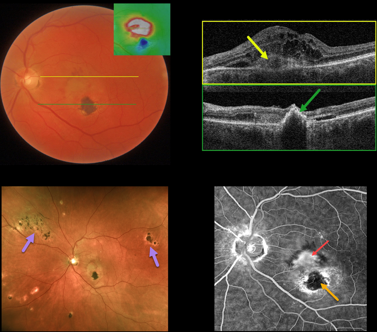
OCULAR HISTOPLASMOSIS
Originally posted on @retina.rocks January 3, 2023
This 62YO male presented with acute vision loss of 20/400 in his left eye. He gave a history of thermal laser in this eye about 25 years earlier for ocular histoplasmosis.
Color photography shows opaque macular fluid with some dots of superior subretinal blood.
OCT map shows central and superior retinal thickening. OCT B-scan through the macular center shows a type 2 macular neovascularization (MNV, located above the RPE) with marked overlying macular edema and trace nasal subretinal fluid. A B-scan through the inferior macular laser scar shows a full-thickness, markedly hyperreflective lesion.
Fluorescein angiography shows a leaking subfoveal MNV with a central hypofluorescent laser scar with surrounding staining.
Optos ultrawidefield imaging provides a broader perspective on macular, peripapillary, and peripheral chorioretinal scarring.
An intravitreal Avastin injection was given, and this will be followed by half-fluence photodynamic therapy to try to minimize the treatment burden.
Learning Points:
Ocular histoplasmosis is caused by the Histoplasmosis capsulatum fungus, which is commonly found in soil contaminated by bird or bat droppings.
It is seen worldwide, but in the United States, it is most often seen in the Ohio-Mississippi River valley. Most people in endemic areas inhale the organism’s spores early in life, with only a few percent developing asymptomatic chorioretinal scars.
A small percentage of these will develop MNV later in life. Treatment is similar to wet AMD, with anti-VEGF therapy usually being the first-line therapy. Unlike AMD, these patients often do not require ongoing injections.
Photodynamic therapy can help minimize treatment burden, and thermal laser therapy remains a good option for more peripheral macular lesions.
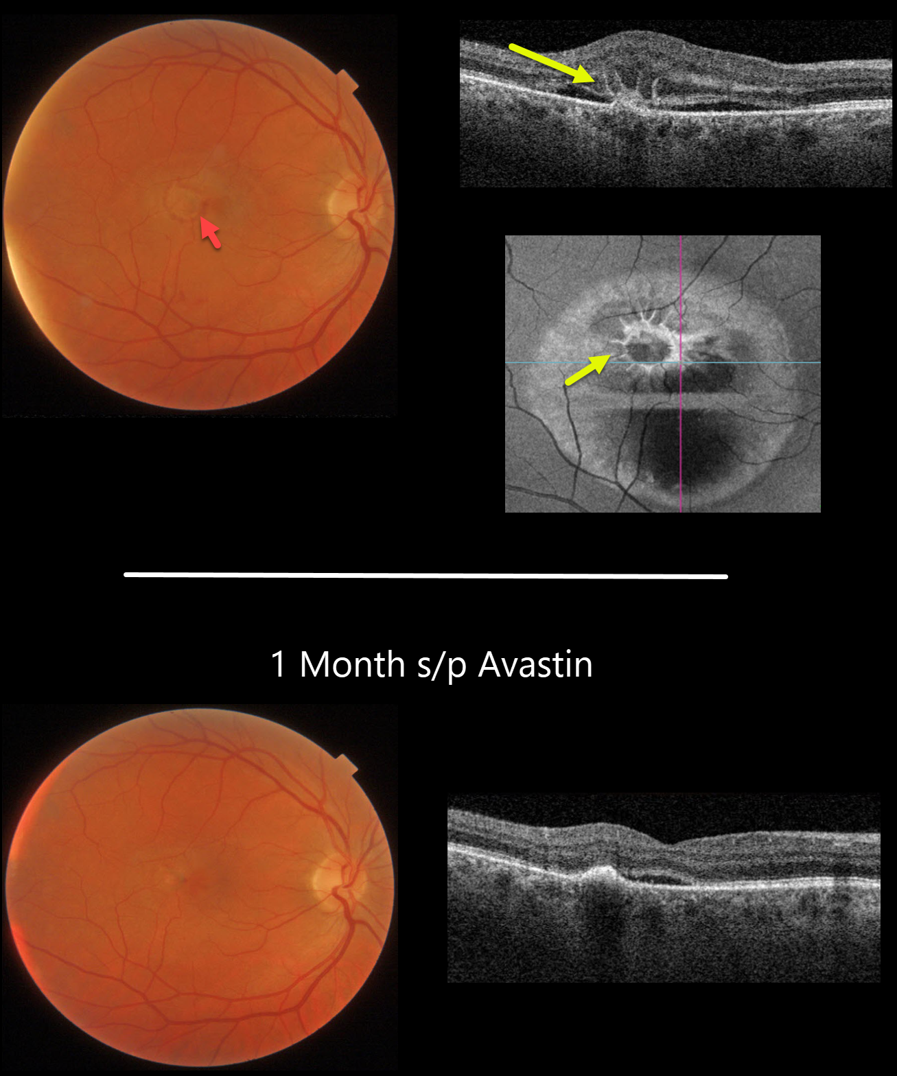
IDIOPATHIC MACULAR NEOVASCULARIZATION (MNV)
Originally posted on @retina.rocks January 2, 2023
This 49YO male presented with acute vision loss of 20/400 in his right eye from an idiopathic macular neovascularization (MNV).
Color photography shows shallow foveal fluid with some areas of subretinal blood. Swept-source OCT shows outer macular edema, shallow subretinal fluid, and radiating hyperreflective lines (pitchfork sign). These radiating lesions are uniquely visualized with en face OCT.
The lesion was virtually dry, with resolution of the pitchfork lesion 1 month after a single intravitreal Avastin injection. Vision improved to 20/80.
Learning Points:
The pitchfork sign was originally described by Hoang et al (Retina 2013;33:1049-1055) as a unique OCT finding in inflammatory MNV.
Falavarajani et al more recently noted that this finding is also associated with non-inflammatory MNV (Ophthalmic Surg Lasers Imaging Retina 2019;50:719-725).
The pathogenesis of the avascular pitchfork spikes is unknown, but may be related to outer retinal traction or Müller cell activation.
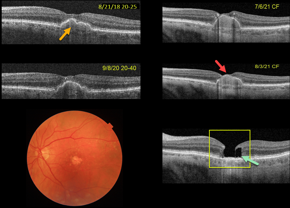
LAMELLAR HOLE AFTER AMD PIGMENT EPITHELIAL DETACHMENT (PED) COLLAPSE
Originally posted on @retina.rocks December 30, 2022
This 75YOF was seen in 2018 with non-neovascular AMD and a central pigment epithelial detachment (PED). Vision was 20/25. She returned on 9/8/20 with 20/40 vision with mild enlargement of the PED.
She complained of decreased vision when seen nearly one year later. Vision had dropped to 20/80. The PED had enlarged to involve full-thickness retina. She elected for an Eylea injection in hopes of possibly treating an occult macular neovascularization vs an avascular PED.
On 7/6/21, vision decreased to counting fingers although the OCT looked fairly stable. An additional Eylea injection followed by half-fluence PDT was given. On 8/3/21, the dome of the PED now extended through the inner retina and vision remained at counting fingers.
On 1/4/22, the PED had completely collapsed, with secondary retinal atrophy and a degenerative lamellar macular hole (LMH).
The progression of her PED through full-thickness retina and subsequent collapse with secondary retinal/RPE atrophy and a LMH are all quite unusual. Fortunately her right eye is 20/20 with intermediate dry AMD.
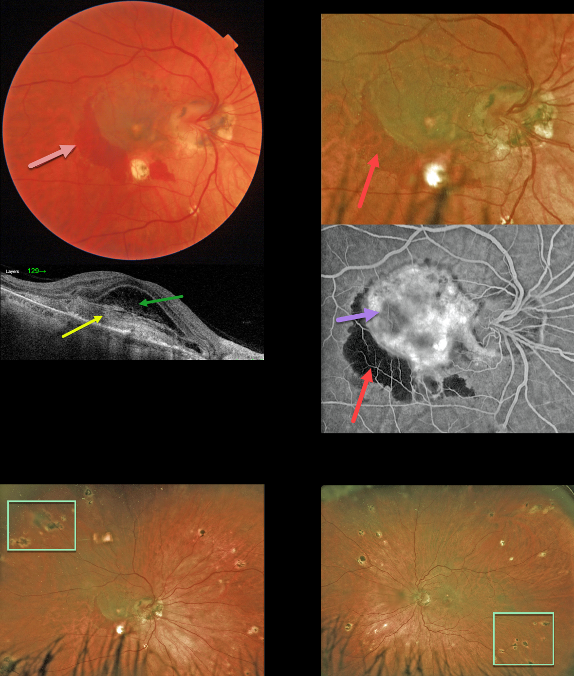
OCULAR HISTOPLASMOSIS
Originally posted on @retina.rocks December 16, 2022
This 52 YO female presented to the clinic with vision of counting fingers OD and 20/25 OS. She reported that her vision had decreased 3 weeks earlier.
Color imaging shows a somewhat pigmented macular neovascularization (MNV) with associated subretinal blood.
Swept-source OCT shows a hyperreflective, laminated type 2 (located above the RPE) MNV. Overlying fibrinous-appearing subretinal fluid vs a bacillary layer detachment is seen.
Optos fluorescein angiography, magnified to show the macular details, show a leaking, hyperfluorescent MNV with adjacent blockage from the subretinal hemorrhage.
Ultrawidefield Optos imaging shows the extent of the bilateral peripheral multifocal chorioretinal scarring. Anti-VEGF therapy was started.
Learning Points:
Ocular histoplasmosis is caused by the Histoplasmosis capsulatum fungus, which is commonly found in soil contaminated by bird or bat droppings.
It is seen worldwide, but in the United States is most often seen in the Ohio-Mississippi River valley. Most people in endemic areas inhale the organism’s spores early in life, with only a few percent developing asymptomatic chorioretinal scars.
A small percentage of these will develop MNV later in life. Treatment is similar to wet AMD, with anti-VEGF therapy usually being the first-line therapy. Unlike AMD, these patients often do not require ongoing injections. Photodynamic therapy can help minimize the treatment burden, and thermal laser is still a good option for more peripheral macular lesions.
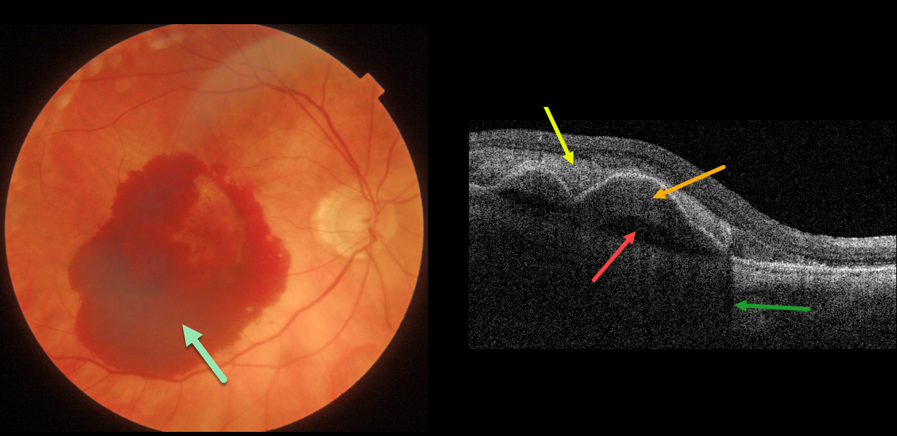
NEOVASCULAR AMD WITH PRECHOROIDAL CLEFT
Originally posted on @retina.rocks December 14, 2022
This 84 YO female with macular degeneration presented with acute vision loss of counting fingers vision in her right eye from neovascular AMD (nAMD).
Fundus photography shows a large central subretinal hemorrhage. Swept-source OCT shows the hyperreflective subretinal blood, which shadows the underlying choroid. Sub-RPE blood vs a type-1 (located beneath the RPE) macular neovascularization (MNV) overlies a prechoroidal cleft.
Anti-VEGF therapy was begun.
Learning Points:
A prechoroidal cleft is a hyporeflective space between the RPE and Bruch’s membrane. These are seen in up to about 20% of treated eyes with nAMD, particularly with type 3 neovascularization (intraretinal MNV) and polypoidal choroidal vasculopathy.
They are usually associated with worse visual acuity due to potential complications including RPE tear and subretinal hemorrhages (Kim et al, Retina 2017;37:2047-2055).
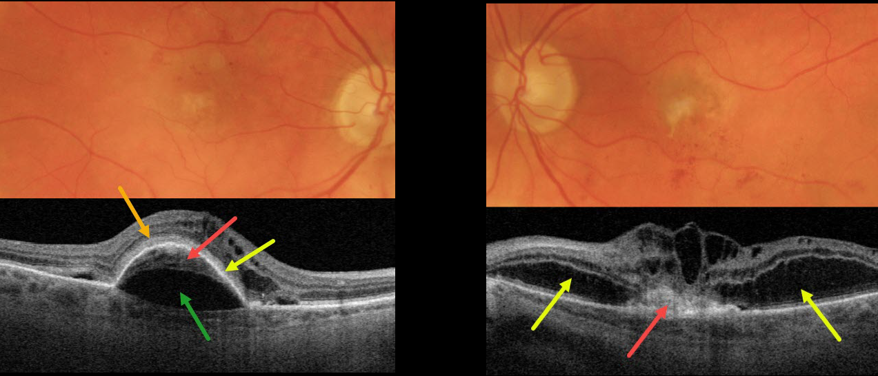
NEOVASCULAR AMD PRECHOROIDAL CLEFT + BACILLARY DETACHMENT
Originally posted on @retina.rocks November 29, 2022
This 87YO patient presented with vision of 20/200 OD and counting finger vision OS from bilateral active neovascular AMD (nAMD).
The right macula has a few dots of central and superior macular blood. OCT scanning shows a type 1 macular neovascularization (MNV, located below the RPE) adherent to an overlying RPE detachment. A prechoroidal cleft separates the MNV from the underlying choroid. Subretinal hyperreflective material (SRHM) lies above the RPE detachment (orange arrow). Intraretinal fluid is noted nasally with trace subretinal fluid on either side of the RPE detachment.
The left macula has faint subfoveal fibrosis with some scattered more inferior subretinal blood. OCT scanning shows a type 2 MNV (located above the RPE), which is tethered to the overlying edematous retina. Bacillary layer detachments are noted nasal and temporal to the MNV.
Learning Points:
A prechoroidal cleft is a hyporeflective space between the RPE and Bruch’s membrane. These are seen in up to about 20% of treated eyes with nAMD, particularly with type 3 neovascularization (intraretinal MNV) and polypoidal choroidal vasculopathy.
They are usually associated with worse visual acuity due to potential complications, including RPE tear and subretinal hemorrhages (Kim et al, Retina 2017;37:2047-2055).
SHRM is an OCT finding that is invisible clinically. This material, located between the neurosensory retina and retinal pigment epithelium (RPE), is thought to consist of fluid, fibrin, blood, and other fibrovascular tissues.
SHRM is a biomarker of active disease that decreases with successful anti-VEGF therapy (Willoughby et al., Ophthalmology 2015;122:1846-1853). Persistent SHRM is associated with an increased incidence of scar formation and worse VA.
Bacillary layer (cone and rod inner and outer segments) detachment appears as a unique dome-shaped collection of intraretinal fluid from photoreceptor splitting. These detachments can be observed in an increasing number of conditions, most classically Vogt-Koyanagi-Harada disease (Cicinelli et al, Ophthalmology Retina 2020;4:454-456).

AMD DISCIFORM SCAR
Originally posted on @retina.rocks November 28, 2022
This 70YO male presented with counting fingers vision in his right eye due to a large disciform scar with radiating inner retinal folds. The radiating folds appear as corrugations on OCT B-scan and as radiating lines on OCT en face.
Learning Points:
Disciform scars were the norm for untreated wet AMD prior to the anti-VEGF era that began in the mid-2000s. New-onset wet AMD diagnosed and treated with appropriate anti-VEGF dosing virtually never goes on to develop these lesions.
There may be progressive macular atrophy following years of therapy, but not this type of fibrotic scarring.
The radiating folds in our patient are caused by contraction of the fibrosed macular neovascularization which is centripetally dragging the overlying neurosensory retina.
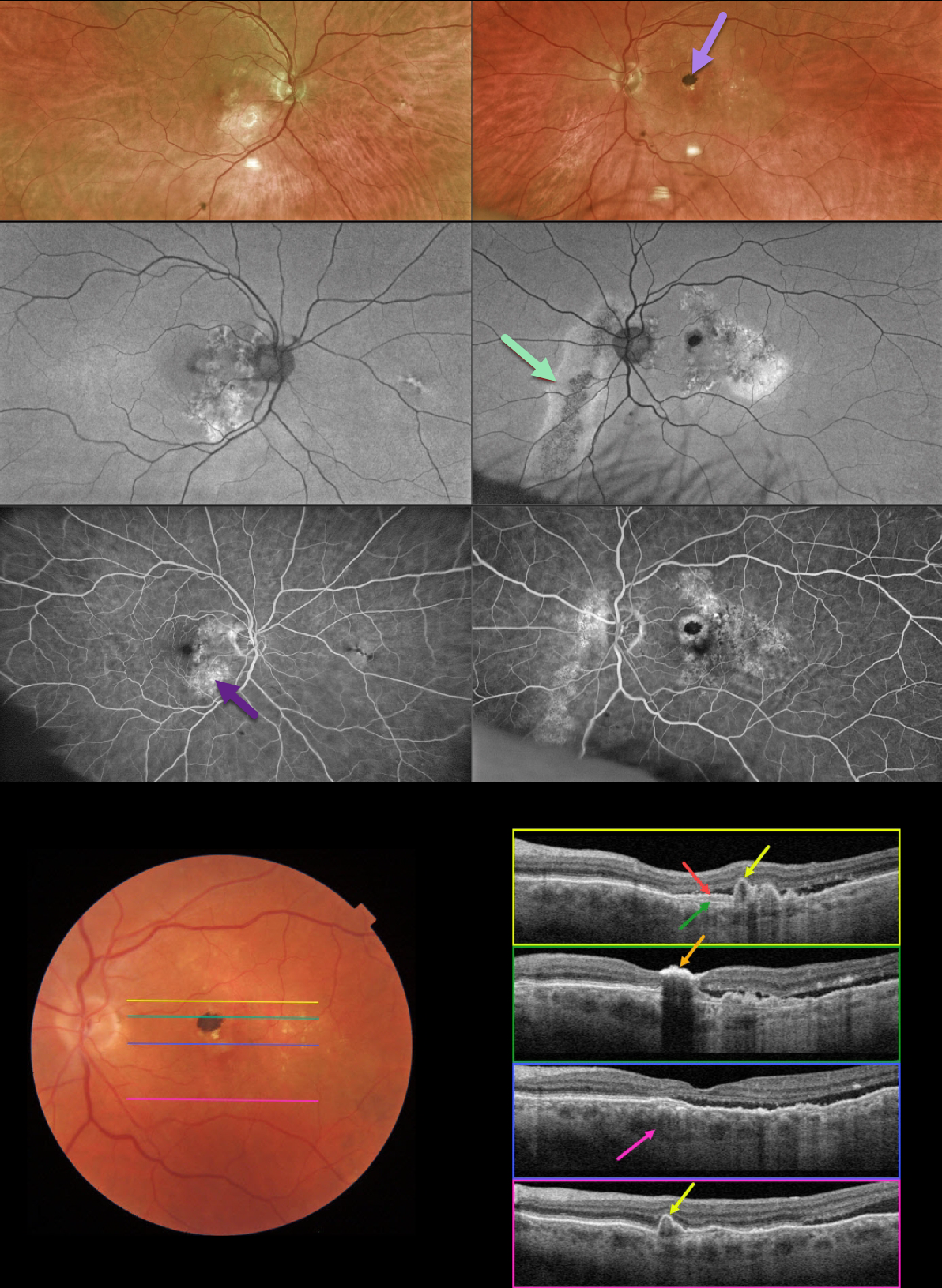
PACHYCHOROID SPECTRUM
Originally posted on @retina.rocks November 22, 2022
This 78YO male was referred for asymptomatic fundoscopic changes. Vision was 20/25 OD and 20/30 OS. He had a history of laser treatment in his left eye decades earlier.
Optos imaging shows bilateral areas of pigmentary loss in each macula, extending inferiorly. A hyperpigmented laser scar is noted in the left superior macula.
Fundus autofluorescence (FAF) shows variable hypo- and hyper-FAF, along with a prominent gutter extending from the left nerve inferonasally.
Fundus fluorescein angiography (FFA) shows window defects within these areas, with blockage from the hyperpigmented laser scar OS.
Swept-source OCT shows numerous findings. Scanning superior to the laser scar shows several choroidal polyps. A shallow elevation of the RPE exposes the underlying Bruch’s membrane. Scanning through the laser scar shows intense hyperreflectivity with posterior shadowing. Scanning through the macular center shows temporal subretinal fluid, along with a thickened choroid. Finally, scanning in the inferior macula shows an additional polyp.
Learning Points:
This case has an overwhelming amount of features from the pachychoroid spectrum, which includes idiopathic central serous retinopathy (ICSC), pachychoroid neovasculopathy, polypoidal choroidal vasculopathy, pachychoroid pigment epitheliopathy, peripapillary pachychoroid syndrome, and choroidal excavation.
The thermal laser treatment many years ago may have been for pachychoroid neovasculopathy, a recently described phenotype (see Pang and Freund, Retina 2015;35:1-9) found in the pachychoroid spectrum. These lesions are particularly common in patients with ICSC, and often remain silent as a flat, irregular RPE elevation on OCT without exudative complications.
Our patient likely had further progression of their pachychoroid disease, resulting in multiple polyps, which are found in polypoidal choroidal vasculopathy (PCV). PCV is a variant of type 1 macular neovascularization, also in the pachychoroid spectrum. PCV is likely distinct from age-related macular degeneration, occurring in a different population (more common in African Americans and Asians), with a relative lack of drusen, and the presence of a thicker choroid.
In addition to the findings above, the patient had a large gutter in his left eye, a common finding in ICSC. Gutters form from pathologies with chronic leakage. Conditions that form gutters other than ICSC include choroidal hemangiomas, choroidal nevi, and uveal melanoma.
For more information on the pachychoroid spectrum, see Cheung et al, Eye 2019;33:14-33 for a great pachychoroid disease review.
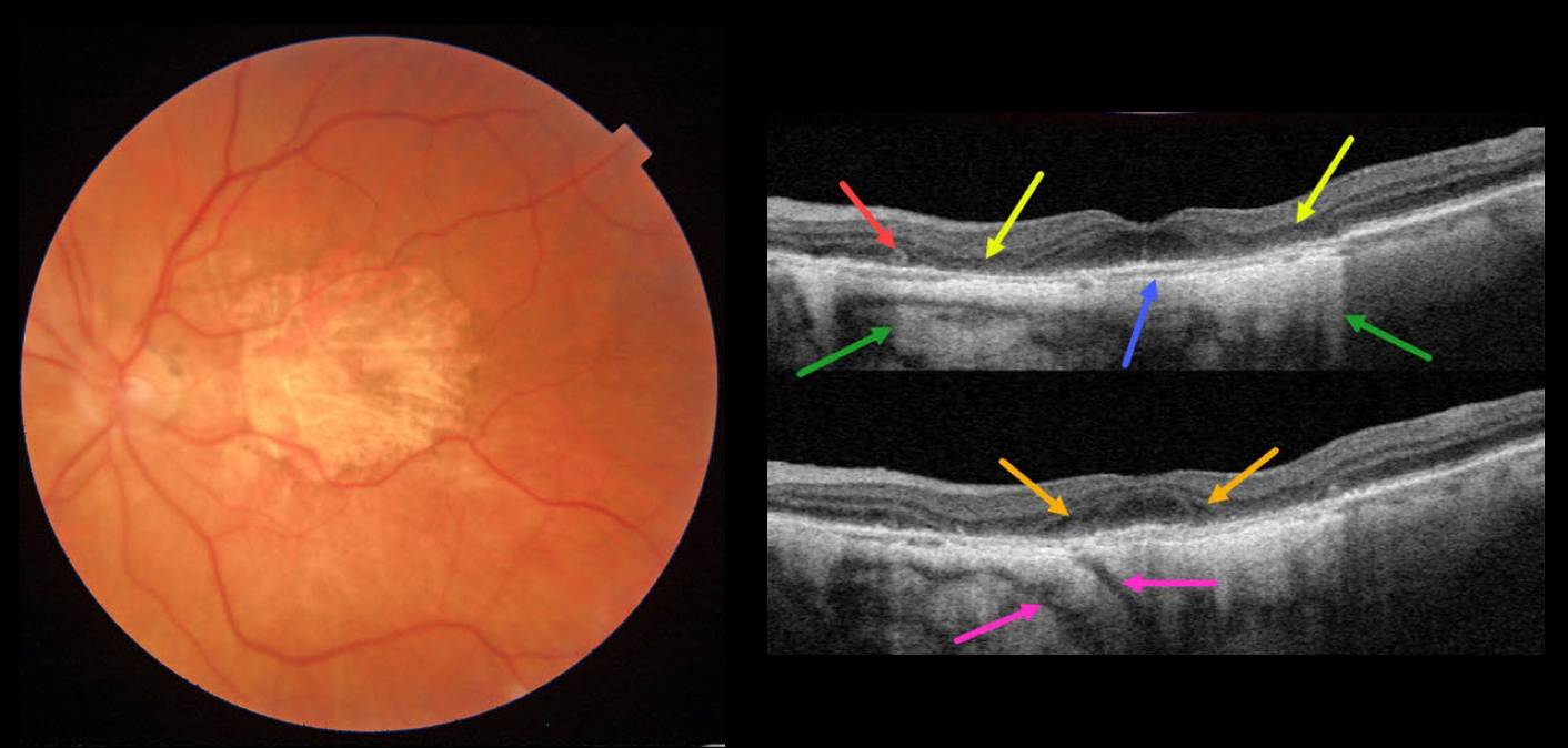
AMD MACULAR ATROPHY
Originally posted on @retina.rocks November 2, 2022
This 84YO female has stable advanced non-exudative AMD with bilateral macular atrophy (MA). There are numerous classic findings on multimodal imaging of her left eye.
Color imaging shows foveal MA. Swept-source OCT shows pericentral outer retinal and RPE atrophy. This atrophy allows for more light to illuminate the underlying choroid and sclera, as well as visualizing two short posterior ciliary arteries posterior to the sclera. The variably atrophic RPE allows for visualization of Bruch membrane, which is normally fused in the RPE-Bruch layer seen in normal eyes. There is a tiny area of outer retinal tubulation (ORT) nasally. Finally, hyporeflective outer plexiform layer wedge defects are noted along the descending outer retinal atrophy.
Wedge defects develop at the boundaries of the degenerating outer plexiform layer in about 75% of MA eyes (Mones et al, Ophthalmology 2012;119:1412-1419).
ORTs represent photoreceptor and Muller cell degeneration (Dolz-Marco et al, Ophthalmology 2017;124:1353-1367). These ORTs have a hyper-reflective border with a central hyporeflective core.
Learning Points:
In 1970, Dr. Gass originally described macular atrophy from AMD as “geographic areas of atrophy” in the setting of “senile macular choroidal degeneration” (Schmitz-Valkenberg, Retina 2016;36:2250-2264).
Today, the terminology is much more specific due to advances in technology and improved visualization of retinal and choroidal structures.
From Sadda et al, Ophthalmology 2018:537-548, the new classification is as follows: Complete RPE and Outer Retinal Atrophy (cRORA), Incomplete RPE and Outer Retinal Atrophy (iRORA), Complete Outer Retinal Atrophy (cORA), and Incomplete Outer Retinal Atrophy (iORA).
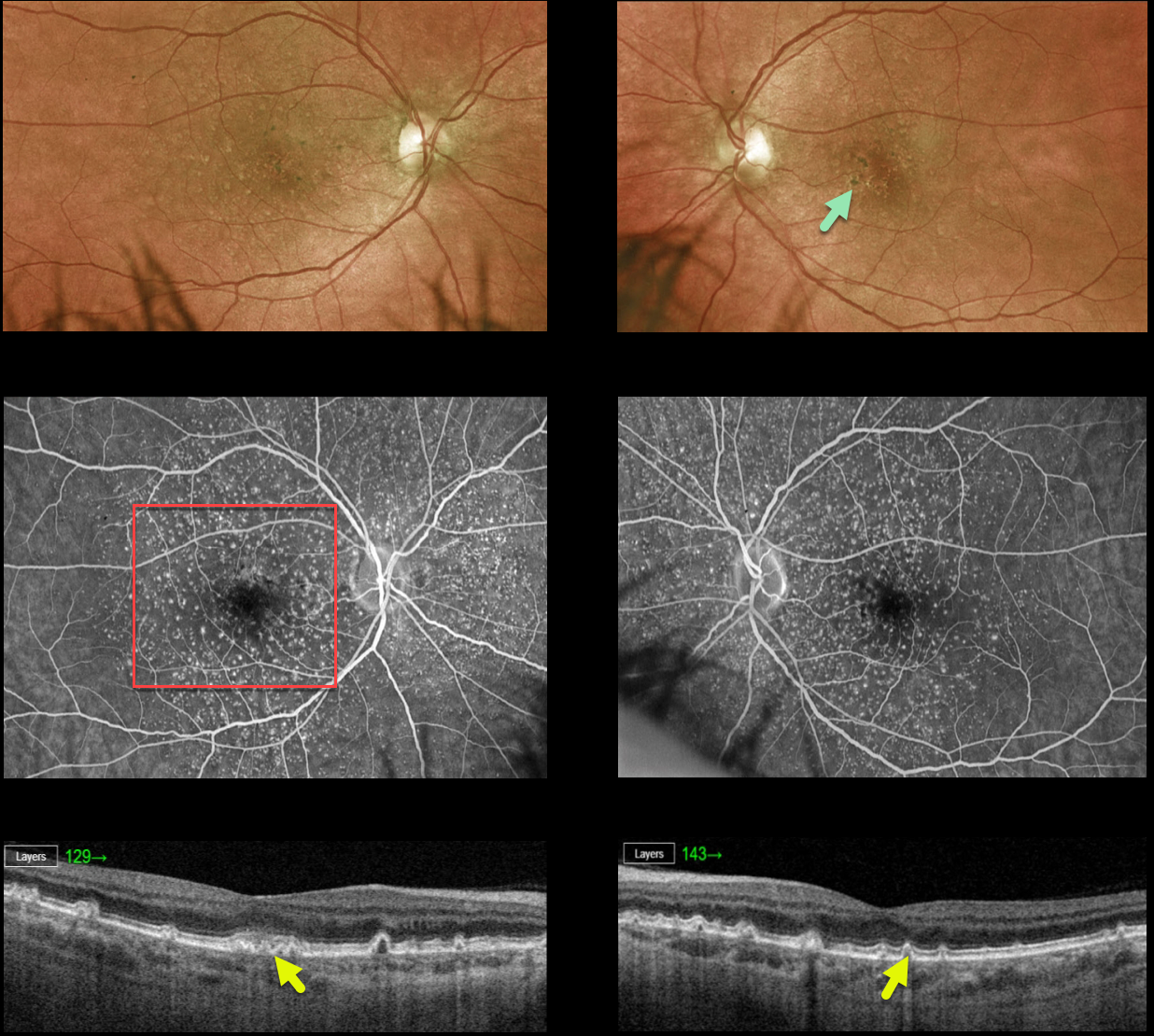
CUTICULAR DRUSEN
Originally posted on @retina.rocks October 26, 2022
This 55YO female is being followed for asymptomatic dry AMD. Vision is 20/30 OD and 20/25 OS.
Optos color imaging shows extensive small drusen with foveal pigment clumping. Fluorescein angiography shows the classic ‘starry sky’ pattern of hyperfluorescent cuticular drusen. Swept-source OCT shows mostly cuticular drusen with arrowhead-like small elevations of the RPE.
Learning Points:
Originally described by Gass as basal laminar drusen (AJO 1985;99:445-459), cuticular drusen were initially thought to represent nodular thickening of the RPE basement membrane.
They appear clinically as numerous, round, yellow-white ‘hard drusen’, and on OCT as small, arrowhead-shaped drusen located above Bruch’s membrane and below the RPE (Spaide and Curcio, Retina 2010;30:1441-1454).
Our patient’s angiographic appearance echoes Dr. Gass’ original description of “a beautiful stars-in-the-sky or Milky-Way pattern” of hyperfluorescence. Cuticular drusen are classically associated with avascular vitelliform lesions, but can also develop macular neovascularization.
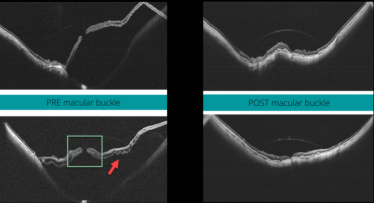
MACULAR HOLE
Veronika Matello and Barbara Parolini
Originally posted on @retina.rocks October 25, 2022
This 58YO highly myopic female presented with 20/400 vision in her left eye. The axial length was 32.45 mm, and her refractive error prior to LASIK surgery was 13D.
Widefield OCT scanning (Canon Xephilio SQ) shows a large macular retinal detachment with (or associated to) a full-thickness macular hole, defined as stage 4c according to the Myopic Traction Maculopathy Staging System (Parolini et al, Eur J Ophthalmol 2021;31:1299-1312).
Pars plana vitrectomy with an ILM flap and macular buckling was performed. One month postoperatively, the retina was flat, and the macular hole closed. Vision was 20/80.
Learning Points:
In our experience, these detachments are totally tractional, with the macular hole developing before or after the retina detaches. The macular buckle treats both the macular schisis and detachment, whereas the vitrectomy with ILM peeling closes the macular hole.
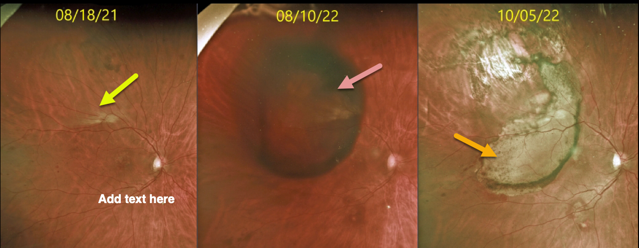
PERIPHERAL EXUDATIVE HEMORRHAGIC CHORIORETINOPATHY
Originally posted on @retina.rocks October 24, 2022
This 58YO diabetic male was seen originally with mild scattered retinal hemorrhages from nonproliferative retinopathy, along with a faint patch of myelinated nerve fiber layer.
He presented one year later after waking with new nasal field loss. Vision was 20/70, and he had a new, large, superotemporal subretinal hemorrhage just outside the macula.
Following 2 monthly intravitreal Avastin injections, the subretinal blood resolved, with secondary subretinal scarring and yellowing devitalized blood. Vision is 20/60.
Learning Points:
Ectopic choroidal neovascularization (CNV) can often masquerade as a choroidal mass or uveal melanoma. It can also cause extensive choroidal/suprachoroidal, subretinal, and vitreous hemorrhage, more commonly in patients who are on blood thinners.
The subretinal blood often spontaneously regresses without treatment, although anti-VEGF injections can be used for symptomatic or increasing exudation.
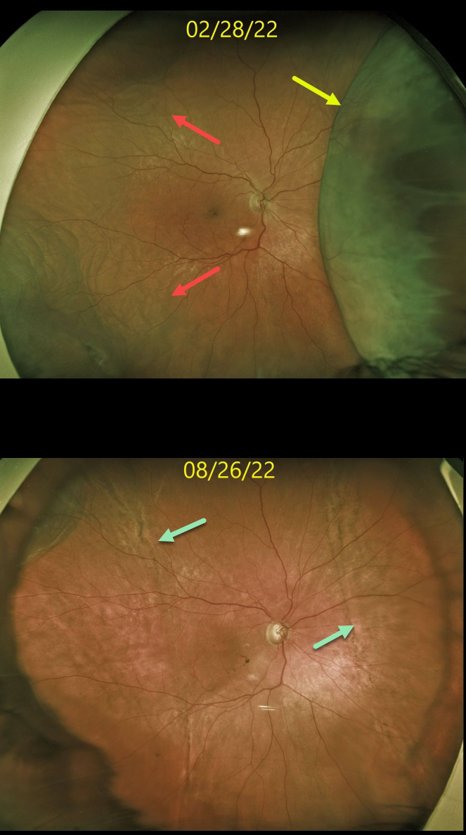
CHOROIDAL DETACHMENT
Originally posted on @retina.rocks October 7, 2022
This 74YO female was receiving periocular and intravitreal steroids for persistent pseudophakic cystoid macular edema (CME). Steroid response prevented further treatment, and she eventually required XenGel glaucoma surgery to control her pressure and allow for continued CME treatment.
Four days following glaucoma surgery, vision was 20/200, and the intraocular pressure (IOP) was 6 mmHg. Optos imaging shows a large nasal choroidal detachment with a shallow temporal choroidal detachment.
Over the next 3 months, her intraocular pressure normalized with total resolution of the choroidals. A ‘high-water’ mark of subretinal pigmentation is seen along the posterior extent of both the prior nasal and temporal choroidal detachments.
Learning Points:
Choroidal detachment is relatively common immediately following glaucoma surgery.
Two mechanisms are believed to be at play: hypotony allows fluid to accumulate in the suprachoroidal space, and inflammation increases the choroidal permeability. The detached ciliary body may also produce less aqueous humor, thus creating a self-perpetuating cycle.
Often, observation is sufficient, but topical steroids and cycloplegics can be helpful in promoting resolution.
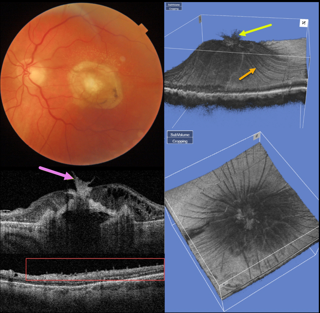
AMD SCAR
Originally posted on @retina.rocks October 4, 2022
This 77YO man presented counting fingers vision OS with what seems at first to be an uneventful fibrotic disciform scar from neovascular AMD. But multimodal imaging shows several interesting findings.
Swept-source OCT through the macular center shows that the central scar extends through the full-thickness retina and becomes incorporated with the attached vitreous.
A 3D macular reconstruction viewed from above also shows how scar contraction is creating radiating retinal folds.
These folds on a B-scan taken through the peripheral macula give the appearance of numerous needle-like projections emanating from the inner retina.
The needle-like projections, created by imaging the radiating inner retinal folds perpendicular to their orientation, mimic findings felt to be pathognomonic for ocular amyloidosis (Kakuhara et al, Ophthalmology Retina 2021;5:1005-1008).
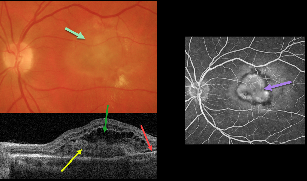
OCULAR HISTOPLASMOSIS
Originally posted on @retina.rocks September 28, 2022
This 54YO female presented with recent vision loss in her left eye. Vision was 20/400 from an ocular histoplasmosis (OHS) related macular neovascularization (MNV).
Optos color imaging shows opaque macular fluid with a few dots of peripheral blood. Multifocal peripheral OHS scars were noted (not shown).
Swept-source OCT shows a type 2 MNV (located above the RPE), overlying outer retinal fluid and trace temporal subretinal fluid.
Fluorescein angiography shows a well-defined subfoveal lesion. Anti-VEGF therapy was recommended.
Learning Points:
Ocular histoplasmosis is caused by the Histoplasmosis capsulatum fungus which is commonly found in soil contaminated by bird or bat droppings.
It is seen worldwide, but in the United States is most often seen in the Ohio-Mississippi River valley. Most people in endemic areas inhale the organism’s spores early in life with only a few percent developing asymptomatic chorioretinal scars.
A small percentage of these will develop MNV later in life. Treatment is similar to wet AMD, with anti-VEGF therapy usually being the first line therapy. Unlike AMD, these patients often do not require ongoing injections.
Photodynamic therapy can help minimize the treatment burden, and thermal laser is still a good option for more peripheral macular lesions.
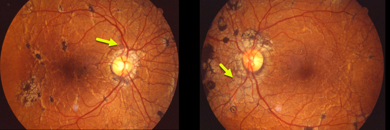
ANGIOID STREAKS
Originally posted on @retina.rocks September 26, 2022
This patient was examined in 1989 and had classic findings of pseudoxanthoma elasticum. Extensive angioid streaks encircle and radiate from the nerves, extending into the peripheral maculas and beyond the arcades.
Angioid streaks are usually less dramatic and are often quite easy to miss since their color and orientation mimic the normal retinal vasculature, hence the term “angioid”.
The orange skin (peaux d’orange) appearance, most pronounced in each temporal macula, is caused by the calcified Bruch’s membrane giving a yellow mottled ‘coquille d’oeuf’ (eggshell) appearance that contrasts with the normal orange RPE and choroidal pigmentation (Spaide, Retina 2015;35:392-397).
Learning Points:
Angioid streaks are known to be associated with systemic conditions such as pseudoxanthoma elasticum, Ehlers-Danlos syndrome, Paget’s disease, sickle cells, or idiopathic (PEPSI).
These patients are at significant risk for macular neovascularization and need to be followed and treated as needed with anti-VEGF agents, as in patients with neovascular AMD.
Genetic testing with pseudoxanthoma would test positive for a pathogenic ABCC6 mutation.
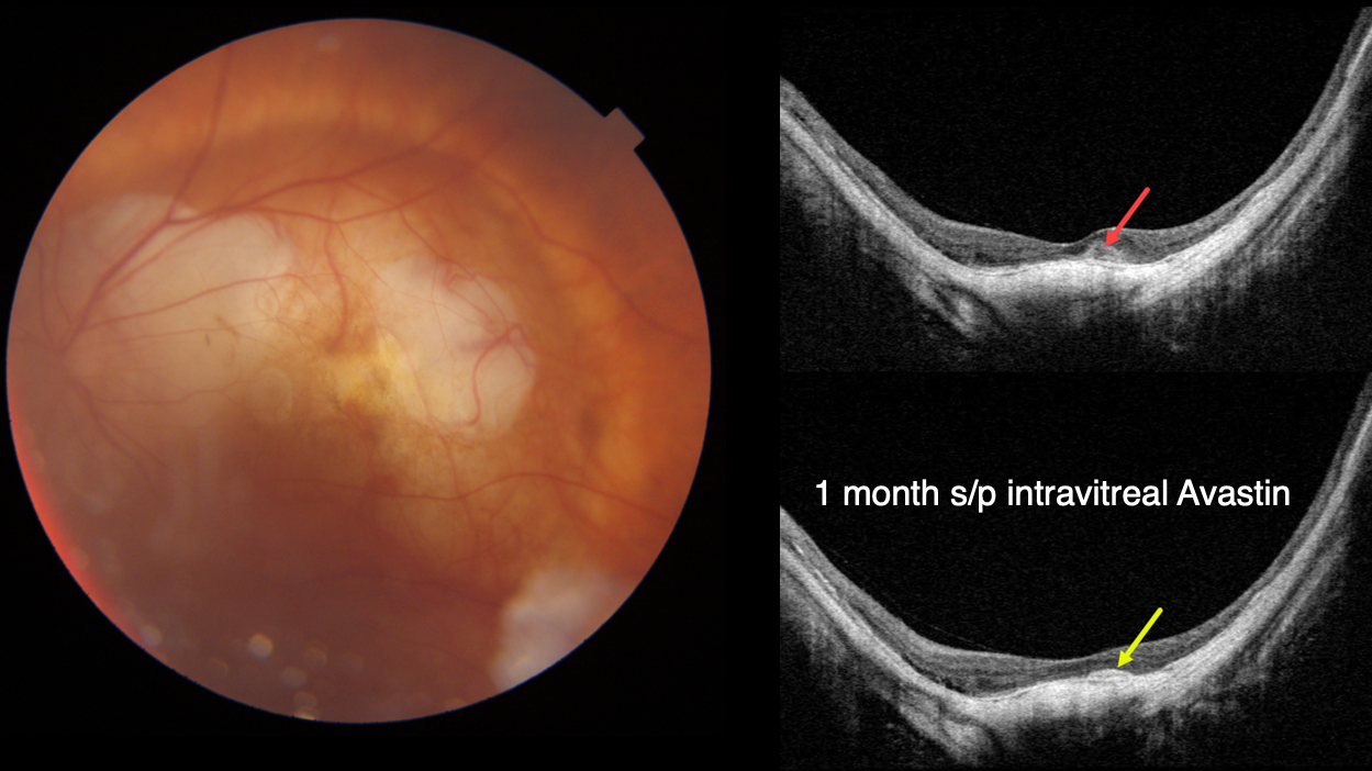
MYOPIC MACULAR NEOVASCULARIZATION
Originally posted on @retina.rocks September 22, 2022
This 92YO female presented with new vision loss of 20/200 in her left eye. She has a history of high myopia, with hand motion vision OD due to a macular staphyloma. Baseline vision in her left eye is 20/40.
Fundus photography shows peripapillary and mostly superior macular atrophy.
Swept-source OCT shows a posterior staphyloma with a dome appearance to the central macula. A new small hyperreflective macular neovascularization (MNV) is noted.
One month following an intravitreal Avastin injection, vision improved to 20/40 with shrinkage of the MNV.
Learning Points:
In our experience, myopic MNV presents quite differently from those found in neovascular AMD.
Significant subretinal blood, subretinal fluid or intraretinal fluid are rarely seen. Often an active MNV will present as a new dot of subretinal blood or pigment.
Occasionally there will just be symptomatic vision loss or metamorphopsia without clinical or OCT findings.
Anti-VEGF therapy is effective, and these lesions can often be managed with PRN therapy.
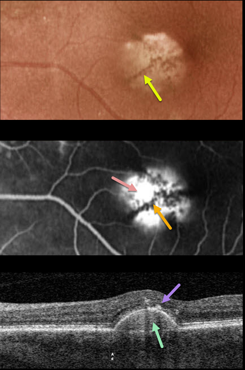
PACHYCHOROID PIGMENT EPITHELIAL DETACHMENT (PED)
Originally posted on @retina.rocks September 13, 2022
This 75YO female has been followed for several years with a stable, asymptomatic chronic retinal pigment epithelial detachment (PED) and 20/40 vision.
Optos color imaging shows a solid, variably depigmented, round PED in the inferonasal region of the left fovea. Radiating linear pigmentation is noted.
Fluorescein angiography shows hyperfluorescent pooling beneath the PED, with blockage from the overlying radiating pigment.
Swept-source OCT shows the PED with a hyperreflective core, overlying hyperreflective foci (representing the clinical radiating pigment), and a relatively thickened choroid for a patient of this age.
Learning Points:
Retinal pigment epithelial detachments are common in patients with AMD and are often associated with macular neovascularization. Non-vascularized PED is also seen as part of the pachychoroid spectrum.
The overlying pigment figure is pathognomonic for a chronic PED, and is seen even when these lesions flatten. Isolated avascular PEDs such as these do not require treatment.
Although our patient was in her mid-70s, there were no drusen and each fundus was otherwise normal. The chronicity of the PED, the lack of other signs of AMD, and the relatively thickened choroid all point to this being an avascular, pachychoroid PED.
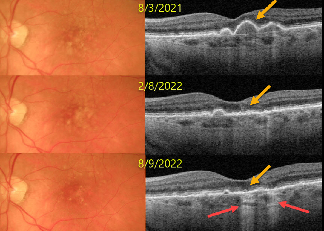
DRY AMD
Originally posted on @retina.rocks September 7, 2022
This 77YO female presented on 8/3/21 with asymptomatic bilateral confluent soft drusen. Vision was 20/30 OU.
Over the next year, the drusen flattened with secondary drusenoid atrophy. Transmission defects from incomplete outer retinal and RPE atrophy are noted at the most recent visit. Vision was 20/20 OU on the 8/9/22 visit.
Learning Points:
Drusen commonly remodel over time, and their collapse can lead to drusenoid atrophy of the RPE, as in our patient. Vision may or may not decrease following drusen regression.
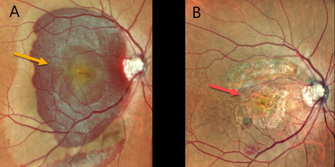
SUBMACULAR HEMORRHAGE
Akansha Sharma and Manish Nagpal
Originally posted on @retina.rocks September 6, 2022
This 73YOF presented with 1 day of acute vision loss in her right eye. Vision was counting fingers OD and hand motion OS.
A large submacular hemorrhage from neovascular AMD occupied the entire right macula. A fibrosed macular scar was noted in her left eye (image not shown).
She was scheduled for vitrectomy with anti-VEGF injection, but unfortunately, was not taken to surgery for about a month from presentation.
Postoperatively, there is dry atrophic macular scarring with virtually complete resolution of all blood, but vision remains at counting fingers from macular atrophy.
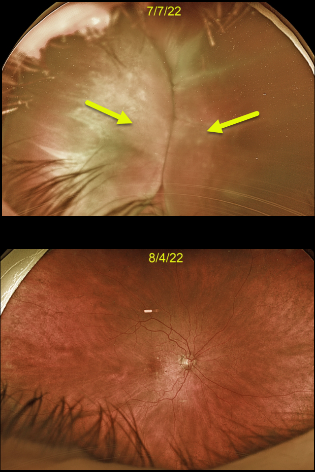
CHOROIDAL DETACHMENT
Originally posted on @retina.rocks August 15, 2022
This 88YO female developed kissing choroidal detachments immediately following Ahmed glaucoma valve placement.
The choroidal detachment spontaneously resolved after 1 month once the intraocular pressure normalized.
Learning Points:
Choroidal detachment is relatively common immediately following glaucoma surgery.
Two mechanisms are believed to be at play: hypotony allows fluid to accumulate in the suprachoroidal space and inflammation increases the choroidal permeability. The detached ciliary body may also produce less aqueous, thus creating a self-perpetuating cycle.
Often, observation is sufficient, but topical steroids and cycloplegics can be helpful to aid in resolution.
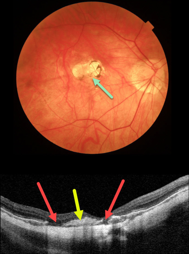
HIGH MYOPIA
Originally posted on @retina.rocks August 11, 2022
This 48YO female has a history of a previously treated myopic macular neovascularization (MNV) in her right eye, which has remained stable with observation for years. Triton color imaging shows an inactive atrophic foveal scar.
Swept source OCT shows an inactive hyperreflective central MNV with adjacent RPE defects. The choroid is severely thinned, consistent with her high myopia.
Learning Points:
An RPE aperture is a rare, recently described OCT finding most commonly seen in non-exudative AMD. Different from an RPE tear, these round RPE discontinuities typically develop from an avascular RPE detachment that remodels over time, resulting in atrophy of the RPE and outer retina.
This remodeling may result from regression of drusenoid material or damage to the RPE due to separation from the choriocapillaris (Querques G et al. Retina. 2016;36:S65-S72).
Some hypothesize that these lesions may represent small RPE micro-tears from underlying type 1 MNV causing increased hydrostatic pressure, leading to damage/atrophy of the RPE.
RPE apertures can also be found in patients with acquired vitelliform lesions (Falavarjani K et al, Canadian J Ophthalmol 2022;57:e91-e94).
We are not aware of a previously described case caused by myopic MNV.
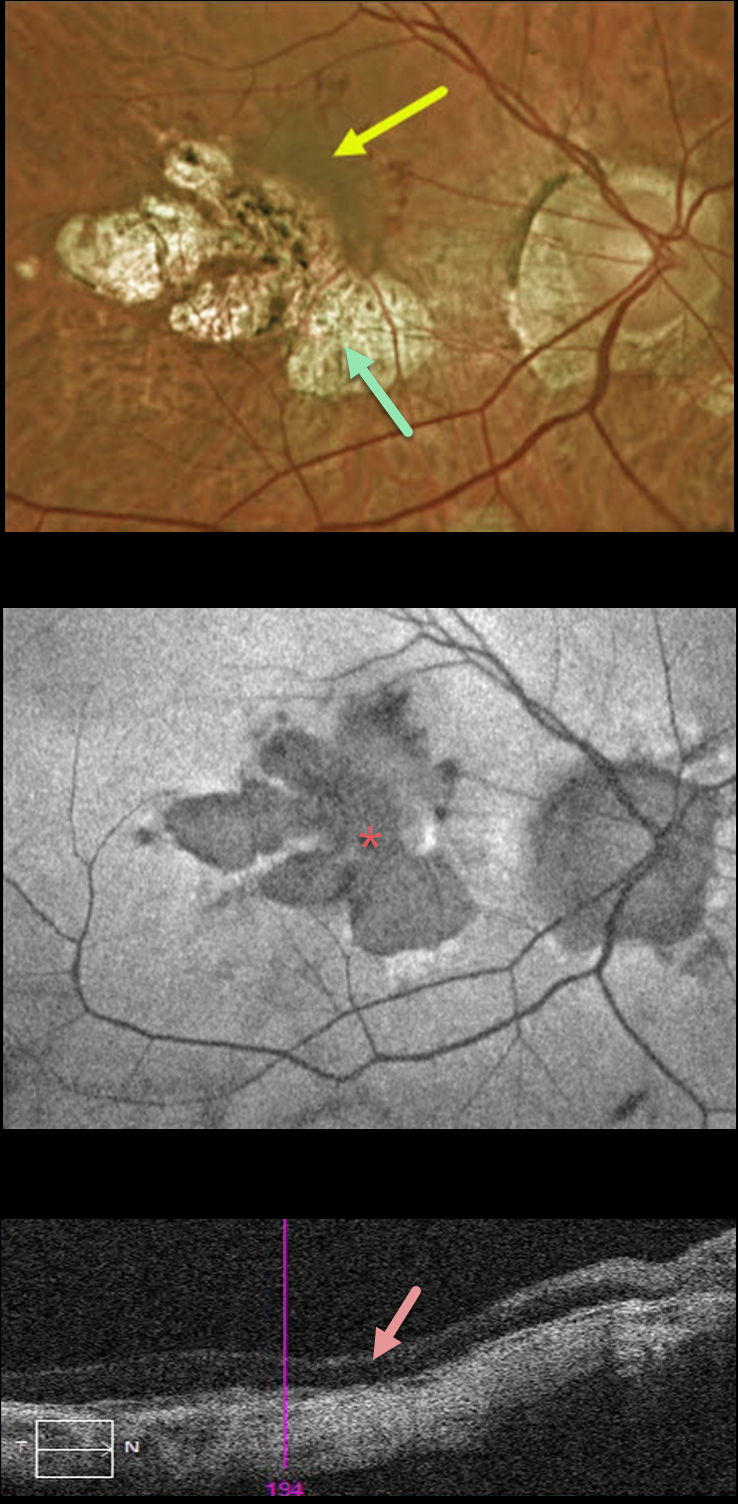
MYOPIC MACULAR NEOVASCULARIZATION
Originally posted on @retina.rocks July 26, 2022
This 51YO female received prior intravitreal injections in her right eye about 5 years earlier, but was subsequently lost to follow-up until she was referred to our office. She felt that her vision had been stable and poor in each eye for years. Vision was counting fingers OD and 20/200 OS.
Optos color imaging of her right eye shows myopic peripapillary and macular scarring extending through the macular center. A pigmented macular neovascularization (MNV) with some dots of subretinal blood extends superonasally from the atrophy. Fundus autofluorescence (FAF) shows areas of hypo-FAF corresponding to the areas of clinical atrophy. Triton swept-source OCT shows central retinal atrophy.
Given the foveal atrophy, severe vision loss, and absence of acute symptoms, we recommended careful observation. We will consider anti-VEGF therapy if the MNV causes increasing exudation.
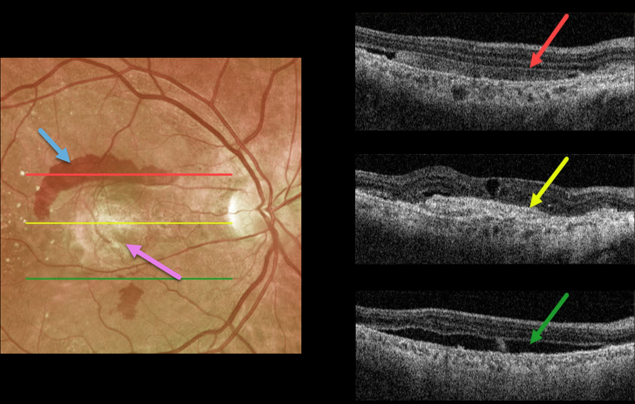
NEOVASCULAR AMD
Originally posted on @retina.rocks July 20, 2022
This 72YO male presented with longstanding decreased vision in his right eye. Vision was counting fingers due to a fibrosed scar from neovascular age-related macular degeneration (nAMD).
Optos imaging shows a central fibrosed macular scar with superior and inferior subretinal blood. Triton swept-source imaging shows multiple cool findings from this single neovascular lesion. Superiorly, the subretinal blood appears as hyperreflective material in the subretinal space. More centrally, the fibrotic scar/type 2 macular neovascularization is hyperreflective. There are also some overlying chronic cystic retinal changes. More inferiorly is shallow, subclinical subretinal fluid.
A therapeutic trial of monthly anti-VEGF injections was initiated, although we’re not sure this will have a visual effect given the subfoveal fibrosis.
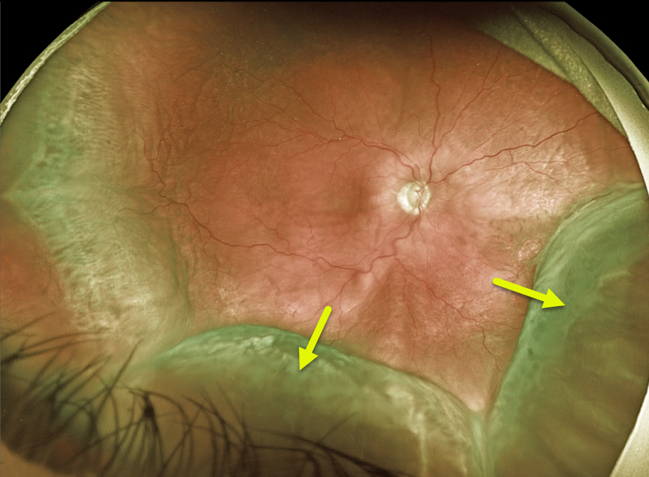
CHOROIDAL DETACHMENT
Originally posted on @retina.rocks July 14, 2022
This 68YO male patient presented with peripheral choroidal detachments following placement of a Baerveldt shunt 1 week earlier. Vision was counting fingers, and pressure was 1 mmHg. All wounds were secure, and the anterior chamber was formed.
Learning Points:
Choroidal detachment is relatively common immediately following glaucoma surgery. Two mechanisms are believed to be at play:
1) hypotony allows fluid to accumulate in the suprachoroidal space, and 2) inflammation increases the choroidal permeability. The detached ciliary body may also produce less aqueous, thus creating a self-perpetuating cycle.
Often, observation is sufficient, but topical steroids and cycloplegics can be helpful in promoting resolution. We are following this patient closely and expect the choroidals to resolve as postoperative pressure increases.
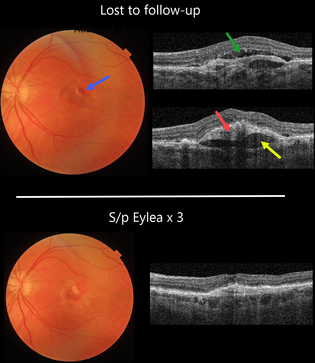
NEOVASCULAR AMD + PRECHOROIDAL CLEFT
Originally posted on @retina.rocks July 8, 2022
This 82YO female was maintaining 20/40 vision in her left eye with intravitreal Eylea every 8 weeks for neovascular AMD. She was then lost to follow-up, with new blood and 20/50 vision.
Swept source OCT shows new subretinal fluid. The hyperreflective sub-RPE type 1 macular neovascularization is elevated above a hyporeflective prechoroidal cleft.
Fortunately, vision returned to 20/30, with complete resolution of all exudative findings, following 3-monthly Eylea injections.
Learning Points:
A prechoroidal cleft is a hyporeflective space between the RPE and Bruch’s membrane. These are seen in up to about 20% of treated eyes with wet AMD, particularly with type 3 neovascularization and polypoidal choroidal vasculopathy. They are usually associated with worse visual acuity due to potential complications, including RPE tear and subretinal hemorrhages.
For a more detailed discussion of prechoroidal clefts, see Kim et al Retina 2017;37:2047-2055.
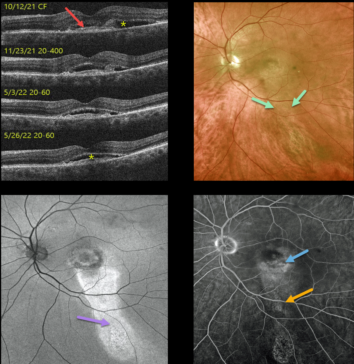
PACHYCHOROID NEOVASCULOPATHY WITH GUTTER
Originally posted on @retina.rocks July 1, 2022
This 69YO male presented with asymptomatic vision loss in his left eye. Vision was 20/30 OD and counting fingers OS.
A small central scar with fluid was present in the left macula (initial photo not shown). OCT scanning shows a shallowly elevated type 1 macular neovascularization with associated subretinal fluid. An intravitreal Avastin injection was given that day, and 1 month later, the fluid had improved. Vision was 20/400. A second intravitreal Avastin was given.
He was then lost to follow-up for about 6 months while he was out of state receiving intravitreal Eylea every 6 weeks. When he returned, his vision was 20/60 with shallow residual subretinal fluid. Optos color imaging shows central macular pigmentary changes with a gutter of inferior pigment loss. Fundus autofluorescence (FAF) more dramatically shows the hyper-FAF inferior gutter. Fluorescein angiography shows mild staining of the foveal MNV with an inferior gutter of window defects.
Due to an upcoming trip to Alaska and Canada, he requested trying photodynamic therapy (PDT) in an attempt to minimize the treatment burden. Several weeks after PDT, vision remained at 20/60, and the subretinal fluid showed slight improvement on OCT. We will see him again when he returns from his travels.
Learning Points:
Pachychoroid neovasculopathy is a recently described phenotype (see Pang and Freund, Retina 2015;35:1-9) found in the pachychoroid spectrum, which includes idiopathic central serous retinopathy (ICSC), pachychoroid neovasculopathy, polypoidal choroidal vasculopathy, pachychoroid pigment epitheliopathy, peripapillary pachychoroid syndrome, and choroidal excavation.
These lesions are particularly common in patients with ICSC and often present as a flat, irregular RPE elevation on OCT without exudative complications. The absence of drusen in our patient’s fellow eye, the thickened choroid, and gutter all point to the diagnosis of pachychoroid neovasculopathy with prior ICSC.
See Cheung et al, Eye 2019;33:14-33 for a great pachychoroid disease review.
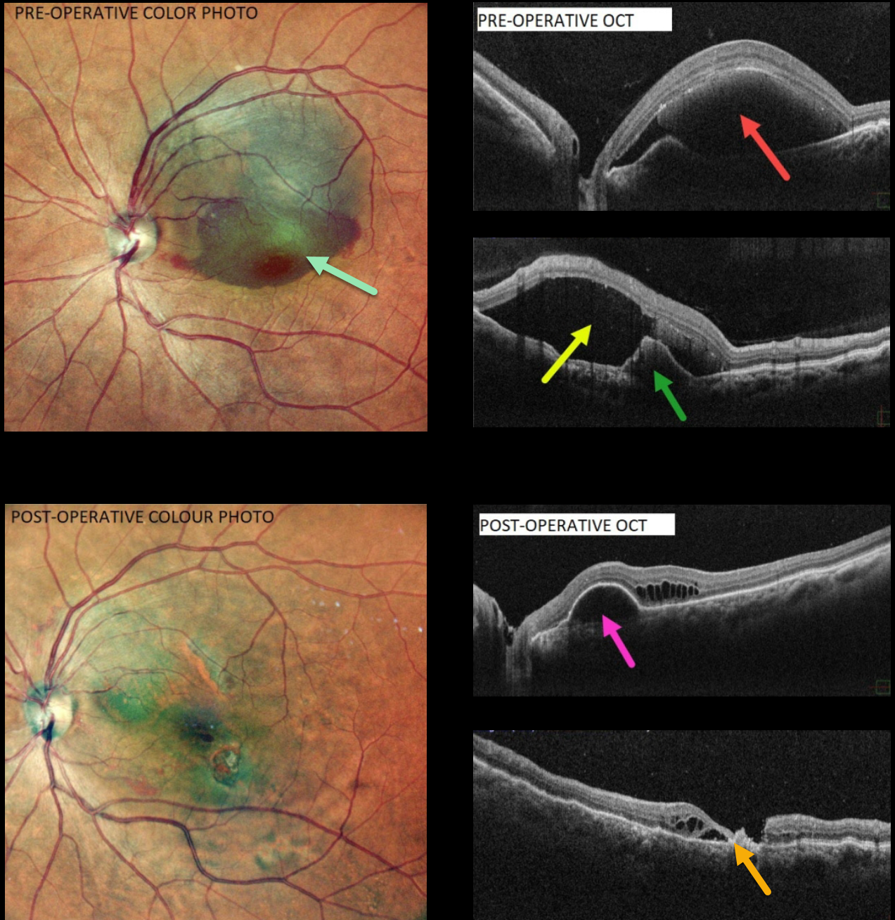
POLYPOIDAL CHOROIDAL VASCULOPATHY
Akansha Sharma and Manish Nagpal
Originally posted on @retina.rocks June 20, 2022
This 60 YO female presented with counting fingers vision from a large central and superior submacular hemorrhage.
OCT scanning shows variable subretinal blood and subretinal fluid along with an RPE detachment. There are no drusen.
Pars plana vitrectomy with drainage of the subretinal blood was performed. TPA was not used. Postoperatively, there is virtually no blood. OCT scanning shows no subretinal blood or fluid. Variable outer retinal cystic changes and atrophy are noted, along with a residual RPE detachment. Vision was 20/120.
Learning Points:
Polypoidal choroidal vasculopathy (PCV) is a variant of type 1 macular neovascularization. PCV is likely distinct from age-related macular degeneration, occurring in a different population (more common in African Americans and Asians), with a relative lack of drusen and a thicker choroid.
The sub-RPE vessels were classically characterized using indocyanine green angiography, although recent literature suggests that OCT is just as effective at diagnosing these vascular polyps. Historically, these lesions were believed to be somewhat resistant to anti-VEGF monotherapy and often required combined treatment with photodynamic therapy. More recent data from the PLANET study suggest that Eylea monotherapy is also effective (AJO 2019;204;80-89).
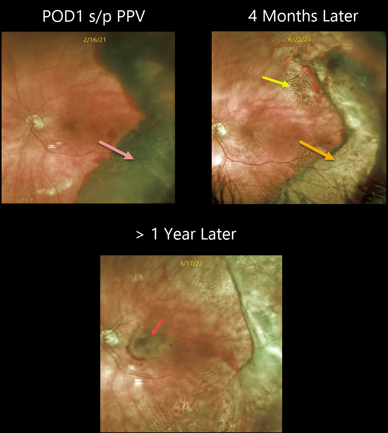
PERIPHERAL EXUDATIVE HEMORRHAGIC CHORIORETINOPATHY
Originally posted on @retina.rocks May 26, 2022
This 80YO female with a known history of dry AMD presented with a 3-week history of poor vision in her left eye. Vision was hand motion from a dense vitreous hemorrhage. B-scan ultrasonography showed a thickened temporal subretinal lesion without retinal detachment (not shown).
One day following vitrectomy, vision improved to 20/100. The temporal subretinal and RPE blood somewhat resembled a uveal malignant melanoma. However, the well-demarcated location and shape of the lesion, along with the posterior rim of subretinal blood, helped establish the diagnosis of a breakthrough vitreous hemorrhage from ectopic choroidal neovascularization (CNV), also known as peripheral exudative hemorrhagic chorioretinopathy (PEHCR).
Four months later, the resolved devitalized blood was turning yellow. The coarse subretinal pigmentation, most prominent superotemporally, is pathognomonic for prior subretinal blood.
She was subsequently lost to follow-up until she presented with a 6-month history of decreased vision. Although the PEHCR ridge continued to contract, vision was counting fingers due to a new macular neovascularization. Anti-VEGF therapy was started.
Learning Points:
PEHCR is an exudative process similar to wet AMD but located in the retinal periphery. Ectopic CNV can often masquerade as a choroidal mass or uveal melanoma.
It can also cause extensive choroidal/suprachoroidal, subretinal, and vitreous hemorrhage, more commonly in patients who are on blood thinners.
The subretinal blood often spontaneously regresses without treatment, although anti-VEGF injections can be used for symptomatic or increasing exudation.
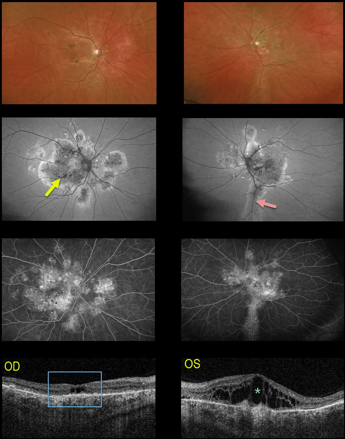
IDIOPATHIC CENTRAL SEROUS CHORIORETINOPATHY
Originally posted on @retina.rocks May 25, 2022
This 64YO male received prior photodynamic therapy in one of his eyes for idiopathic central serous chorioretinopathy (ICCS) and was subsequently lost to follow-up for 10-15 years. He presented with 20/400 vision bilaterally.
Multimodal imaging shows classic findings for chronic ICSC. Optos color imaging shows macular and peripapillary pigmentary changes with a suggestion of an inferior gutter OS.
Fundus autofluorescence better shows the bilateral pigmentary changes and inferior gutter OS, and fluorescein angiography shows variable chorioretinal staining.
Triton swept-source OCT shows bilateral thickened choroid, dry central cavitation-like changes OD, and marked cystoid macular edema OS.
Photodynamic therapy was performed for the left eye, although we are not certain this will improve his vision, given the chronicity of these findings.
Learning Points:
Macular edema is an uncommon finding with typical ICSC, likely due at least in part to the external limiting membrane acting as a barrier to subretinal fluid migrating into the retina. Eyes with chronic ICSC, however, can develop retinal edema as in this case.
Gutters are commonly seen in pathology with chronic subretinal leakage, including central serous retinopathy, choroidal hemangioma, choroidal nevus, and uveal melanoma. These pigmentary changes are usually best imaged with FAF.
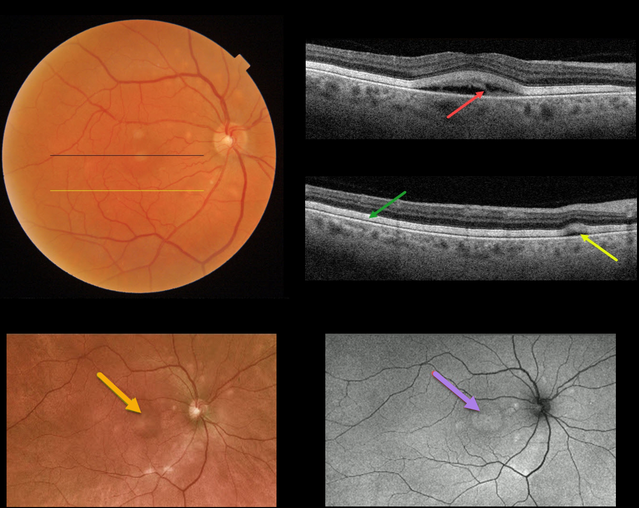
ACUTE EXUDATIVE POLYMORPHOUS VITELLIFORM MACULOPATHY
Originally posted on @retina.rocks May 18, 2022
This 72YO male presented with recent difficulties with dark adaptation and 20/60 vision in his right eye. His left eye was enucleated 2 years earlier for a uveal malignant melanoma.
Triton color imaging shows multiple yellowish subretinal round lesions scattered around the optic nerve and macula.
Triton Swept Source OCT through the fovea shows a partially scrambled vitelliform lesion. An OCT B-scan through the inferior macula shows the outer retina diffusely infiltrated by hyperreflective vitelliform material, along with a small serous detachment.
Optos imaging shows an ultrawide view of the smaller and larger central vitelliform lesions, and fundus autofluorescence (FAF) shows hyper-FAF from the corresponding vitelliform lesions. ICG and FA were both normal.
Learning Points:
Acute exudative polymorphous vitelliform maculopathy (AEPVM) is a rare condition characterized by acute vision loss from multifocal serous retinal detachments that later progress to subretinal vitelliform lesions.
Initially, the serous detachments can mimic central serous retinopathy, including choroidal thickening. The condition is often self-limiting, and vision usually recovers within several months. The yellow vitelliform lesions may take 1-2 years to resolve completely.
These patients need a systemic workup for metastatic disease, since paraneoplastic retinopathy, including metastatic melanoma, can mimic idiopathic AEPVM.
Sadly, preliminary metastatic workup for this patient showed two possible liver metastases.
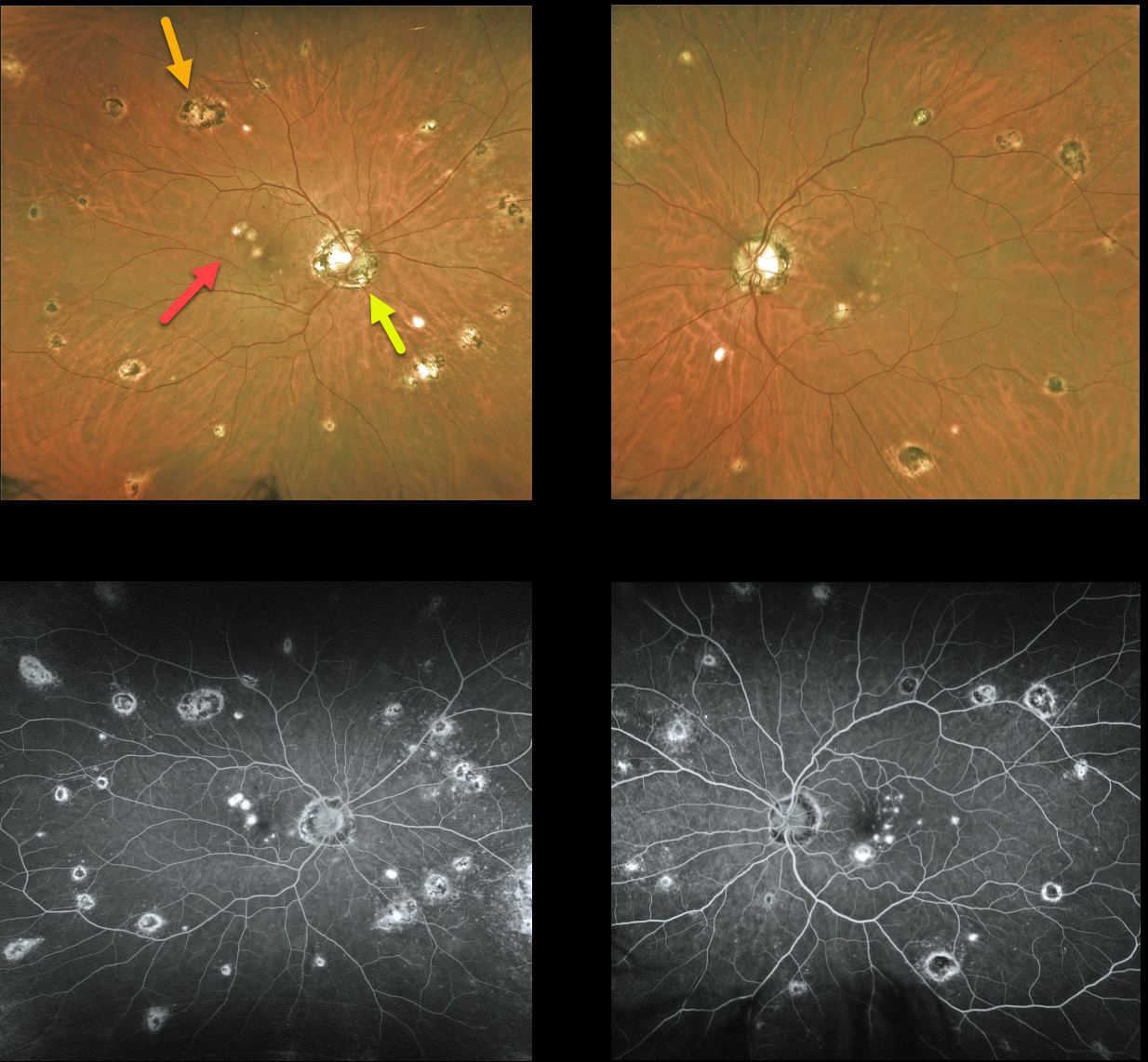
OCULAR HISTOPLASMOSIS
Originally posted on @retina.rocks May 13, 2022
This 45YO female has classic bilateral findings for atrophic ocular histoplasmosis (OHS), including the triad of peripapillary scarring, macular lesions, and scattered peripheral scars. Vision is 20/25 OD and 20/30 OS. All scars stain on fluorescein angiography.
We like to add a fourth criterion, a quiet vitreous, to this classic triad. The presence of vitreous inflammation or new lesions is not seen in OH and would suggest an alternative condition, such as multifocal choroiditis.
Learning Points:
Atrophic OHS does not require treatment, but patients with macular or peripapillary scarring need to be monitored due to the risk for macular neovascularization.
Treatment is similar to wet AMD, with anti-VEGF therapy usually being the first-line therapy. Unlike AMD, these patients often do not require ongoing injections.
Photodynamic therapy can help minimize treatment burden, and thermal laser therapy remains a good option for more peripheral macular lesions.
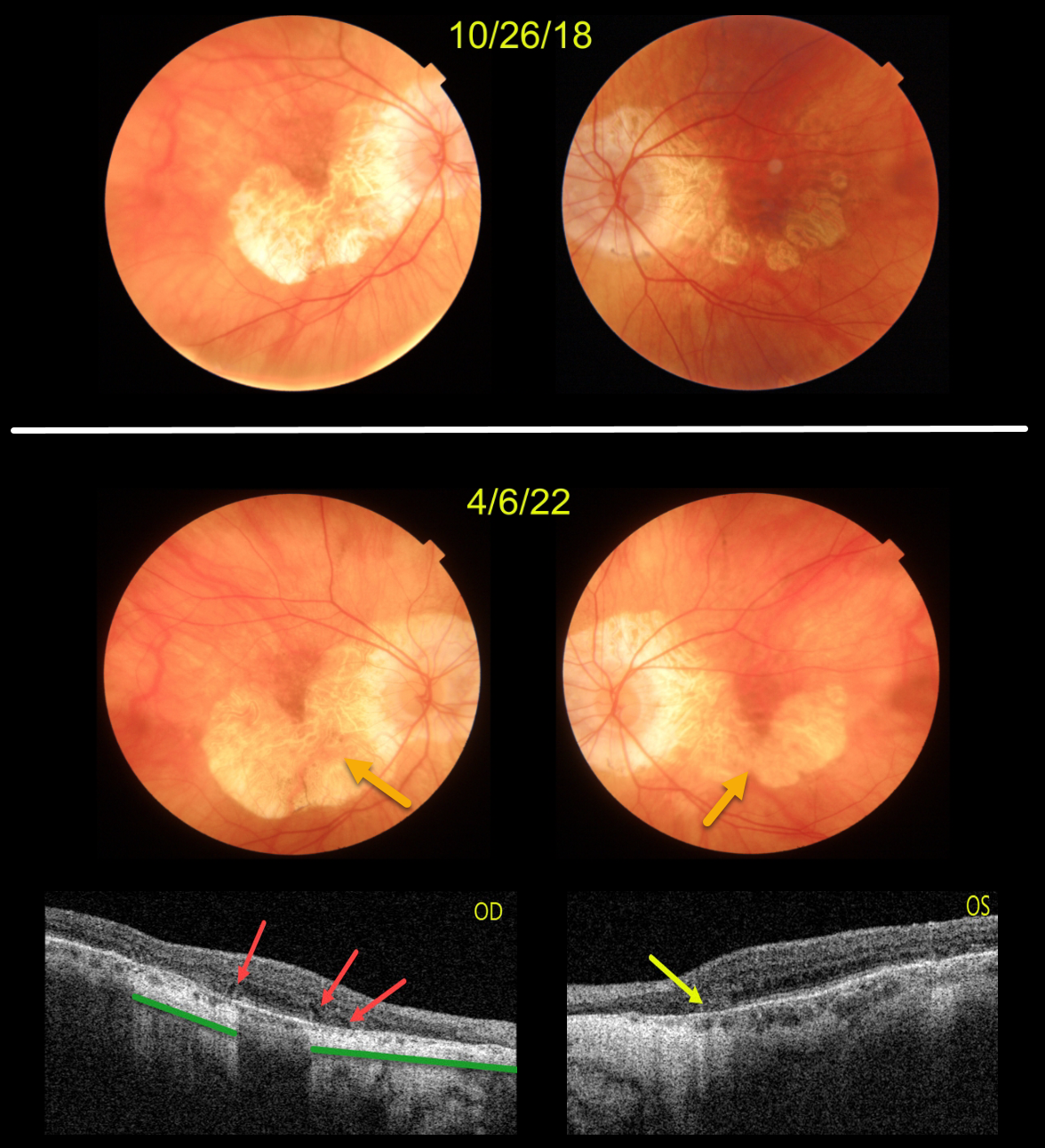
AMD MACULAR ATROPHY
Originally posted on @retina.rocks April 28, 2022
This patient originally presented in 2018 with vision of 20/40 OD and 20/50 OS. Areas of AMD-related macular atrophy (MA) skirted each macular center.
Four years later, vision had decreased to 20/100 OU due to progressive atrophy. Comparing the two images from 2018 to 2022, there is a bilateral linear enlargement for all margins of the atrophy, particularly in the left eye.
Triton swept-source OCT B-scans show classic findings for MA. The right eye has multiple wedge defects, hyporeflective wedges which develop at the boundaries of the degenerating outer plexiform layer in about 75% of MA eyes (Mones et al, Ophthalmology 2012;119:1412-1419). In addition, loss of the outer retina and RPE allows more OCT light to be transmitted into the choroid, resulting in distinct areas of choroidal and scleral hyperreflectivity.
There is a tiny area of outer retinal tubulation (ORT) nasally in the left eye, which represents photoreceptor and Muller cell degeneration (Dolz-Marco et al, Ophthalmology 2017;124:1353-1367). These ORTs have a hyper-reflective border with a central hyporeflective core.
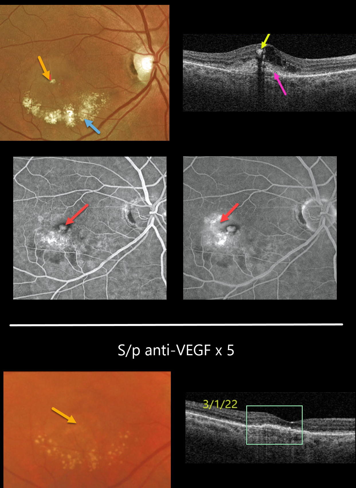
MACULAR NEOVASCULARIZATION (MNV)
Originally posted on @retina.rocks April 22, 2022
This 64YO female presented with this symptomatic unilateral exudative lesion in her right eye. Vision was 20/200.
Optos color imaging shows a small inner retinal yellow-white nodule with inferior subretinal lipid.
Triton swept-source OCT shows hyperreflectivity of this nodule with posterior shadowing, surrounding inner retinal fluid, and a hyperreflective macular neovascularization (MNV) located both below (type 1) and above (type 2) the RPE.
Optos fluorescein angiography shows a hyperfluorescent lesion with late leakage. The left eye showed mild macular drusen and pigmentary changes (not shown). This patient’s MNV is somewhat atypical given the extent of lipid exudation.
With monthly anti-VEGF therapy, the exudation resolved and vision improved to 20/60 following 5 intravitreal injections. The yellow nodular inner retinal lesion also completely resolved.
We therefore believe this most likely represented an unusual collection of inner macular lipids rather than a type 3 MNV.
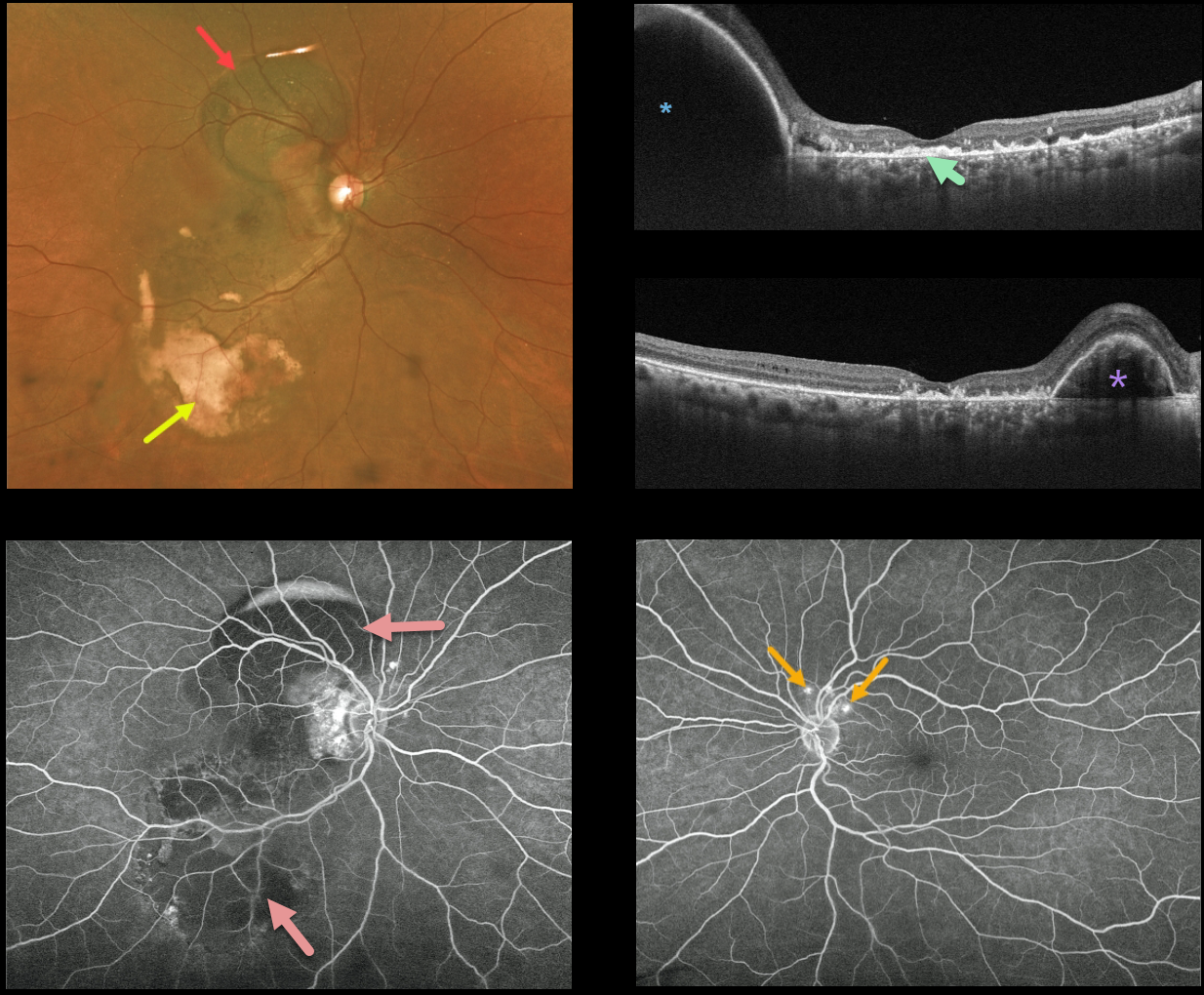
POLYPOIDAL CHOROIDAL VASCULOPATHY
Originally posted on @retina.rocks April 6, 2022
This 59YO female patient presented with variable mostly peripheral submacular blood in her right eye and counting fingers vision. Optos imaging shows the extent of the subretinal blood, which is dark superiorly and yellow inferiorly.
Triton swept-source OCT shows variable outer retinal and subretinal hyperreflective material from resolved submacular blood, and this is seen clinically as the coarse subretinal pigment most evident in the inferotemporal macula. A large RPE detachment (PED) is seen superiorly in the vertical scan, with a smaller PED nasally in the horizontal scan.
Fluorescein angiography shows mostly blockage from the subretinal blood. Several small asymptomatic polyps are noted superior to the left nerve.
Learning Points:
Polypoidal choroidal vasculopathy (PCV) is a variant of type 1 macular neovascularization. PCV is likely distinct from age-related macular degeneration, occurring in a different population (more common in African Americans and Asians), with a relative lack of drusen and a thicker choroid.
The sub-RPE vessels were classically characterized using indocyanine green angiography, although recent literature suggests that OCT is just as effective at diagnosing these vascular polyps.
Historically, these lesions were believed to be somewhat resistant to anti-VEGF monotherapy, often requiring combined treatment with photodynamic therapy.
More recent data from the PLANET study suggest that Eylea monotherapy is also effective (AJO 2019;204:80-89).
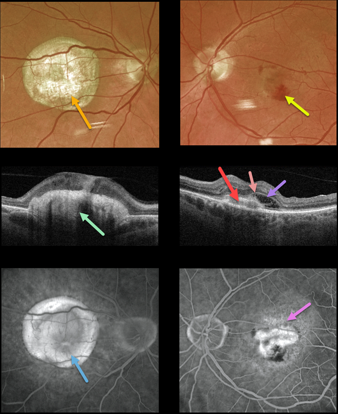
NEOVASCULAR AMD
Originally posted on @retina.rocks March 22, 2022
This 77YO male presented with counting fingers vision OD from a large fibrotic disciform scar and 20/200 vision OS from a new macular neovascularization.
The disciform scar is contracted and fibrotic, with marked hyperreflectivity on swept-source OCT. The MNV in his left eye is seen as opaque fluid with some subretinal blood. The type 2 MNV (located above the RPE) is noted on the OCT B-scan. There is also a bacillary detachment and a hyperreflective line indicating a possible pitchfork sign.
Optos fluorescein angiography shows diffuse staining of the subretinal scarring OD and leakage from the MNV OS.
Learning Points:
Disciform scars were the norm for untreated wet AMD before the anti-VEGF era that began in the mid-2000s. New-onset wet AMD diagnosed and treated with appropriate anti-VEGF dosing virtually never goes on to develop these lesions. There may be progressive macular atrophy following years of therapy, but not this type of fibrotic scarring.
An excellent review of wet AMD fibrotic lesions is in Souied et al’s AOS thesis (AJO 2020;214:151-171).
The pitchfork sign was originally described by Hoang et al (Retina 2013;33:1049-1055) as a unique OCT finding in inflammatory MNV. Falavarajani et al more recently noted that this finding is also associated with non-inflammatory MNV (Ophthalmic Surg Lasers Imaging Retina 2019;50:719-725).
The pathogenesis of the avascular pitchfork spikes is unknown, but may be related to outer retinal traction or Müller cell activation.
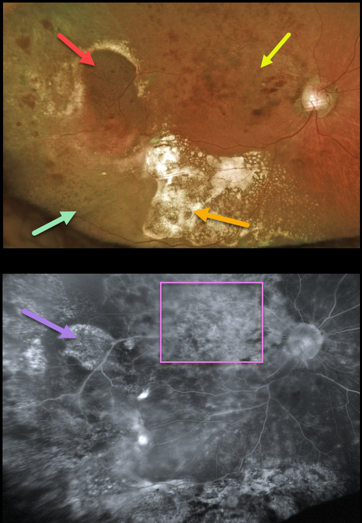
PERIPHERAL EXUDATIVE HEMORRHAGIC CHORIORETINOPATHY
Originally posted on @retina.rocks March 21, 2022
This 94YO female presented with ectopic choroidal neovascularization (CNV), also known as peripheral exudative hemorrhagic chorioretinopathy (PEHCR) in her right temporal periphery. Inferior to this lesion are clumps of subretinal pigment. Confluent subretinal lipid is also noted more inferiorly.
Vision was 20/400 due to macular edema from an unrelated major superotemporal branch retinal vein occlusion (BRVO). The inner retinal hemorrhages from the BRVO extend into the region of the ectopic subretinal lesion, somewhat confusing the clinical picture.
Fluorescein angiography shows variable diffuse staining of the ectopic CNV and secondary pigmentary changes, along with diffuse leakage from the BRVO.
Learning Points:
PEHCR is an exudative process similar to wet AMD but located in the retinal periphery. Ectopic CNV can often masquerade as a choroidal mass or uveal melanoma. It can also cause extensive choroidal/suprachoroidal, subretinal, and vitreous hemorrhage, more commonly in patients who are on blood thinners.
The subretinal blood often spontaneously regresses without treatment, although anti-VEGF injections can be used for symptomatic or increasing exudation. The inferior subretinal pigment in our patient is a classic finding for resolved subretinal blood.
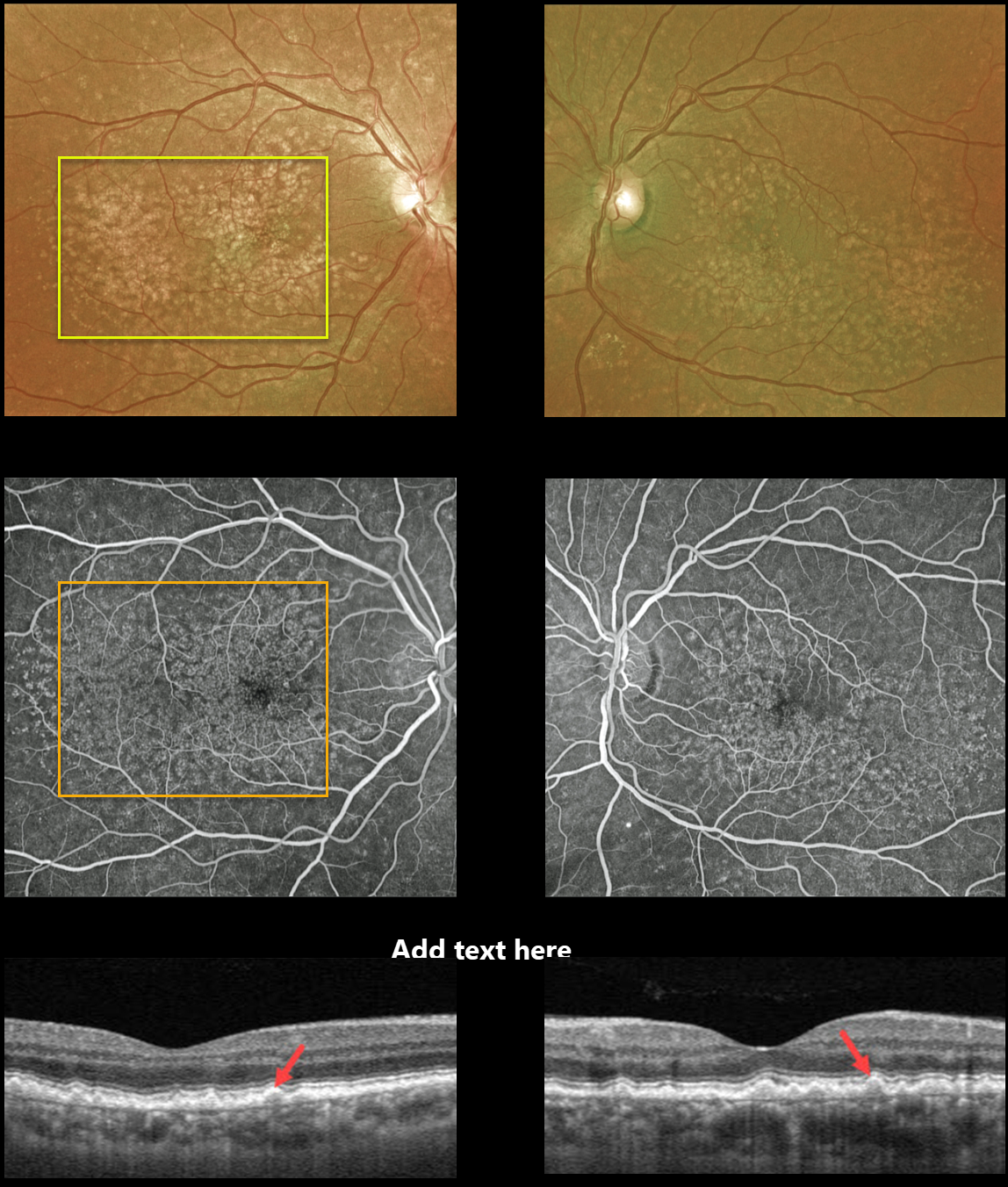
CUTICULAR DRUSEN
Originally posted on @retina.rocks March 17, 2022
This 62YO male presented with 20/20 vision OU and asymptomatic macular drusen. Optos photos of both eyes show numerous scattered cuticular drusen throughout each macula.
Fluorescein angiography shows a dramatic bilateral starry sky pattern of innumerable hyperfluorescent drusen.
Triton swept-source OCT reveals multiple tooth-like cuticular drusen.
Learning Points:
Cuticular drusen, formerly known as basal laminar drusen, appear as numerous, round, yellow-white punctate accumulations located above Bruch’s membrane and below the RPE.
Originally described by Gass as a unique entity (AJO 1985;99;445-459), they are now thought to fall at least in part within the AMD spectrum. They are classically associated with avascular vitelliform lesions, but can also cause macular neovascularization.
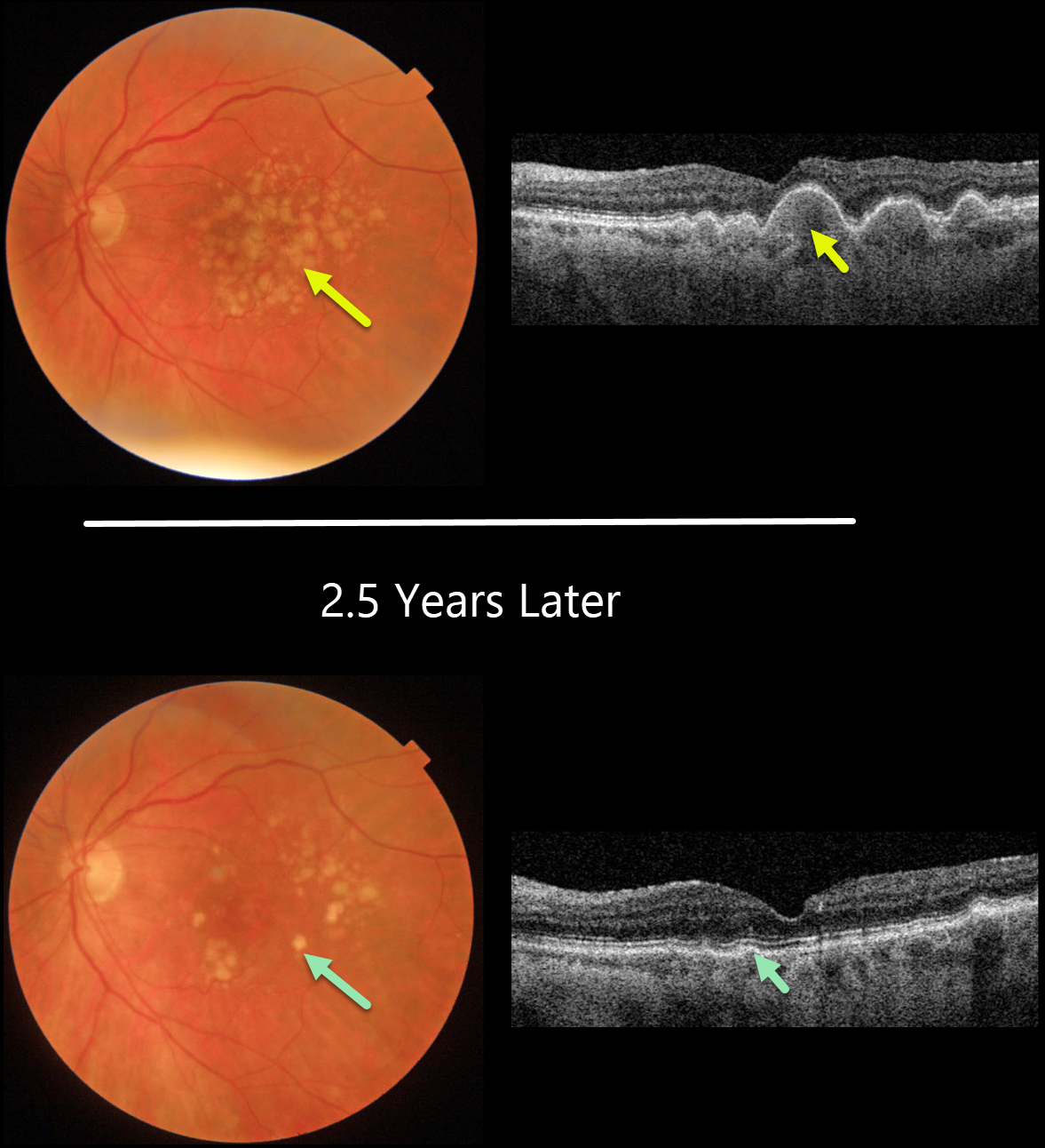
DRUSEN RESOLUTION
Originally posted on @retina.rocks March 16, 2022
This 77YO female presented with diffuse, large, confluent soft drusen in both eyes (only left eye pictured). Vision was 20/50.
Two and a half years later, her macular examinations appeared quite different with marked drusen resolution. OCT shows near-complete flattening of all drusen, with irregularity of the outer retinal bands and RPE. Vision actually improved to 20/25.
Learning Points:
Drusen commonly remodel over time, and their collapse can lead to drusenoid atrophy of the RPE.
Although our patient’s outer retina is certainly abnormal on OCT, her clinical macular findings are remarkably benign given the extent of drusen several years ago.
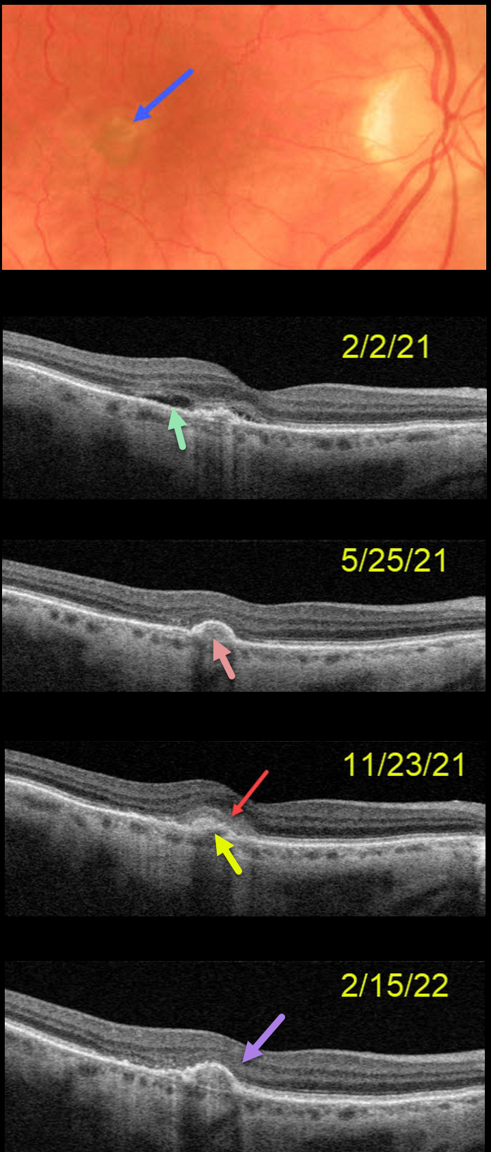
SUBRETINAL HYPERREFLECTIVE MATERIAL (SHRM)
Originally posted on @retina.rocks March 14, 2022
This 78YO female originally presented on 2/2/21 with decreased vision in her right eye from a treatment-naive macular neovascularization (MNV) with subretinal fluid on OCT.
The lesion became completely inactive after several monthly anti-VEGF injections (5/25/21).
She was then lost to follow-up due to other health issues and returned on 11/23/21 with an active lesion with overlying subretinal hyperreflective material (SHRM).
After several additional monthly injections, all exudation resolved with the disappearance of the SHRM (2/15/22).
Learning Points:
SHRM is an OCT finding that is invisible clinically. This material, located between the neurosensory retina and retinal pigment epithelium (RPE), is thought to consist of fluid, fibrin, blood, and other fibrovascular tissues.
SHRM is a biomarker of active disease that decreases with successful anti-VEGF therapy (see Willoughby et al., Ophthalmology 2015;122:1846-1853).
Persistent SHRM is associated with an increased incidence of scar formation and worse VA.
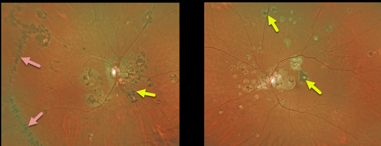
OCULAR HISTOPLASMOSIS
Originally posted on @retina.rocks March 2, 2022
This 66YO female with a history of ocular histoplasmosis has extensive bilateral macular, peripapillary, and peripheral scarring. The right eye shows a large hyperpigmented temporal streak lesion. Vision is stable at counting vision OD and 20/40 OS.
Learning Points:
The incidence of streak lesions in OHS, also known as Schlaegel lines, is estimated at 5% (Arch Ophthalmol 1981;99:246-248), and in our experience, they are more common in areas of more extensive peripheral scarring. They appear as linear aggregations of midperipheral OHS scars that run parallel to the ora serrata.
They somewhat resemble lattice degeneration in location and appearance, but do not increase the patient’s risk of ocular complications.

SUBRETINAL DRUSENOID DEPOSITS (SDD)
Originally posted on @retina.rocks February 23, 2022
This 77YO female presented with vision of 20/20 OD and 20/30 OS and extensive bilateral subretinal drusenoid deposits.
Swept-source OCT shows numerous hyperreflective deposits in the outer retina located just above the RPE. En face OCT dramatically shows the extent of the dot-like drusenoid material.
Learning Points:
Subretinal drusenoid deposits (SDD) are polymorphous light-gray interconnected accumulations located above the RPE. They are present in about one-quarter of older adults with healthy maculas and in more than half of those with early- to intermediate-stage dry AMD. They can appear as an array of dots (as in our patient), interconnected reticular bands, or confluent lesions (Suzuki and Spaide, AJO 2014;157:1005-1012). The dot variant is associated with macular neovascularization (MNV) and the confluent variant with macular atrophy (Zhou et al, Ophthalmology 2016;123:1530-1540).
Clinically, they are often mistaken for conventional drusen and consist of a similar yet distinct lipid-rich material to that found in soft drusen. They are localized to rods, unlike typical soft drusen, which are more central and cone-based. As with drusen, SDDs are dynamic and can grow, shrink, resolve, or even recur.
Since they are located above the RPE, they are best visualized with blue or green illumination. On OCT, they appear in 3 stages (Zweifel et al, Ophthalmology 2010;117:303-312), growing upwards from the RPE towards the ELM (external limiting membrane): stage 1, diffuse deposition of granular hyperreflective material between the RPE and the EZ (ellipsoid zone), stage 2, material alters the EZ contour, and stage 3, conical appearance breaking through the EZ.
Rod function seems to be preferentially affected, and there is an associated increased risk of delayed dark adaptation, macular atrophy, and types 2 and 3 MNV. In fact, type 2 neovascularization in the setting of AMD occurs almost solely in eyes with subretinal drusenoid deposits.
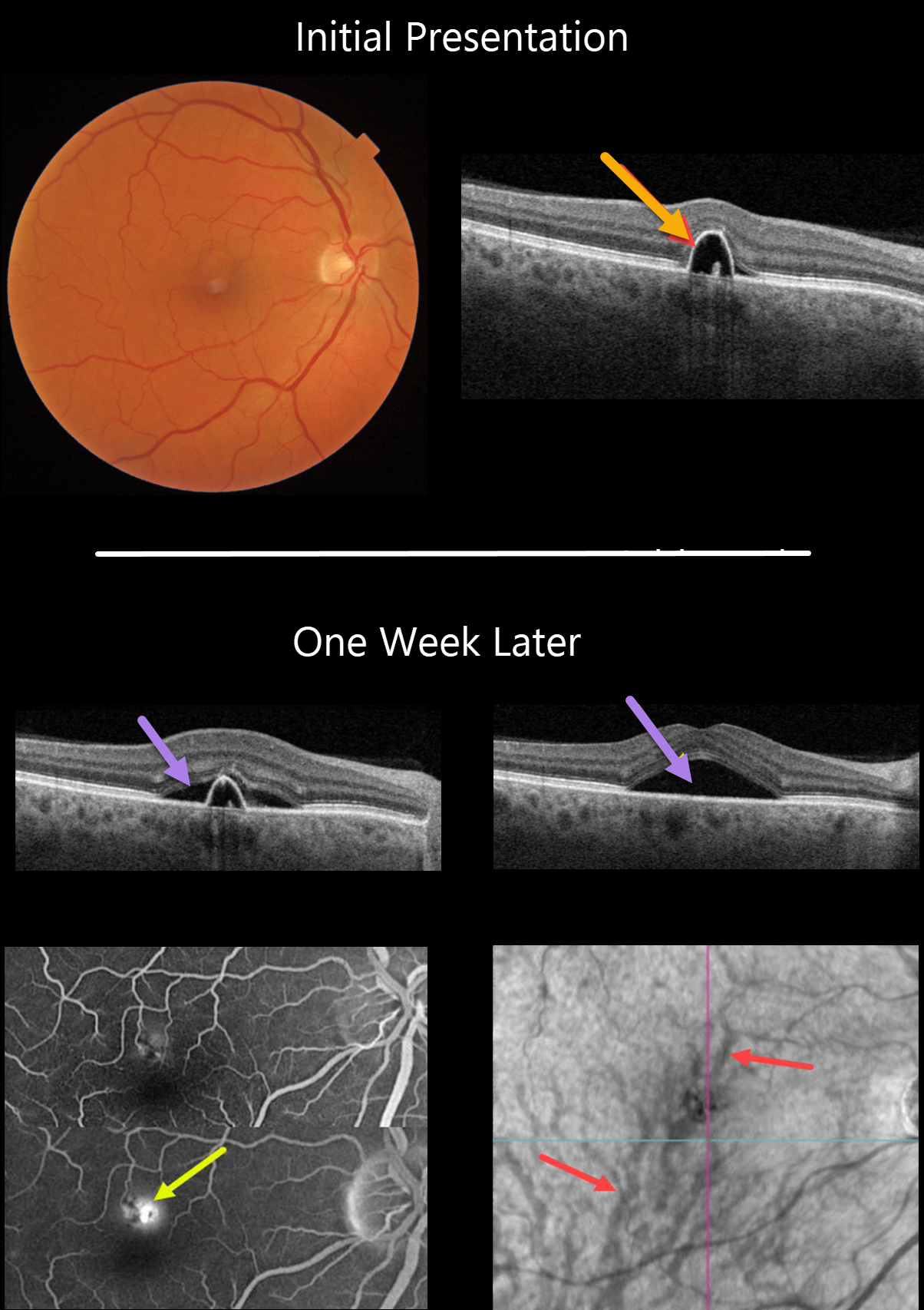
IDIOPATHIC CENTRAL SEROUS CHORIORETINOPATHY
Originally posted on @retina.rocks February 16, 2022
This 55YO male presented on 1/24/22 with a small symptomatic pigment epithelial detachment (PED) in the superior right fovea. Vision was 20/30.
One week later, he was subjectively worse, although vision remained at 20/30. There was a new small blister of foveal subretinal fluid, best captured on swept-source OCT.
Optos fluorescein angiography shows a typical blot leak along the dome of the PED. En face OCT shows a choroidal venous anastomosis.
Learning Points:
The choroidal venous system, as with the neurosensory retinal vasculature, usually respects the horizontal meridian. With pachychoroid disorders, these anastomoses develop, and in this patient’s case, the inferior vortex system drains nearly the entire macula.
For a wonderful review highlighting the concept of venous overload choroidopathy, see Spaide et al, Progress Retinal Eye Research 2022;86;100973.
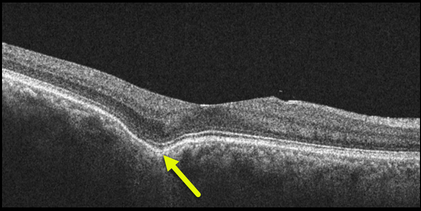
CHOROIDAL EXCAVATION
Will Gibson
Originally posted on @retina.rocks February 9, 2022
This 56YO highly myopic male has 20/20 vision in his right eye with an asymptomatic temporal foveal focal choroidal excavation (FCE). No treatment was recommended.
Learning Points:
Focal choroidal excavation, originally described by Jampol et al. in 2006, is likely part of the pachychoroid spectrum, as it is usually associated with a thickened choroid and pachyvessels.
The FCE can be observed, although symptomatic manifestations, including active central serous retinopathy or macular neovascularization, often require treatment.
An excellent review is by Verma et al, Br J Ophthalmol 2021;105:1043-1048.
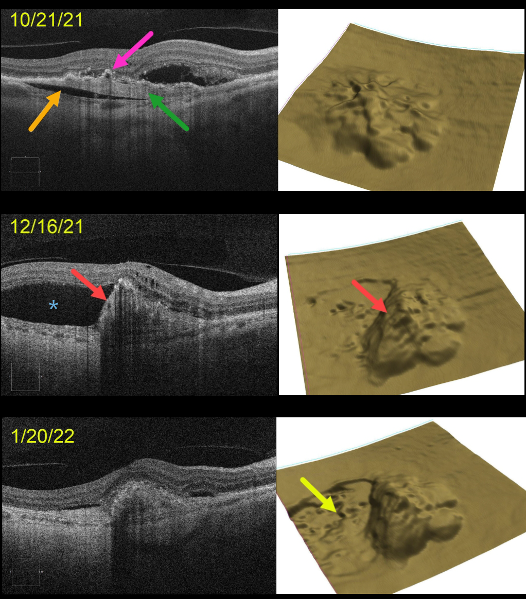
RPE TEAR
Originally posted on @retina.rocks February 2, 2022
This 71YO female presented with a treatment-naive, age-related type 1 macular neovascularization (MNV). The already contracting MNV is elevated above the underlying choroid (prechoroidal cleft), and the overlying RPE is irregularly contracted and adherent to the MNV. The retinal pigment epithelium (RPE) map shows a 3-dimensional view of the RPE elevation. An anti-VEGF injection was given that day on 10/21/21.
On 12/16/21, vision was unchanged at 20/400 despite a new asymptomatic temporal RPE tear with marked overlying new subretinal fluid. An additional anti-VEGF injection was given.
On 1/20/22, vision was counting fingers. OCT scanning shows near-total resolution of all intra- and subretinal fluid. The RPE map nicely shows the crater of devoid RPE.
Learning Points:
A tear of the RPE is a fairly common occurrence in RPE detachment (PED) associated with type 1 MNV (located below the RPE). The neovascularization can spontaneously contract or involute following treatment. It then puts traction on the overlying RPE, which can then rip at the opposite edge of the PED.
This traction was already evident at our patient’s initial pre-injection visit, including the prechoroidal cleft, which is associated with a worse visual prognosis (see Kim et al, Retina 2017;37:2047-2055).
Like a cheap window blind, the torn RPE retracts and scrolls towards the MNV. This leaves a depigmented region where the RPE was and is now absent, along with a relatively straight, pigmented subretinal lesion extending from the scrolled RPE edge.
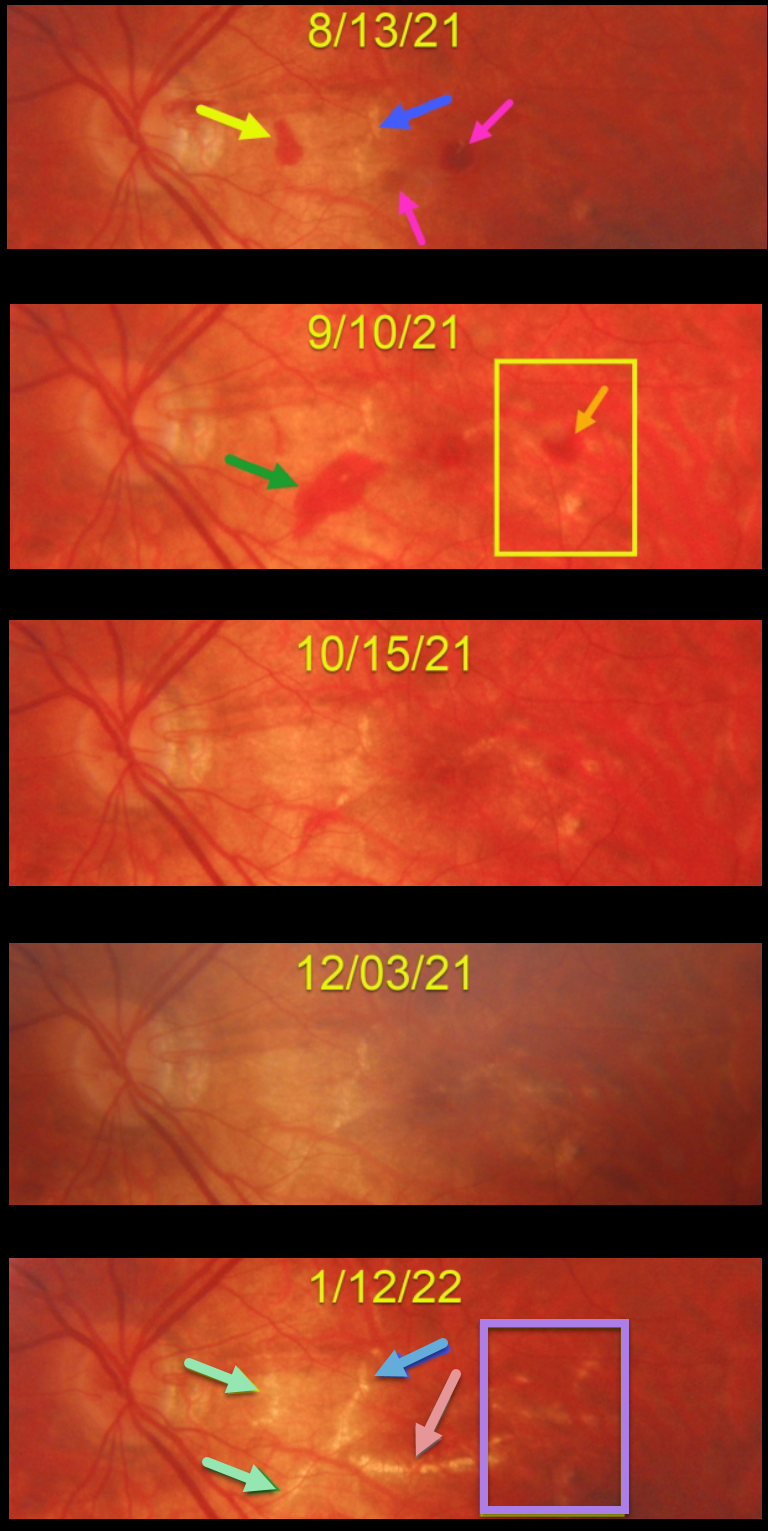
HIGH MYOPIA WITH AVASCULAR RETINAL HEMORRHAGES AND LACQUER CRACK FORMATION
Originally posted on @retina.rocks January 31, 2022
This 23YO high myopic female presented on 8/13/21 with some symptomatic central and paracentral scotomas. Vision was 20/80.
There were a few round subretinal hemorrhages near the central macula, along with an asymptomatic nasal macular hemorrhage. A few small lacquer cracks were noted.
She was then monitored monthly with an unusual rapid succession of new and resolving hemorrhages with subsequent lacquer crack development.
On 9/10/21, the prior hemorrhages were resolving, but a new area of subretinal blood was noted along the inferior edge of the previously noted lacquer crack. There was also a new hemorrhage in the distal macula. These hemorrhages all resolved by the 12/3/21 exam.
When last seen on 1/12/22, in the areas of prior subretinal blood, there are new lacquer cracks, enlargement of the originally noted lacquer crack, and a large new horizontal lacquer crack inferior to the fovea. In the region of the distal temporal macular hemorrhage noted on 9/10/21, there are numerous new lacquer cracks. Vision was 20/60, and we continue to monitor her for macular neovascularization (MNV).
Learning Points:
Lacquer cracks are a common finding in patients with high myopia. They consist of breaks in Bruch’s membrane due to the outpouching/stretching of the pathologically myopic eye and increase the risk for MNV.
In some cases, similar to this patient, they can have small avascular subretinal hemorrhages, not associated with neovascularization, and are monitored. These are best confirmed with fluorescein angiography. In cases with an active membrane, they are treated with anti-VEGF injections.
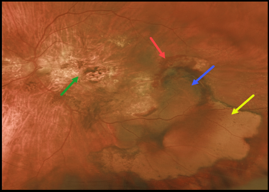
PERIPHERAL EXUDATIVE HEMORRHAGIC CHORIORETINOPATHY
Originally posted on @retina.rocks January 28, 2022
This 77YO female has been followed for years with vision of 20/200 OD and counting fingers OS from inactive age-related disciform scars. She then came in with sudden symptomatic vision loss and floaters in her left eye.
Vision was hand motion OS from a breakthrough vitreous hemorrhage from an inferotemporal ectopic choroidal neovascularization (CNV) that grew from the edge of the original macular scar. She was not taking anticoagulants or aspirin.
Following two monthly intravitreal Avastin injections, vision remained at hand motion. The vitreous hemorrhage had cleared, and the subretinal blood was resolving.
Optos imaging shows the original macular scarring, along with varying stages of resolving rub-RPE blood, subretinal blood, and devitalized, yellow subretinal blood.
Learning Points:
Ectopic CNV, also known as peripheral exudative hemorrhagic chorioretinopathy, is an exudative process similar to wet AMD but located in the retinal periphery.
Ectopic CNV can often masquerade as a choroidal mass or uveal melanoma. It can also cause extensive choroidal/suprachoroidal, subretinal, and vitreous hemorrhage, more commonly in patients who are on blood thinners.
The subretinal blood often spontaneously regresses without treatment, although anti-VEGF injections can be used for symptomatic or increasing exudation.
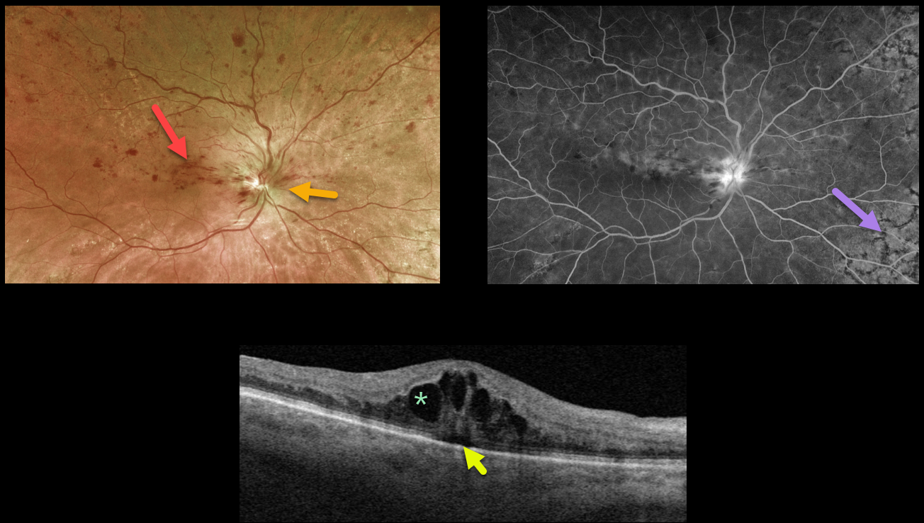
HRVO
Originally posted on @retina.rocks January 13, 2022
This 81YO female presented with 20/60 vision from a symptomatic superior hemiretinal vein occlusion (HRVO). Optos imaging shows foveal and superior inner retinal hemorrhages with a swollen optic nerve, and fluorescein angiography shows a well-perfused occlusion.
Swept-source OCT confirms moderate cystic macular edema with a trace amount of underlying subretinal fluid.
Age-related peripheral reticular degeneration of the RPE is best seen angiographically as an incidental finding.
The patient is receiving ongoing anti-VEGF injections to control her macular edema. Intravitreal injections (anti-VEGF or steroids) are the treatment of choice, although grid laser may help decrease the treatment burden.
Learning Points:
Branch retinal vein occlusions are virtually always caused by a retinal artery compressing a retinal vein within its common adventitial sheath.
Some people are born with an anomalous two-trunked central retinal vein that independently drains the superior and inferior retinal hemispheres.
HRVO, similar to central retinal vein occlusion, is caused by compression of the retinal vein posterior to the lamina cribrosa (see Hayreh, Arch Ophthalmol 1980;98:1600-1609).
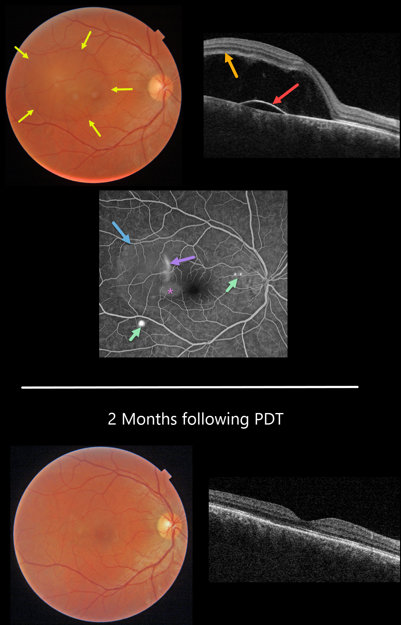
IDIOPATHIC CENTRAL SEROUS CHORIORETINOPATHY
Originally posted on @retina.rocks January 12, 2022
This 33YO male presented with recent central blurred vision in his right eye. He is a powerlifter and self-injects testosterone with possible anabolic steroids. Vision was 20/70 OD.
Triton color fundus photography shows a large serous macular detachment. Triton swept-source OCT shows a large serous neurosensory retinal detachment with an underlying shallow RPE detachment.
Optos fluorescein angiography shows classic findings for idiopathic central serous chorioretinopathy (ICSC), including peripheral blot leaks and a more central smokestack leak. Fluorescein leaks through a physiologic break in the RPE and accumulates in the subretinal space, giving rise to the smokestack, which is found in only 10% of cases. The fluorescein dye then diffuses and pools within the subretinal space, highlighting the extent of the serous detachment. Fluorescein also pools beneath the PED. In this case, subretinal pooling is almost always associated with an RPE detachment.
Most cases of central serous chorioretinal atrophy have a single unilateral leak. Multifocal cases are commonly associated with systemic steroids.
Our patient underwent PDT therapy, and vision returned to 20/20 two months later with dramatic resolution of the subretinal fluid and RPE detachment. Since he is asymptomatic with good vision in this eye, we continue to observe. He is trying to lower the amount of injected testosterone to lower the risk of recurrent ICSC.
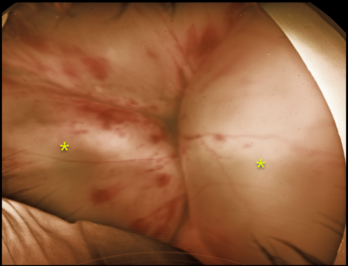
CHOROIDAL DETACHMENT
Originally posted on @retina.rocks December 23, 2021
This 25YO male patient presented to our practice with no light perception vision in his left eye. He had lost vision following an injury from a cat three years prior.
Optos imaging shows large kissing choroidal detachments with scattered intraretinal hemorrhages.
He has severe neovascular glaucoma with significant pain, and is considering cyclophotocoagulation vs enucleation.
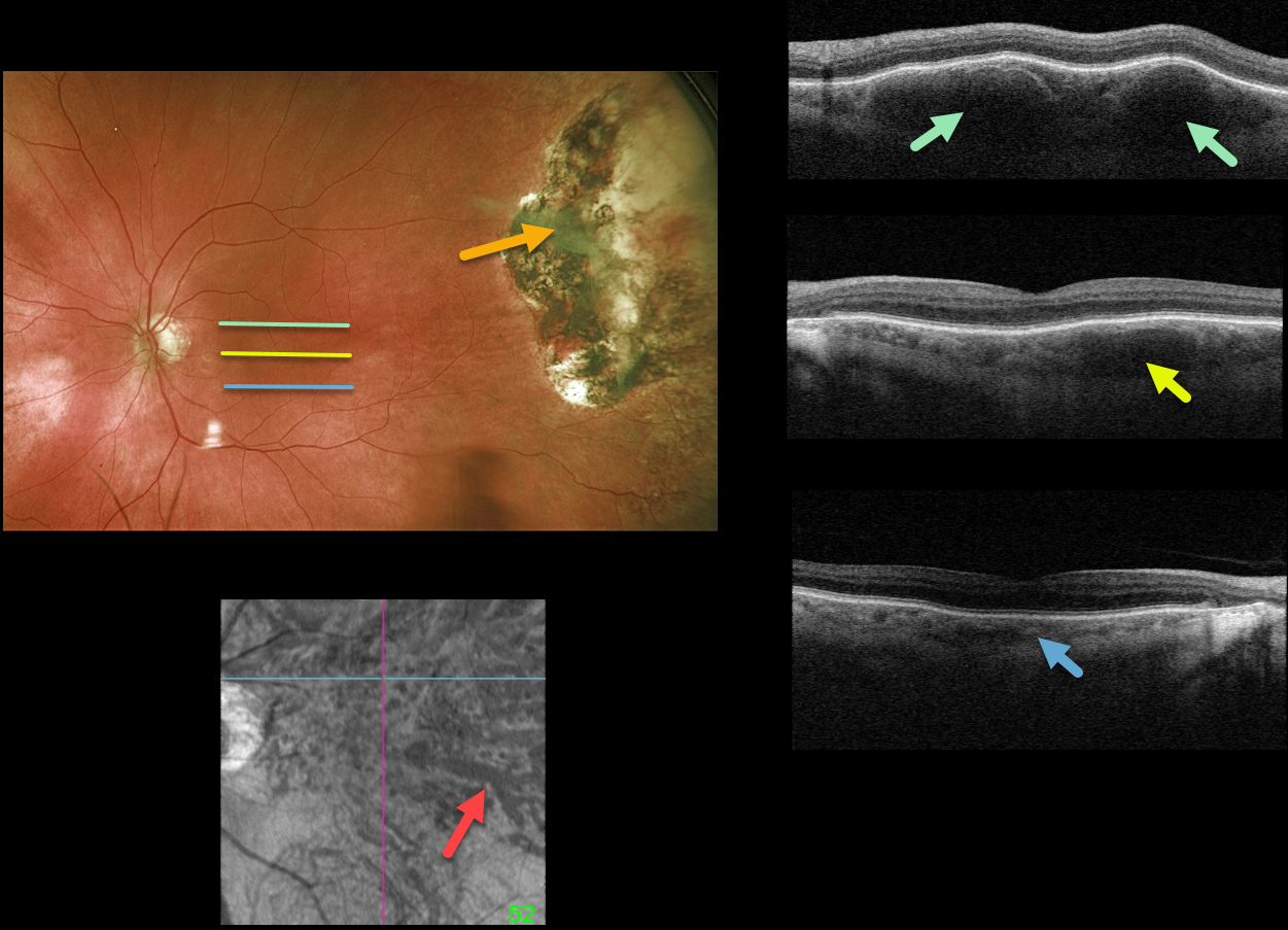
PERIPHERAL EXUDATIVE HEMORRHAGIC CHORIORETINOPATHY + PACHYCHOROID
Originally posted on @retina.rocks December 22, 2021
This 85YO male underwent cryotherapy 7 years earlier for a symptomatic hemorrhagic ectopic choroidal neovascularization in his left eye. Optos imaging shows the current appearance of the inactive cryotherapy scarring.
Although the macula has a normal fundoscopic appearance, Triton swept-source OCT scanning is far from normal. B-scans are shown through the top, central, and inferior macula. Although the inferior macular choroid appears relatively normal, a thickened outer choroid (pachyvessels) is noted more centrally, and is huge superiorly. En face imaging through this abnormal choroid is shown.
Learning Points:
The pachychoroid spectrum is a collection of conditions with a thick (“pachy”) choroid. These pachyvessels can cause a range of disorders, including idiopathic central serous chorioretinopathy, pachychoroid pigment epitheliopathy, choroidal excavation, pachychoroid neovasculopathy, polypoidal choroidal vasculopathy, pachydrusen, and peripapillary pachychoroid syndrome.
They are also associated with ectopic neovascularization (aka peripheral exudative hemorrhagic chorioretinopathy), although the choroid in these cases shows increased thickening towards the temporal periphery (see Shroff et al, Retina 2021;41:1518-1525).

CHOROIDAL TRANSPLANT
Barbara Parolini and Veronika Matello
Originally posted on @retina.rocks December 21, 2021
This patient is a teacher who suffered from a wet AMD submacular hemorrhage. Despite six anti-VEGF injections, vision dropped to 20/200. She was 58 years old and unable to read.
After a long discussion regarding the pros vs cons of surgery, she underwent autologous choroidal transplantation in 2013.
The surgical procedure included:
1) Complete pars plana vitrectomy
2) Creation of a temporal retinal detachment
3) Peripheral 200-degree retinotomy
4) Macular neovascularization (MNV) removal
5) Feeder vessel endodiathermy
6) Designing the peripheral choroidal patch with endodiathermy
7) Cutting and isolation of a full-thickness autologous RPE and choroid transplant (under perfluorocarbon liquid, PFCL)
8) Translocation of the patch under the fovea (under PFCL)
9) Transfer of PFCL from under to over the retina with retinal reattachment
10) Peripheral laser
11) PFCL exchange for 1000cs silicone oil.
Eight years later, a Clarus 500 widefield photograph shows the intact submacular choroidal autograft and a white area of bare sclera in the temporal quadrant, which was the harvesting site for the autograft.
Canon Xephilio S1 widefield OCT shows a fairly normal retina overlying the graft. Xephilio A1 angioOCT shows the intact choroidal graft vessels arranged in a different direction compared to the native surrounding choroidal vasculature.
When last examined, near vision was 20/30. She recently developed an MNV in the contralateral eye, which is being treated with anti-VEGF injections.
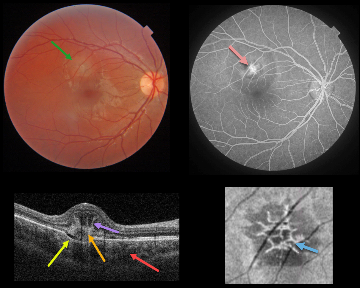
IDIOPATHIC MACULAR NEOVASCULARIZATION (MNV)
Originally posted on @retina.rocks December 20, 2021
This healthy 14YO female presented with acute vision loss in her right eye. Vision was 20/40. Triton color photography shows a depigmented subretinal lesion in the superior macula.
Swept-source OCT shows a constellation of fascinating findings. A type 2 macular neovascularization is seen overlying a choroidal excavation. The choroid is thickened.
The most dramatic findings are the unusual linear, radiating, hyperreflective lesions extending anteriorly from the MNV, also known as the “pitchfork sign.” En face OCT through this lesion shows a spiky, wreath-like pattern of hyperreflectivity.
The MNV was confirmed on fluorescein angiography. Intravitreal anti-VEGF was begun.
Learning Points:
The pitchfork sign was originally described by Hoang et al (Retina 2013;33:1049-1055) as a unique OCT finding in inflammatory MNV. Falavarajani et al more recently noted that this finding is also associated with non-inflammatory MNV (Ophthalmic Surg Lasers Imaging Retina 2019;50:719-725). The pathogenesis of the avascular pitchfork spikes is unknown, but may be related to outer retinal traction or Müller cell activation.
Focal choroidal excavation, originally described by Jampol et al in 2006, is likely part of the pachychoroid spectrum and is associated with type 2 MNV. An excellent review is by Chung et al (Retina 2017;37;199-221).
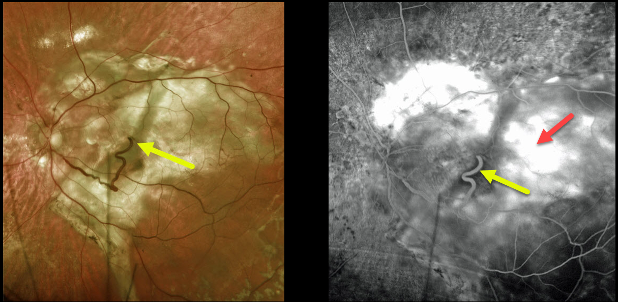
NEOVASCULAR AMD + RETINAL CHOROIDAL ANASTAMOSIS
Originally posted on @retina.rocks November 25, 2021
This 74YO female has a large complex disciform scar from end-stage age-related macular degeneration (AMD).
A very prominent retinal vein drains directly into the substance of the scar inferiorly, indicating a chorioretinal anastomosis. Fluorescein angiography shows diffuse staining of the subretinal scarring.
Learning Points:
Under normal conditions, the retinal and choroidal circulations remain separate and distinct. Rarely, the two circulations connect directly through a chorioretinal anastomosis.
These can occur in chorioretinal scars (typically from inactive toxoplasmosis lesions), in macular telangiectasia, and in disciform scars.
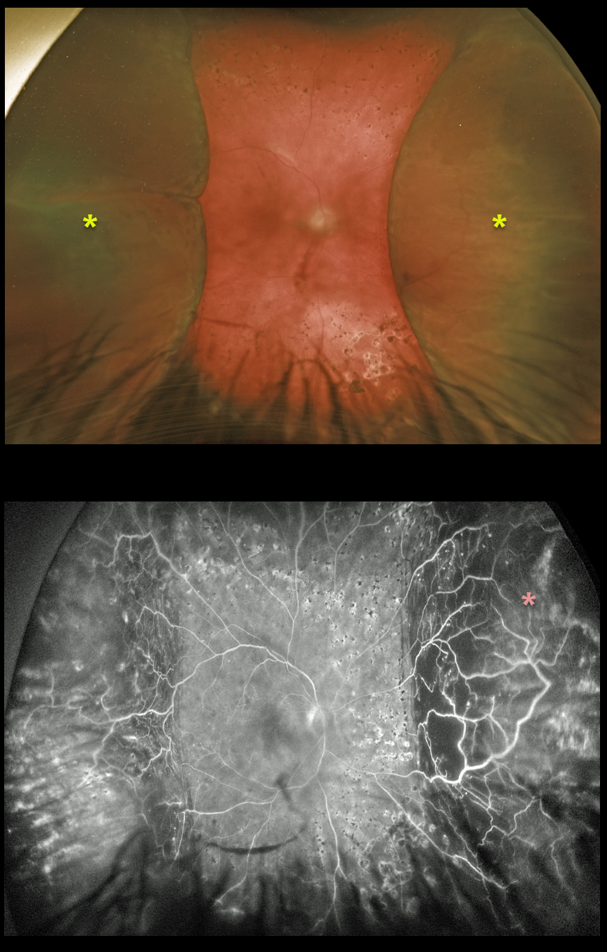
CHOROIDAL DETACHMENT
Originally posted on @retina.rocks November 24, 2021
This 49YOM underwent trabeculectomy surgery elsewhere 6 weeks earlier.
Peripheral choroidal detachments are noted on Optos imaging. Fluorescein angiography shows significant peripheral ischemia.
Recent panretinal photocoagulation was started due to proliferative diabetic retinopathy (neovascularization not visualized due to the choroidals).
Learning Points:
Choroidal detachment is relatively common after glaucoma surgery. Two mechanisms are believed to be at play: hypotony allows fluid to accumulate in the suprachoroidal space, and inflammation increases the choroidal permeability. The detached ciliary body may also produce less aqueous humor, thus creating a self-perpetuating cycle.
Often, observation is sufficient, but topical steroids and cycloplegics can help promote resolution.
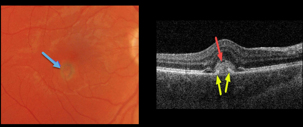
IDIOPATHIC MACULAR NEOVASCULARIZATION (MNV)
Originally posted on @retina.rocks November 10, 2021
This 30YO male presented with acute unilateral vision loss of 20/50 in his right eye. Ocular examinations were normal bilaterally except for a small area of retinal edema with a subretinal pigment ring in the right inferotemporal fovea.
Swept-source OCT shows a hyperreflective macular neovascularization (MNV) mostly above the RPE. Elevated RPE partially extends into the midportion of the MNV.
In this case, due to our patient’s acute symptoms, we chose to begin anti-VEGF therapy.
Learning Points:
Type 2 MNV often have RPE cells that migrate along its anterior surface, and this appears clinically as a pigment ring. Although the RPE never completely envelops the MNV, its presence often helps to stabilize the lesion’s exudation. In this case, due to our patient’s acute symptoms, we chose to begin anti-VEGF therapy.
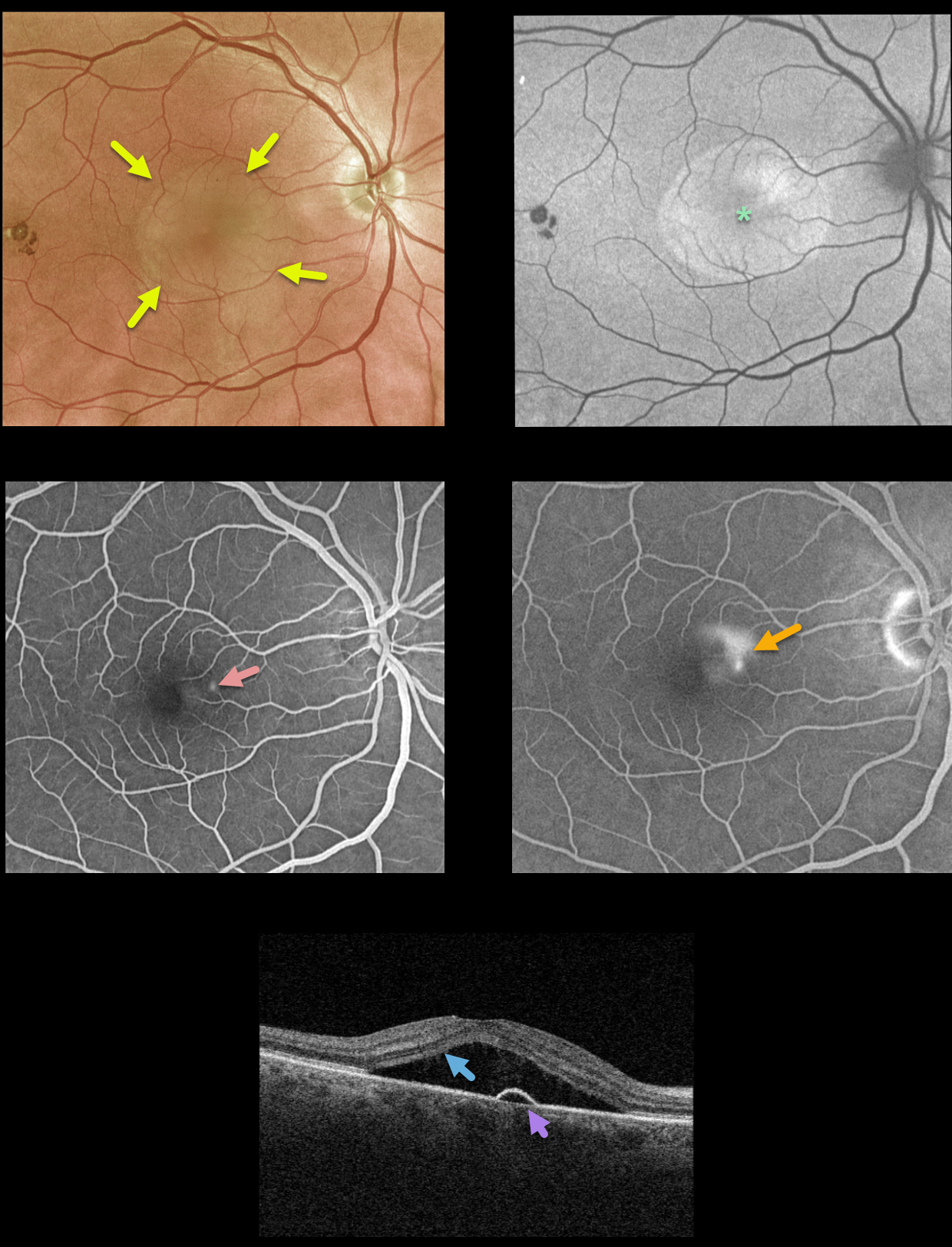
IDIOPATHIC CENTRAL SEROUS CHORIORETINOPATHY
Originally posted on @retina.rocks November 1, 2021
This 61YO male presented with classic findings for idiopathic central serous chorioretinopathy (ICSC). There is a large serous macular detachment that is diffusely hyperautofluorescent.
Fluorescein angiography shows a small dot of early hyperfluorescence with a late smokestack leak.
Triton swept-source OCT shows the serous detachment with a small underlying RPE detachment near the area of angiographic leakage.
Learning Points:
‘Smokestack’ leakage, which only occurs in about 10% of active ICSC cases, is due to a physiologic break in the retinal pigment epithelium (RPE), allowing the fluorescein to leak through the RPE and rise into the subretinal space.
Most ICSC serous detachments spontaneously resolve within 3 months and do not require treatment. Patients who can’t wait 3 months (e.g., pilots or retinal surgeons) or those with persistent symptomatic detachments are best treated with photodynamic therapy.
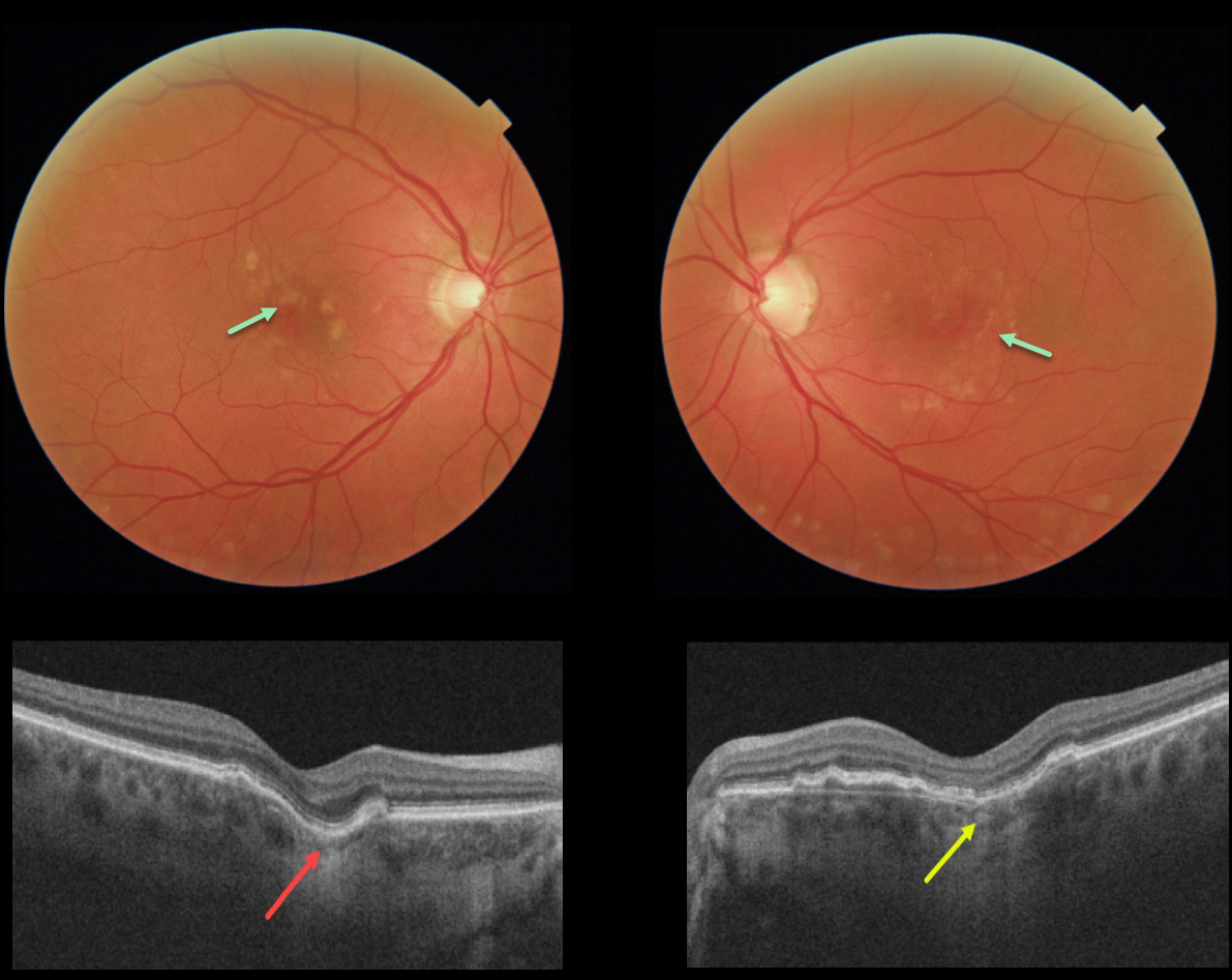
FOCAL CHOROIDAL EXCAVATION
Originally posted on @retina.rocks October 6, 2021
This 60YO female has a history of idiopathic central serous chorioretinopathy that was previously treated successfully with photodynamic therapy in her left eye. Bilateral macular drusen are noted.
OCT shows a definite focal choroidal excavation (FCE) in her right eye, and a possible FCE in her left eye.
Learning Points:
Focal choroidal excavation, originally described by Jampol et al. in 2006, is likely part of the pachychoroid spectrum, as it is usually associated with a thickened choroid and pachyvessels.
The FCE can be observed, although symptomatic manifestations, including active central serous retinopathy or macular neovascularization, often require treatment.
Spaide and Ryan noted this finding in about two-thirds of patients with central serous retinopathy (Am J Ophthalmology 2015;160;1211-1216). An excellent review is by Chung et al (Retina 2017;37;199-221).
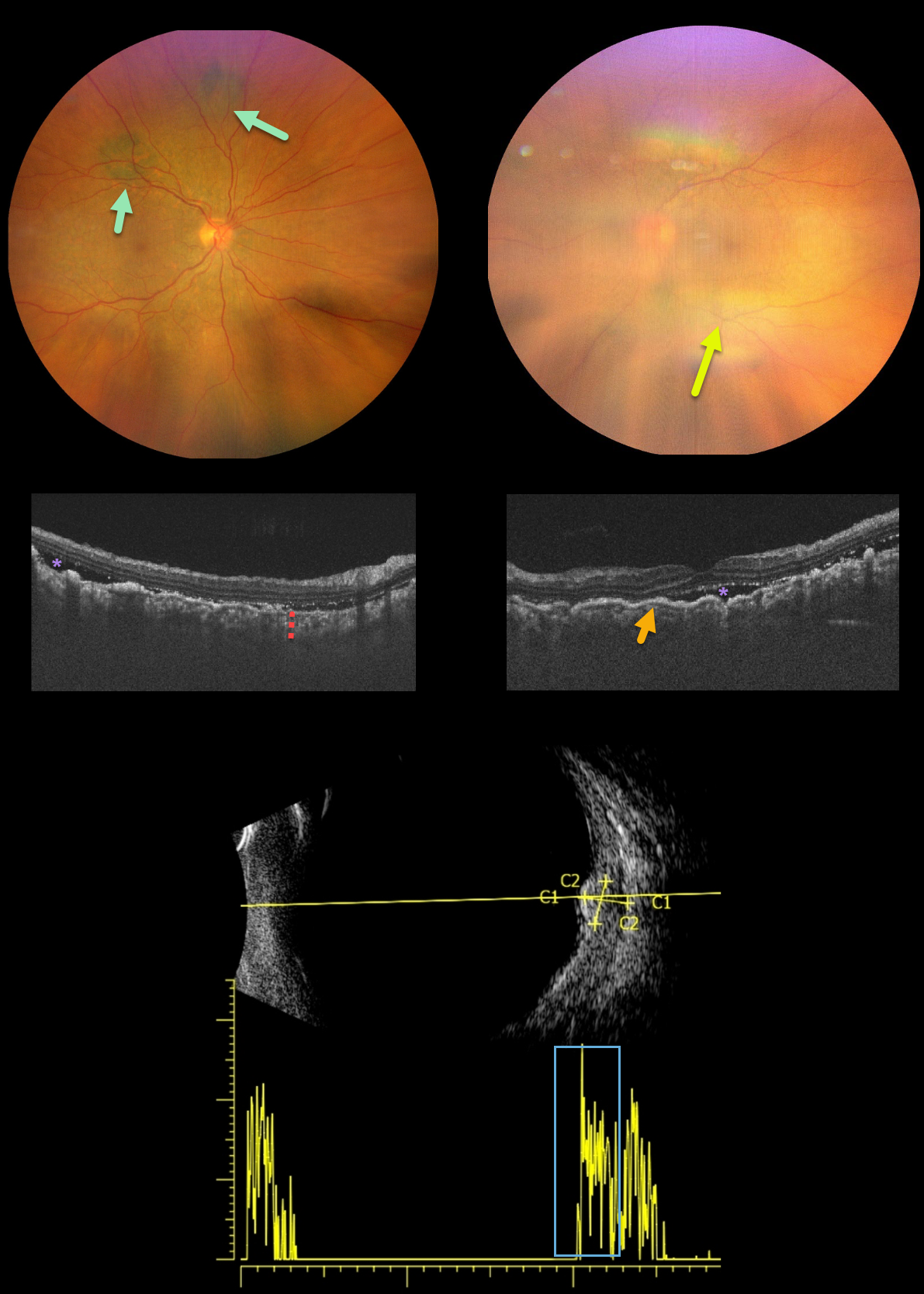
BILATERAL DIFFUSE UVEAL MELANOCYTIC PROLIFERATION (BDUMP)
Originally posted on @retina.rocks September 29, 2021
This is a 62YO female presented with bilateral vision loss over the past 3 months. Her vision was 20/200 OD and counting fingers OS compared to 20/20 OU at baseline. She was diagnosed with stage 4 ovarian cancer one month earlier.
New multifocal, elevated, pigmented choroidal lesions are noted superonasally in the right eye. View of the left fundus is limited due to a significant progressive cataract.
OCT reveals bilateral choroidal thickening with overlying folds and some subretinal fluid. B-scan ultrasonography through one of the choroidal lesions in the right eye shows moderate homogenous internal reflectivity.
Learning Points:
Initially described by Machemer in 1966, the five cardinal features of bilateral diffuse uveal melanocytic proliferation (BDUMP) include:
(1) multiple round or oval subtle red patches at the level of the RPE
(2) a striking pattern of multifocal areas of fluorescein angiographic hyperfluorescence from these lesions
(3) multiple slightly elevated pigmented and nonpigmented uveal melanocytic tumors with diffuse uveal thickening
(4) exudative retinal detachment
(5) rapidly progressive cataracts (Gass et al, Arch Ophthalmology 1990;108:527-533).
The thickened choroid and pigmentary changes are secondary to infiltration by benign uveal melanocytes, possibly in response to a primary visceral tumor (ovarian and uterine carcinomas in women, lung carcinoma in men) that produces melanocytic growth factors. Vision typically deteriorates to near blindness within a year of presentation. The serous detachments can resolve with plasmapheresis, although the overall prognosis is generally poor, with a mean survival of about 1 year from the time of ocular diagnosis.
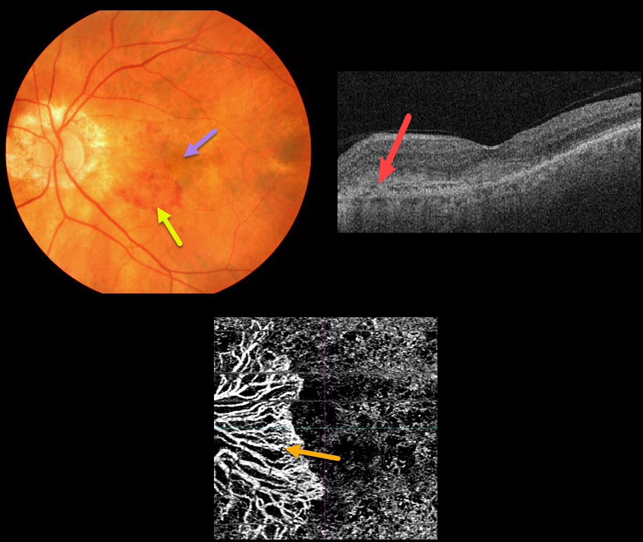
MACULAR NEOVASCULARIZATION (MNV)
Originally posted on @retina.rocks September 27, 2021
This 86YO male presented with symptomatic macular neovascularization (MNV).
Color imaging shows nasal patches of subretinal blood and more central subfoveal pigment. OCT scanning shows a hyperreflective MNV above the RPE (type 2 MNV). The peripapillary MNV is seen on OCT angiography.
Learning Points:
Macular neovascularization is also known by other terms, including choroidal neovascular membrane and subretinal neovascular membrane.
However, since these vessels are now known not to necessarily arise just from the choroid, the proper term is macular neovascularization (see Consensus on Neovascular Age-Related Macular Degeneration Nomenclature Study Group Ophthalmology 2020;127;616-636).
MNV is classified according to the location of the new vessels in relation to the RPE and neurosensory retina: type 1 (below the RPE), type 2 (between the RPE and neurosensory retina, and type 3 (within the neurosensory retina).
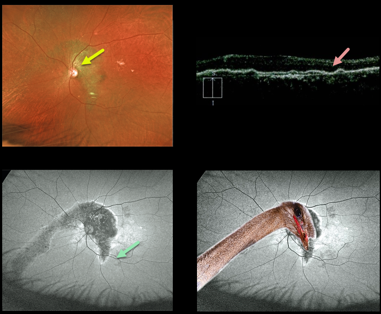
OSTRICH MACULOPATHY
Originally posted on @retina.rocks September 24, 2021
This 79YO male has counting-fingers vision in his left eye, likely due to complications of chronic central serous retinopathy.
Optos color imaging shows increased subretinal pigmentation extending around the nerve superiorly and into the macula and inferonasal periphery.
Fundus autofluorescence (FAF) more dramatically shows these changes as hypo-FAF. OCT scanning shows diffuse thinning of the overlying neurosensory retina.
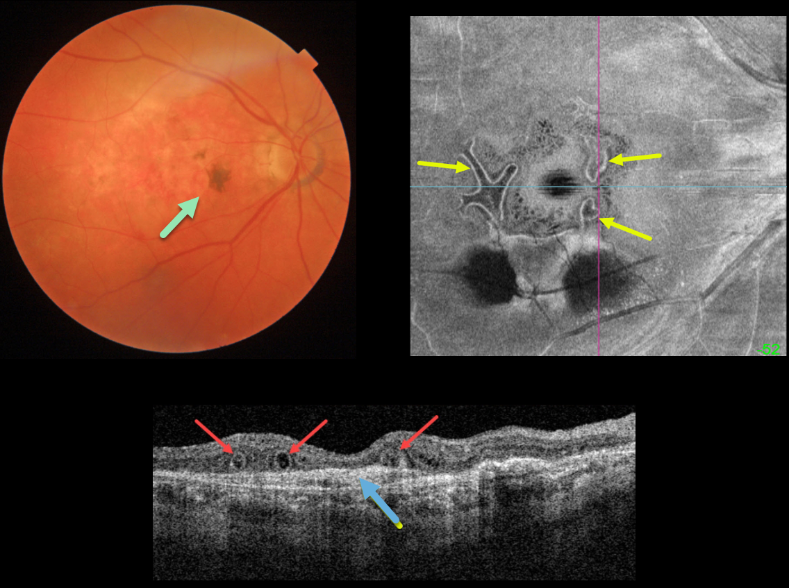
OUTER RETINAL TUBULATION FOLLOWING ONGOING ANTI-VEGF FOR NEOVASCULAR AMD
Originally posted on @retina.rocks September 23, 2021
This is an 86YO female who has stable dry macular scarring from wet age-related macular degeneration with ongoing anti-VEGF injections.
Triton swept-source OCT shows numerous outer retinal tubulations (ORT) overlying an inactive type 1 macular neovascularization (MNV). These tubules are more dramatically seen with en face imaging of the outer retina.
Learning Points:
Outer retinal tubulation is often noted overlying inactive MNV with ongoing anti-VEGF therapy and should not be confused with exudative fluid or cysts, which lack a hyperreflective border.
ORT, initially described by Zweifel et al (Arch Ophthalmol 2009;127:1596-1602), is a neurodegenerative condition of the photoreceptors and Muller cells associated with atrophy affecting the outer retina and retinal pigment epithelium, including advanced AMD and inherited retinal diseases.
The outer hyperreflective band likely represents inner segment mitochondria undergoing fission and translocation toward the nucleus (Litts et al, Retina 2018;38:445-461).
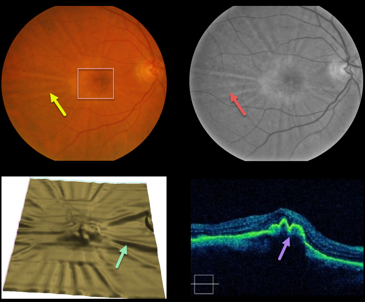
MACULAR NEOVASCULARIZATION (MNV)
Originally posted on @retina.rocks September 14, 2021
This patient developed radiating chorioretinal folds from an involuted type 1 macular neovascularization (MNV, located below the RPE) following multiple intravitreal Avastin injections.
These folds are imaged on the color and red-free photos, as well as on the OCT thickness map and B-scans. Remarkably, vision has been stable at 20/30 for years.
Learning Points:
Macular neovascularization is also known by other terms, including choroidal neovascular membrane and subretinal neovascular membrane.
However, since these vessels are now known not to necessarily arise just from the choroid, the proper term is macular neovascularization (see Consensus on Neovascular Age-Related Macular Degeneration Nomenclature Study Group Ophthalmology 2020;127;616-636).
MNV is classified according to the location of the new vessels in relation to the RPE and neurosensory retina: type 1 (below the RPE), type 2 (between the RPE and neurosensory retina, and type 3 (within the neurosensory retina).
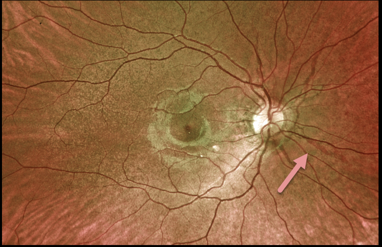
ANGIOID STREAKS
Originally posted on @retina.rocks September 1, 2021
This 19YO female presented with these asymptomatic fundoscopic changes. Vision was 20/25 OU. Optos imaging shows angioid streaks radiating from each optic nerve.
Our patient denied any past medical or family history of pseudoxanthoma elasticum. Genetic testing for the ABCC6 mutation is pending.
Learning Points:
Angioid streaks are often quite easy to miss since their color and orientation mimic the normal retinal vasculature, hence the term ‘angioid.’
The orange skin (peaux d’orange) appearance, most pronounced in each temporal macula, is caused by the calcified Bruch’s membrane giving a yellow mottled ‘coquille d’oeuf’ (eggshell) appearance that contrasts with the normal orange RPE and choroidal pigmentation (Spaide, Retina 2015;35;392-397).
These patients are at significant risk for macular neovascularization (MNV) and need to be followed and treated as needed with anti-VEGF agents, as in patients with AMD.
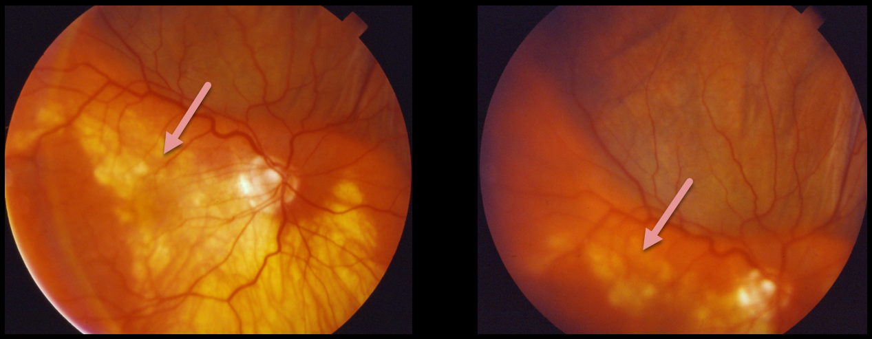
SUPRACHOROIDAL HEMORRHAGE
Originally posted on @retina.rocks August 20, 2021
This patient presented with a pigmented, well-defined, and irregularly lumpy superior subretinal mass due to a suprachoroidal hemorrhage. This simulated a uveal melanoma.
Unfortunately, we don’t have any further information or images for this patient. However, we suspect that the blood spontaneously resolved with normalization of the fundus appearance.
Learning Points:
Patients presenting with suprachoroidal hemorrhage will usually have a history of recent intraocular surgery, trauma, or Valsalva. A clinical clue to this being a hemorrhage and not a melanoma includes the often somewhat lumpy and corrugated appearance, as in this patient.
See Chu and Green for a good review of suprachoroidal hemorrhage (Surv Ophthalmol 1999;43:471-486).
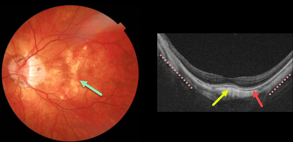
HIGH MYOPIA
Originally posted on @retina.rocks August 19, 2021
This high myope has extensive dry myopic atrophic pigmentary changes noted on Triton color imaging.
Triton swept-source OCT shows typical myopic concave posterior bowing of the eye wall, except more centrally, where there is a convex elevation beneath the fovea. The choroid is also markedly thinned throughout the posterior pole.
Learning Points:
Originally described in 2008 by Gaucher et al (AJO 2008;145:909-914), a dome-shaped macula is a macular convexity found in eyes with high myopia, often associated with a posterior staphyloma. It can be associated with avascular subretinal fluid and macular neovascularization.
See Kumar et al for an excellent recent review of this entity (Surv Ophthalmology 2021;66:560-571).
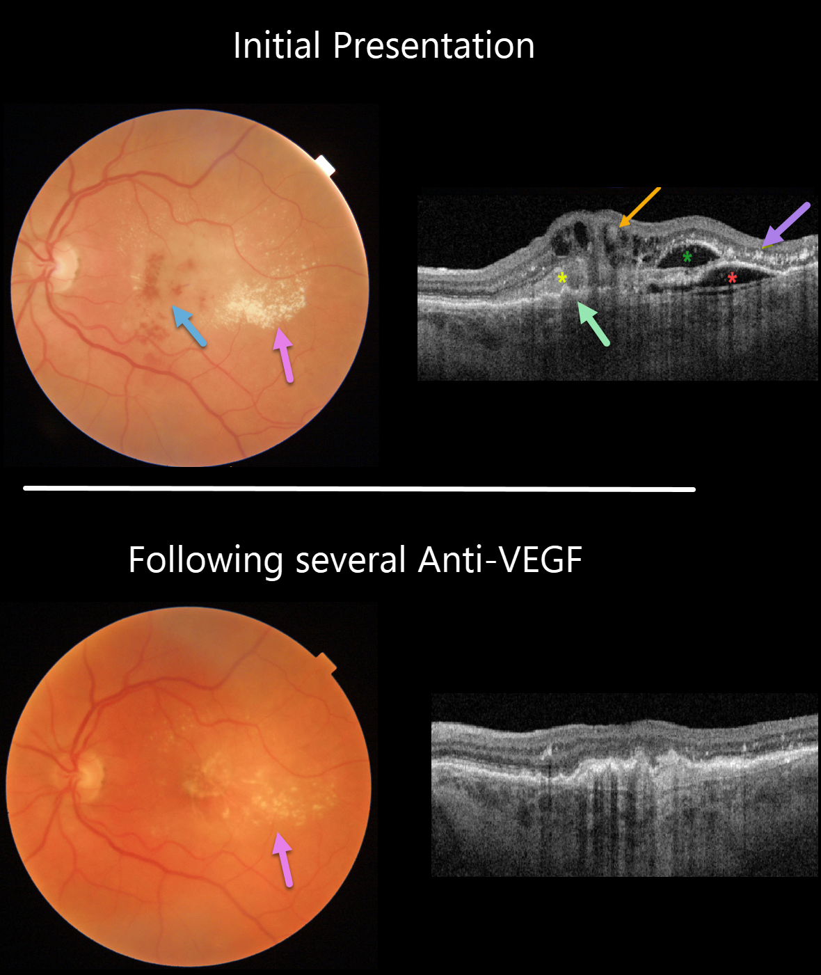
RETINAL ANGIOMATOUS PROLIFERATION (RAP)
Originally posted on @retina.rocks August 18, 2021
This 74YO man presented with counting fingers vision from previously untreated wet age-related macular degeneration (AMD).
Triton color imaging and swept-source OCT beautifully highlight this complex macular neovascularization (MNV). There are scattered inner and deep retinal hemorrhages, opaque fluid, and temporal lipid.
OCT shows an irregular shallow elevation of the RPE more nasally, possibly indicating a sub-RPE (type 1 MNV), with overlying subretinal hyperreflective material. More temporally, there is a more prominent area of RPE elevation that is more hyporeflective with an overlying pocket of intraretinal fluid vs a bacillary layer detachment. Finally, central inner retinal thickening and cysts are seen, indicating intraretinal neovascularization (type 3 MNV), also known as a retinal angiomatous proliferation (RAP) lesion. The lipid appears as hyperreflective clumps in the outer plexiform and outer nuclear layers.
The fluid and blood regressed, with significantly decreased lipid, after several monthly Avastin injections (image 2). Vision improved to 20/400.
Learning Points:
Unlike more typical age-related MNV that start either below (type 1 MNV) or occasionally above (type 2 MNV) the RPE, RAP lesions originate in the retina.
These incompetent vessels leak fluid and blood into the surrounding tissue. Their intraretinal location is a biomarker for RAP lesions.
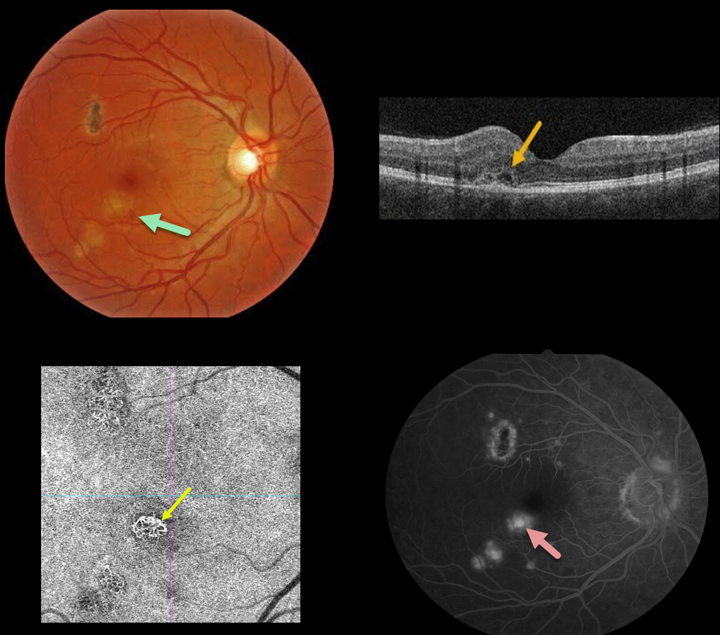
OCULAR HISTOPLASMOSIS
Originally posted on @retina.rocks August 16, 2021
This 30YO male presented with 20/25 vision in his right eye from an ocular histoplasmosis-related macular neovascularization (MNV). Some fluid and blood were associated with a macular histoplasmosis scar in the inferotemporal fovea.
A type 2 MNV, located above the RPE, was noted on OCT. The MNV was best imaged on OCT angiography, with macular leakage noted on fluorescein angiography.
Anti-VEGF therapy was recommended.
Learning Points:
Ocular histoplasmosis is caused by the fungus Histoplasma capsulatum, which is commonly found in soil contaminated by bird or bat droppings. It is found worldwide, but in the United States, it is most common in the Ohio-Mississippi River valleys.
The vast majority of people in endemic areas inhale the organism’s spores early in life, and only a few percent will develop asymptomatic chorioretinal scars.
A small percentage of these will develop MNV later in life. Treatment is similar to wet AMD, with anti-VEGF therapy usually being the first-line therapy. Unlike AMD, these patients often do not require ongoing injections.
Photodynamic therapy can help minimize treatment burden, and thermal laser therapy remains a good option for more peripheral macular lesions.
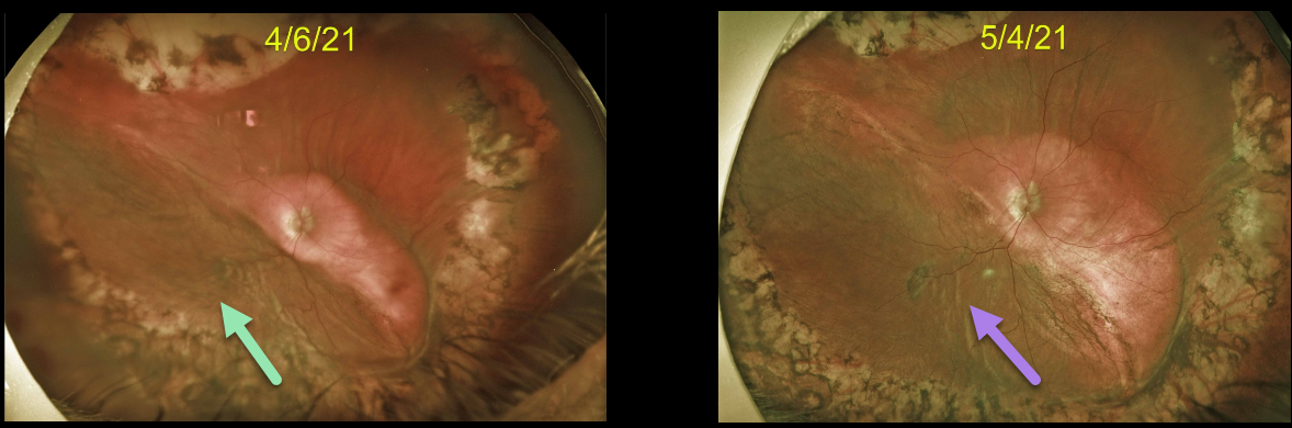
CHOROIDAL DETACHMENT
Originally posted on @retina.rocks August 4, 2021
This 75YO male developed a large choroidal detachment 2 months following a complex vitrectomy for an intraocular lens exchange. There was no wound leak, and the intraocular pressure was 5.
He returned one month later with a spontaneously improving choroidal detachment.
Learning Points:
Choroidal detachment is relatively common after surgery with hypotony. Two mechanisms are believed to be at play: hypotony allows fluid to accumulate in the suprachoroidal space, and inflammation increases the choroidal permeability.
The detached ciliary body may also produce less aqueous humor, thus creating a self-perpetuating cycle.
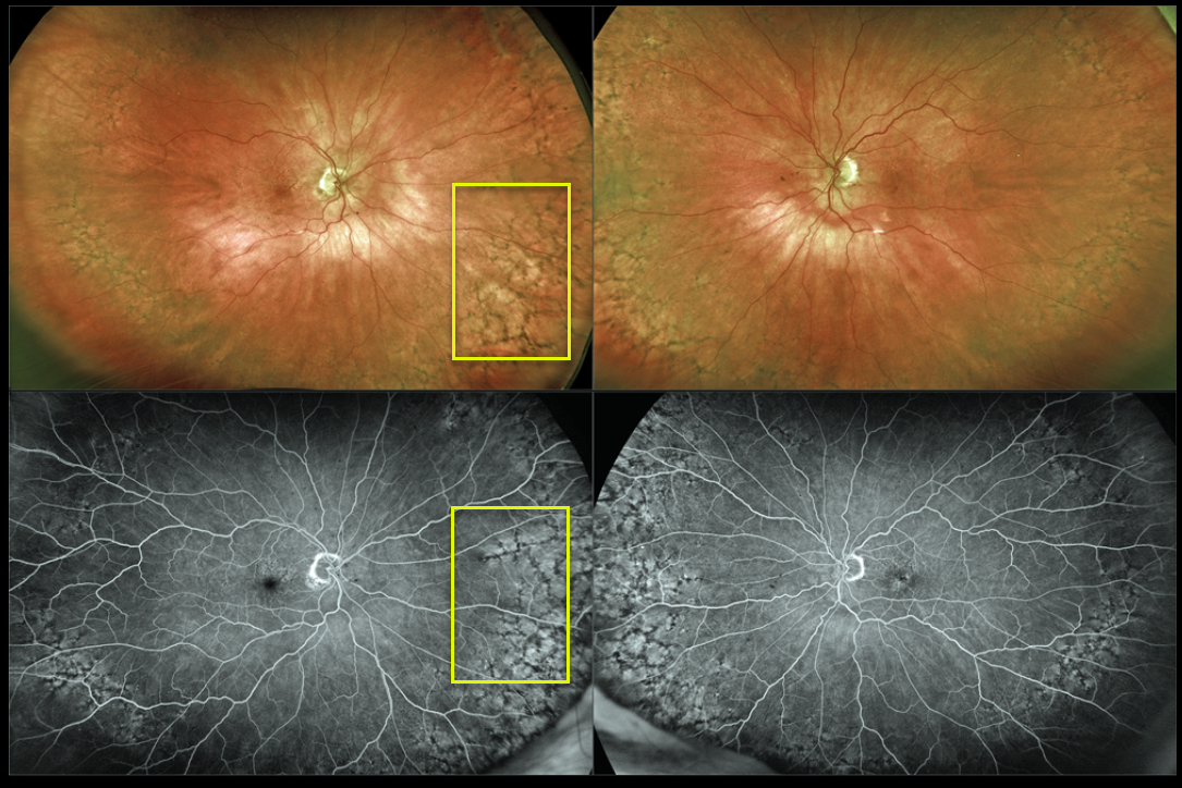
PERIPHERAL RETICULAR DEGENERATION OF THE RPE
Originally posted on @retina.rocks June 22, 2021
This 87YO female shows prominent bilateral peripheral reticular degeneration of the RPE (PRDRPE) with Optos ultra-widefield imaging and fluoroscein angiography.
The reticular changes at first glance may appear similar to the ‘bone spicules’ found with retinitis pigmentosa, which are caused by disinhibited RPE cells migrating along retinal capillaries following photoreceptor degeneration.
Learning Points:
The findings in PRDRPE are part of the age-related macular degeneration (AMD) spectrum and are subretinal and located in the periphery. A recent meta-analysis by Forshaw et al found PRDRPE to be present in 68-83% of eyes with AMD (Ophthalmology Retina 2019;734-743).
It is associated with decreased dark adaptation, which may help explain why many patients with AMD experience difficulty with night vision (see Lains et al, AJO 2018;190;113-124).
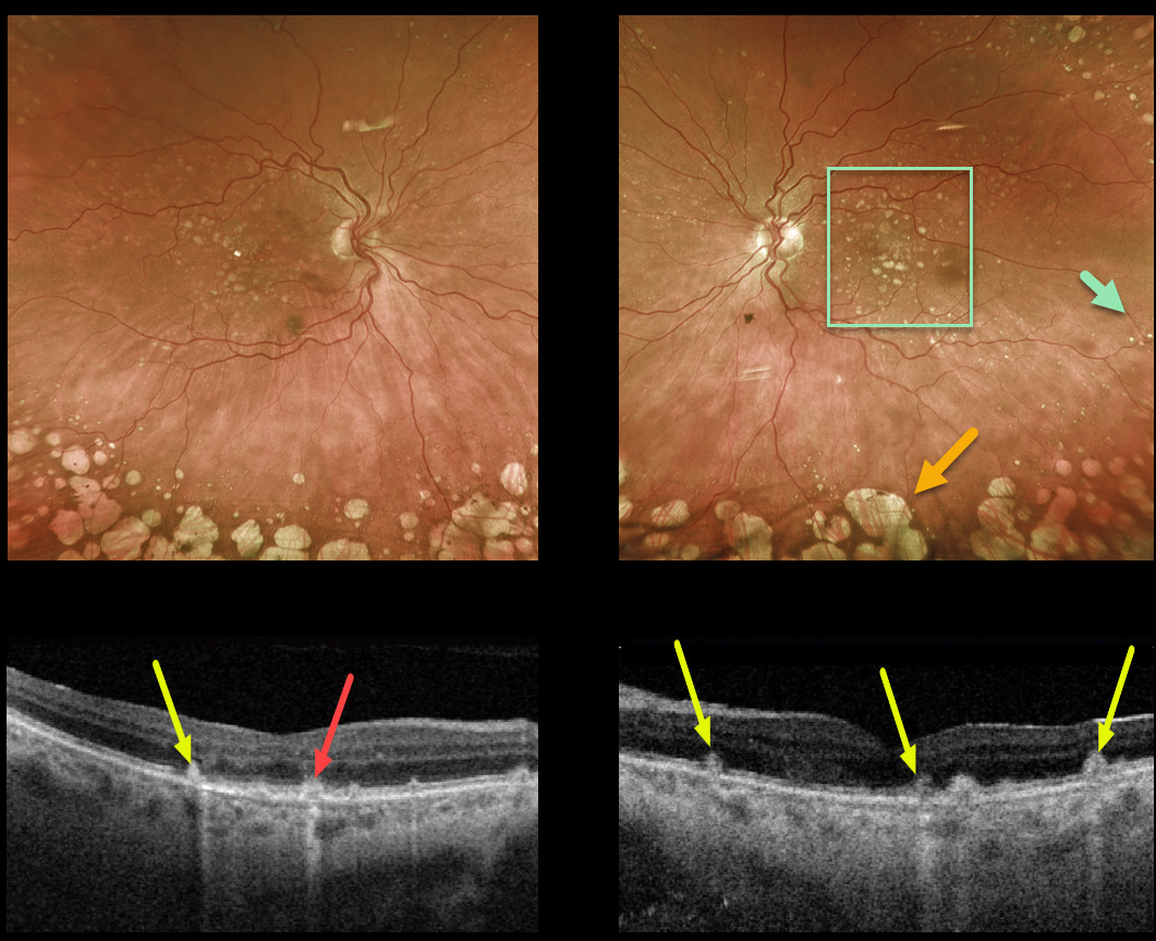
DRY AMD
Originally posted on @retina.rocks June 11, 2021
This 69YO female presented with 20/50 vision OU and bilateral mixed macular and peripheral drusen, along with extensive peripheral cobblestone degeneration.
Triton swept-source OCT shows a mixture of drusen and subretinal drusenoid deposits.
Learning Points:
Macular drusen and subretinal drusenoid deposits are some of the hallmark features of non-exudative age-related macular degeneration.
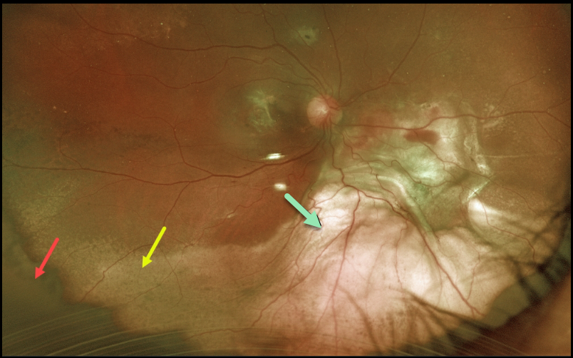
PERIPHERAL EXUDATIVE HEMORRHAGIC CHORIORETINOPATHY
Originally posted on @retina.rocks May 31, 2021
This is a beautiful Optos image of a 67YO female with counting finger vision 1-week following pars plana vitrectomy for a breakthrough vitreous hemorrhage.
There is a rim of dark yellow vitreous skirt, resolving devitalized subretinal blood, and elevated subretinal fibrotic scarring.
Learning Points:
Ectopic choroidal neovascularization (CNV), also known as peripheral exudative hemorrhagic chorioretinopathy, is an exudative process similar to wet AMD but located in the retinal periphery.
Ectopic CNV can often masquerade as a choroidal mass or uveal melanoma. It can also cause extensive choroidal/suprachoroidal, subretinal, and vitreous hemorrhage, more commonly in patients who are on blood thinners.
The subretinal blood often spontaneously regresses without treatment, although anti-VEGF injections can be used for symptomatic or increasing exudation.
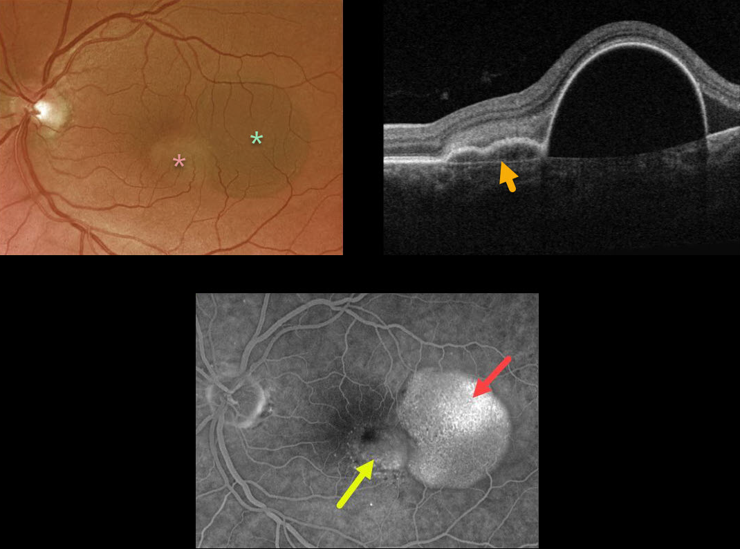
PIGMENT EPITHELIAL DETACHMENT (PED)
Originally posted on @retina.rocks April 26, 2021
This 57YO female presented with counting-finger vision from a large RPE detachment (PED). The PED was located in the temporal macula, with a smaller notch of irregular RPE elevation more centrally.
Triton swept-source OCT shows a shallow PED at the notch overlying Bruch’s membrane. Optos fluorescein angiography shows late uneven filling of the PED and late speckled staining of the notch.
Anti-VEGF therapy was recommended.
Learning Points:
Originally described by Gass (Retina 1984;4:205-220), a notched PED is a sign of an occult macular neovascularization (MNV). The notch represents a type 1 MNV, which tethers the overlying RPE from the surrounding more highly elevated PED. Hydrostatic leakage from the MNV may contribute to the larger PED.
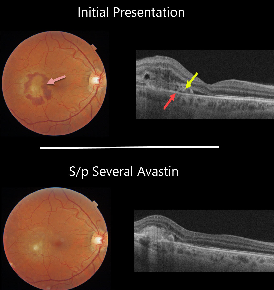
OCULAR HISTOPLASMOSIS
Originally posted on @retina.rocks April 21, 2021
This 44YO male presented with a symptomatic macular neovascularization (MNV) from ocular histoplasmosis. Vision was 20/40.
Subretinal blood surrounds the MNV in the temporal macula. On OCT, the MNV is located above the RPE, indicating a type 2 lesion. Some retinal thickening is noted above the MNV.
The blood and fluid resolved completely following several additional monthly Avastin injections.
Learning Points:
There are three types of MNV: type 1 MNV is below the RPE, type 2 is between the RPE and outer segments, and type 3 is intraretinal.
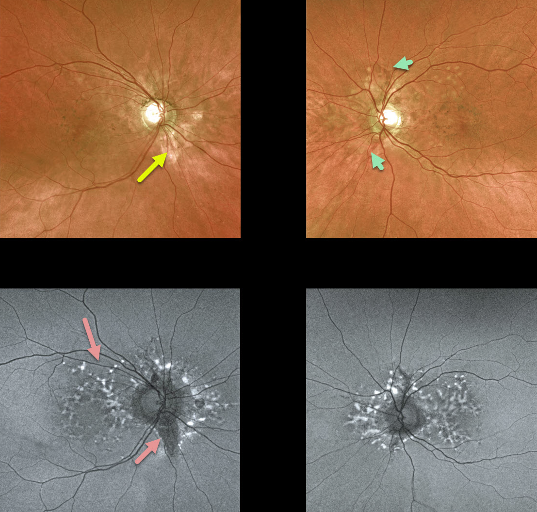
ANGIOID STREAKS
Originally posted on @retina.rocks April 1, 2021
We have been following this 54YO female with pseudoxanthoma elasticum (PXE) for many years. Vision is 20/20 bilaterally, and fortunately, she remains asymptomatic.
She has bilateral macular peripapillary pigmentary changes. Angioid streaks, always tough to find unless the clinician actively looks for them, are not prominent, although a few are visible in her left eye.
Fundus autofluorescence (FAF) shows more dramatic changes, including scattered hyper- and hypo-FAF peripapillary and macular spots.
Her sister became legally blind many years ago from bilateral macular neovascularization (MNV).
Learning Points:
Patients with angioid streaks and pseudoxanthoma elasticum (PXE) can present with retinal findings similar to those of pattern dystrophies.
These patients are at significant risk for macular neovascularization and need to be followed and treated as needed with anti-VEGF agents, as in patients with AMD.
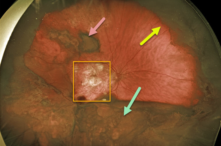
PERIPHERAL EXUDATIVE HEMORRHAGIC CHORIORETINOPATHY
Originally posted on @retina.rocks March 30, 2021
This 80YO male had 20/80 vision in his left eye with ongoing Eylea injections for wet age-related macular degeneration (AMD). Vision was hand motion in his right eye due to an inactive disciform scar.
Several months ago, he complained of decreased vision OD and was found to have a dense vitreous hemorrhage. About 6 weeks later, he underwent vitrectomy in this eye to try to restore his peripheral vision.
One day postoperatively, Optos photography showed the residual vitreous skirt, subretinal blood, and elevated choroidal/suprachoroidal yellow blood.
Learning Points:
Ectopic choroidal neovascularization (CNV), also known as peripheral exudative hemorrhagic chorioretinopathy, is an exudative process similar to wet AMD but located in the retinal periphery.
Ectopic CNV can often masquerade as a choroidal mass or uveal melanoma. It can also cause extensive choroidal/suprachoroidal, subretinal, and vitreous hemorrhage, more commonly in patients who are on blood thinners (our patient was on Pradaxa).
The subretinal blood often spontaneously regresses without treatment, although anti-VEGF injections can be used for symptomatic or increasing exudation.
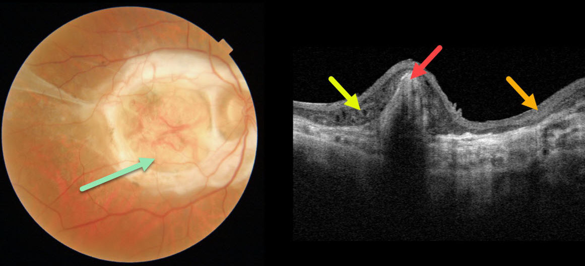
SUBRETINAL FIBROSIS
Originally posted on @retina.rocks March 11, 2021
This 69YO male has counting-finger vision in his right eye from a fibrotic disciform scar due to wet age-related macular degeneration.
Triton swept-source OCT scanning shows hyperreflective subretinal scarring, variable disorganized thinned overlying retinal layers, and outer retinal thickening.
Learning Points:
This type of end-stage wet AMD was the norm prior to the anti-VEGF era, which began in 2005. Fortunately, with early and timely intravitreal injections, disciform scars are now virtually only seen in untreated eyes.
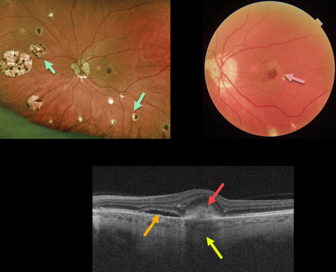
OCULAR HISTOPLASMOSIS
Originally posted on @retina.rocks March 10, 2021
This 65YO presented with 20/400 vision in the left eye. Optos imaging shows classic ocular histoplasmosis (OHS) peripapillary, macular, and peripheral chorioretinal scarring. A more close-up macular image from the Triton swept-source OCT shows a greenish macular neovascularization (MNV) with associated subretinal blood.
The OCT B-scan shows subretinal fluid and hyperreflective subretinal blood, which causes choroidal shadowing.
Learning Points:
Ocular histoplasmosis is caused by the Histoplasmosis capsulatum fungus, which is commonly found in soil contaminated by bird or bat droppings. It is found worldwide, but in the United States, it is most common in the Ohio-Mississippi River valleys.
The vast majority of people in endemic areas inhale the organism’s spores early in life, and only a few percent will develop asymptomatic chorioretinal scars. A small percentage of these will develop MNV later in life.
Originally described by Woods and Wahlen in 1959, the findings include the classic triad of peripapillary atrophy, peripheral multifocal punched-out scars, and a macular scar or neovascularization.
We like to add a fourth criterion: a quiet vitreous, since numerous inflammatory disorders can simulate ocular histoplasmosis.
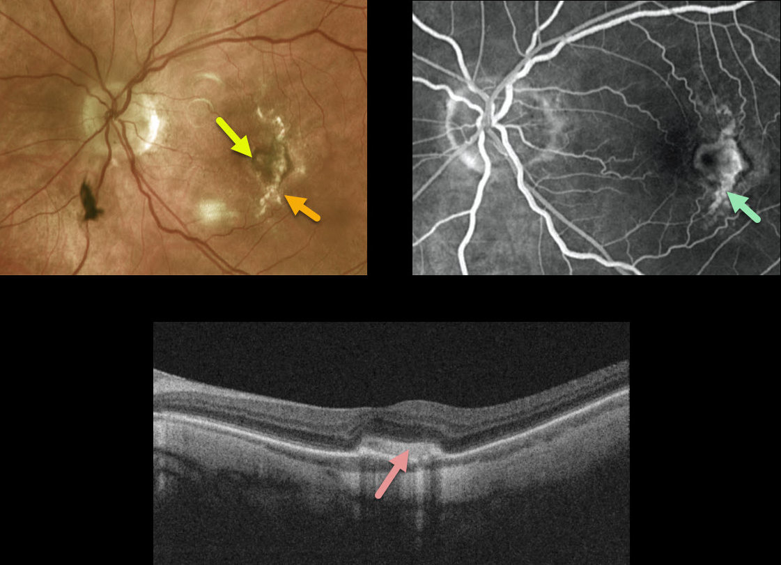
MYOPIC MACULAR NEOVASCULARIZATION
Originally posted on @retina.rocks February 24, 2021
This 69YO female came in for a routine exam with a few months of vision loss in her left eye.
There is a new small area of subfoveal pigment adjacent to an old curvilinear scar more temporally. Although this temporal scar appeared to represent a choroidal rupture, she denied any prior trauma.
Axial length is 27.30 mm in this eye, nearly 1 mm greater than her fellow eye, indicating that this scar is likely a large and unusual lacquer crack.
Fluorescein angiography (FA) shows staining of the temporal scar and subfoveal pigment. An OCT B-scan through the subfoveal pigment shows a type 2 macular neovascularization (MNV) without fluid.
Learning Points:
Myopic MNV often presents as a pigmented subretinal lesion, often with minimal to no fluid due to the underlying thinned choroid.
In our experience, the presence or absence of new blood or new visual symptoms is more useful than the OCT, which usually shows no fluid, or FA, which often shows no leakage. Anti-VEGF injections were therefore recommended.
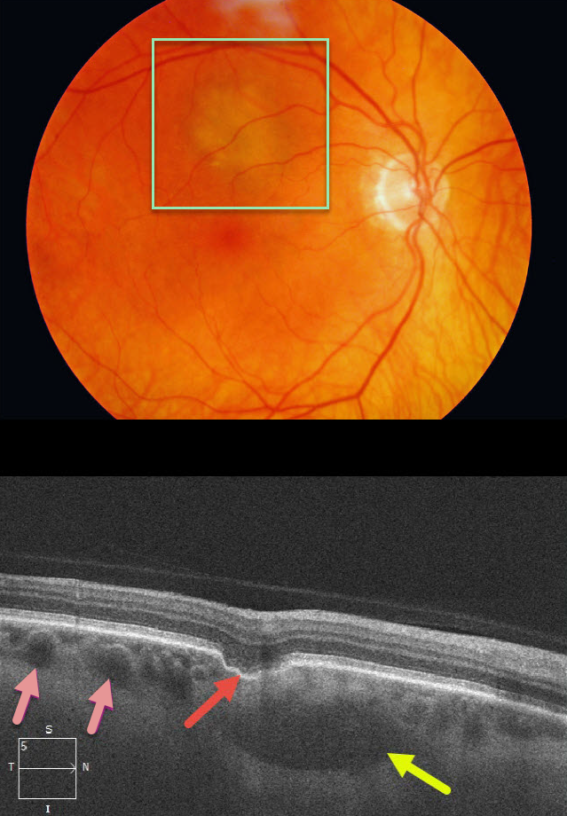
CHOROIDAL EXCAVATION
Originally posted on @retina.rocks February 17, 2021
This patient was referred for an asymptomatic, slightly depigmented subretinal lesion in the superior right macula. OCT through this lesion reveals a focal choroidal excavation, choroidal pachyvessels, and probable loculated outer choroidal fluid.
Learning Points:
Focal choroidal excavation (FCE), originally described by Jampol et al in 2006, is likely part of the pachychoroid spectrum since it is usually associated with a thickened choroid and pachyvessels. The FCE can be observed, although symptomatic manifestations, including active central serous retinopathy or macular neovascularization, often require treatment.
Our patient also showed accumulation of fluid in the outer choroid. Spaide and Ryan noted this finding in about two-thirds of patients with central serous retinopathy (Am J Ophthalmology 2015;160;1211-1216). The presence of an FCE, pachyvessels, and loculated posterior choroidal fluid in our patient is intriguing and supports the association between pachychoroid disorders and FCE.
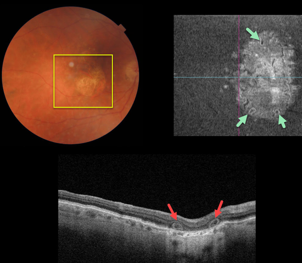
OUTER RETINAL TUBULATION
Originally posted on @retina.rocks February 16, 2021
This 80YO female was 20/200 in her right eye due to central macular atrophy from atrophic age-related macular degeneration (AMD).
Triton swept-source optical coherence tomography showed outer retinal tubulation (ORT) in the outer retinal layers. The extent of these tubular structures was best visualized on en face imaging.
Learning Points:
Outer retinal tubulation, initially described by Zweifel et al (Arch Ophthalmol 2009;127;1596-1602), is a neurodegenerative condition of the photoreceptors and Muller cells associated with atrophy affecting the outer retina and retinal pigment epithelium, including advanced AMD and inherited retinal diseases.
ORT is often seen overlying inactive macular neovascularization in the setting of ongoing anti-VEGF therapy and should not be confused with exudative fluid or cysts, which lack a hyperreflective border.
The outer hyperreflective band likely represents inner segment mitochondria undergoing fission and translocation toward the nucleus (Litts et al Retina 2018;38;445-461).
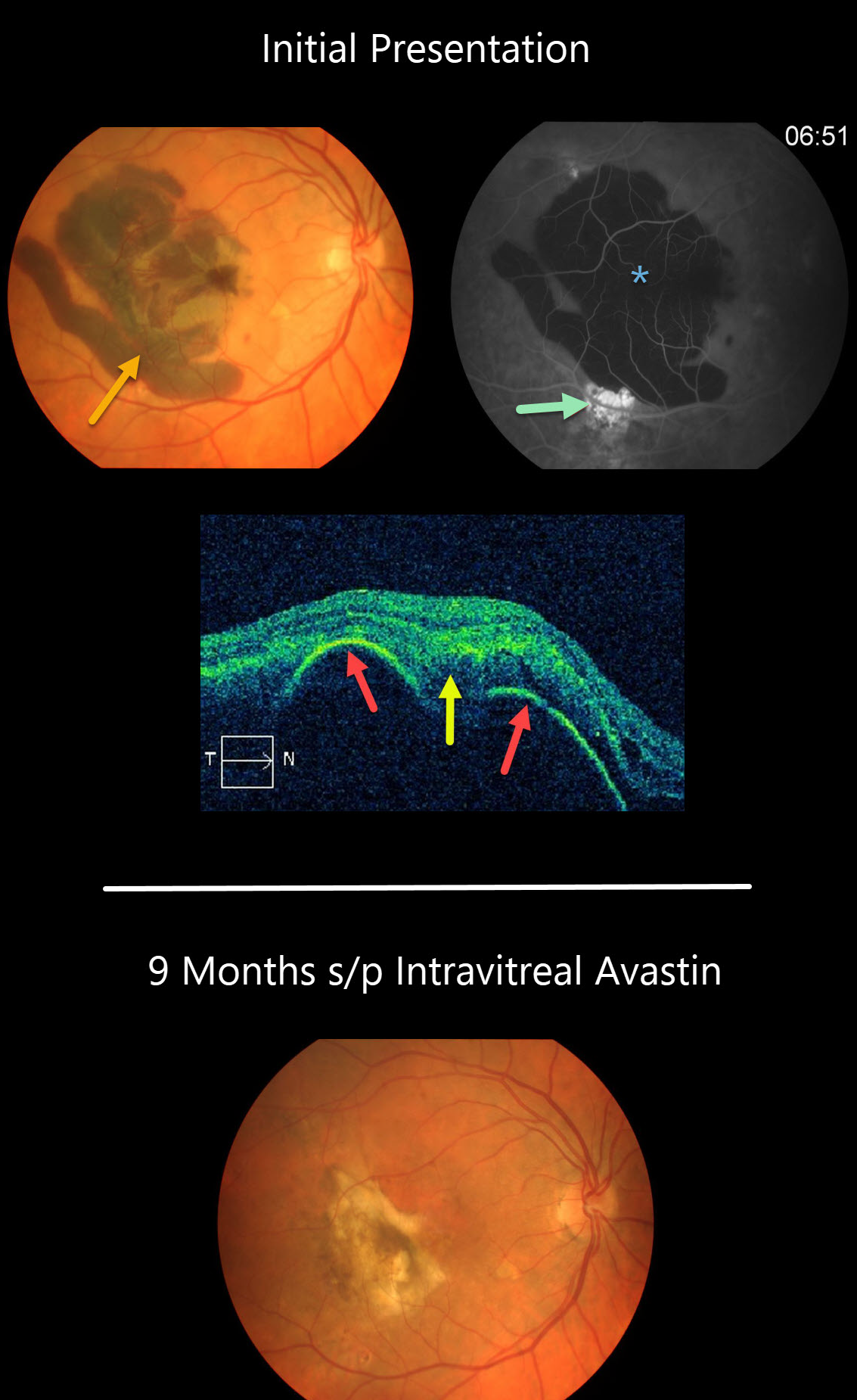
POLYPOIDAL CHOROIDAL VASCULOPATHY
Originally posted on @retina.rocks February 8, 2021
This patient presented with a large acute submacular hemorrhage in the right eye.
The blood caused diffuse blockage on fluorescein angiography, and the source of the blood may have been from the area of subretinal hyperfluorescence inferiorly.
OCT scanning shows a large retinal pigment epithelium (RPE) detachment with notched thumb-like projections with subretinal hyperreflectivity from the blood.
Our patient’s subretinal blood resolved following several intravitreal Avastin injections.
Learning Points:
Polypoidal choroidal vasculopathy (PCV) is a variant of type 1 macular neovascularization. PCV is likely distinct from age-related macular degeneration, occurring in a different population (more common in African Americans and Asians) and characterized by a relative lack of drusen and a thicker choroid.
The sub-RPE vessels were classically characterized using indocyanine green angiography, although recent literature suggests that OCT is just as effective at diagnosing these vascular polyps. Historically, these lesions were believed to be somewhat resistant to anti-VEGF monotherapy and often required combined treatment with photodynamic therapy. More recent data from the PLANET study suggest that Eylea monotherapy is also effective (AJO 2019;204;80-89).
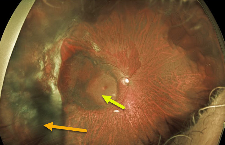
PERIPHERAL EXUDATIVE HEMORRHAGIC CHORIORETINOPATHY
Originally posted on @retina.rocks January 15, 2021
This 71YO male had a vitrectomy in 2018 for a breakthrough vitreous hemorrhage from ectopic choroidal neovascularization (CNV). He did well with 20/40 vision until a recurrent vitreous hemorrhage developed two years later.
The vitreous cleared with several monthly intravitreal Avastin injections, but vision remains at hand motion due to submacular blood, some of which is yellow and devitalized. We discussed repeat vitrectomy with subretinal TPA, but he elected for in-office anti-VEGF injections. He is not on any systemic blood thinners.
Learning Points:
Ectopic CNV, also known as peripheral exudative hemorrhagic chorioretinopathy, is a degenerative and exudative process similar to wet AMD but located in the retinal periphery.
Ectopic CNV can often masquerade as a choroidal mass or uveal melanoma. It can also cause extensive subretinal and vitreous hemorrhage, more commonly in patients who are on blood thinners.
The subretinal blood often spontaneously regresses without treatment, although anti-VEGF injections can be used for symptomatic or increasing exudation.
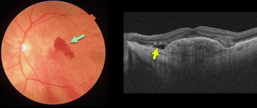
NEOVASCULAR AMD
Originally posted on @retina.rocks January 14, 2021
This patient with wet age-related macular degeneration (AMD) was being treated with PRN anti-VEGF injections. She came in for her regular examination without new symptoms and with 20/70 vision.
Although the OCT line scan shows some fairly subtle new sub-retinal fluid, the color photo dramatically shows new blood.
Learning Points:
There is currently some debate about the utility of performing full dilated examinations when patients are being followed with ongoing anti-VEGF therapy.
New blood is quite easy to miss on an OCT without a fundus photograph or examination. In our hands, having a high-quality fundus photograph at each visit, regardless of whether the patient is in for just an injection or a full examination, is invaluable.
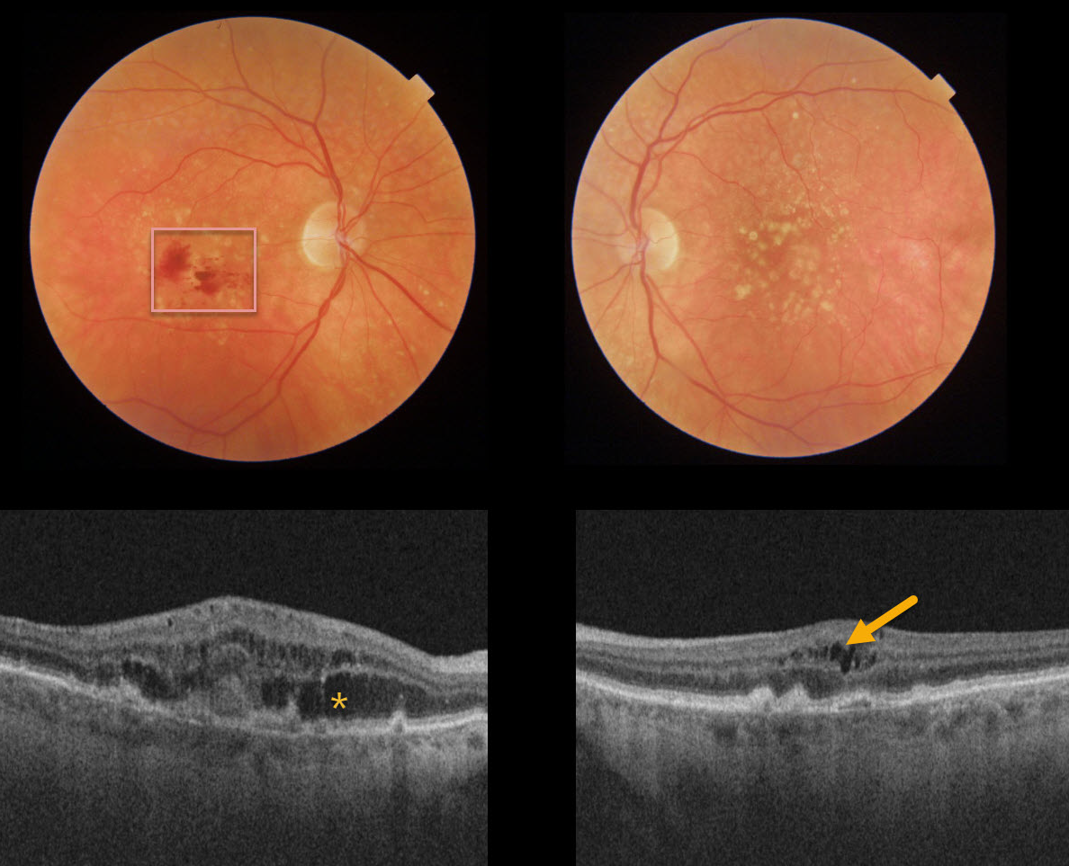
RETINAL ANGIOMATOUS PROLIFERATION (RAP)
Originally posted on @retina.rocks January 5, 2021
Our patient has age-related macular degeneration with a type 3 macular neovascularization (MNV), also known as retinal angiomatous proliferation (RAP).
Her fundus photo reveals inner retinal hemorrhages of her right eye and variable intraretinal fluid on OCT. Our patient showed no obvious exudation clinically, but the small amount of intraretinal fluid on OCT in the left eye indicates a subclinical RAP in this eye as well.
Bilateral anti-VEGF therapy was started, and we expect that she will do well.
Learning Points:
Unlike more typical age-related MNV that start either below (type 1 MNV) or occasionally above (type 2 MNV) the RPE, RAP lesions originate within the retina.
These incompetent vessels leak fluid and blood into the surrounding tissue.
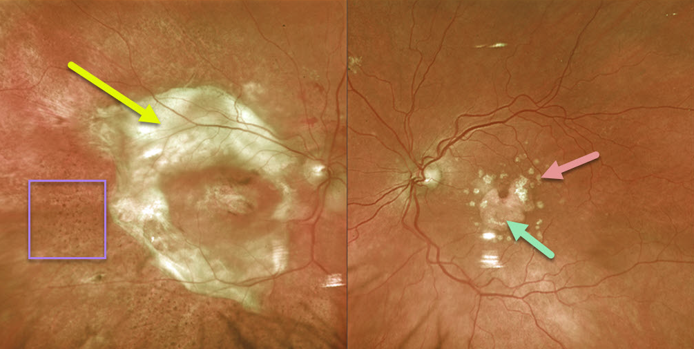
DISCIFORM SCAR
Originally posted on @retina.rocks December 15, 2020
This patient with age-related macular degeneration (AMD) has a large fibrotic disciform scar in the right eye and dry AMD in the left eye with macular atrophy skirting the foveal center and more distal calcified drusen.
The coarse pigmentary changes noted inferiorly and temporally in the right eye are a classic tell-tale finding for prior resolved subretinal blood (see Hussnain et al Retina 2019;39:1925-1935).
Learning Points:
Disciform scars were the norm for untreated wet AMD prior to the anti-VEGF era that began in the mid-2000s.
New-onset wet AMD diagnosed and treated with appropriate anti-VEGF dosing virtually never goes on to develop these lesions.
There may be progressive macular atrophy following years of therapy, but not this type of fibrotic scarring.
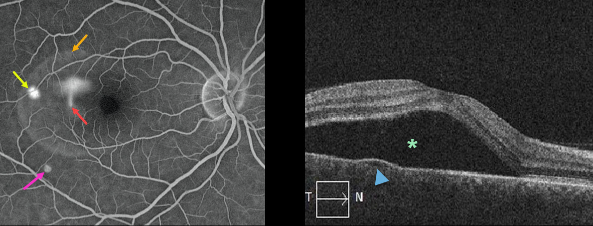
IDIOPATHIC CENTRAL SEROUS CHORIORETINOPATHY
Originally posted on @retina.rocks December 14, 2020
This 44YO male presented with 20/60 vision in his right eye. The left eye was normal with 20/20 vision.
His fluorescein angiogram (FA) shows classic findings for idiopathic central serous chorioretinopathy (ICSC). There is the more common blot leak, as well as the classic “smokestack” leak, which occurs in only about 10% of cases.
The FA also demonstrates the extent of the serous detachment; there is also a tiny RPE detachment without leakage in the distal inferior macula.
The OCT shows both the serous detachment and the small pigment epithelial detachment.
Learning Points:
Fluorescein leaks through a physiologic break in the retinal pigment epithelium (RPE) and rises in the subretinal space, giving the smokestack.
The fluorescein dye then diffuses and pools within the subretinal space, highlighting the extent of the serous detachment.
Most ICSC serous detachments spontaneously resolve within 3 months and do not require treatment. Patients who can’t wait the 3 months (i.e., pilots or surgeons), or those with persistent symptomatic detachments, are best treated with photodynamic therapy.
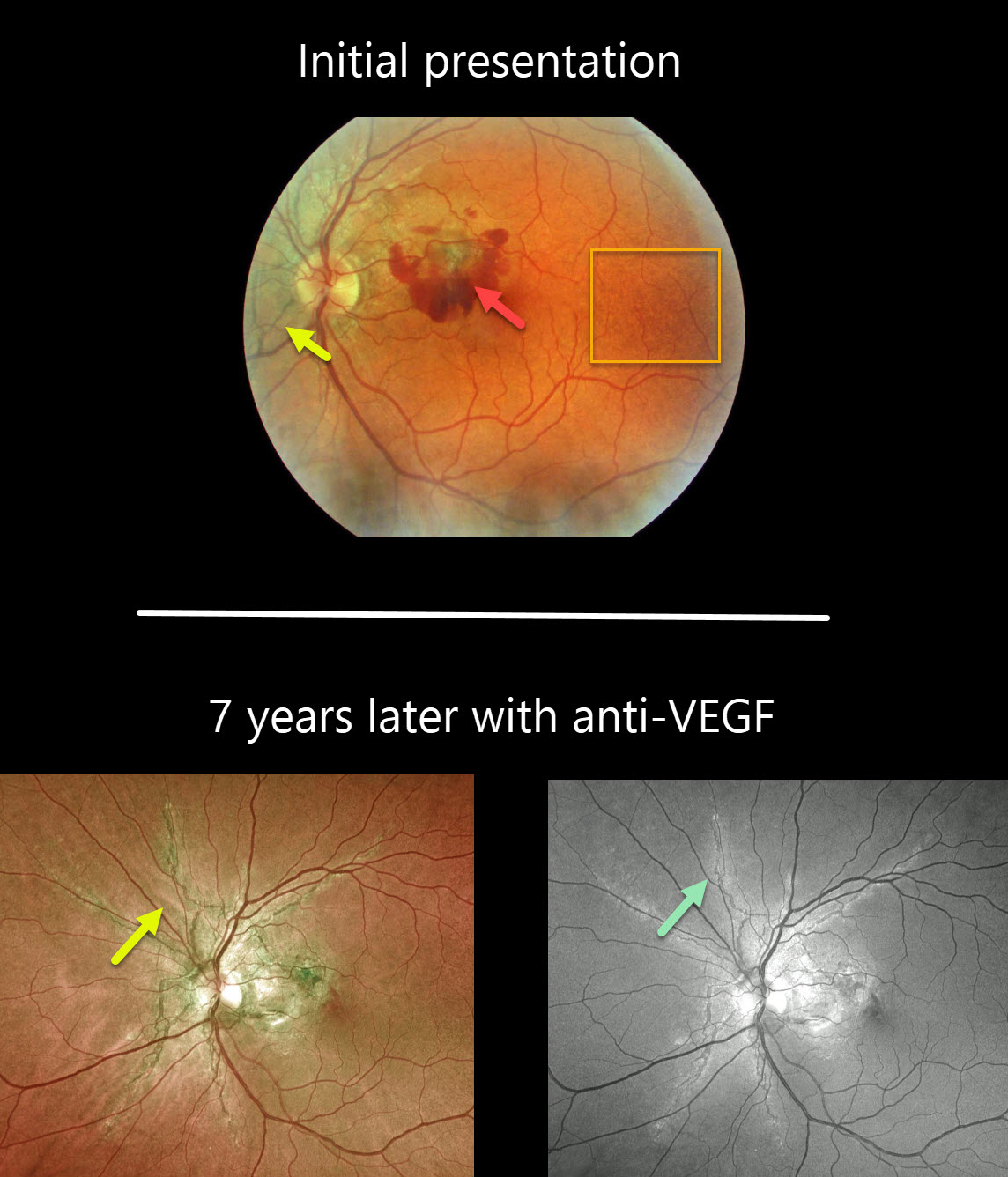
ANGIOID STREAKS
Originally posted on @retina.rocks November 26, 2020
This patient initially presented with dramatic pigmented angioid streaks and acute vision loss from macular neovascularization.
The classic peau d’orange (orange skin) pigmentary changes are seen in the temporal macula.
Seven years later, vision is 20/25 with ongoing anti-VEGF therapy. The angioid streaks are best imaged with the Optos green channel.
Learning Points:
Macular neovascularization is a complication of virtually any disorder that affects Bruch’s membrane, the most common being age-related macular degeneration, ocular histoplasmosis, idiopathic, degenerative myopia (lacquer cracks), trauma (choroidal rupture), and angioid streaks.
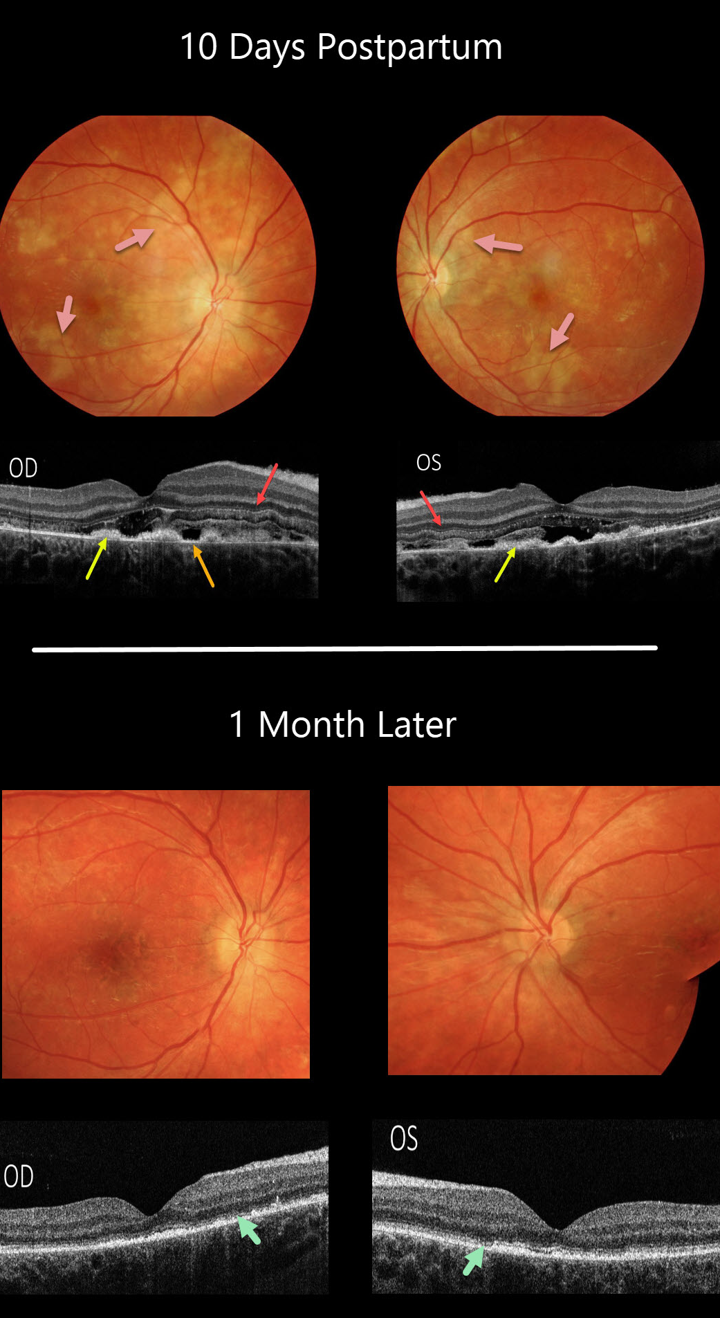
TOXEMIA
Originally posted on @retina.rocks November 9, 2020
This patient was toxemic before delivery, with rapid bilateral vision loss and severely elevated blood pressure. When we examined her 10 days postpartum, her vision was counting fingers bilaterally.
There are multiple patches of creamy white, ischemic RPE and outer retina scattered throughout both posterior poles.
OCT scanning shows normal inner retinal bands extending from the internal limiting membrane to the inner nuclear layer, but the photoreceptor layers showed multiple additional hypo- and hyper-reflective bands, outer retinal disorganization, retinal edema, thickened RPE, and RPE loss with visible Bruch’s membrane.
One month later, vision remarkably improved to 20/25 bilaterally. The creamy multifocal lesions resolved, with residual pigmentary changes.
The prior OCT outer retinal changes resolved, with residual outer retinal and RPE atrophy.
Learning Points:
Toxemia causes choroidal ischemia with secondary RPE dysfunction, which can cause exudative retinal detachments.
Acute outer retinal ischemia causes these transient, bizarre, and pathognomonic OCT changes.
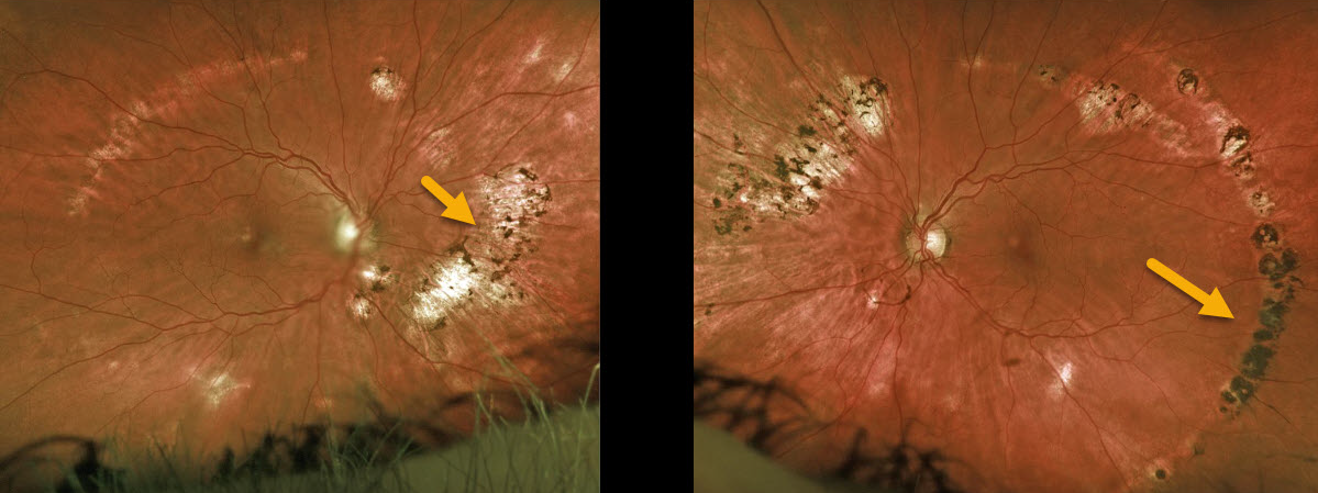
OCULAR HISTOPLASMOSIS
Originally posted on @retina.rocks October 29, 2020
This is a beautiful example of bilateral streak lesions in a patient with the ocular histoplasmosis syndrome (OHS).
Learning Points:
The incidence of streak lesions (Schlaegel lines) in OHS is estimated at about 5% (Arch Ophthalmol 1981;99:246-248), and in our experience, they are more common in areas of more extensive peripheral scarring.
They appear as linear aggregations of midperipheral OHS scars that run parallel to the ora serrata.
They somewhat resemble lattice degeneration in location and appearance, but do not increase the patient’s risk of ocular complications.
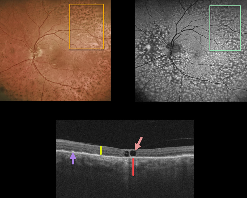
PERIPAPILLARY PACHYCHOROID SYNDROME WITH UNILATERAL BDUMP
Originally posted on @retina.rocks October 15, 2020
While being followed for peripapillary pachychroid syndrome (PPS) with periodic photodynamic therapy for symptomatic foveal fluid OS, this patient presented with new diffuse pigmentary deposits in the left eye only.
Fundus autofluorescence shows a dramatic pattern of hyperautofluorescent and hypofluorescent spots scattered throughout the fundus. These findings, although unilateral, look very similar to those in the paraneoplastic condition, bilateral diffuse uveal melanocytic proliferation (BDUMP).
Macular OCT scanning shows a diffusely thickened choroid when compared to the neurosensory retina. Other OCT findings here include a few large central foveal cysts, outer retinal atrophy, and RPE changes.
Our patient has no known prior cancer diagnoses, and recently had a negative CT abdominal scan and chest x-ray. He continues to be followed closely by his internist since BDUMP often precedes the diagnosis of a systemic malignancy by months to years.
Learning Points:
Typical PPS findings include thickened choroid, chorioretinal folds, shorter axial lengths, and hyperopia.
In BDUMP, the thickened choroid and pigmentary changes are secondary to infiltration by benign uveal melanocytes, possibly in response to a primary visceral tumor that produces melanocytic growth factors.
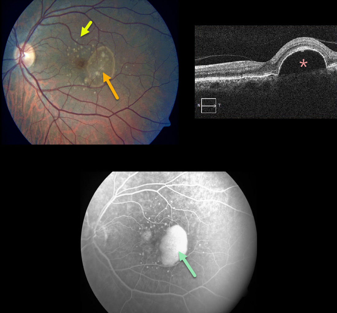
PIGMENT EPITHELIAL DETACHMENT (PED)
Originally posted on @retina.rocks October 13, 2020
This patient presented with 20/25 vision in her left eye with significant Amsler grid changes. There is an irregular RPE detachment (PED) with variably sized drusen, and OCT confirms a large PED.
Fluorescein angiography shows a slightly uneven pattern of pooling beneath the PED.
We discussed starting treatment due to her symptoms, but for now, we have elected for close observation. There is a risk for an RPE tear with or without treatment
Learning Points:
PEDs associated with AMD are often associated with macular neovascularization (MNV).
Angiographic clues for MNV include a notched PED or uneven angiographic filling. Fluorescein angiography is probably still better than OCT angiography for detecting MNV.
Anti-VEGF therapy can effectively flatten these PEDs even when they are presumably avascular.
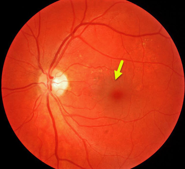
TOXEMIA
Originally posted on @retina.rocks October 7, 2020
Our patient was referred for asymptomatic macular pigmentary changes. Vision was 20/20 OD and 20/25 OS. On further questioning, she reported severe vision loss with toxemia just before her now 16 YO daughter was born.
Learning Points:
Preeclampsia (toxemia of pregnancy), which usually develops in the third trimester, manifests as severe hypertension, swelling of the legs, feet, and hands, and proteinuria. When seizures develop, the condition is called eclampsia. This may lead to serious and possibly fatal effects for both the mother and child.
Patients may develop hypertensive retinopathy. Choroidal ischemia can cause infarction of the RPE with exudative and hemorrhagic retinal detachment.
These changes usually resolve rapidly with delivery, although they can cause chorioretinal changes that mimic numerous entities, including central serous retinopathy and macular/retinal dystrophies.
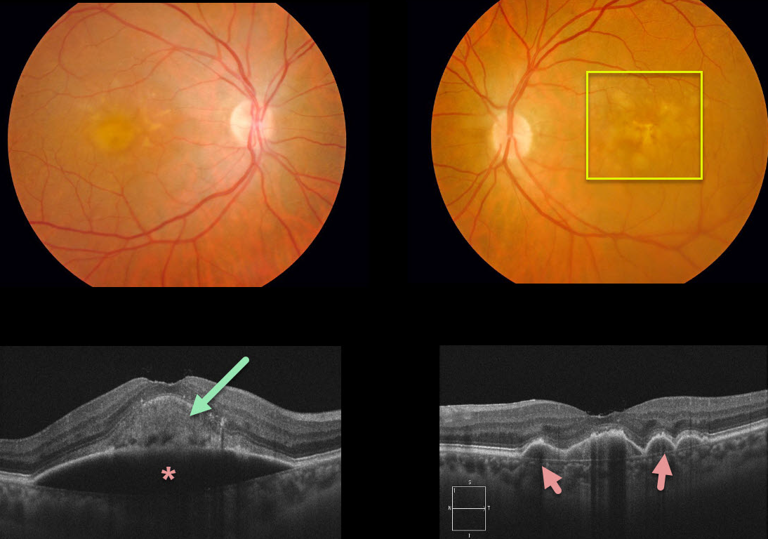
PIGMENT EPITHELIAL DETACHMENT (PED)
Originally posted on @retina.rocks September 30, 2020
This patient had been followed for age-related macular degeneration (AMD) with bilateral confluent soft drusen. She then developed a large acquired vitelliform lesion overlying a retinal pigment epithelial detachment (PED) in her right eye. There are several PEDs also shown in the left eye.
Learning Points:
Vitelliform lesions are commonly found with other disorders, including retinal dystrophies (Best’s disease and macular dystrophies), cuticular drusen, AMD, and vitreomacular traction.
The yellow subretinal material is felt to be debris from shed photoreceptor outer segments. These lesions markedly autofluoresce on fundus autofluorescence.
A great reference to learn more about AVLs is Freund et al Retina 2011;31:13-25.
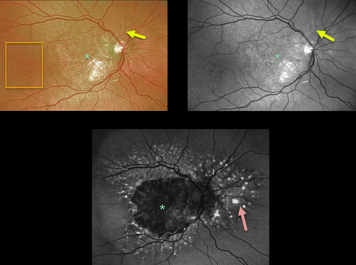
ANGIOID STREAKS
Originally posted on @retina.rocks August 21, 2020
This patient has angioid streaks that radiate from the optic nerve. These are very easy to miss unless they are explicitly looked for.
Optos color and green-channel photographs of the right eye show central macular atrophy with more temporal peaux d’orange changes. The left eye (not pictured) showed identical findings.
Fundus autofluorescence (FAF) shows widespread hyper-FAF lesions surrounding the central hypo-FAF macula. These multifocal hyper-FAF lesions are typical of pattern dystrophies and can somewhat simulate fundus flavimaculatus if they extend outside the macula.
This patient is monitored regularly due to the risk of developing macular neovascularization, although that is fairly unlikely given the macular atrophy.
Learning Points:
Patients with angioid streaks and pseudoxanthoma elasticum (PXE) can present with retinal findings similar to those of pattern dystrophies.
These patients are at significant risk for macular neovascularization (MNV) and need to be followed and treated as needed with anti-VEGF agents, as in patients with AMD.
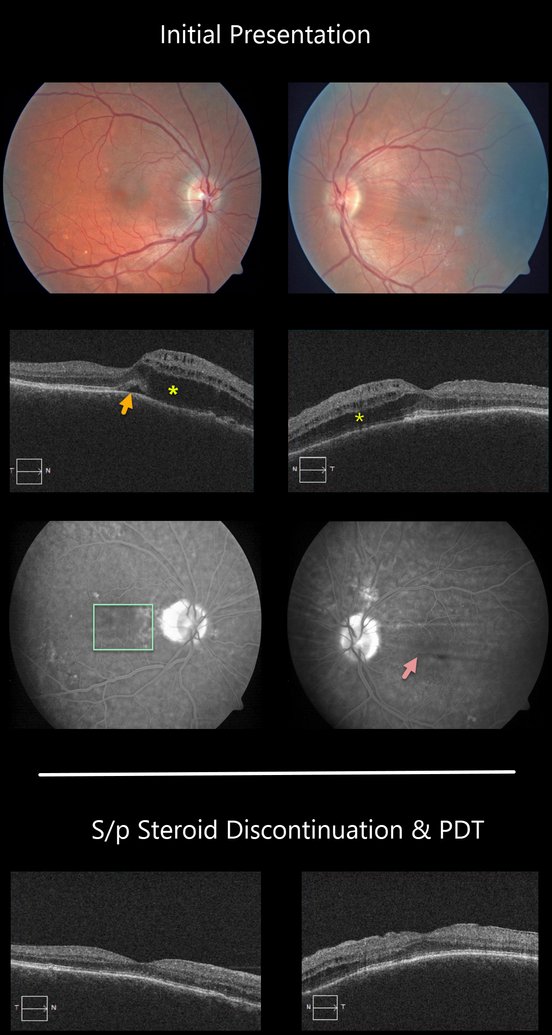
PERIPAPILLARY PACHYCHOROID SYNDROME
Originally posted on @retina.rocks August 18, 2020
This patient has peripapillary pachychoroid syndrome (PPS), a recently described bilateral condition characterized by nasal macular intraretinal and subretinal fluid.
These eyes can look very similar to those of patients with optic pit maculopathy, but without the pit. Additional findings include a thicker nasal macular choroid than temporal macular choroid, choroidal folds (75% of cases), shorter axial lengths (<23mm), and hyperopia.
There is no angiographic evidence of leakage despite fluid noted on OCT.
The patient worsened when initially placed on steroids, but made a tremendous improvement after steroids were discontinued, followed by photodynamic therapy.
Learning Points:
PPS is part of the pachychoroid spectrum, where patients have a thick choroid. Probably the most common and well-known pachychoroid disease is idiopathic central serous chorioretinopathy. Just like ICSC, this condition worsens with steroid use.
See Phasukkijwatana et al Retina, 2018;38:1652-1657 for more on PPS.
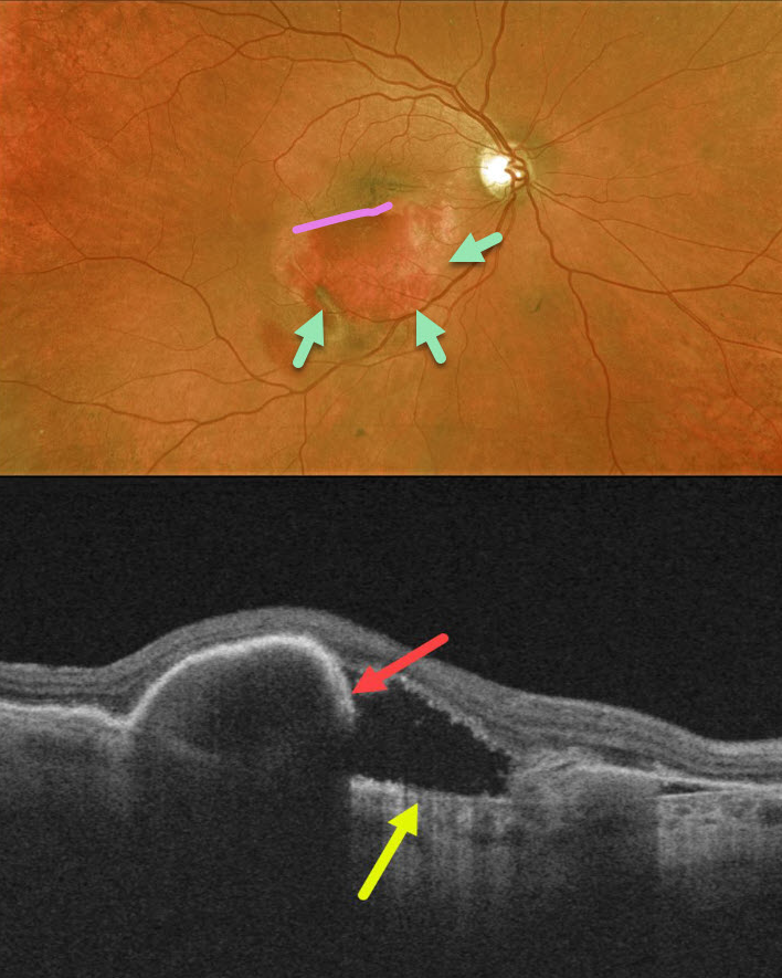
RPE TEAR
Originally posted on @retina.rocks August 11, 2020
Our patient had a retinal pigment epithelial (RPE) tear 2 weeks following an injection for wet age-related macular degeneration. The edges of the bare choroid and the edge of the retracted RPE can be visualized.
OCT shows bare Bruch’s membrane and the retracted, torn RPE.
Learning Points:
A tear of the RPE is a fairly common finding in RPE detachment (PED) associated with type 1 macular neovascularization (MNV), which is located below the RPE.
The neovascularization can spontaneously contract or involute following treatment. It then puts traction on the overlying RPE, which can then rip at the opposite edge of the PED. Like a cheap window blind, the torn RPE retracts and scrolls towards the MNV. This leaves a depigmented region where the RPE was and is now absent, along with a pigmented, relatively straight subretinal lesion extending from the scrolled RPE edge.
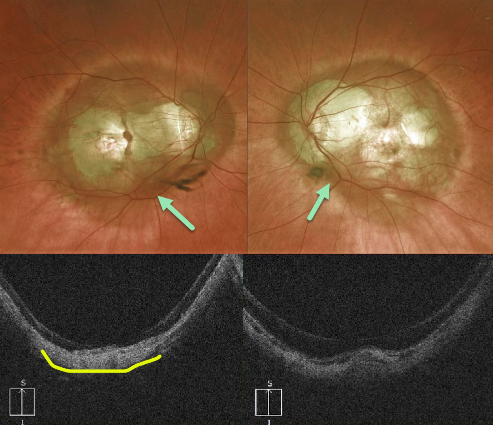
HIGH MYOPIA
Originally posted on @retina.rocks July 27, 2020
Our highly myopic patient has a fairly symmetrical bilateral posterior staphyloma that encircles each optic nerve and macula. Vision is counting fingers OD and 20/50 OS.
OCT demonstrates steep scleral curvature and outpouching.
Learning Points:
Posterior staphyloma is a weakening, thinning, and outpouching of the sclera most commonly seen in degenerative myopia.
See Moriyama et al Ophthalmology 2011;118;1626-1637 for some amazing 3D MRI reconstructions of myopic staphylomas.
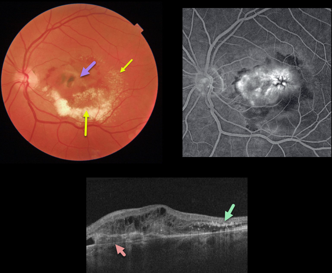
IDIOPATHIC MACULAR NEOVASCULARIZATION (MNV)
Originally posted on @retina.rocks July 24, 2020
This patient was being followed for an inactive nasal macular chorioretinal scar with 20/25 vision. After being lost to follow-up for many months, he presented with counting-finger vision from an active type 1 macular neovascularization.
The macular OCT shows a complex lesion with the MNV located primarily beneath the RPE. The lipid precipitated out in the subretinal space and follows Henle’s layer temporally.
Learning Points:
The amount of lipid layered in the subretinal space is unusual in MNV. Extensive submacular lipid like this is more common with Coats disease or retinal capillary hemangiomas.
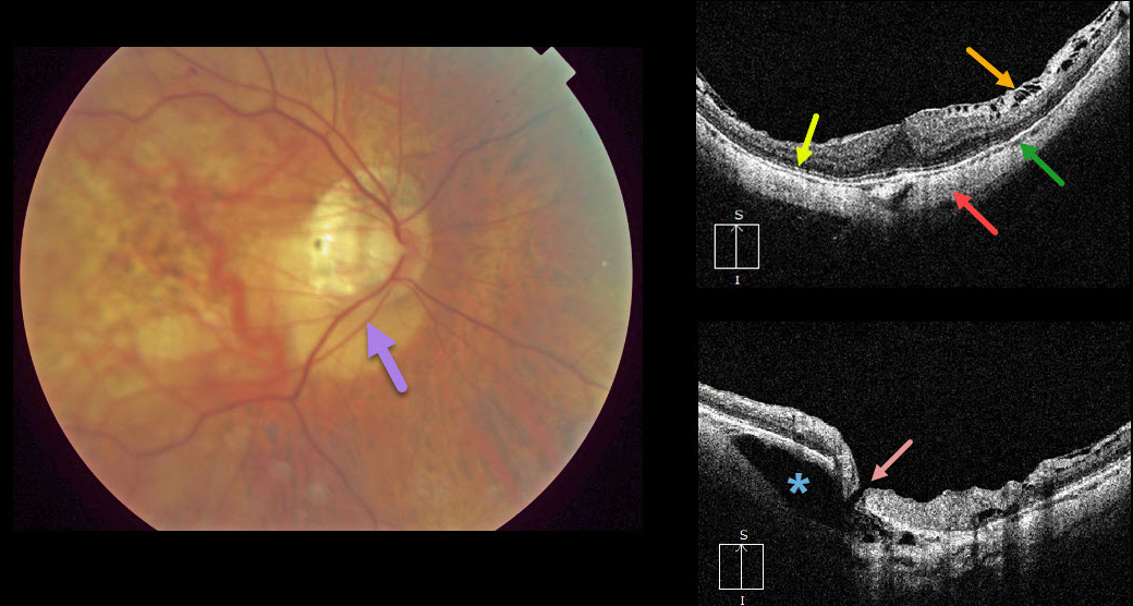
HIGH MYOPIA & INTRACHOROIDAL CAVITATION
Originally posted on @retina.rocks July 9, 2020
Peripapillary intrachoroidal cavitation is a relatively rare abnormality most commonly found in high myopia. Clinically, these subretinal lesions have an orange coloration and are usually located inferior to the optic nerve.
OCT scanning of the central macula shows a variety of typical myopic findings, including posterior bowing of the sclera, variable attenuation of the ellipsoid zone, markedly thinned choroid, and a variable epiretinal membrane with inner retinal schisis most prominent superiorly.
OCT scanning centered over the optic nerve reveals the intrachoroidal cavitation. There also seems to be a tiny full-thickness retinal defect that communicates directly with the cavitation.
Learning Points:
To learn more about intrachoroidal cavitations and high myopia, see Spaide et al Retina 2012;32;1037-1044.
See Ikuno Retina 2017;37;2347-2351 for a nice overview of macular findings in high myopia.
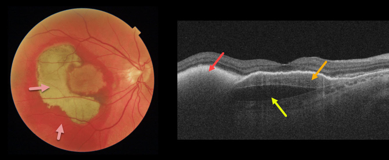
MACULAR NEOVASCULARIZATION (MNV)
Originally posted on @retina.rocks July 8, 2020
This patient’s vision remained 20/100 one month following vitrectomy with subretinal tissue plasminogen activator.
Surgery accomplished its goal of displacing most of the subfoveal blood, which is variably red and devitalized (yellow).
OCT shows the remaining subretinal hyperreflective blood, the causative sub-RPE type 1 macular neovascularization, and a subfoveal prechoroidal cleft.
Learning Points:
A prechoroidal cleft is a hyporeflective space between the RPE and Bruch’s membrane.
These are seen in up to about 20% of treated eyes with wet AMD, particularly with type 3 neovascularization and polypoidal choroidal vasculopathy.
They are usually associated with worse visual acuity due to potential complications, including RPE tear and subretinal hemorrhages.
For a more detailed discussion of prechoroidal clefts, see Kim et al Retina 2017;37;2047-2055.
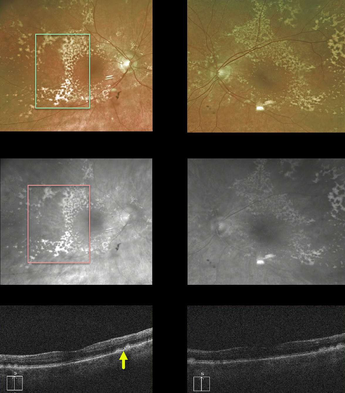
DRUSEN
Originally posted on @retina.rocks July 7, 2020
This 83YO female has extensive, confluent drusen outside the perifoveal area.
The drusen, located beneath the RPE, are well visualized with the Optos red channel. Subretinal drusenoid deposits (not present), which form above the RPE, would disappear with the red channel.
OCT also demonstrates drusen.
Learning Points:
Sub-RPE drusen are a hallmark feature of non-exudative age-related macular degeneration.
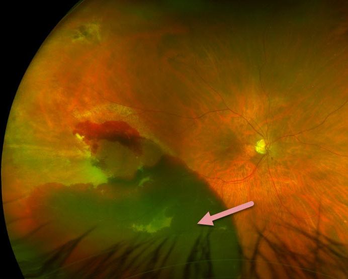
PERIPHERAL EXUDATIVE HEMORRHAGIC CHORIORETINOPATHY
Originally posted on @retina.rocks June 12, 2020
This 65YO female has a fresh peripheral subretinal hemorrhage.
The subretinal blood often regresses spontaneously, as occurred in this patient (later images not shown).
Learning Points:
Peripheral exudative hemorrhagic chorioretinopathy (PEHCR) is essentially choroidal neovascularization occurring in the retinal periphery.
PEHCR can be mistaken for uveal melanoma because the dark subretinal blood can simulate a pigmented choroidal tumor.
PEHCR seems to share features with polypoidal choroidal vasculopathy.
Anti-VEGF therapy is helpful when needed. Patients can also develop a breakthrough vitreous hemorrhage, which also sometimes requires vitrectomy.
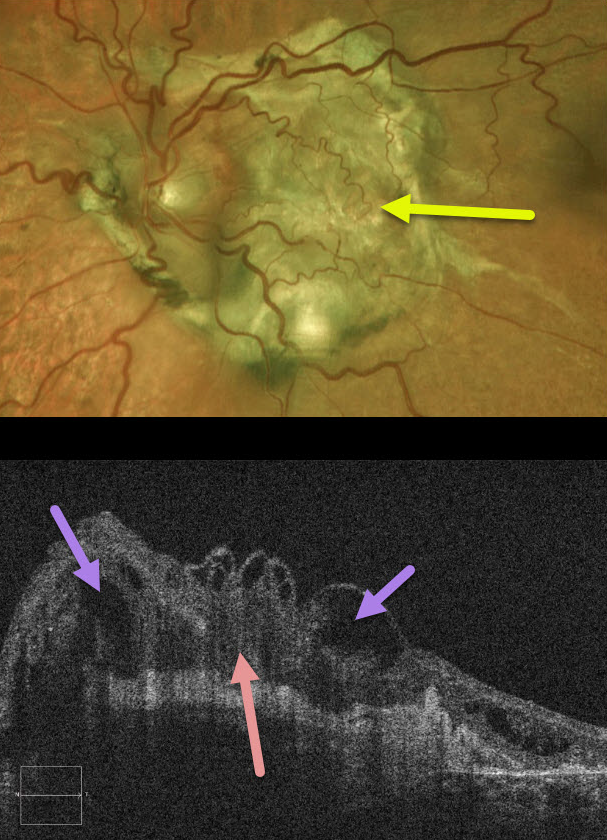
DISCIFORM SCAR
Originally posted on @retina.rocks June 9, 2020
This patient has a large subretinal fibrotic disciform scar from end-stage wet age-related macular degeneration.
OCT shows marked overlying retinal disorganization and cystic changes.
Learning Points:
These fibrotic scars are usually the result of untreated macular neovascularization. These were more the rule than the exception prior to the introduction of anti-VEGF therapy in the mid-2000’s.
With early and aggressive treatment, this type of outcome is now rarely, if ever, seen.
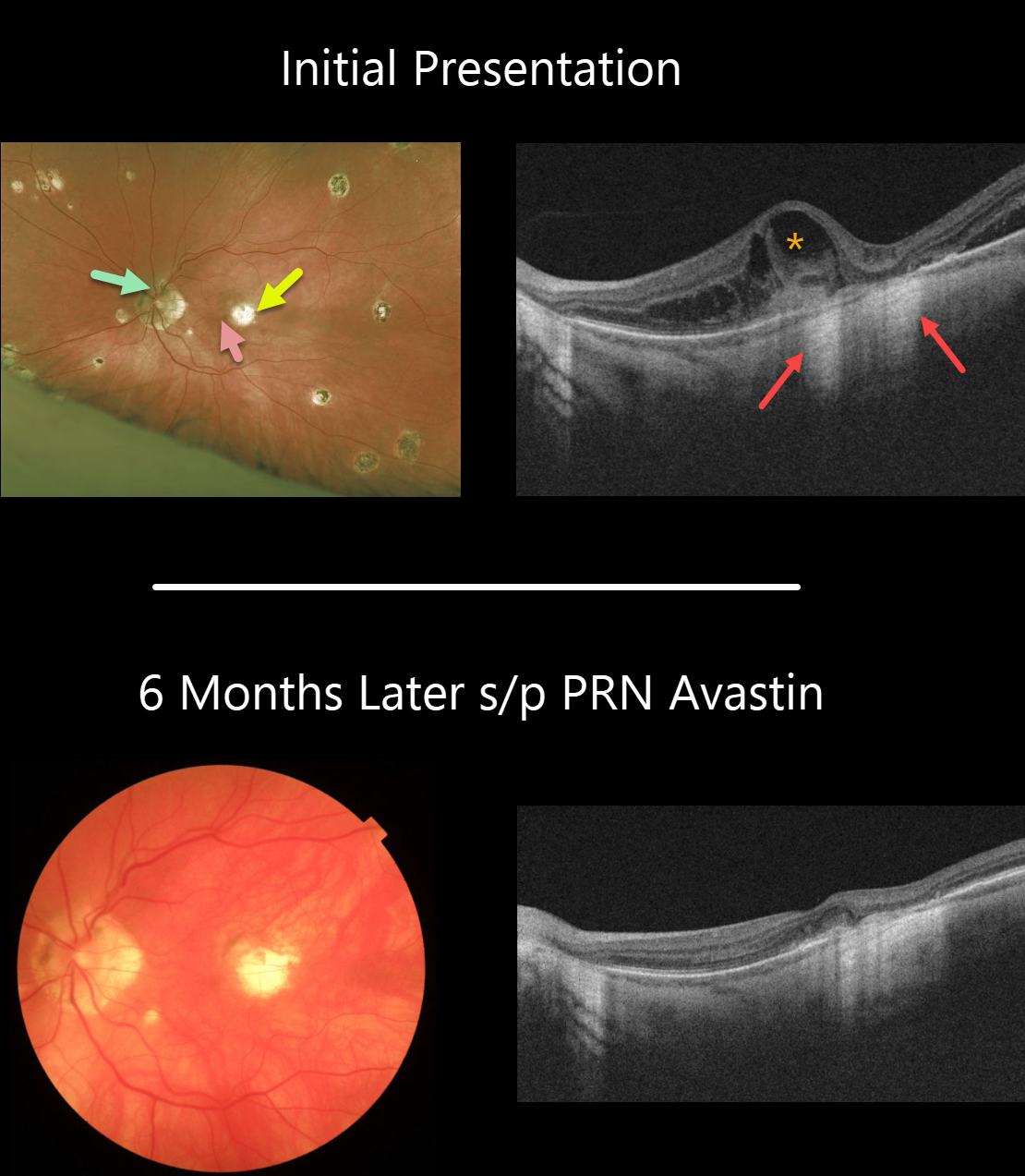
OCULAR HISTOPLASMOSIS
Originally posted on @retina.rocks May 29, 2020
Our patient has 20/20 vision OD and 20/400 OS. He has classic peripapillary atrophy, macular and peripheral scarring.
He came in with symptomatic vision loss in his left eye with a subtle nasal foveal subretinal hemorrhage from an acute macular neovascularization (MNV) adjacent to a pre-existing chorioretinal scar.
The OCT line scan surprisingly shows significant intraretinal fluid overlying the hemorrhage and temporal to the clinical scar. The loss of the RPE at the chorioretinal scar allows OCT to show increased hyperreflectance of the underlying choroid and sclera. This is the opposite of shadowing.
Six months later, vision in the left eye is 20/200, and the blood and fluid remain completely resolved with PRN Avastin.
Learning Points:
Ocular histoplasmosis is caused by the Histoplasmosis capsulatum fungus, which is commonly found in soil contaminated by bird or bat droppings. It is found worldwide, but in the United States, it is most common in the Ohio-Mississippi River valleys.
The vast majority of people in endemic areas inhale the organism’s spores early in life, and only a few percent will develop asymptomatic chorioretinal scars. A small percentage of these will develop MNV later in life.
Originally described by Woods and Wahlen in 1959, the findings include the classic triad of peripapillary atrophy, peripheral multifocal punched-out scars, and a macular scar or neovascularization.
We like to add a fourth criterion: a quiet vitreous, since numerous inflammatory disorders can simulate ocular histoplasmosis.
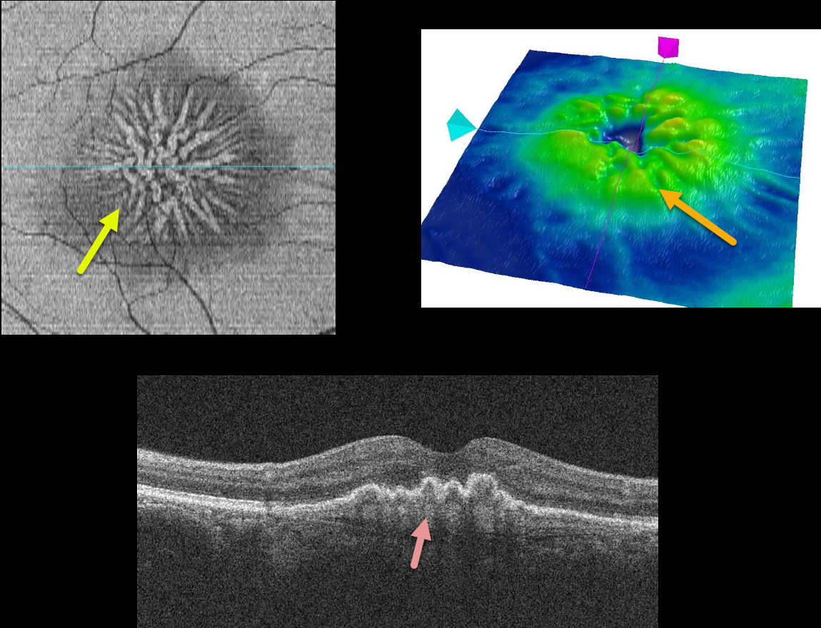
MACULAR NEOVASCULARIZATION (MNV)
Originally posted on @retina.rocks May 15, 2020
This patient is receiving ongoing intravitreal Avastin injections for a type 1 macular neovascularization (MNV) from wet AMD.
The injections caused the MNV to involute and contract, dragging the overlying RPE into radiating radial folds. This is best seen in the cool OCT en face view. The RPE folds are also seen in the OCT retinal map and OCT B-scan.
Learning Points:
Macular neovascularization is known by many other terms, including choroidal neovascular membrane, subretinal neovascular membrane, etc.
However, since these vessels are now known not to necessarily arise just from the choroid, the proper term is macular neovascularization (see Consensus on Neovascular Age-Related Macular Degeneration Nomenclature Study Group Ophthalmology 2020;127;616-636).
MNV is classified according the location of the new vessels in relation to its location relative to the RPE and neurosensory retina: type 1 (below the RPE), type 2 (between the RPE and neurosensory retina, and type 3 (within the neurosensory retina).
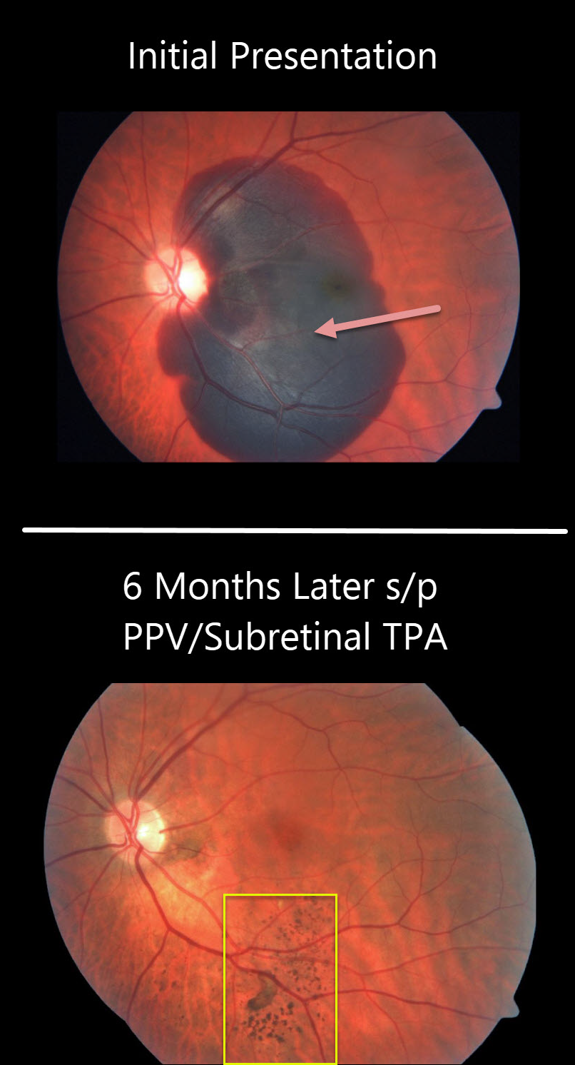
IDIOPATHIC MACULAR NEOVASCULARIZATION (MNV)
Originally posted on @retina.rocks April 29, 2020
The large submacular hemorrhage is likely from a macular neovascularization from polypoidal choroidal vasculopathy.
The patient was treated with vitrectomy, subretinal tissue plasminogen activator (TPA), and fluid air exchange. The TPA helped liquefy the clot, which was then hydraulically displaced inferiorly outside the macula by the gas bubble.
At six months post-op, the central macula is remarkably normal with multifocal clumps of subretinal pigment more inferiorly.
Learning Points:
Multifocal subretinal pigment is a classic finding for regressed subretinal blood. This pigment is hypoautofluorescent without RPE atrophy (see Hussnain et al, Retina 2019;39;1925-1935).
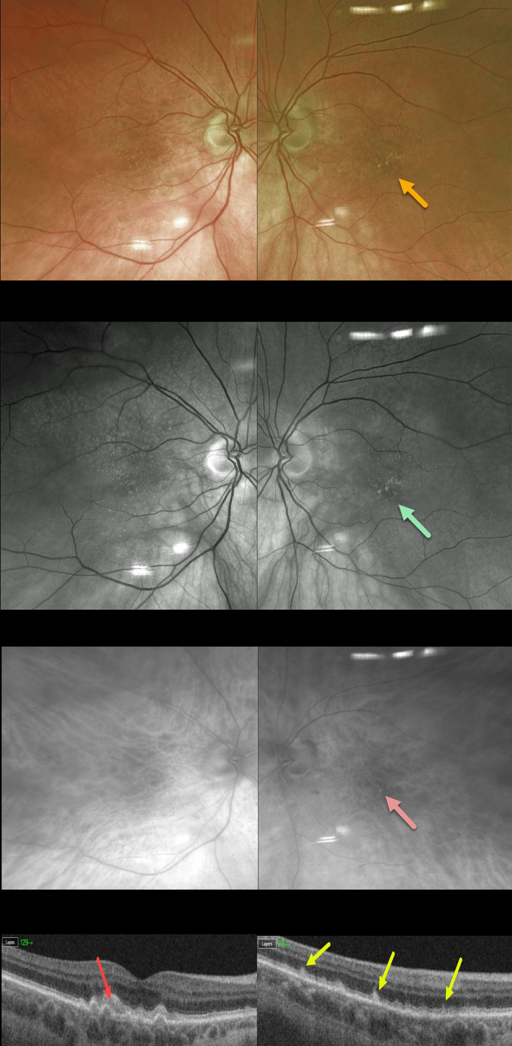
SUBRETINAL DRUSENOID DEPOSITS (SDD)
Originally posted on @retina.rocks April 22, 2020
This 86YO female has dry AMD and has subretinal drusenoid deposits (SDD) located above the RPE layer.
Since they are located above the RPE, they are best visualized with blue or green illumination. Notice how the SDD are easily visible on the Optos green channel but disappear in the red channel. Typical drusen (not pictured) would be best seen in the red channel.
The OCT images of a similar (but different patient) illustrate various stages of SDD with outer retinal atrophy.
Learning Points:
Subretinal drusenoid deposits (SDD) are polymorphous light-gray interconnected hypoautofluorescent accumulations located above the RPE. They are present in about one quarter of older adults with healthy maculas and in more than half of those with early to intermediate dry AMD.
Clinically they are often mistaken for conventional drusen and consist of the same lipid-rich membranous debris found in soft drusen. As with drusen, SDD are dynamic and can grow, shrink, resolve, or even recur.
SDDs can appear on OCT as they appear in 3 stages (Zweifel et al, Ophthalmology 2010; 117:303-312) growing upwards from the RPE towards the ELM:
Stage 1- diffusegranular hyperreflective material between the RPE and EZ
Stage 2- material alters the EZ contour
Stage 3- conical, breaking through the EZ
Rod function seems to be preferentially affected, and there is an associated increased risk of delayed dark adaptation, macular pattern dystrophy-like subretinal vitelliform deposits (image 9), geographic atrophy and types 2 and 3 macular neovascularization.
In fact, type 2 neovascularization in the setting of AMD occurs almost solely in eyes with subretinal drusenoid deposits.
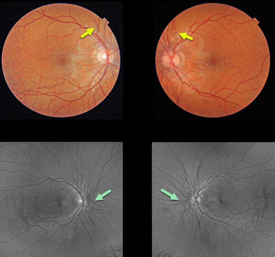
ANGIOID STREAKS
Originally posted on @retina.rocks April 7, 2020
This patient with pseudoxanthoma elasticum (PXE) has classic extensive angioid streaks with peaux d’orange (orange skin). The orange skin appearance is caused by the calcified Bruch’s membrane giving a yellow mottled appearance that contrasts with the normal orange RPE and choroidal pigmentation. The green channel (532 nm) Optos images best show these findings in the RPE/Bruch’s membrane.
Learning Points:
Although angioid streaks can be seen in other disorders, including age-related and sickle-cell disease, the findings in our patient are pathognomonic for PXE.
These patients are at significant risk for macular neovascularization (MNV) and need to be followed and treated as needed with anti-VEGF agents, as in patients with AMD. Fortunately our patient has no MNV with normal macular OCTs (not shown).
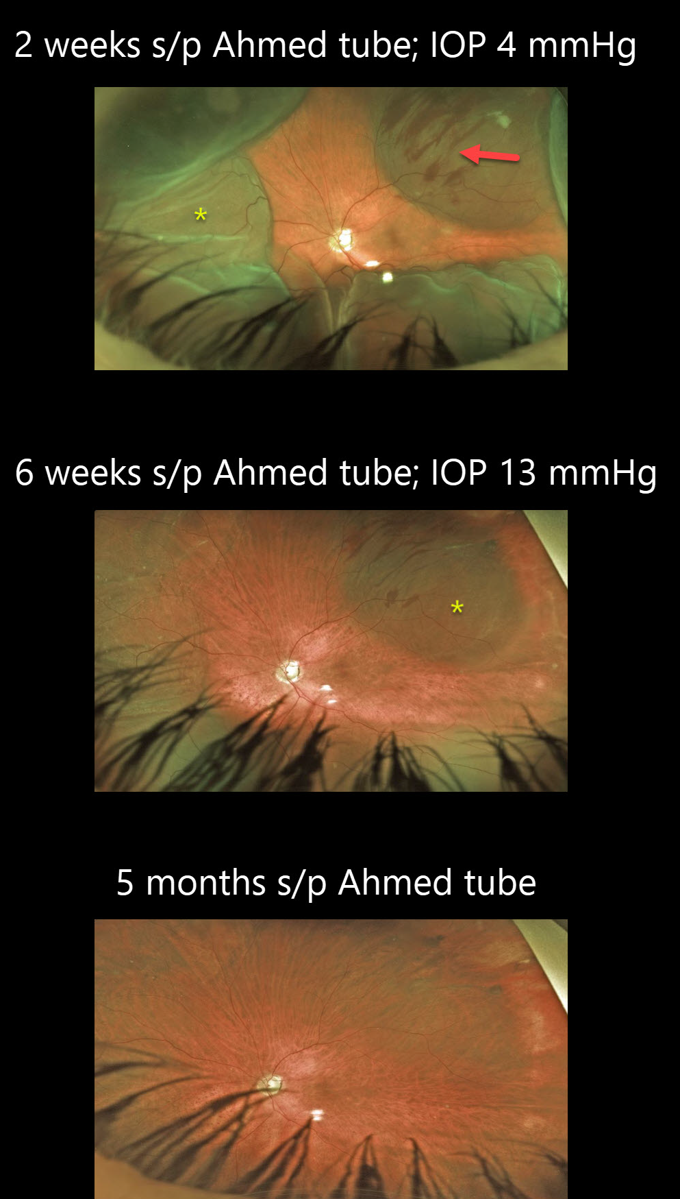
CHOROIDAL DETACHMENT
Originally posted on @retina.rocks March 30, 2020
This patient had an uncomplicated Ahmed tube surgery 2 weeks prior, but he did not stop his IOP-lowering medication nor take his prescribed anti-inflammatories and atropine.
His IOP was 4 mmHg, which led to choroidal detachments. The subretinal blood noted superotemporally is likely iatrogenic from an inadvertent overly deep suture used to secure the Ahmed explant.
Aggressive anti-inflammatories and atropine drops were started, and he was asked to stop all IOP-lowering drops.
The choroidals continued to resolve at 6 weeks as IOP continued to climb to 13mmHg. Five months post-op the choroidal detachment and subretinal blood completely resolved.
Learning Points:
Choroidal detachment is relatively common after glaucoma filtration surgery. Two mechanisms are believed to be at play:
(1) Hypotony allows fluid to accumulate in the suprachoroidal space
(2) Inflammation increases the choroidal capillary permeability.
The detached ciliary body may also produce less aqueous, thus creating a self-perpetuating hypotony.
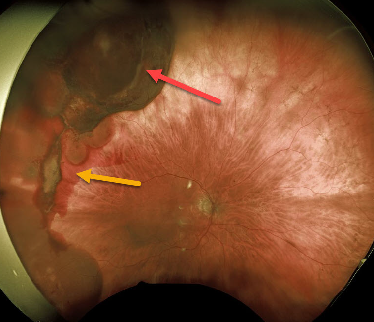
TRAUMA
Originally posted on @retina.rocks March 26, 2020
This patient presented with unilateral subretinal and suprachoroidal hemorrhages from a fall 1 month earlier causing blunt facial trauma.
Vision was normal and she was asymptomatic. She is being followed without treatment.
Learning Points:
There will likely be at least one choroidal rupture visible as the overlying blood clears over time. Subretinal blood following blunt trauma virtually always indicates the presence of an underlying choroidal rupture.
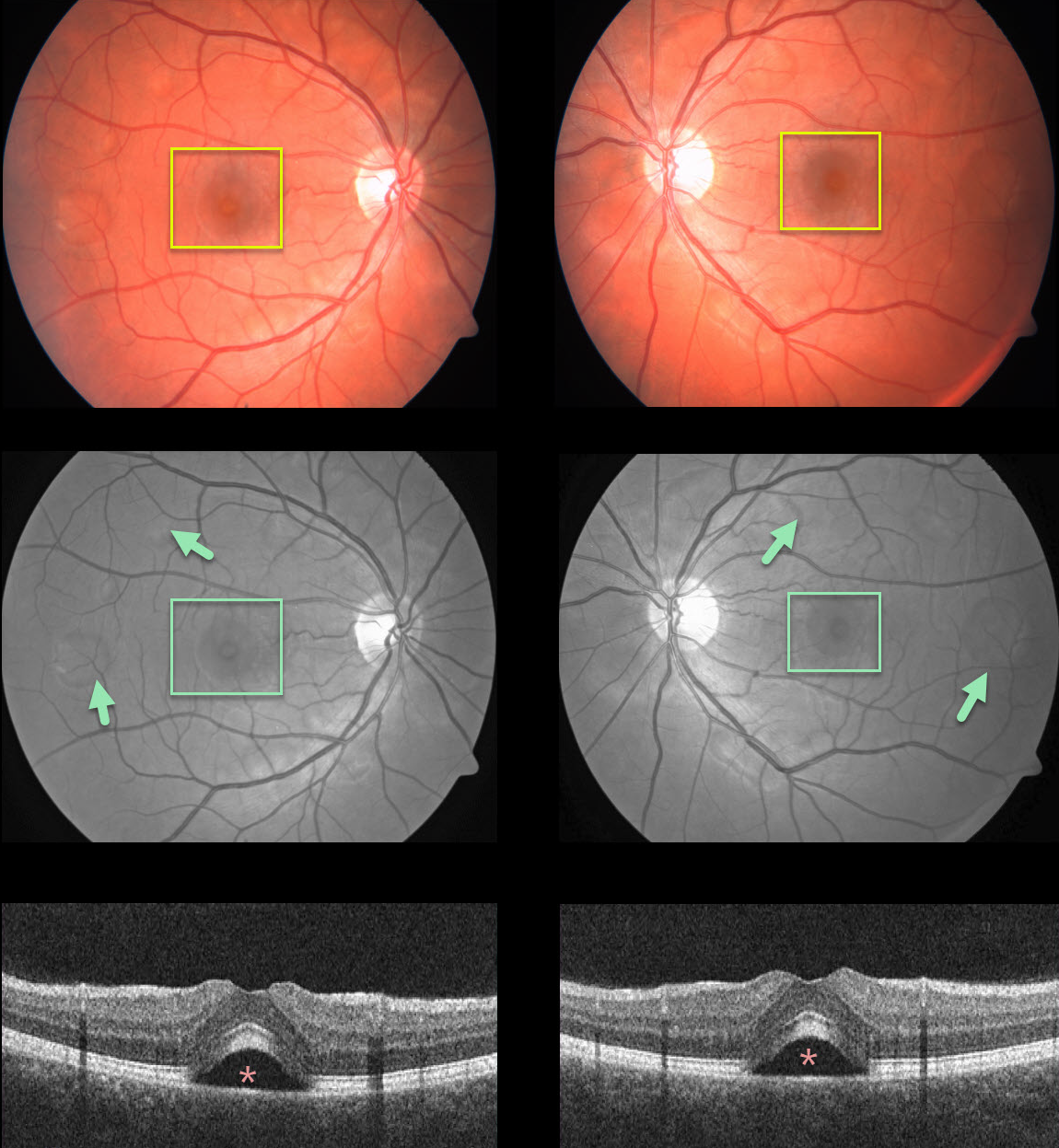
ACUTE EXUDATIVE POLYMORPHOUS VITELLIFORM MACULOPATHY
Originally posted on @retina.rocks March 2, 2020
Our patient presented with bilateral multifocal serous retinal detachments, which are better seen on red-free fundus imaging and OCT. Unfortunately, we don’t have any further clinical information.
Learning Points:
Acute exudative polymorphous vitelliform maculopathy (AEPVM) is a rare condition characterized by acute vision loss from multifocal serous retinal detachment that later progress to subretinal vitelliform lesions.
Initially the serous detachments can mimic central serous retinopathy, including thickening of the choroid. The yellow vitelliform lesions may take 1-2 years to completely resolve.
The condition is often self-limiting, and vision usually recovers within several months.
These patients need a systemic workup for metastatic disease, since paraneoplastic retinopathy, including metastatic melanoma, can mimic AEPVM.
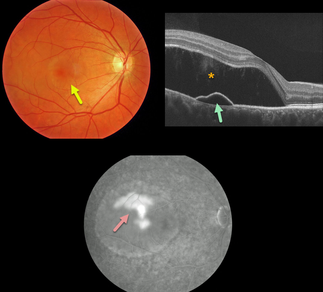
IDIOPATHIC CENTRAL SEROUS CHORIORETINOPATHY
Originally posted on @retina.rocks February 5, 2020
This patient has a large serous retinal detachment from idiopathic central serous chororetinopathy (ICSC).
OCT demonstrates the serous retinal detachment and a pigment epithelial detachment.
The late phase fluorescein angiography hows classic “smokestack” leakage.
Learning Points:
ICSC is part of a larger spectrum of pachychoroid diseases. Pachychoroid, meaning “thick” choroid, is secondary to large dilated Haller vessels.
Increased hydrostatic pressure from these dilated vessels can lead to RPE dysfunction, causing serous detachments and macular neovascularization.
The “smokestack” leakage pattern on fluorescen angiography is actually seen in only 10% of eyes with active ICSC.
Some cases resolve on their own, but in chronic cases, photodynamic therapy (PDT) is the gold standard for treatment.
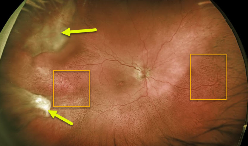
PERIPHERAL EXUDATIVE HEMORRHAGIC CHORIORETINOPATHY
Originally posted on @retina.rocks January 30, 2020
This patient has involuted neovascularization with subretinal fibrosis in the temporal and superotemporal periphery.
The coarse pigment clumping seen throughout the temporal, inferior and nasal retina is pathognomonic for prior resolved subretinal blood.
Fortunately the entire process spares the macular center, leaving the patient with normal central vision.
Learning Points:
Ectopic choroidal neovascularization, also known as peripheral exudative hemorrhagic chorioretinopathy, is a degenerative and exudative process like wet AMD but found in the retinal periphery.
Ectopic neovascularization can appear like a choroidal mass or uveal melanoma and is often misdiagnosed as such. It can also cause subretinal and vitreous hemorrhage.
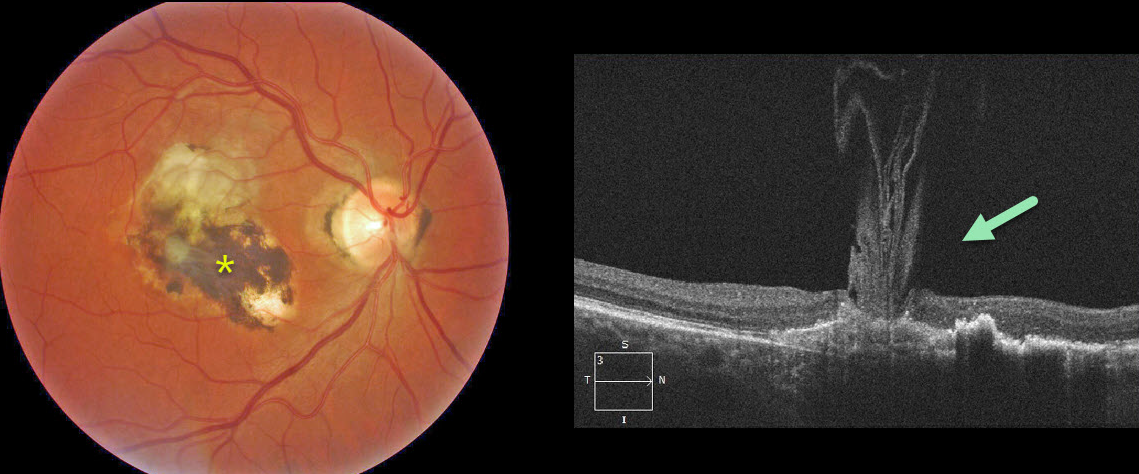
VOLCANIC MACULOPATHY
Originally posted on @retina.rocks January 20, 2020
The OCT scan shows unusual vitreous strands adhering to an inactive histoplasmosis scar resembling an erupting volcano.


