

Disorganization of Inner Retinal Layers (DRIL)
<< Back to Cases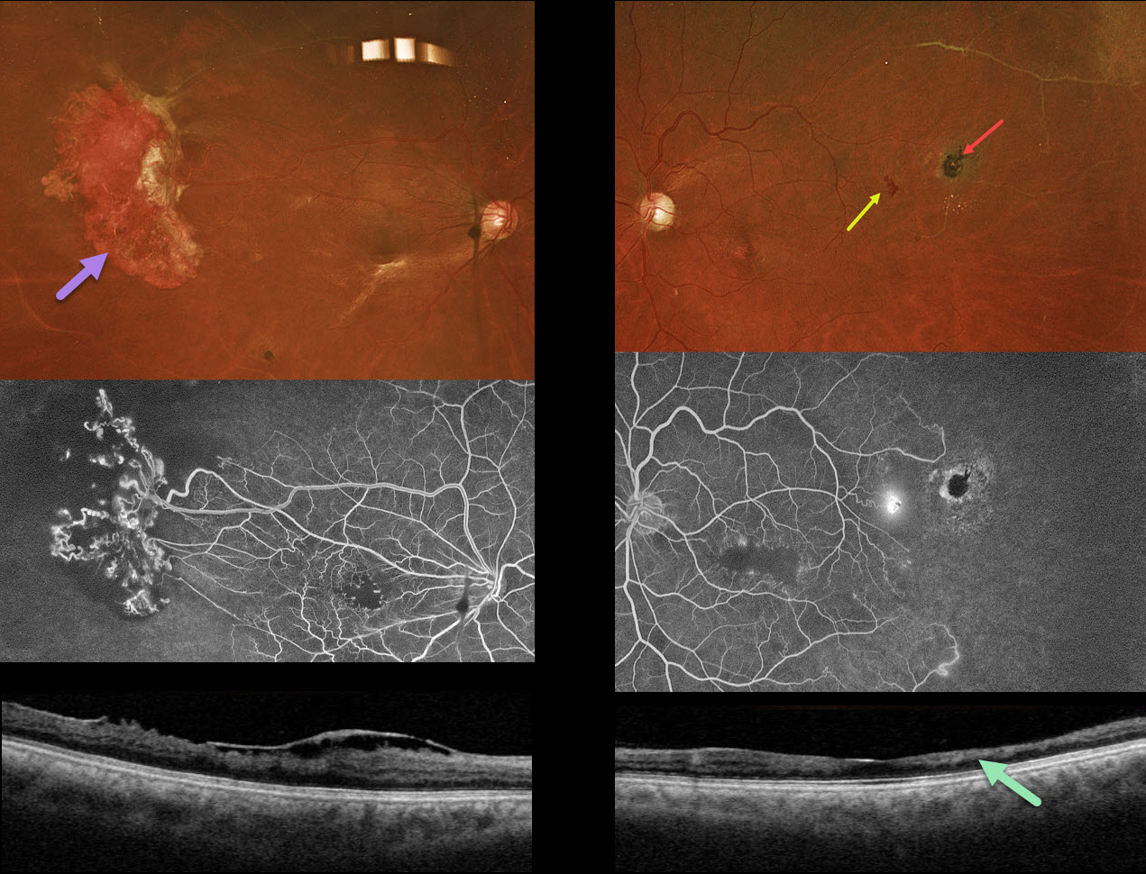
SICKLE CELL RETINOPATHY
Originally posted on @retina.rocks May 5, 2026
This 44YO male with a known history of sickle cell disease was referred for a possible detached retina. Vision has decreased over the past few years. Vision was 20/200 OU.
Optos color RGB imaging shows a large area of active neovascularization in the right temporal midperiphery associated with extensive angiographic retinal ischemia. Similar ischemia is noted OS with a small patch of leaking neovascularization. There is a small, more distal black sunburst lesion. The foveal avascular zones are enlarged with surrounding telangiectasia. This is reflected in bilateral inner retinal thinning with disorganization of inner retinal layers on OCT scanning (DRIL). An epimacular membrane is also noted OD.
Learning Points:
Patients with sickle cell disease develop symptoms throughout their bodies as stiffened, sickled red blood cells block capillary beds. In the eye, this can cause proliferative sickle cell retinopathy (PSR) with sea-fan-shaped areas of peripheral retinal neovascularization developing at the junction of perfused and ischemic retina. Increasing macular ischemia correlates with peripheral angiographic nonperfusion, as seen in our patient (Han et al., Ophthalmology Retina 2019;2:599-605). Complications include vitreous hemorrhage and tractional retinal detachment. Although patients with SS disease tend to have more systemic complications, SC disease is usually associated with more severe ocular findings.
Treatment with sectoral (Farber et al Arch Ophthalmology 1991;109:363-367) or circumferential (Kimmel et al Ophthalmology 1986;93:1429-1434) scatter laser photocoagulation is somewhat controversial since, unlike more typical proliferative retinopathies (ie from diabetes and branch retinal vein occlusions), these new vessels tend to auto-infarct with a good natural history for untreated disease (Downes et al Ophthalmology 2005;112:1869-1875). Based on the active appearance of the large temporal neovascularization OD, we elected for prophylactic scatter laser throughout the ischemic retina.
TUBERCULAR ISCHEMIC VASCULITIS
The European VitreoRetina Society (EVRS) and Abhishek Upadhyaya
Originally posted on @retina.rocks November 28, 2025
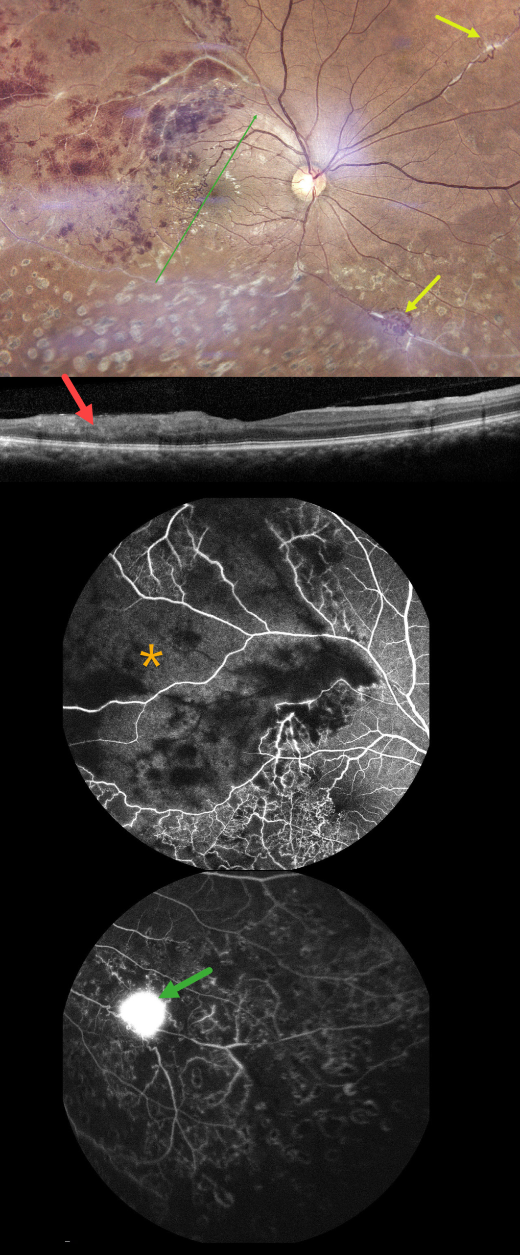
This 40YO male presented with 10 days of vision loss in his right eye. There was a history of prior laser treatment to this eye elsewhere. Vision was 20/40 OD and 20/20 in his normal OS.
Fundus photography shows superotemporal retinal hemorrhages consistent with a branch retinal vein occlusion (BRVO), with a possible additional BRVO inferotemporally. Collateral vessels are noted in the temporal fovea with scatter laser scarring in the inferior hemiretina. Skip vasculitis can be seen in the superotemporal quadrant along with venous sheathing in other areas as well. There are a few areas of fibrosed peripheral neovascularization (NV). OCT scanning shows variable temporal macular thinning with disorganization of retinal inner layers (DRIL). Fluorescein angiography shows profound ischemia within the superotemporal BRVO, along with vascular leakage and peripheral ischemia. Leakage from the Inferonasal NVE can be seen in the late stages.
Systemic workup was consistent with Ocular Tuberculosis (TB), including a positive Mantoux skin test (12mm), positive Quantiferon TB testing, and calcified lung nodules on contrast-enhanced computed tomography. He was started on oral steroids and antitubercular therapy, and scatter laser was planned for the areas of angiographic ischemia.
Learning Points:
The differential for occlusive peripheral retinal vasculitis (Huvard et al, Ophthalmology Retina 2022;6:43-48) includes TB, rheumatologic disorders/systemic vasculitides (granulomatosis with polyangiitis, Bechet’s disease, systemic lupus, etc.), idiopathic retinal vasculitis and neuroretinitis, and sarcoidosis. Historically, Eales disease has been used to describe a variety of heterogeneous disorders, including tuberculous vasculitis, and, due to its ambiguity, we prefer not to use this term.
BRVO WITH COLLATERALS AND DRIL
Malvika Singh, Akansha Sharma and Manish Nagpal
Originally posted on @retina.rocks August 19, 2025
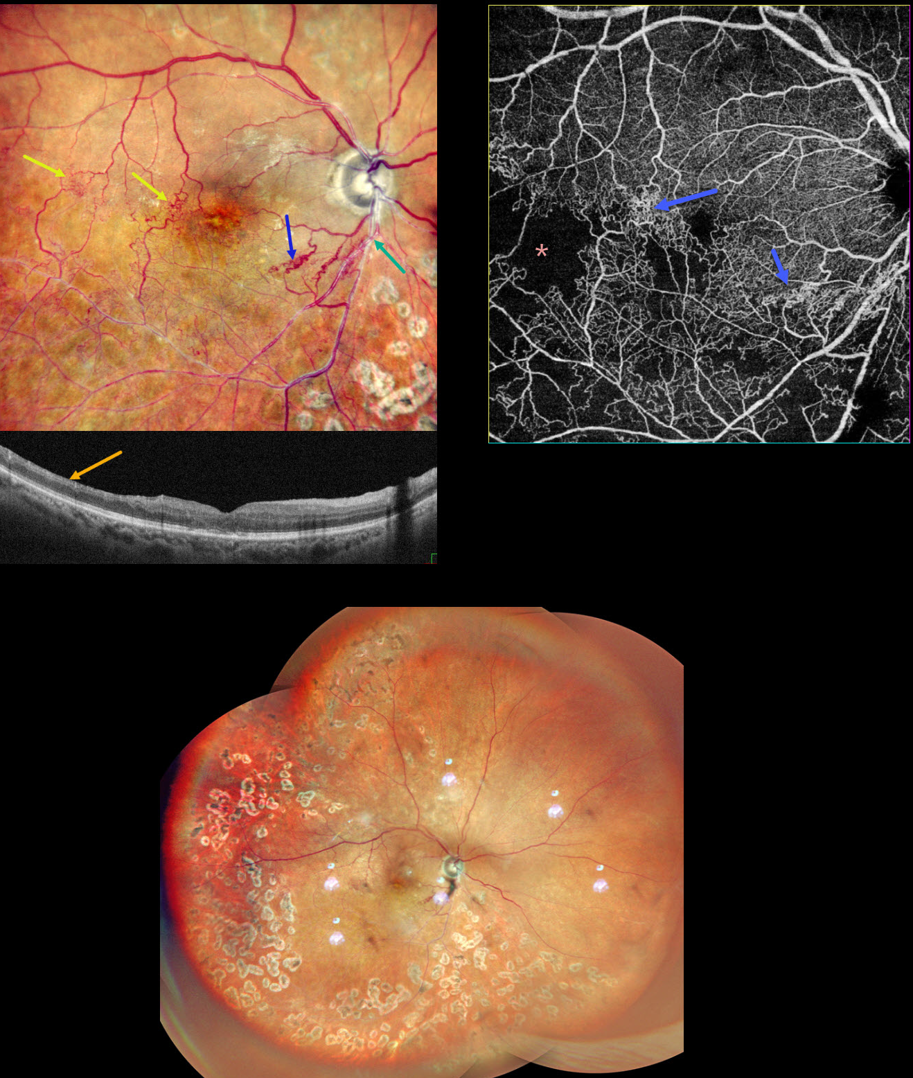
This 65YO male presented with a branch retinal vein occlusion (BRVO) in his right eye and 20/40 vision. He was treated elsewhere about a year ago with 3 intravitreal injections and scatter laser 7 years earlier.
Pseudocolor SLO shows an old major inferotemporal ischemic BRVO, which arises from an arteriovenous crossing just inferior to the disc. Extensive collateral vessels bypass the occlusion and cross the temporal horizontal raphe. OCT scanning shows thinned, featureless inner retina (disorganization of retinal inner layers, DRIL). Scatter laser is noted temporally and inferiorly. OCT angiography shows variable capillary loss throughout the BRVO distribution and highlights the collateral vessels.
Learning Points:
Collateral vessels are dilated, pre-existing vessels that bypass an occlusion. These are most commonly seen in the posterior pole following retinal venous occlusions and are exceedingly rare with arterial occlusions. The endothelial tight junctions are intact, so they don’t leak angiographically. This helps differentiate them from neovascularization, which does leak.
DRIL is seen in various retinal vascular disorders, including diabetic macular edema and retinal vein occlusions. It is generally associated with worse vision and increased anti-VEGF treatment burden (see Babiuch et al, JAMA Ophthalmol 2019;137:38-46).
PROLIFERATIVE DIABETIC RETINOPATHY (PDR)
Sehrish Momin and Haroon Tayyab
Originally posted on @retina.rocks July 31, 2023
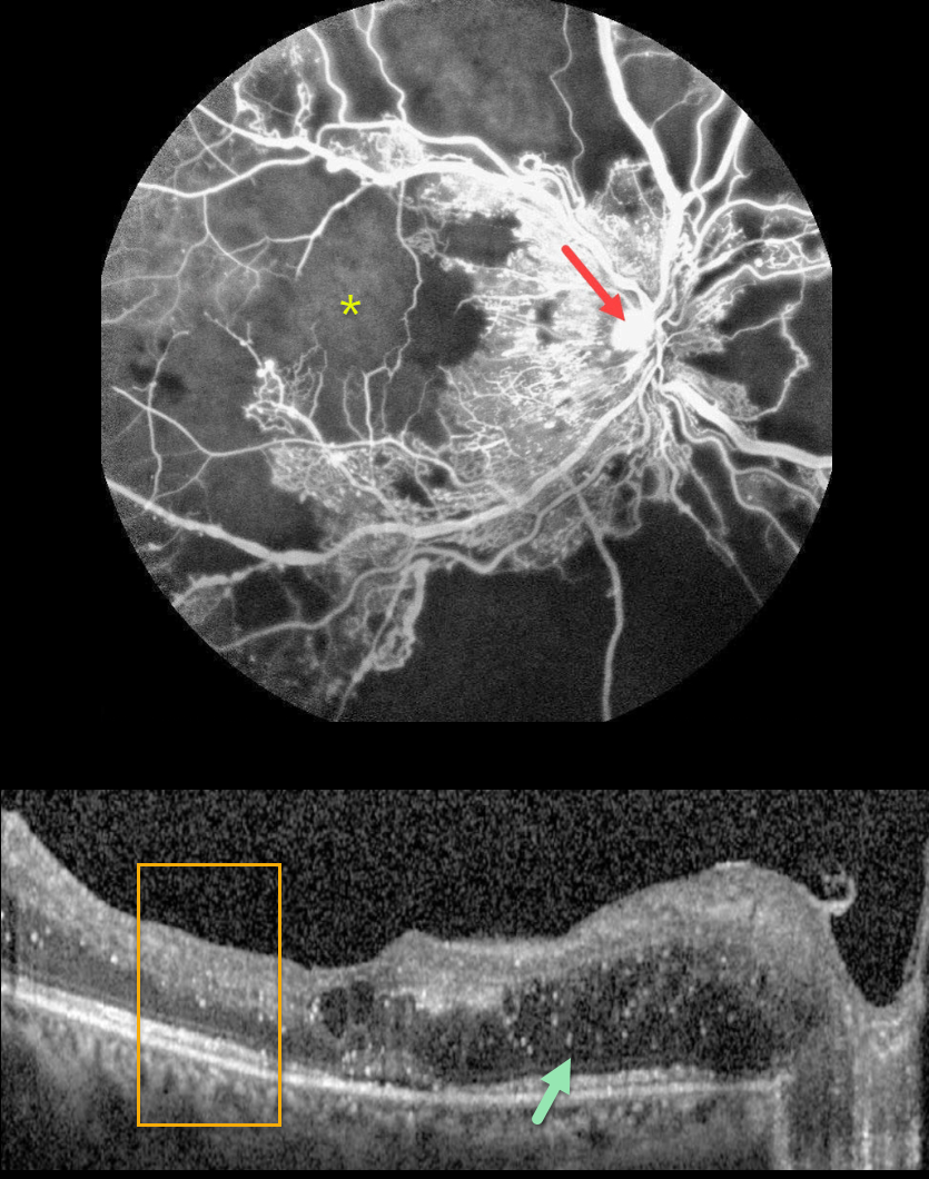
This 27YO female with a history of uncontrolled type 1 diabetes presented with bilateral vision loss. Vision was 20/40 OD and 20/200 OS.
Fluorescein angiography shows severe capillary loss that extends through the central macula. The remaining vessels are telangiectatic, especially bordering the areas of frank loss, and an area of leaking disc neovascularization is noted.
OCT scanning shows mostly nasal edema of the outer nuclear layer with numerous suspended hyperreflective particles (hyperreflective foci, HRF), along with more central cysts and trace subretinal fluid. The inner retinal layers have lost some of their normal hyper- and hypo-reflective structures (disorganization of the retinal inner layers, DRIL). Similar findings were noted in her left eye (images not shown).
Monthly anti-VEGF therapy was recommended although the patient was subsequently lost to follow-up.
Learning Points:
Anti-VEGF therapy has become the standard treatment for central-involved diabetic macular edema (CI-DME), with the vast majority of eyes showing improvements in macular thickness and vision.
Although we expect our patient’s edema to improve if she returns for treatment, we are more guarded regarding her final acuity since the capillary loss is permanent. Eyes with CI-DME and baseline central DRIL have a worse visual prognosis (Sun et al, JAMA Ophthalmol 2014;123:1309-1316).
HRF are small dot-like retinal opacities found in some eyes with diabetic macular edema. Although their pathogenesis is debated, they likely represent extravasated lipoproteins that can later become clinically evident lipid exudates (Ganne et al, Indian J Ophthalmol 2021;69:3208-3217).
Although HRF numbers decrease with treatment, it is unclear whether they serve as a biomarker for predicting visual outcome (Huang et al, Ophthalmology Retina 2022;6:814-827).
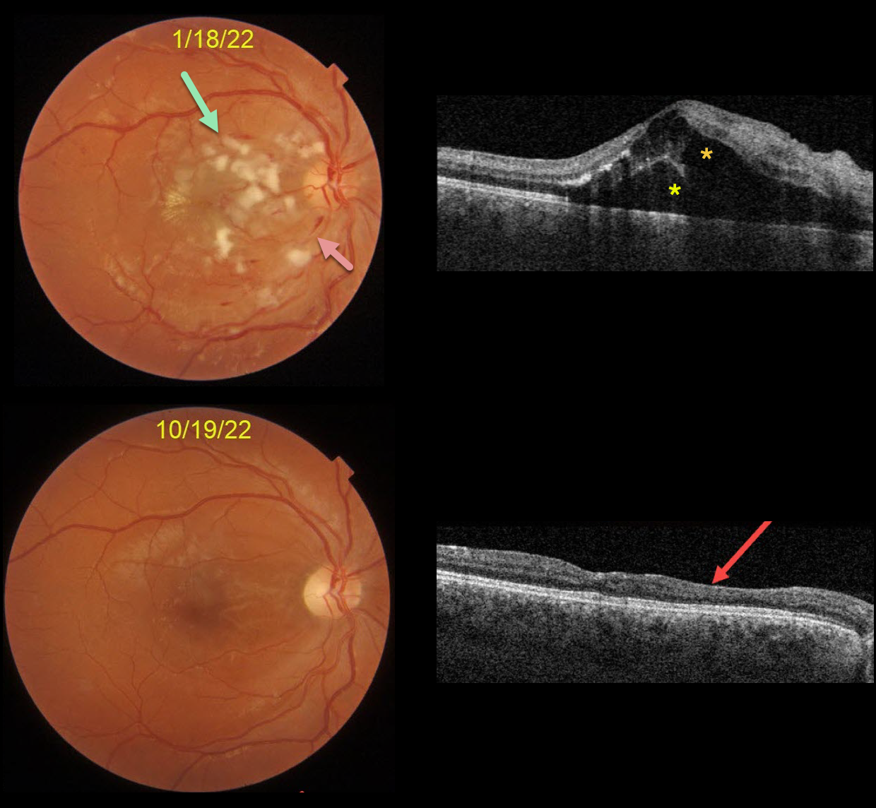
HYPERTENSIVE RETINOPATHY
Originally posted on @retina.rocks November 24, 2022
This is a follow-up from our 2/4/22 post of a patient with malignant hypertension.
This 22YO male presented on 1/18/22 with subacute vision loss of 20/200 OD and 20/50 OS. Blood pressure was 169/135. Color imaging shows nerve fiber layer infarcts (cotton-wool spots) and mild inner retinal hemorrhages.
OCT scanning shows mostly outer macular edema emanating from the nerve, with foveal subretinal fluid.
Nine months later, the macular and OCT findings have mostly normalized. OCT scanning shows thinning with disorganization of retinal inner layers (DRIL) nasally OD.
Learning Points:
Malignant hypertension, defined as blood pressure above 180 systolic and/or 120 diastolic, is a life-threatening medical emergency. Eye doctors are in a unique position to often diagnose this condition.
Patients will often present with bilateral optic nerve swelling, flame-shaped retinal hemorrhages, and, with more chronic disease, lipid precipitates in the nasal macular outer plexiform layer.
Since the macular fluid emanates from the optic nerve, the macular thickening is always worse nasally (as in our case), and is an important clinical clue for this diagnosis.
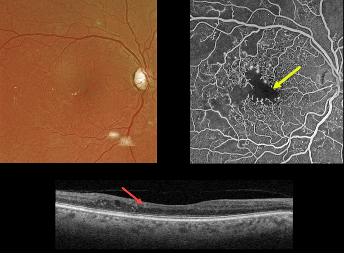
DIABETIC MACULAR ISCHEMIA
Originally posted on @retina.rocks November 23, 2022
This 67YO male has a history of type 2 diabetes. Vision is 20/40.
Optos color imaging is fairly unremarkable, with some mild temporal foveal lipid. However, fundus fluorescein angiography shows foveal ischemia with an enlarged, irregular foveal avascular zone along with more peripheral superotemporal retinal ischemia.
Swept-source OCT shows a thinned, relatively featureless inner retina, also known as disorganization of inner retinal layers (DRIL).
Observation was recommended. The ischemic retina and DRIL will not improve with anti-VEGF therapy.
Learning Points:
DRIL is seen in various retinal vascular disorders, including diabetic macular edema and retinal vein occlusions. It is generally associated with worse vision and increased anti-VEGF treatment burden (see Babiuch et al, JAMA Ophthalmol 2019;137:38-46).
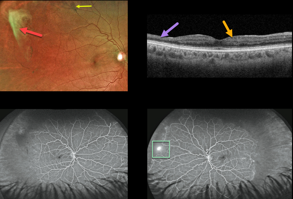
SICKLE CELL RETINOPATHY
Originally posted on @retina.rocks October 21, 2022
This 39YO African American male presented with asymptomatic proliferative sickle cell retinopathy with spontaneously avulsed bilateral superotemporal peripheral fibrosed retinal neovascularization (sea fans). A small midperipheral black sunburst lesion is seen superotemporally in his right eye.
Swept-source OCT shows variable inner retinal thinning and disorganization of the retinal inner layers (DRIL).
Fluorescein angiography shows a patch of retinal neovascularization OS at the junction of perfused and non-perfused retina.
Since these lesions often auto-infarct, as they did in our patient, we elected to observe the small area of neovascularization in his right eye. He is also at low risk for a vitreous hemorrhage or traction detachment since the vitreous is already detached in this area.
Learning Points:
Patients with sickle cell disease develop symptoms throughout their bodies as stiff, sickled red blood cells block capillary beds. In the eye, this can cause proliferative sickle cell retinopathy (PSR) with sea-fan-shaped areas of peripheral retinal neovascularization developing at the junction of perfused and ischemic retina.
The variable macular thinning noted on OCT is caused by microinfarcts to the retinal microcirculation. Enlargement of the foveal avascular zone is a common OCT angiographic finding (Fares, AJO 2021;224:7-17).
The black sunburst lesions are thought to occur from intraretinal RPE migration in response to photoreceptor damage from intraretinal and subretinal hemorrhage (see Ausdourian et al, BJO 1975;59:710-716).
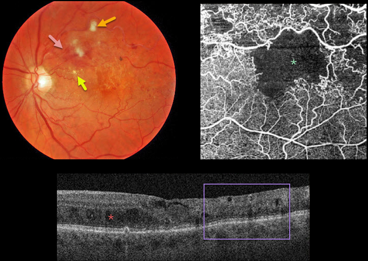
BRVO WITH MACULAR ISCHEMIA
Originally posted on @retina.rocks June 10, 2022
This 79YO male presented with a major superotemporal branch retinal vein occlusion (BRVO) and counting fingers vision.
Fundus photography shows superior macular hemorrhages with inner retinal ischemia (cotton-wool spots) and some nasal lipid. The vessels, especially around the superotemporal fovea, are nonperfused.
The clinically suspected foveal ischemia is confirmed on OCT angiography. Spectral-domain OCT shows disorganization of the inner retinal layers (DRIL), particularly within the clinically ischemic temporal macula, along with scattered inner and outer retinal cysts.
Learning Points:
DRIL is seen in various retinal vascular disorders, including diabetic macular edema and retinal vein occlusions. It is generally associated with worse vision and increased anti-VEGF treatment burden (see Babiuch et al, JAMA Ophthalmol 2019;137:38-46).
Since the patient has no central foveal thickening or retinal neovascularization, we are observing for now. Anti-VEGF therapy will not restore the foveal circulation.


