

Dissociated Retinal Nerve Fiber Layer
<< Back to Cases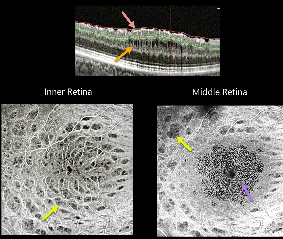
MACULAR PUCKER
Seif Anwar
Originally posted on @retina.rocks January 4, 2022
This 52YO pseudophakic female with an idiopathic macular pucker and 20/60 vision underwent pars plana vitrectomy (PPV) with internal limiting membrane (ILM) peeling. Vision improved to 20/30 after surgery, followed 6 months later by a gradual decrease to 20/50.
SOLIX OCT B-scan imaging shows multiple inner surface irregularities and thickening, with middle-layer cavitations.
En face OCT of the inner retina shows multiple concentric dark spots (dimples), characteristic of dissociated optic nerve fiber layer (DONFL). En face imaging through the mid-retinal region shows a central pattern of tiny hyporeflective cysts, with still-visible dimples more peripherally.
Learning Points:
PPV with membrane peeling is frequently performed to manage many vitreomacular interface disorders, including vitreomacular traction, macular hole, and epiretinal membrane.
While ILM peeling decreases epiretinal membrane recurrence and enhances macular hole closure, it also causes mechanical trauma to the inner retina, as evidenced by DONFL.
Many mechanisms have been proposed, including Müller cell damage from direct contact with surgical instruments, damage to the deep inner retinal layer, damage from the visualization dye, or tractional thinning of the temporal retina.


