

Eales Disease + Idiopathic Ischemia/Neovascularization
<< Back to Cases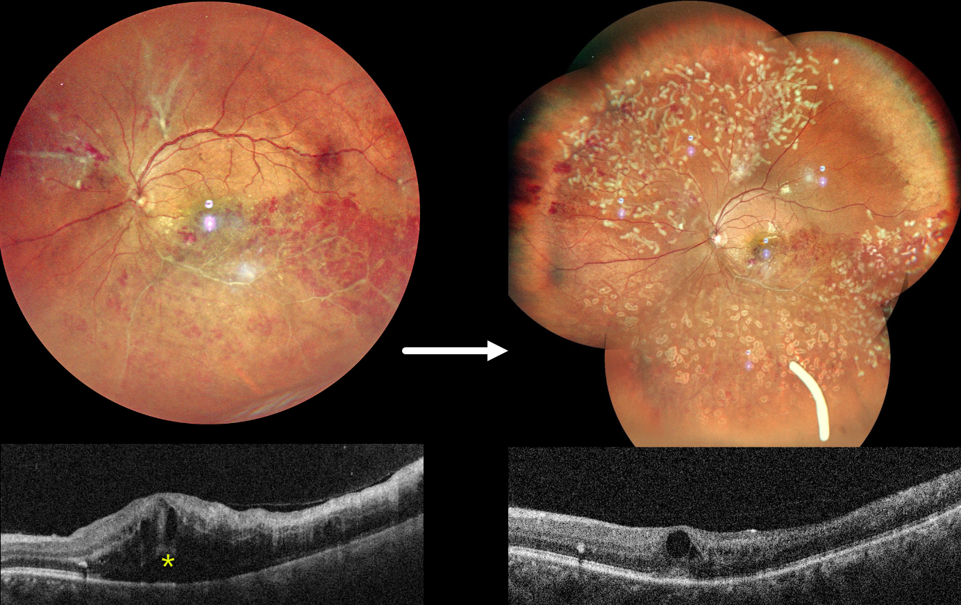
OCCLUSIVE RETINAL VASCULITIS
Anjana Mirajkar and Manish Nagpal
Originally posted on @retina.rocks July 23, 2024
This healthy 32YO male presented on 4/23/24 with 2 months of decreased vision in his left eye. Family history was negative. A prior workup by a prior doctor was negative, including protein electrophoresis and homocysteine levels. He failed a short trial of oral steroids. Vision was 20/20 in his healthy right eye and counting fingers in his left eye.
Fundus photography shows an ischemic inferotemporal branch retinal vein occlusion with moderate, mostly peripheral retinal hemorrhages. The superotemporal retinal vein is diffusely beaded, and an apparent independent ischemic BRVO is seen superonasally, with probable preretinal blood. Marked macular edema is seen on OCT. Unfortunately, fluorescein angiography is not available.
An Ozurdex injection was given for macular edema, and two sessions of panretinal photocoagulation were performed for proliferative disease. On 6/3/24, vision improved to 20/120, and the macular edema was improving.
EALES DISEASE
Narciso Atienza Jr.
Originally posted on @retina.rocks March 28, 2024
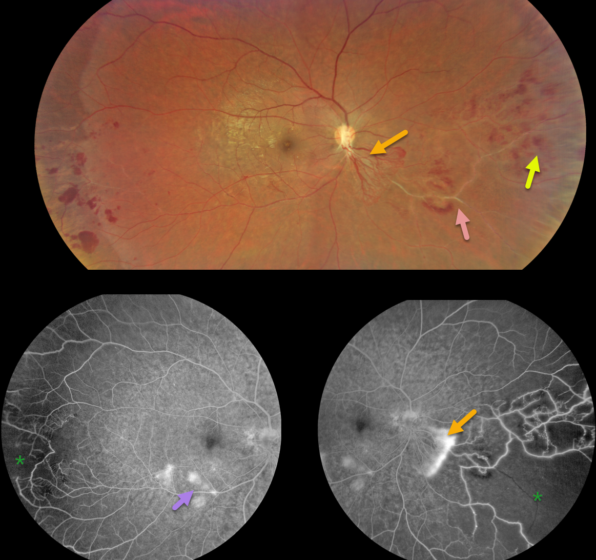
This healthy 31YO male presented with 6 months of blurred vision. He was previously diagnosed by a general ophthalmologist with a branch vein occlusion in the OD. Vision was 20/30 in his right eye and 20/20 in his left eye.
Color imaging shows active neovascularization extending inferonasally from the nerve, with more distal retinal hemorrhages and vascular sheathing.
Fluorescein angiography shows leaking neovascularization with retinal ischemia nasally and temporally. Some vascular leakage is noted in the inferotemporal macula.
Learning Points:
First described by British ophthalmologist Henry Eales in 1880, Eales disease is characterized by sharply circumscribed peripheral regions of distal ischemia, often with retinal neovascularization at the junction of the perfused and ischemic retina. It is more common in males in their 20’s to 30’s and may be associated with a prior exposure to tuberculosis. Most patients are asymptomatic unless they develop a vitreous hemorrhage. Scatter laser to ischemic retina and intravitreal anti-VEGF injections are effective for retinal neovascularization.
It is likely a heterogeneous disorder and is a diagnosis of exclusion, needing first to rule out the multitude of other disorders that can cause peripheral retinal ischemia and neovascularization. The location of our patient’s neovascularization is a bit unusual for Eales, which is usually more peripheral. However, since his systemic work-up, including chest X-ray, was normal except for a positive PPD, we feel Eales is a reasonable diagnosis. A scatter laser to the ischemic retina has been planned and scheduled.
UNKNOWN RETINAL ISCHEMIA
Originally posted on @retina.rocks October 6, 2022
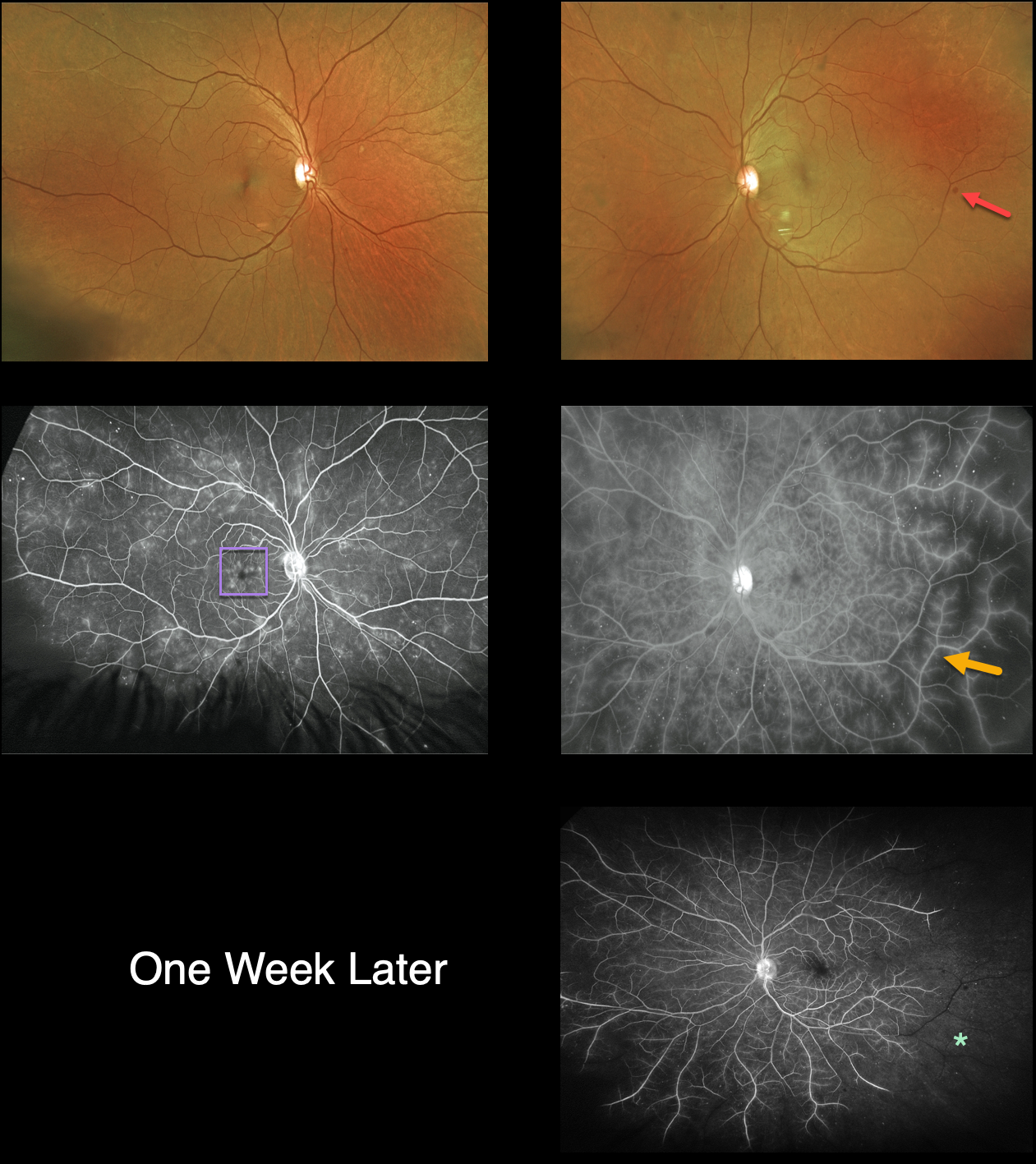
This 57YO female presented with 2 weeks of blurred vision. There was a 10-year history of type 2 diabetes and prior use of intravenous crack cocaine, though she claimed to have been clean for 18 years. Vision was 20/30 OD and 20/60 OS. The intraocular pressures were normal, but diffuse rubeosis was seen in her left eye.
Optos color imaging is fairly unremarkable, except for a few scattered bilateral dot-blot retinal hemorrhages.
Fluorescein angiography, however, shows diffuse vascular staining and ischemia, especially temporarily in her left eye. Cystoid leakage is also seen in her right macula.
Intravitreal Avastin was injected into her left eye. One week later, vision decreased to counting fingers OS, and the rubeosis had completely resolved. However, there was a marked increase in retinal ischemia. Unfortunately, she was then lost to follow-up.
Learning Points:
The rapid progression of the retinal ischemia in her left eye would be quite unusual for diabetes, especially immediately following intravitreal Avastin.
Although we are not certain of the underlying etiology, possible concurrent cocaine use in combination with diabetic endothelial damage may have been a contributing factor.
EALES DISEASE
Originally posted on @retina.rocks October 21, 2020
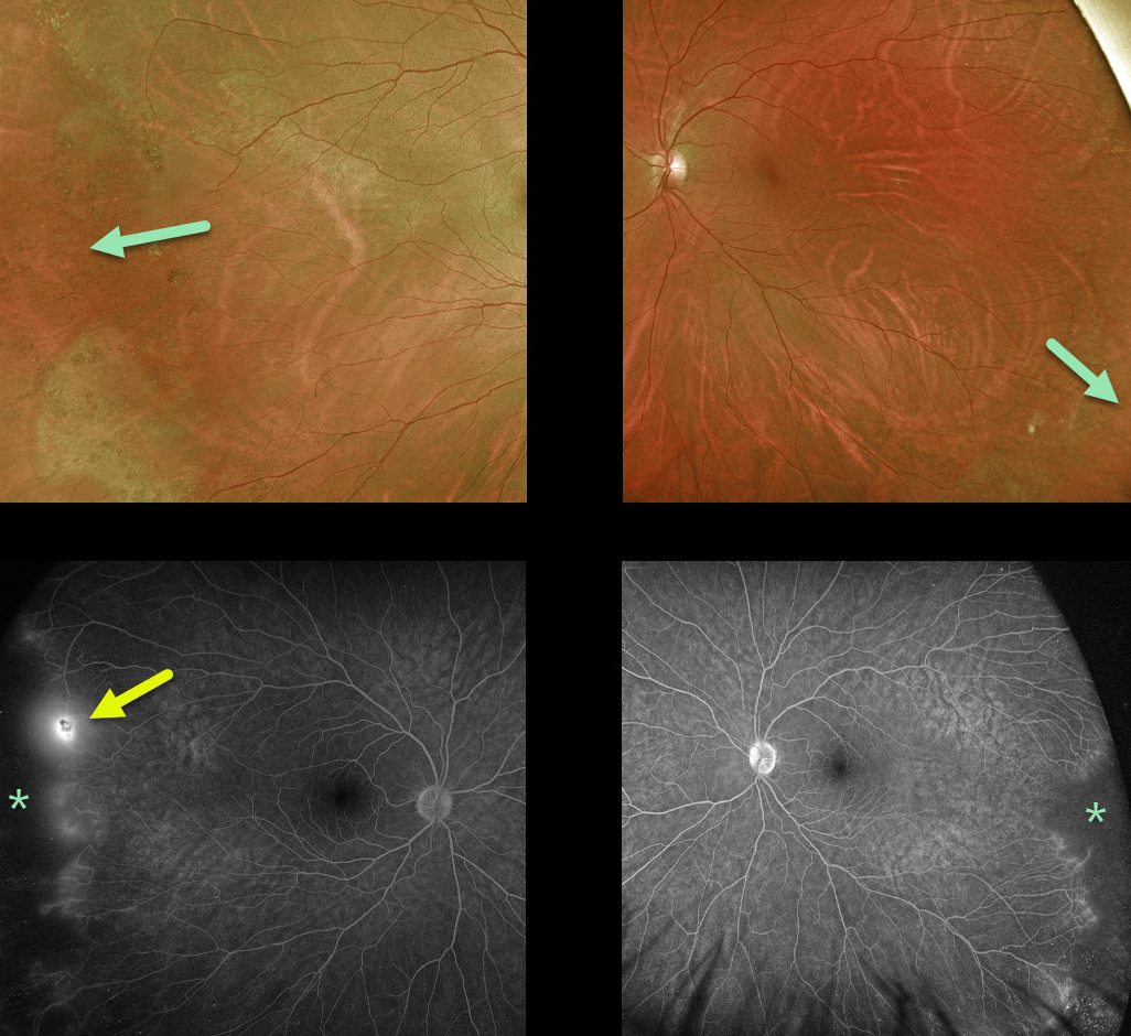
This healthy 50YO male presented with asymptomatic bilateral peripheral retinal ischemia and temporal neovascularization OD seen on fluorescein angiography.
Learning Points:
Eale’s disease is an idiopathic peripheral occlusive retinal vasculopathy that can later develop peripheral neovascularization.
It is more common in males in their 20’s to 30’s, and may be associated with prior tuberculosis exposure. Most patients are asymptomatic unless they develop a vitreous hemorrhage. There is no associated uveitis.
It is likely a heterogeneous disorder and is a diagnosis of exclusion, needing first to rule out the multitude of other disorders that can cause peripheral retinal ischemia and neovascularization.
We have seen many patients with these findings since integrating Optos ultrawidefield imaging into our practice, and therefore suspect that these peripheral retinal findings are much more common than previously described.
Peripheral retinal microvascular abnormalities have also recently been described with ultrawidefield imaging, and may represent a normal variant in this retinal vascular watershed zone (see Shah et al, Retina 2016;36;1087-1092).
EALES DISEASE
Originally posted on @retina.rocks June 17, 2020
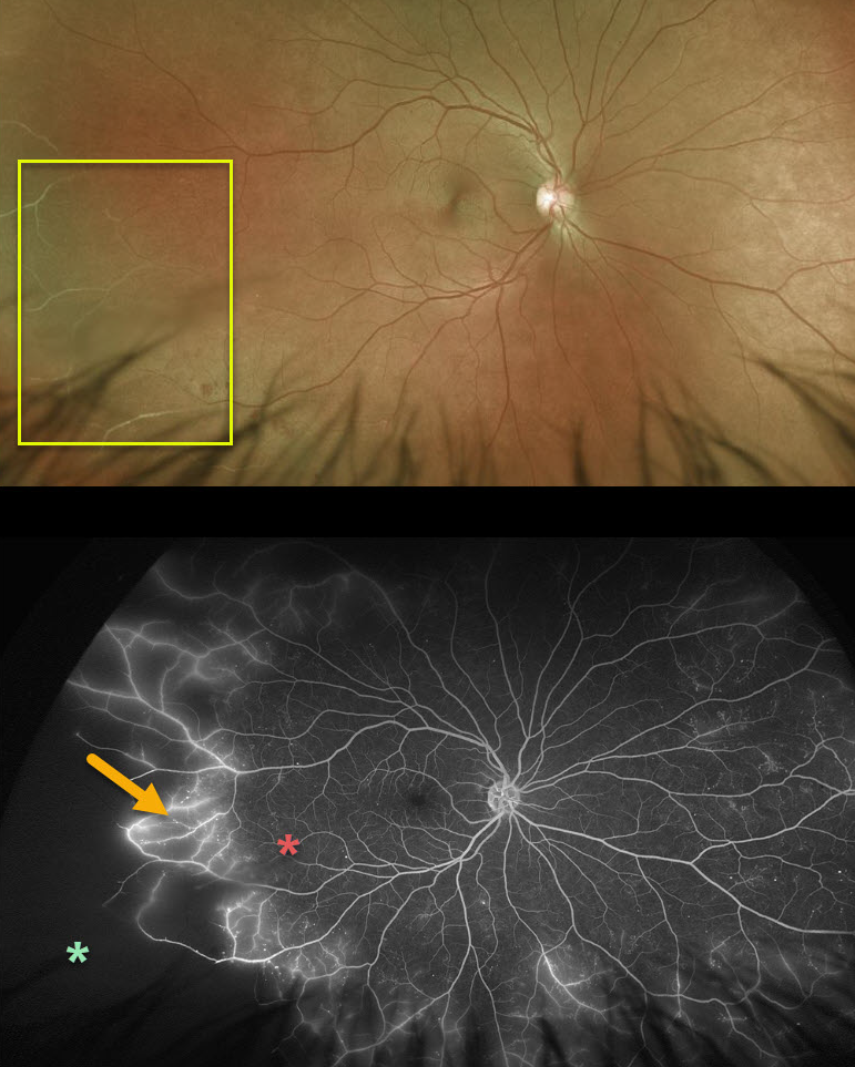
Eale’s disease is our best guess for this 44YO male patient. Eale’s disease is characterized by peripheral, sharply circumscribed regions of ischemia. Fluorescein angiogram demonstrates leakage of retinal vessels at the junction of perfused and ischemic retina.
For now, we are carefully observing the ischemic retina, but we will treat this region with scatter laser if neovascularization develops.
Learning Points:
Eale’s disease is an idiopathic peripheral occlusive retinal vasculopathy that can later develop peripheral neovascularization. It is more common in males in their 20’s to 30’s, and may be associated with prior tuberculosis exposure. Most patients are asymptomatic unless they develop a vitreous hemorrhage.
It is likely a heterogeneous disorder and is a diagnosis of exclusion, needing first to rule out the multitude of other disorders that can cause peripheral retinal ischemia and neovascularization.
We have seen many patients with these findings since integrating Optos ultrawidefield imaging into our practice, and therefore suspect that these peripheral retinal findings are much more common than previously described.
Peripheral retinal microvascular abnormalities have also recently been described with ultrawidefield imaging, and may represent a normal variant in this retinal vascular watershed zone (see Shah et al, Retina 2016;36;1087-1092).
EALES DISEASE
Originally posted on @retina.rocks January 6, 2020
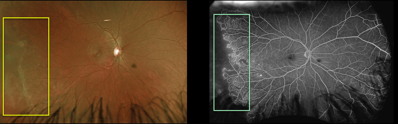
In this patient’s right eye, there is a classic sharp margin between perfused and nonperfused retina. The fellow eye (not pictured) is relatively normal.
Angiography shows some vascular leakage at the margin of the perfused and nonperfused retina.
Ultra-widefield imaging in our practice has dramatically increased the detection of Eale’s disease.
Learning Points:
Eales’ disease is an idiopathic peripheral occlusive vasculopathy that can cause retinal neovascularization and vitreous hemorrhage.
It is characterized by peripheral sharply circumscribed regions of ischemia, often with retinal neovascularization at the junction of perfused and ischemic retina.


