

Endophthalmitis
<< Back to Cases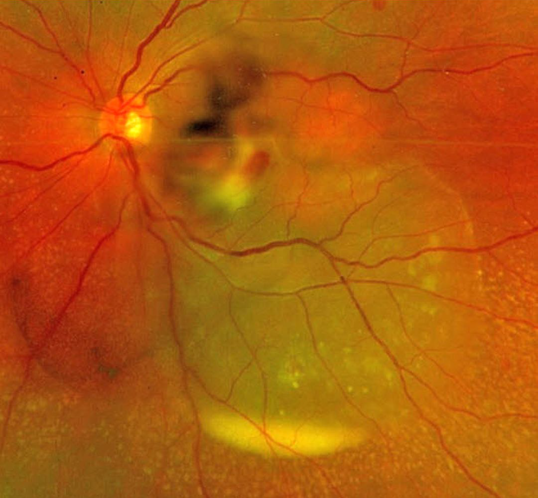
SEPTIC RETINAL EMBOLUS WITH SUBRETINAL HYPOPYON
The European VitreoRetinal Society (EVRS) and Mohit Dogra
Originally posted on @retina.rocks February 6, 2026
This previously healthy 37YO female presented to us for a second opinion regarding 4 days of decreased vision in her left eye. There was a recent history of a urinary tract infection secondary to E. coli. Vision was 20/20 in her normal OD and 20/400 OS. There was a moderate non-granulomatous uveitis with vitreous cells.
Optos color RG imaging shows opaque white retina in the inferonasal macula with overlying retinal and vitreous blood. An exudative retinal detachment extends inferotemporally with an inferior subretinal hypopyon.
She was referred back to the initially treating ophthalmologist and was subsequently lost to follow-up.
Learning Points:
Posterior segment chorioretinitis from septic embolization is a rare event, most commonly found in patients with bacterial endocarditis. Our patient presented with a constellation of findings, including retinitis, retinal and vitreous hemorrhage, and an exudative retinal detachment with an inferior subretinal hypopyon.
Subretinal hypopyon is characterized by yellowish inflammatory material settling inferiorly in the subretinal space. It has been documented in infectious (bacterial and fungal endophthalmitis, acute retinal necrosis, tuberculosis, syphilis), inflammatory (sympathetic ophthalmia), and neoplastic (leukemia, lymphoma) disorders.
CANDIDA ENDOPHTHALMITIS
Santosh Bhide
Originally posted on @retina.rocks October 11, 2023
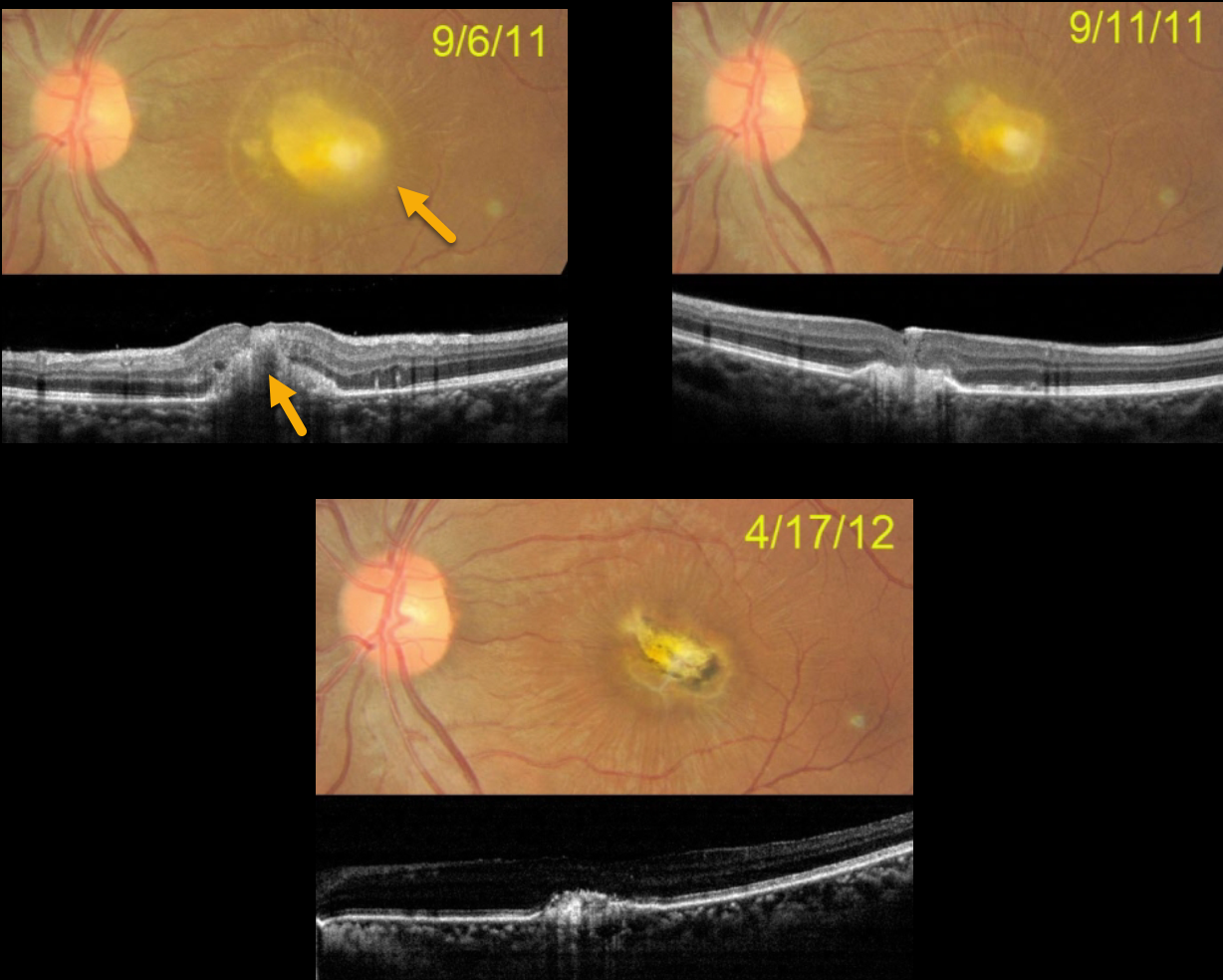
In 2011, this 27YO female was recently hospitalized and treated with intravenous amphotericin. Upon discharge, she was examined for vision loss in her left eye. Vision was hand motion.
Color photography shows a thickened yellow subfoveal lesion that extends through the full-thickness retina on OCT. One week later, vision improved to counting fingers, and the lesion is flattening and contracting. Seven months later, a small contracted foveal scar remains, with 4/60 vision. She was subsequently lost to follow-up.
Learning Points:
Ocular candidiasis is characterized by single or multiple focal yellow-white chorioretinal lesions with overlying vitritis, vitreous haze, vitreous abscess (fluff balls or string of pearls), and other more generalized presentations of infection and inflammation, such as endophthalmitis, hypopyon, scleritis, exudates, and anterior chamber cells. Ocular candidiasis can be caused by exogenous or endogenous sources. Exogenous routes include trauma and inoculation from surgical procedures. Endogenous ocular candidiasis arises from hematogenous seeding of chorioretinal blood vessels via fungemia, indwelling catheters, a severely compromised immune system, or broad-spectrum antibiotic abuse, etc.
Treatment includes removal of the inciting source of candidiasis (i.e., indwelling catheter) and systemic therapy with possible intravitreal antifungal therapy.
Routine ophthalmic screening for known systemic candida sepsis is not recommended unless there are suggestive signs or symptoms for ocular involvement (see Breazzano et al, Ophthalmology 2011;129:73-76).
MACULAR PUCKER FOLLOWING ENDOPHTHALMITIS
Originally posted on @retina.rocks March 31, 2022
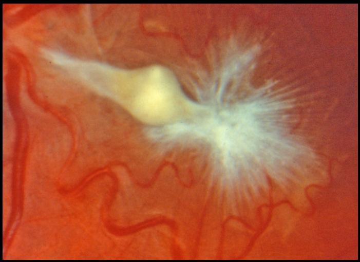
This eye shows a severely contracted and complex epimacular membrane in a patient who recovered from prior endophthalmitis. Unfortunately, we don’t have any further clinical information.
CANDIDA ENDOPHTHALMITIS
Giuseppina Monteleone
Originally posted on @retina.rocks January 25, 2022
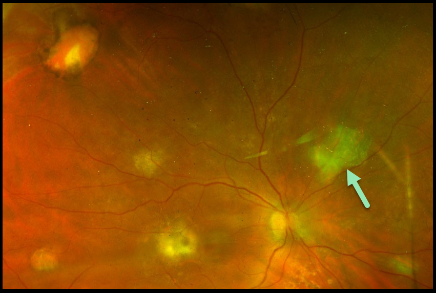
In October 2021, this 73YO emaciated woman presented for a routine cataract evaluation. Vision was 20/600 with moderate cell and flare bilaterally. Posterior segments were grossly normal, but the views were limited due to vitreous opacification.
There was a history of colon cancer treated with chemotherapy in 2018. In 2019, she suffered a cerebral hemorrhage and was later evacuated via craniotomy. In 2020, she developed a bowel obstruction during chemotherapy treatment, and total parenteral nutrition was initiated. In August 2021, a central line infection caused Candida albicans sepsis. Voriconazole was administered until blood cultures came back negative, and she became afebrile.
Based on the clinical history, she was felt to have bilateral fungal endophthalmitis despite the negative blood cultures and negative in-office vitreous and aqueous stains and cultures.
Fluconazole 400 mg/day and intravitreal Amphotericin B (0.01 mg/0.1 ml) were administered, with improvement in anterior segment inflammation but no change in the vitritis.
Eventually, a diagnostic vitrectomy was performed in the right eye, revealing yellowish retinal foci, typical for endogenous Candida albicans endophthalmitis, although Gram stain and cultures were negative.
Vision improved to 20/100 in the right eye. We plan on performing vitrectomy in her left eye when her clinical condition allows.
CANDIDA ENDOPHTHALMITIS
Originally posted on @retina.rocks December 31, 2021
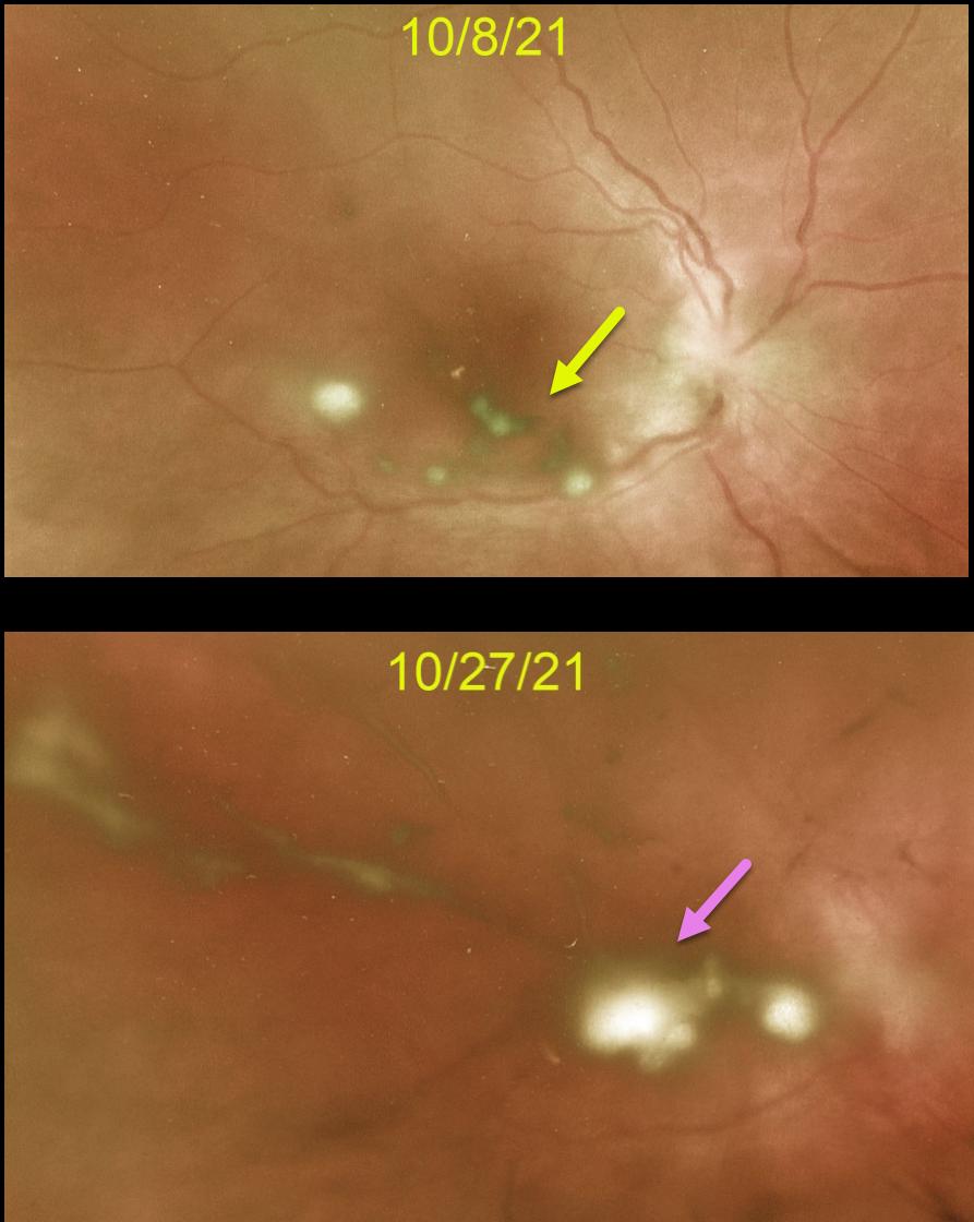
This 37YO male presented with a 4-day history of right eye pain and blurred vision. There was no prior ocular history, surgery, recent trauma, or intravenous drug use.
Vision was counting fingers OD and 20/25 in his normal OS. Moderate cell and flare were noted OD, along with a tiny hypopyon. Optos imaging shows multiple posterior inflammatory puff balls suggestive of endogenous fungal endophthalmitis.
We ordered emergent blood work to rule out other infectious causes before initiating intravitreal or systemic antifungal therapy. Unfortunately, he was completely noncompliant about going for testing despite daily calls.
He subsequently returned to the office 2 weeks later with increased vitreous opacities. On further questioning, he gave a history of recent low back pain and inflammation, where 6 weeks earlier he underwent surgery with metal plates. We told him to go to the ER for candida sepsis workup immediately.
However, he went home and continued his noncompliance, which finally led him to our local teaching hospital 1 week later. We assume the infectious source was the hardware implanted in his back.
Learning Points:
Ocular candidiasis is characterized by single or multiple focal yellow-white chorioretinal lesions with overlying vitritis, vitreous haze, vitreous abscess (fluff balls or string of pearls), and other more generalized presentations of infection and inflammation, such as endophthalmitis, hypopyon, scleritis, and anterior chamber cells.
Exogenous or endogenous sources can cause ocular candidiasis. Exogenous routes include trauma and inoculation from surgical procedures.
Endogenous ocular candidiasis arises from hematogenous seeding of the chorioretinal blood vessels caused by fungemia, indwelling catheters, severely compromised immune systems, and abuse of broad-spectrum antibiotics.
Treatment includes removal of the inciting source of candidiasis (e.g., an indwelling catheter) and systemic therapy, with possible intravitreal antifungal therapy.
Routine ophthalmic screening for known systemic candida sepsis is not recommended unless there are suggestive signs or symptoms for ocular involvement (see Breazzano et al, Ophthalmology 2011;129:73-76).
CANDIDA ENDOPHTHALMITIS
Originally posted on @retina.rocks May 20, 2021
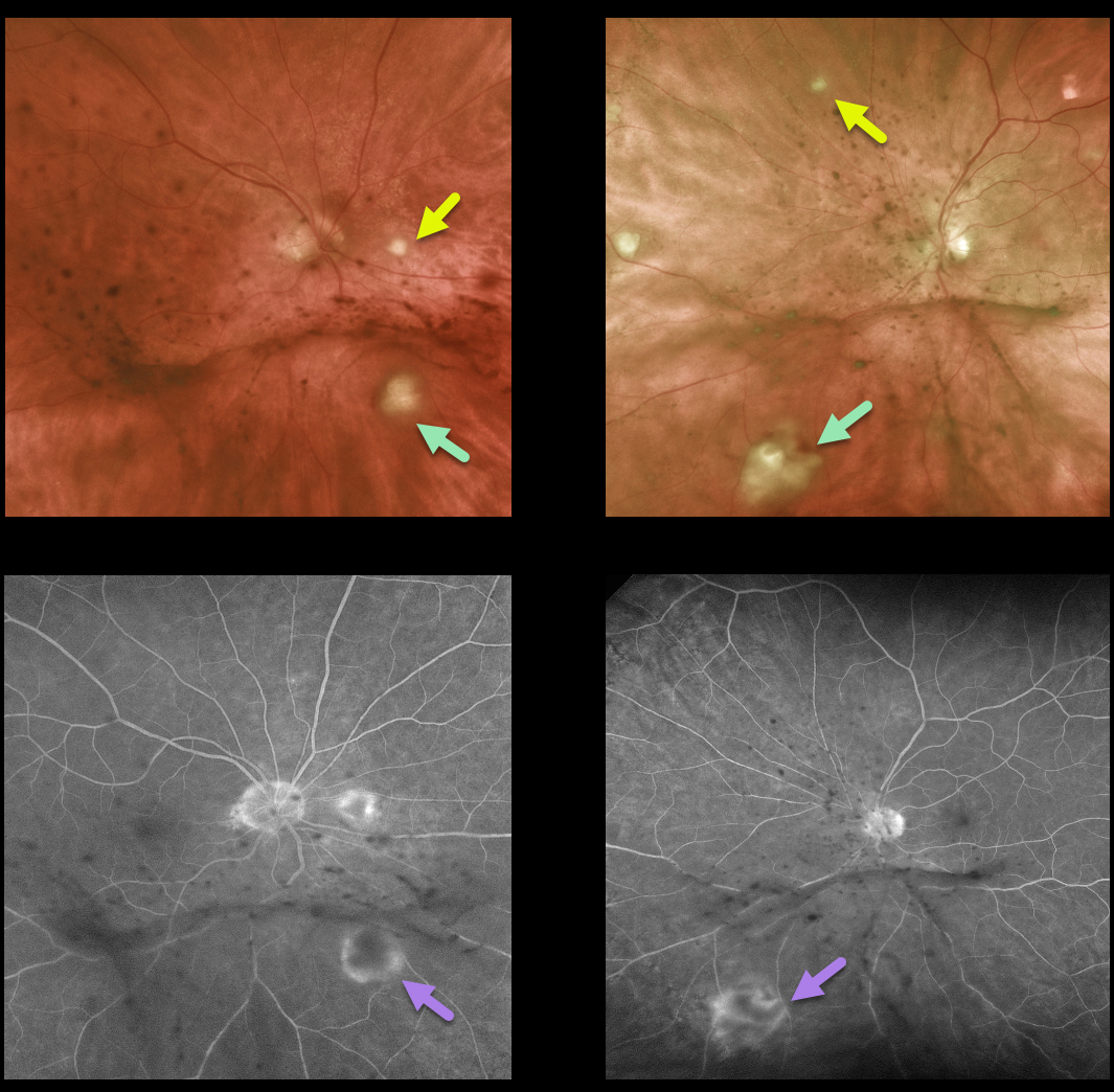
This 75 yo retired physician presented with decreased vision and floaters for about 8 weeks. Vision was 20/25 OD and 20/70 OS. Anterior segments were normal except for mild nuclear sclerosis.
There are multifocal areas of yellow-white chorioretinal inflammation spreading into the overlying vitreous, along with clumps of inflammatory deposits on the back of each hyaloid. Fluorescein angiography reveals early blockage with late surrounding staining of these inflammatory lesions.
He gave a history of non-Hodgkin’s lymphoma, which was in remission. He still had a port in place despite having completed chemotherapy treatments. He was also being treated with oral prednisone and azithromycin for organizing pneumonia and had active oral candidiasis.
Based on his clinical findings, immunosuppression, chemotherapy port, and oral thrush, we suspected bilateral candidal endophthalmitis, although we were also concerned about an atypical presentation for intraocular lymphoma.
Diagnostic vitrectomy was performed. Cytology was negative for lymphoma, and Gram stain and fungal cultures were also surprisingly negative. Blood cultures were also negative.
Despite the negative workup, we still felt that fungal endophthalmitis was the most likely diagnosis and started oral fluconazole. Since the port was no longer in use, this was removed and did, in fact, grow out Candida. The intraocular inflammation subsequently improved.
Learning Points:
In many cases of endophthalmitis, no infectious agent is identified, and often the diagnosis is made clinically. Empiric treatment often includes intravitreal antibiotics. In this case, despite negative blood cultures and vitreous biopsy, our clinical suspicion ultimately led to the correct diagnosis and treatment.


