

Exudative Retinal Detachment (RD)
<< Back to Cases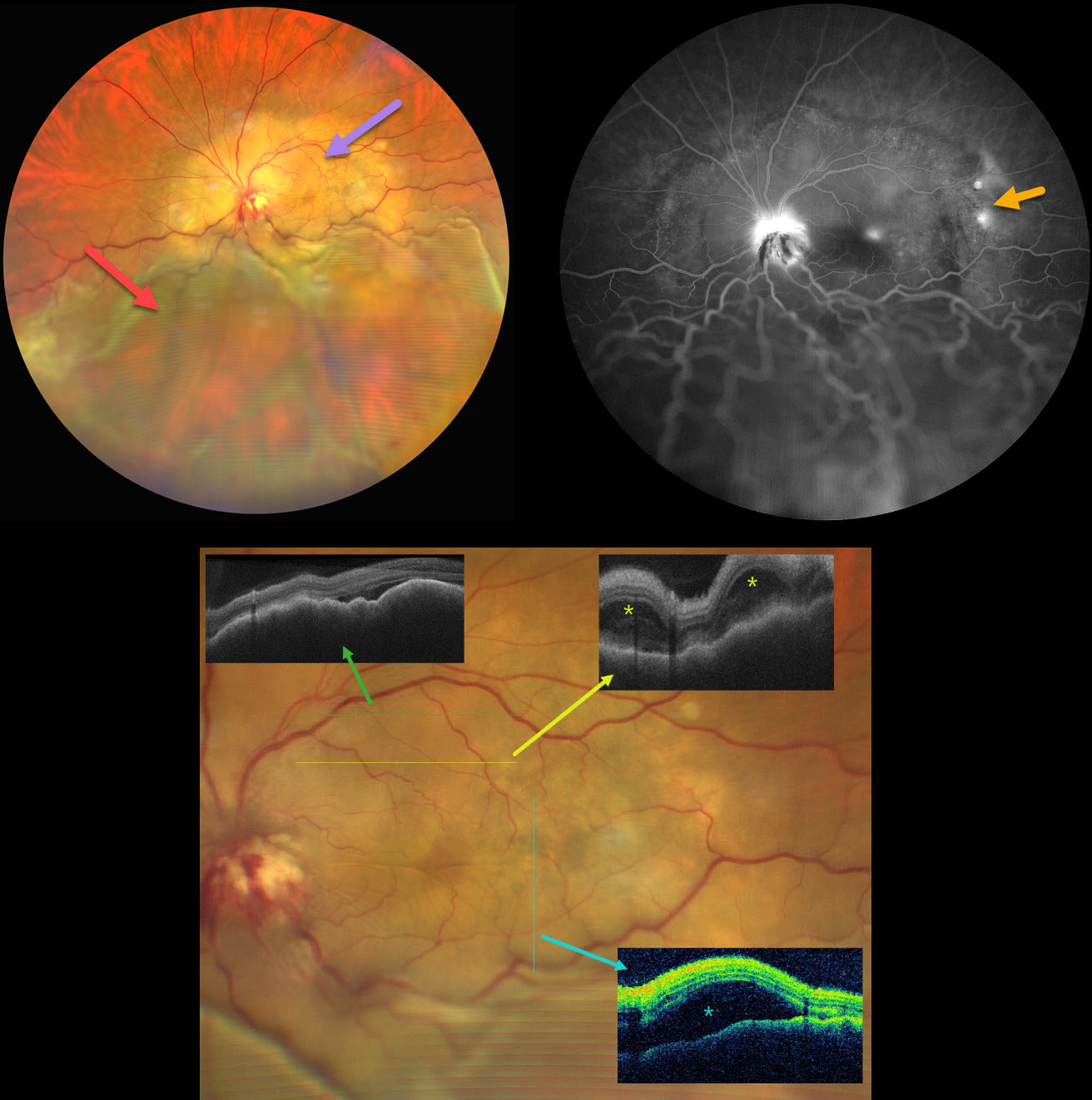
CHOROIDAL METASTASES WITH EXUDATIVE RETINAL DETACHMENT
Ayushi Gupta and Vishal Agrawal
Originally posted on @retina.rocks June 18, 2025
This 47YO female with a known history of metastatic breast carcinoma presented with several weeks of decreased vision in her left eye. She was on active chemotherapy and hormonal therapy. Vision was no light perception OD following trauma 30 years earlier, and hand motion OS.
Ultrawidefield color photography shows a large, amelanotic yellow choroidal lesion involving the entire macula and peripapillary choroid, which extends into the midperiphery. A large secondary inferior exudative retinal detachment involves the entire inferior hemisphere. This fluid shifted when laid supine. The optic disc is hyperemic with indistinct margins and associated blood.
OCT scanning shows a thickened, undulating choroid with variable overlying subretinal fluid and bacillary layer detachments. Fluorescein angiography shows multifocal subretinal leaks, choroidal lesion staining with possible overlying pooling, and intense nerve leakage.
Learning Points:
Although uveal melanoma is the most common primary intraocular malignancy, metastatic choroidal tumors are the most common intraocular malignancies. Lung and breast cancer are the most common sources.
Our patient had a known history of metastatic disease when she presented with this large lesion occupying her entire posterior pole. The extent of the metastasis and the overlying exudation are atypical in our experience. She was immediately referred to her oncologist for external-beam radiation, but was lost to follow-up.


