

Familial Exudative Vitreoretinopathy (FEVR)
<< Back to Cases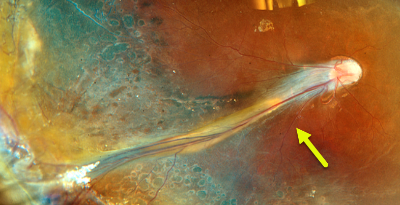
FAMILIAL EXUDATIVE VITREORETINOPATHY (FEVR)
João Pedro Marques
Originally posted on @retina.rocks March 23, 2026
This healthy 6YO boy from Guinea-Bissau with long-standing low vision was referred due to a family history of familial exudative vitreoretinopathy (FEVR) in an older brother and maternal uncle. Vision was 20/400 OD and no light perception OS.
Fundus examination OD showed a falciform retinal fold, straightening of the temporal retinal vascular arcades, and temporal macular dragging. There was a dense white cataract OS, with a total funnel-shaped retinal detachment noted on ultrasonography. Scatter laser and peripheral cryotherapy were applied throughout the ischemic retina. Postoperative Optos color RGB imaging shows laser and cryotherapy scarring.
Familial variant testing confirmed the presence of the pathogenic c.362G>A p.(Arg121Gln) variant in hemizygosity in the NDP gene (NM_000266.4), establishing the diagnosis of X-linked FEVR.
Learning Points:
Originally described by Criswick and Schepens (AJO 1969;58:578-594), familial exudative vitreoretinopathy (FEVR) can be inherited as an autosomal-dominant, recessive, or X-linked trait with high penetrance and variable expressivity. There are numerous genes associated with FEVR, including LRP5, FZD4, NDP, TSPAN12, ZNF408, CTNNB1 and KIF11 [Tao et al, Invest Ophthalmol Vis Sci 2021;62(15);4].
FEVR is characterized by peripheral temporal retinal avascularity, lipid exudation, peripheral neovascularization, tractional retinal detachment, and temporal dragging of the macula and retinal vessels. These findings are somewhat similar to those seen in retinopathy of prematurity.
POSSIBLE FAMILIAL EXUDATIVE VITREORETINOPATHY (FEVR)
Mattie Adams
Originally posted on @retina.rocks May 29, 2025
This healthy 9YO boy was referred for an asymptomatic abnormal right optic nerve. There was no ocular family history. Vision was 20/150 OD and 20/30 in his normal OS.
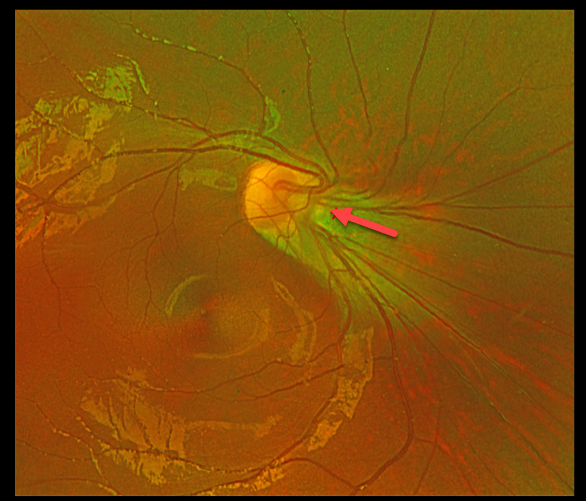
Optos color RG imaging shows an anomalous right optic nerve, which is tilted and displaced superonasally from its apparent normal location. The fovea is markedly ectopic, located about 60 degrees inferotemporal to the nerve. Genetic testing revealed a heterozygous FZD4 variant of uncertain significance.
Learning Points:
Originally described by Criswick and Schepens (AJO 1969;58:578-594), familial exudative vitreoretinopathy (FEVR) can be inherited as an autosomal-dominant, recessive, or X-linked trait with high penetrance and variable expressivity. There are numerous genes associated with FEVR, including LRP5, FZD4, NDP, TSPAN12, ZNF408, CTNNB1 and KIF11 [Tao et al, Invest Ophthalmol Vis Sci 2021;62(15);4].
FEVR is characterized by peripheral temporal retinal avascularity, lipid exudation, neovascularization, tractional retinal detachment, and temporal dragging of the macula and retinal vessels. These findings are somewhat like those found with retinopathy of prematurity.
FAMILIAL EXUDATIVE VITREORETINOPATHY (FEVR)
Barbara Parolini, Veronika Matello, Giulia Freschi, and Roberta Penzani
Originally posted on @retina.rocks January 23, 2024
This otherwise healthy 8YO male presented with a chronic history of vision loss in his right eye. Vision was counting fingers OD and 20/25 OS.
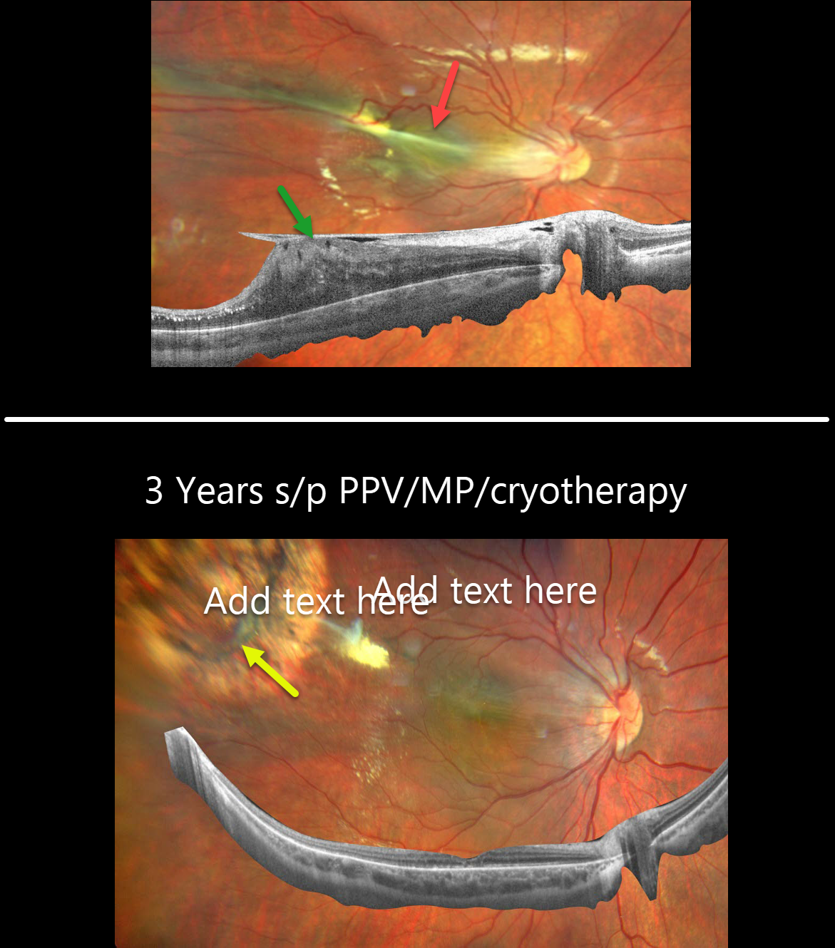
Fundus photography shows dragging of the optic nerve and macula temporally towards an area of dilated, anomalous retinal vessels. Widefield OCT of the right shows an adherent epiretinal membrane elevating and dragging the nerve and macula temporally.
Pars plana vitrectomy with membrane peeling and cryotherapy of the area with anomalous vessels was performed. Three years later, vision improved to 20/25 OD. The macula is clinically and on OCT completely flat, with a dry cryotherapy scar noted in the superotemporal midperiphery.
This Eyecare Clinic (in Brescia, Italy) case was submitted by Barbara Parolini, Veronika Matello, Giulia Freschi, and Roberta Penzani.
Learning Points:
Originally described by Criswick and Schepens (AJO 1969;58:578-594), familial exudative vitreoretinopathy (FEVR) can be inherited as an autosomal-dominant, recessive, or X-linked trait with high penetrance and variable expressivity. There are numerous genes associated with FEVR, including LRP5, FZD4, NDP, TSPAN12, ZNF408, CTNNB1 and KIF11 [Tao et al, Invest Ophthalmol Vis Sci 2021;62(15);4].
FEVR is characterized by peripheral temporal retinal avascularity, lipid exudation, neovascularization, tractional retinal detachment, and temporal dragging of the macula and retinal vessels. These findings are somewhat similar to those found with retinopathy of prematurity (ROP).
FAMILIAL EXUDATIVE VITREORETINOPATHY (FEVR)
Originally posted on @retina.rocks October 3, 2023
This 22YO female presented with a lifelong history of stable severe bilateral vision loss, congenital nystagmus, and a prior inoperable retinal detachment in her left eye. There was no past medical or family history of eye disease.
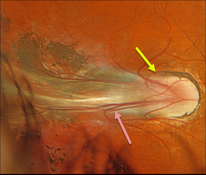
Optos color RGB imaging of her right eye shows temporal dragging of the disc and vessels from a falciform retinal fold extending towards the periphery. Secondary surrounding pigmented chorioretinal scarring is noted.
The left eye had a dense white cataract with no view of the posterior pole. Genetic testing was positive for a pathogenic KIF11 mutation.
Learning Points:
Originally described by Criswick and Schepens (AJO 1969;58;578-594), familial exudative vitreoretinopathy (FEVR) can be inherited as an autosomal-dominant, recessive, or X-linked trait with high penetrance and variable expressivity.
FEVR is characterized by peripheral temporal retinal avascularity, lipid exudation, neovascularization, tractional retinal detachment, and temporal dragging of the macula and retinal vessels. These findings are somewhat similar to those found with retinopathy of prematurity (ROP).
There are numerous genes associated with FEVR, including LRP5, FZD4, NDP, TSPAN12, ZNF408, CTNNB1 and KIF11 [Tao et al, Invest Ophthalmol Vis Sci 2021;62(15);4].
The exact mechanism by which KIF11 mutations cause FEVR is not fully understood. However, it is thought that mutations in the KIF11 gene disrupt the transport of vesicles and organelles essential for retinal blood vessel development.
FAMILIAL EXUDATIVE VITREORETINOPATHY (FEVR)
Originally posted on @retina.rocks December 20, 2022
This 53YO male came in for an initial examination since both of his sons and one of his nephews have been followed by us for years with familial exudative vitreoretinopathy (FEVR).
Both sons developed peripheral retinal neovascularization that responded well to scatter laser. Their father was visually asymptomatic.
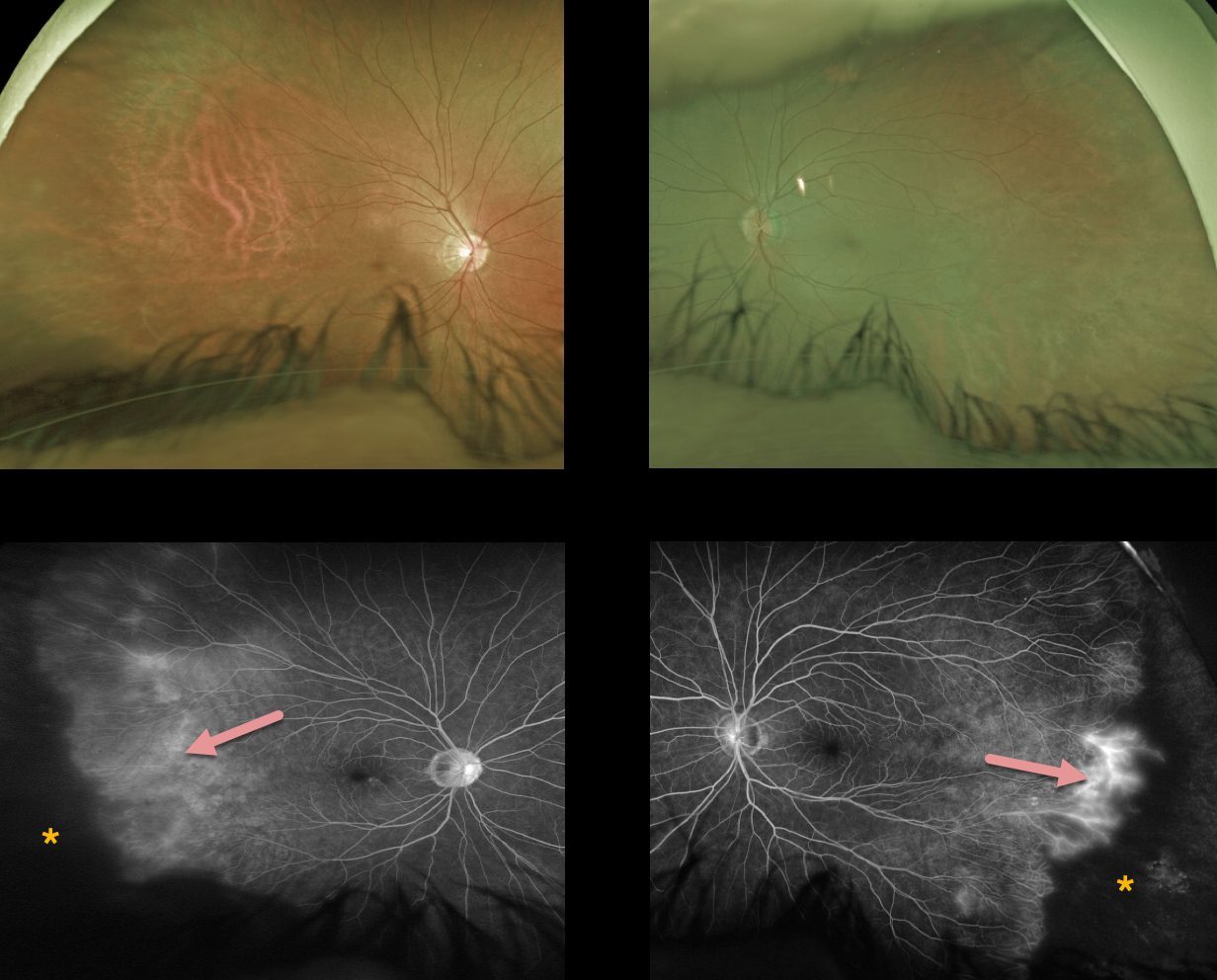
Optos color imaging shows normal posterior poles with featureless, ischemic temporal retinas. Fluorescein angiography shows marked bilateral peripheral temporal ischemia with leaking vessels at the border of the perfused and ischemic retina.
Prophylactic scatter laser to the ischemic retina was recommended to help prevent retinal neovascularization.
Learning Points:
Originally described by Criswick and Schepens (AJO 1969;58:578-594), familial exudative vitreoretinopathy (FEVR) can be inherited as an autosomal-dominant, recessive, or X-linked trait with high penetrance and variable expressivity.
FEVR is characterized by peripheral temporal retinal avascularity, lipid exudation, neovascularization, tractional retinal detachment, and temporal dragging of the macula and retinal vessels. These findings are somewhat similar to those found with retinopathy of prematurity (ROP).
Kashani et al found that peripheral retinal vascular findings are highly prevalent in asymptomatic FEVR relatives (Ophthalmology 2014;121:262-268), as was the case with our patient. They therefore recommended widefield angiographic screening in immediate relatives.
FAMILIAL EXUDATIVE VITREORETINOPATHY (FEVR)
Akansha Sharma and Manish Nagpal
Originally posted on @retina.rocks September 15, 2022
This 20YO female presented with a 2-month history of vision loss in her right eye. Vision was counting fingers.
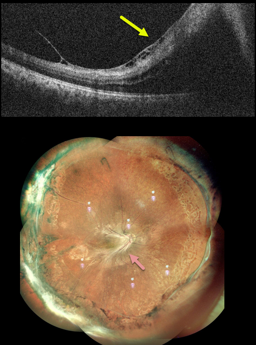
The macular vessels were dragged temporally with a taut membrane elevating the fovea, as demonstrated on OCT. Fluorescein angiography showed bilateral encircling peripheral ischemia with retinal neovascularization (not shown).
Encircling peripheral scatter laser was performed for her left eye. Pars plana vitrectomy with membrane peeling and intravitreal gas was performed for her right eye.
Two months following surgery, all traction remained relieved, and the retina was completely attached.
FAMILIAL EXUDATIVE VITREORETINOPATHY (FEVR)
Originally posted on @retina.rocks June 23, 2021
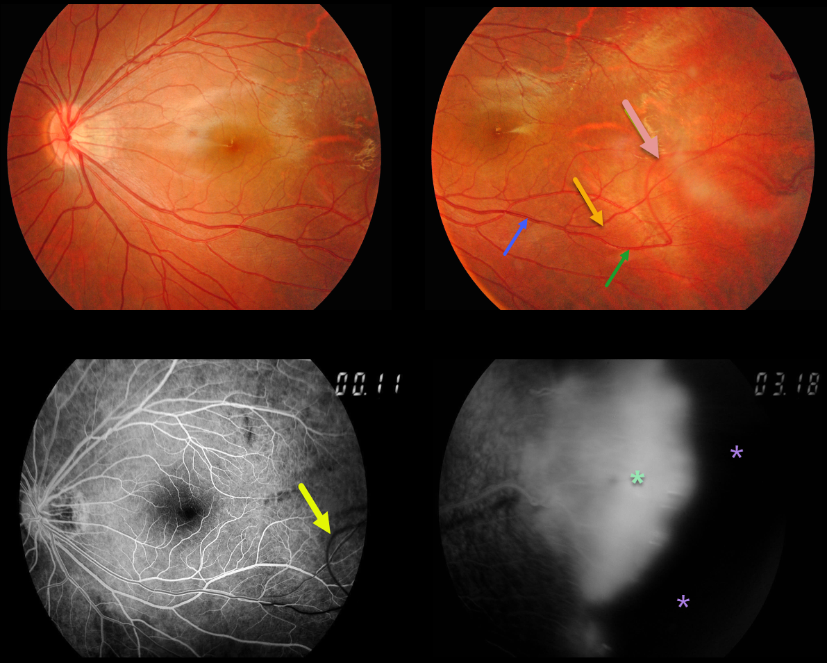
Our patient’s macular retinal vessels are dragged temporally. His brother has similar retinal findings.
The major inferotemporal vein branches into smaller superior and inferior venules. The inferior venule makes an abrupt turn more distally, arching superiorly before extending into the temporal periphery.
Arteries cross veins, veins cross arteries, but arteries never cross arteries, and veins never cross veins.
So why does this inferior venule eventually cross the superior venule? Although we don’t have any stereoscopic images, we suspect that vitreous traction pulled the inferior venule anteriorly and superiorly.
This is also evident on fluorescein angiography (FA), where the superior vessel, which has still not completely filled, is clearly anterior to the other venules, which have laminar filling. FA also shows leaking neovascularization and more peripheral retinal ischemia.
Learning Points:
Originally described by Criswick and Schepens (AJO 1969;58;578-594), familial exudative vitreoretinopathy (FEVR) can be inherited as an autosomal-dominant, recessive, or X-linked trait with high penetrance and variable expressivity.
FEVR is characterized by peripheral temporal retinal avascularity, lipid exudation, neovascularization, tractional retinal detachment, and temporal dragging of the macula and retinal vessels. These findings are somewhat similar to those found with retinopathy of prematurity (ROP).


