

Focal Scleral Nodule
<< Back to Cases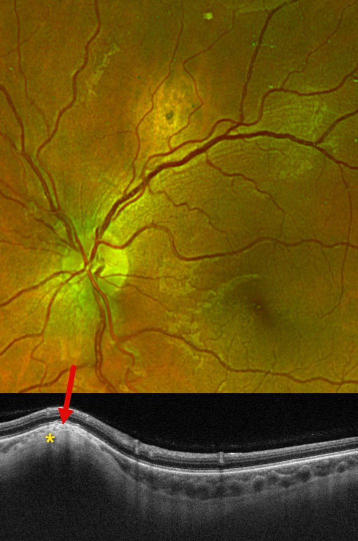
FOCAL SCLERAL NODULE
Originally posted on @retina.rocks February 13, 2025
This 29YO female presented with this asymptomatic amelanotic lesion above the right superotemporal arcade. Optos color RG imaging shows an apparent mostly depigmented chorioretinal scar. However, Triton swept-source OCT scanning shows hyperreflective scleral thickening, which compresses the overlying choroid and elevates the retina.
Learning Points:
Focal scleral nodule, previously known as solitary idiopathic choroiditis (SIC) and unifocal helioid choroiditis, presents as a single, yellow-white, elevated nodular, avascular subretinal lesion. On OCT, it arises from the inner sclera, causing marked choroidal compression. These lesions require no treatment, although they may rarely show slight growth (Fung and Li, Retinal Cases & Brief Reports 2024;18:526-528).
FOCAL SCLERAL NODULE
Janelle Adeniran
Originally posted on @retina.rocks March 6, 2024
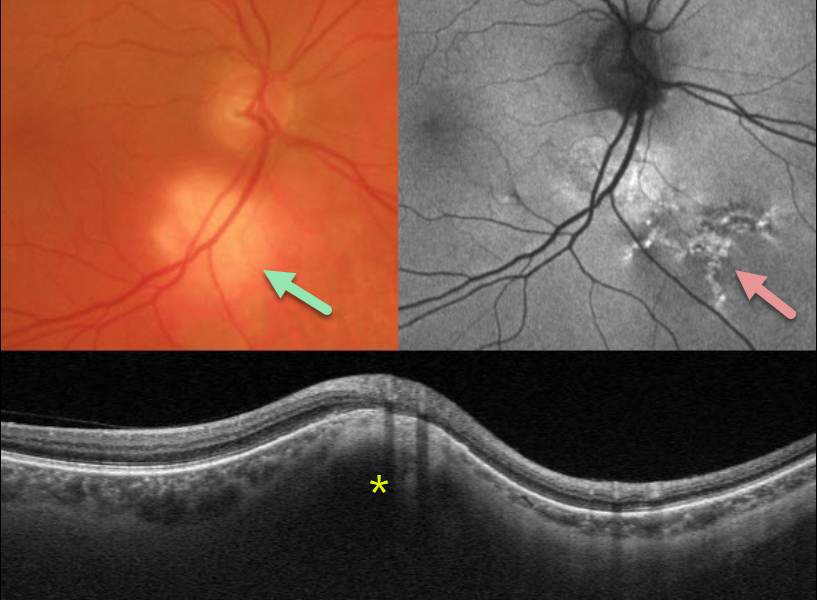
This 73YO male presented with this asymptomatic amelanotic lesion inferior to the right optic nerve. Optos fundus autofluorescence (FAF) shows overlying hyper-FAF with some dendritic radiating lines of hyper- and hypo-FAF extending inferonasally.
Triton swept-source OCT scanning through this region shows hyperreflective scleral thickening, which compresses the overlying choroid and elevates the retina.
Learning Points:
Focal scleral nodule, previously known as solitary idiopathic choroiditis (SIC) and unifocal helioid choroiditis, presents as a single, yellow-white, elevated nodular, avascular subretinal lesion. On OCT, it arises from the inner sclera, causing marked compression of the choroid.
For the largest series to date and the rationale behind its renaming, see Fung et al, Ophthalmology 2020;127:1567-1577.
FOCAL SCLERAL NODULE
Originally posted on @retina.rocks November 6, 2023
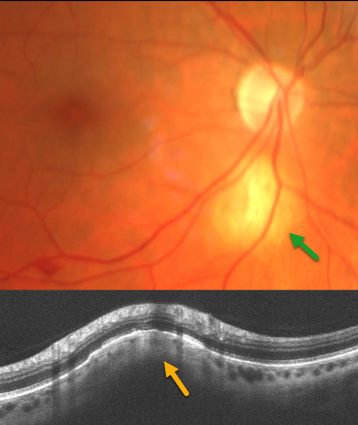
This patient presented with an asymptomatic amelanotic lesion inferior to the right optic nerve. OCT scanning through this region shows hyperreflective scleral thickening, which compresses the overlying choroid and elevates the retina.
Learning Points:
Focal scleral nodule, previously known as solitary idiopathic choroiditis (SIC) and unifocal helioid choroiditis, presents as a single, yellow-white, elevated nodular, avascular subretinal lesion. On OCT, it arises from the inner sclera, causing marked compression of the choroid.
For the largest series to date and the rationale behind its renaming, see Fung et al, Ophthalmology 2020;127:1567-1577.
FOCAL SCLERAL NODULE
Ryan Kern and Alex Hynes
Originally posted on @retina.rocks May 11, 2023
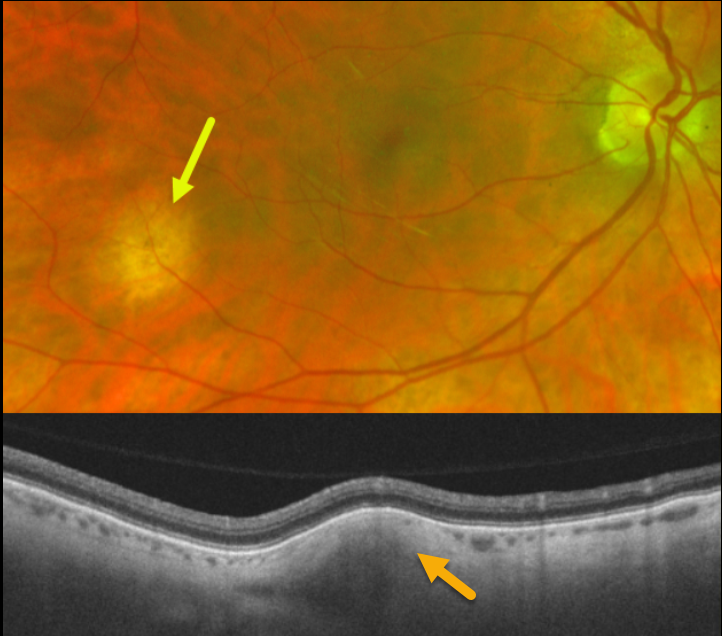
Optos color imaging shows an amelanotic subretinal lesion in the right inferotemporal midperiphery. Swept-source OCT through this lesion shows hyperreflective scleral thickening, which compresses the overlying choroid.
Learning Points:
Focal scleral nodule (previously known as solitary idiopathic choroiditis [SIC] and unifocal helioid choroiditis), presents as a single, yellow-white, elevated nodular, avascular subretinal lesion. On OCT it arises from the inner sclera, causing marked choroidal compression.
For the largest series to date and the rationale behind the renaming of this entity, see Fung et al, Ophthalmology 2020;127:1567-1577.
FOCAL SCLERAL NODULE
Originally posted on @retina.rocks March 7, 2023
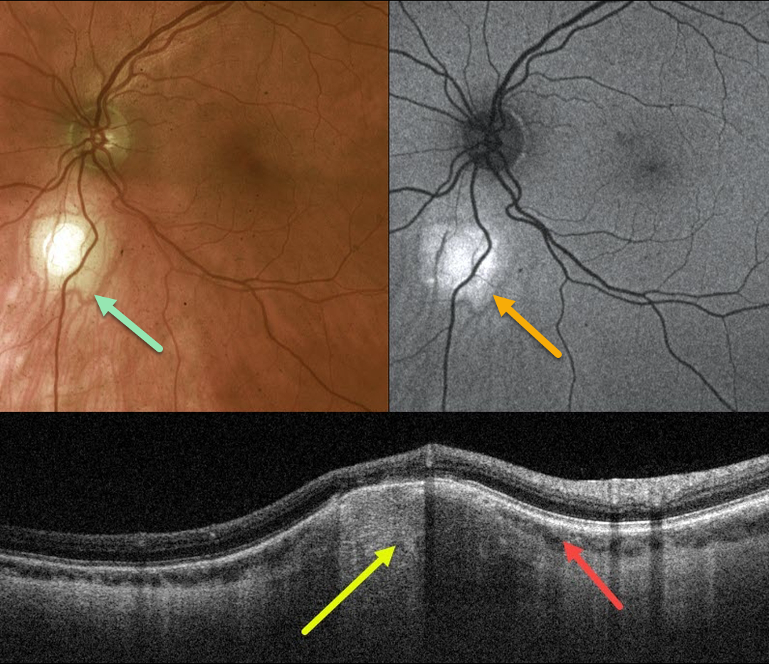
This 45YO healthy male was referred for an asymptomatic lesion in his left eye. Vision was 20/20 OU.
Optos color imaging shows an amelanotic subretinal lesion just inferior to the left optic nerve. Fundus autofluorescence (FAF) shows this lesion to be hyper-FAF.
Swept-source OCT through this lesion shows a hyperreflective scleral thickening that compresses the overlying choroid.
Learning Points:
Focal scleral nodule (previously known as solitary idiopathic choroiditis [SIC] and unifocal helioid choroiditis), presents as a single, yellow-white, elevated nodular, avascular subretinal lesion. On OCT it arises from the inner sclera, causing marked choroidal compression.
For the largest series to date and the rationale behind the renaming of this entity, see Fung et al, Ophthalmology 2020;127:1567-1577.
FOCAL SCLERAL NODULE
Originally posted on @retina.rocks October 28, 2021
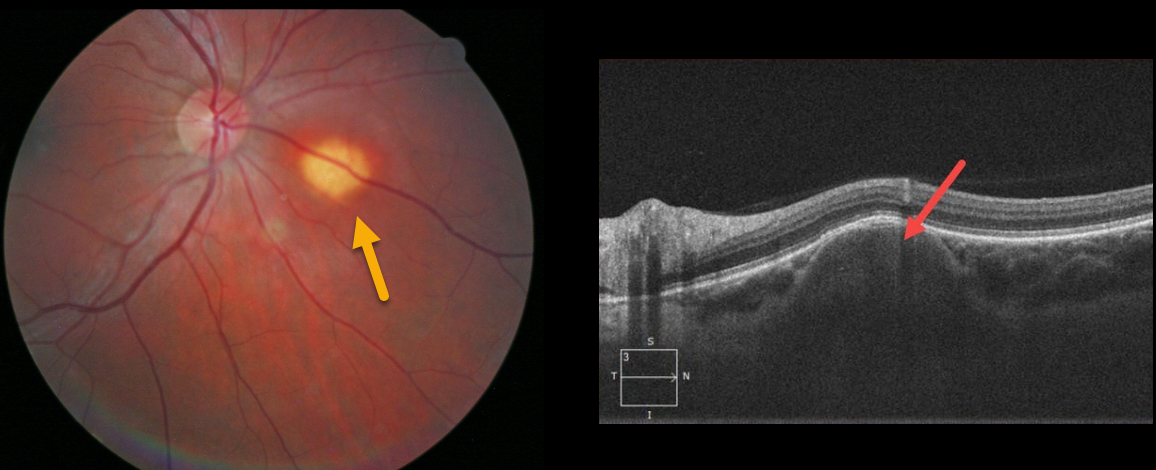
This patient presented with an asymptomatic yellowish, nodular subretinal lesion inferonasal to the right optic nerve.
Spectral domain optical coherence tomography shows that the lesion originates from the sclera.
Learning Points:
Focal scleral nodule (previously known as solitary idiopathic choroiditis [SIC] and unifocal helioid choroiditis) presents as a single, yellow-white, elevated nodular, avascular subretinal lesion. On OCT, it arises from the inner sclera, causing marked choroidal compression.
For the largest series to date and the rationale behind the renaming of this entity, see Fung et al, Ophthalmology 2020;127:1567-1577.


