

Frosted Branch Angiitis
<< Back to Cases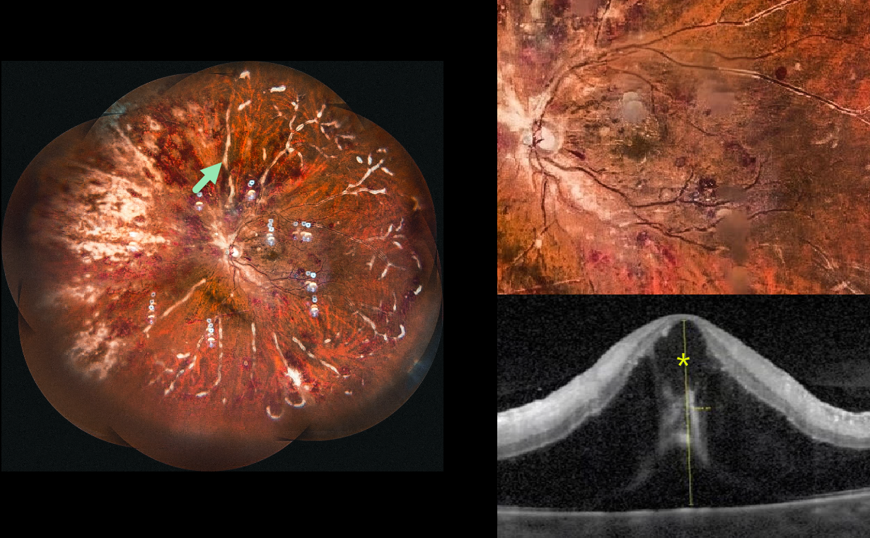
FROSTED BRANCH ANGIITIS
The European VitreoRetinal Society (EVRS) and Nitin Ramanujam
Originally posted on @retina.rocks July 3, 2026
This 45YO female with a known history of poorly controlled systemic lupus erythematosus (SLE) presented to us for a third opinion with 10 days of bilateral vision loss. Vision was 20/100 OD and 20/200 OS.
Ultrawidefield imaging of her left eye shows extensive retinal hemorrhages with a frosted branch appearance to the retinal veins. Marked cystoid edema and subfoveal fluid are noted on OCT. Identical findings were seen in her right eye (not shown). She was immediately lost to follow-up.
Learning Points:
Frosted branch angiitis is a retinal vasculitis that gets its name from the involved vessels resembling branches of a tree that are “frosted” with snow (Kleiner et al, AJO 1988;106:27-34). Although these findings may be isolated, they are often considered a phenotype found in many inflammatory conditions, most commonly CMV retinitis. Inflammation is usually highly responsive to steroids, and the visual prognosis is generally good, although some may develop secondary neovascularization in the anterior and posterior segments due to widespread ischemia.
Although frosted branch angiitis has not been specifically linked to SLE, retinal vasculitis (arteriolar vasculitis with cotton wool spots, retinal hemorrhages, and vascular occlusions) is a known lupus finding (Gao et al, Lupus 2017;26:1182-1189).
POSSIBLE EPIDEMIC RETINITIS
Sharat Hegde
Originally posted on @retina.rocks June 2, 2025
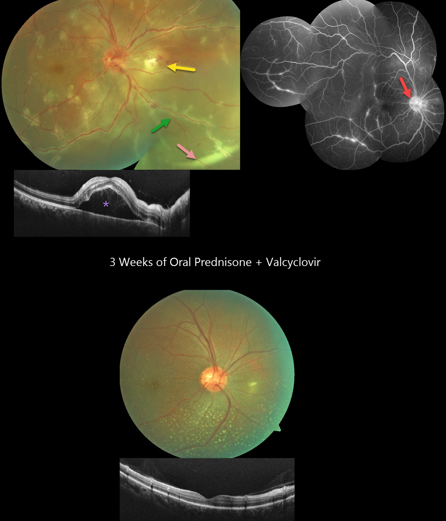
This healthy 36YO male presented with 3 days of floaters and decreased vision in his right eye. Vision was 20/60 OD and 20/20 in his healthy OS. The right eye contained mild anterior chamber cells and moderate vitreous cells.
Fundus photography shows a swollen nerve, a frosted branch pattern of vasculitis involving the retinal veins, an area of retinitis just nasal to the nerve, and a subretinal hypopyon in the inferonasal midperiphery. Some multifocal areas of outer retinal inflammation are noted, along with some retinal hemorrhages. OCT scanning shows variable hyperreflective foveal subretinal fluid. Fluorescein angiography shows variable venous staining and leakage, including from the disc and in nasal retinitis.
The workup included elevated ESR and CRP levels, along with an increased IgG titer for Varicella zoster virus. Mantoux testing, syphilis, Bartonella, typhus, HIV, and chest X-ray were all negative. Although very atypical for viral uveitis, we started oral prednisone and valaciclovir. After one week, the hypopyon resolved, with decreased vasculitis and new scattered flecks of subretinal lipid throughout the posterior pole. By three weeks, vision improved to 20/20 with continued funduscopic improvement.
Learning Points:
Epidemic (post-fever) retinitis is an infectious or para-infectious entity that develops in immunocompetent individuals several weeks following a bacterial, viral, or protozoal infection. Causes include Dengue, Rickettsiosis, West Nile, Leptospirosis, Chikungunya, Typhoid, and Ebola. Patients present with sudden, painless vision loss.
Fundus findings include unilateral or bilateral unifocal or multifocal patches of retinitis with possible nerve involvement, serous macular detachment, and vasculitis or frosted branch angiitis. There is usually a favorable response to steroids and treating the underlying infectious cause with appropriate systemic therapy. See Mahendradas et al (Indian J Ophthalmol 2020;68:1775-1786) for an excellent post-fever retinitis review.
Frosted branch angiitis is a retinal vasculitis that gets its name from the involved vessels resembling branches of a tree that are “frosted” with snow (Kleiner et al, AJO 1988;106:27-34). Although these findings may be isolated, they are often considered a phenotype found in many inflammatory conditions, most commonly CMV retinitis. The inflammation is usually highly responsive to steroids, and the visual prognosis is generally good, although some may develop secondary neovascularization in the anterior and posterior segments due to widespread ischemia.
Subretinal hypopyon is characterized by yellowish inflammatory material settling inferiorly in the subretinal space. It has been documented in infectious (bacterial and fungal endophthalmitis, acute retinal necrosis, tuberculosis, syphilis), inflammatory (sympathetic ophthalmia), and
POST-VIRAL FROSTED BRANCH ANGIITIS
Ayushi Gupta and Vishal Agrawal
Originally posted on @retina.rocks April 7, 2025
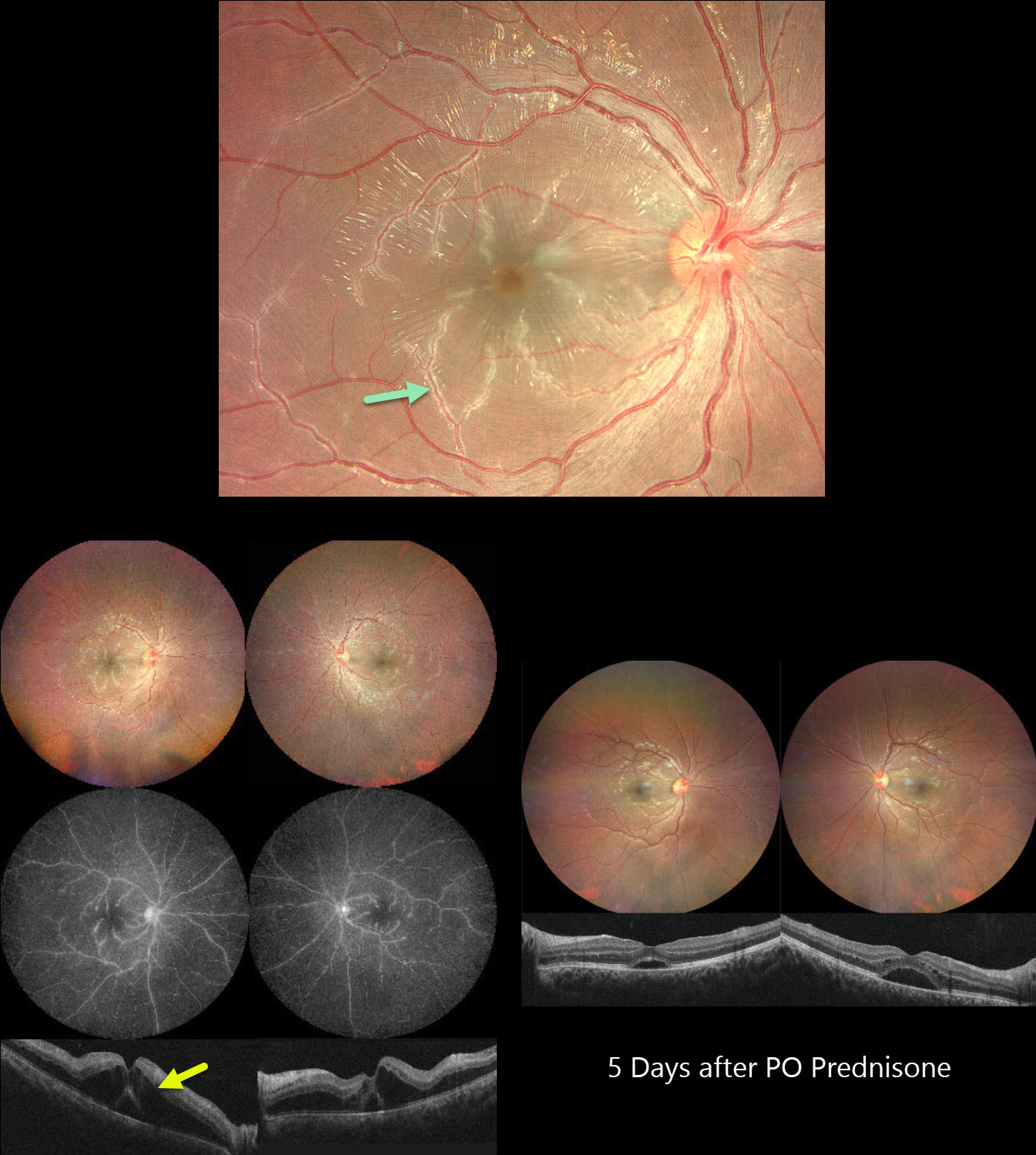
This previously healthy 21YO male developed bilateral vision loss 1 week following a viral illness. Vision was 20/100 OU.
Color photography shows a bilateral frosted-branch appearance of the retinal veins, most prominent in the maculas. Fluorescein angiography shows diffuse leakage and staining of the venous vascular tree. OCT scanning shows marked bilateral outer nuclear layer edema with foveal subretinal fluid. He was started on oral prednisone 60mg daily. Five days later vision improved to 20/40 OU. The venous changes are dramatically better with decreasing macular fluid. A slow steroid taper began.
Learning Points:
Frosted branch angiitis is a retinal vasculitis that gets its name from the involved vessels resembling branches of a tree that are “frosted” with snow (Kleiner et al, AJO 1988;106:27-34). Although these findings may be isolated, they are often considered a phenotype found in many inflammatory conditions, most commonly CMV retinitis. The inflammation is usually highly responsive to steroids, and the visual prognosis is generally good, although some may develop secondary neovascularization in the anterior and posterior segments due to widespread ischemia.
FROSTED BRANCH ANGIITIS
Originally posted on @retina.rocks June 15, 2023
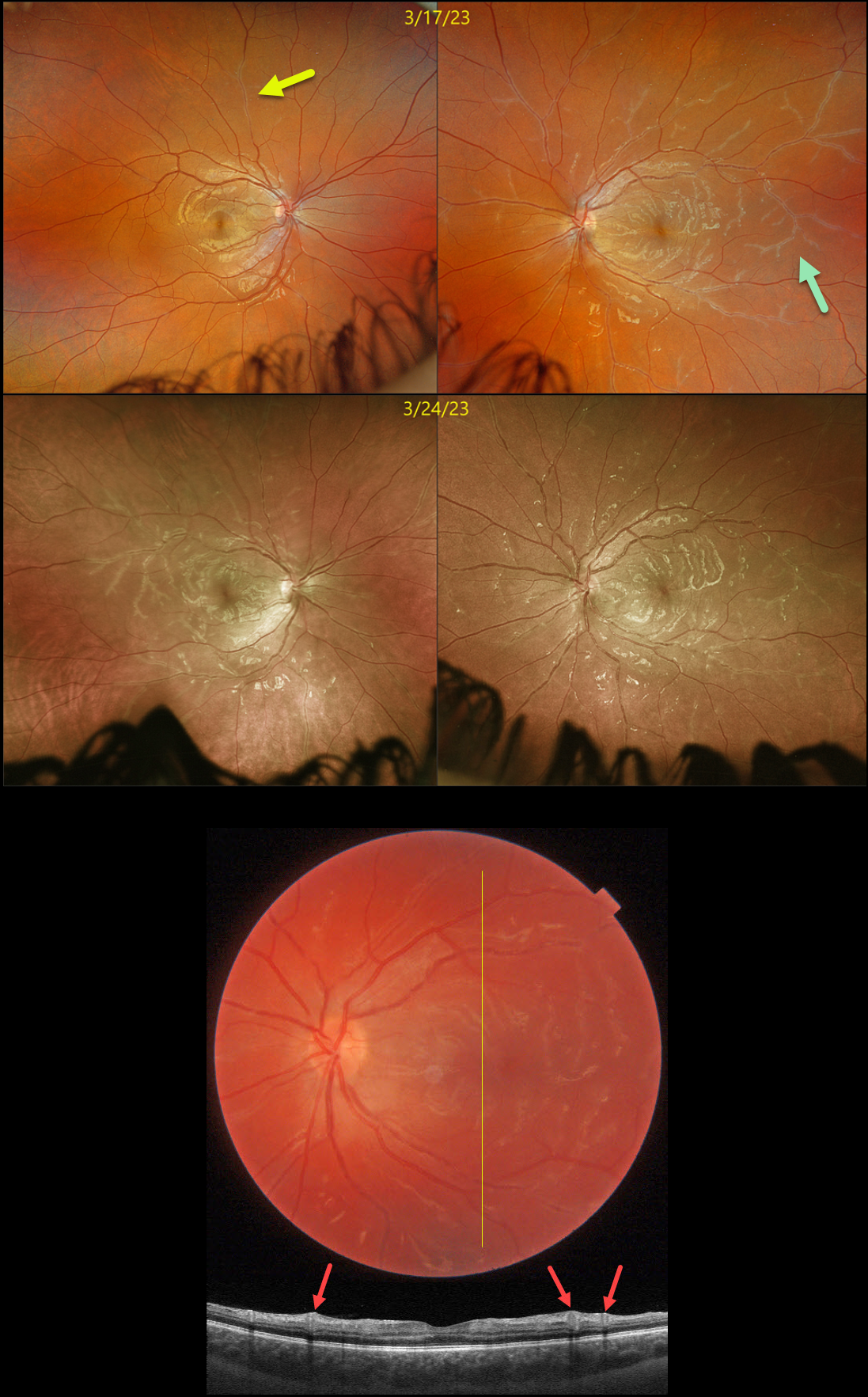
This healthy 16YO female presented with several days of bilateral blurred vision. There was no past ocular or medical history. Vision was 20/25 OD and 20/30 OS. Anterior segments were normal.
Optos imaging of her right eye shows a frosted branch appearance to a retinal vein in the superior midperiphery, with more diffuse vasculitis seen in her left eye.
Swept-source OCT through the left macula shows some hyperreflective inner retinal thickening around the inflamed veins. Bloodwork for Bartonella, syphilis, and sarcoid was ordered.
She returned one week later with new blurred vision in her left eye. Vision was 20/25 OU. Although findings were stable OS, new diffuse frosted branch changes were present in the OD. Her bloodwork returned negative, and she was started on oral prednisone 60mg daily.
Learning Points:
Frosted branch angiitis is a retinal vasculitis that gets its name from the involved vessels resembling branches of a tree that are “frosted” with snow (Kleiner et al, AJO 1988;106:27-34).
Although these findings may be isolated, they are often considered a phenotype found in a multitude of inflammatory conditions, most commonly CMV retinitis. The inflammation is usually very responsive to steroids, and the visual prognosis is generally good, although some patients may develop secondary anterior and posterior segment neovascularization due to widespread ischemia.
FROSTED BRANCH ANGIITIS FROM SARCOIDOSIS
David Kilpatrick
Originally posted on @retina.rocks October 11, 2021
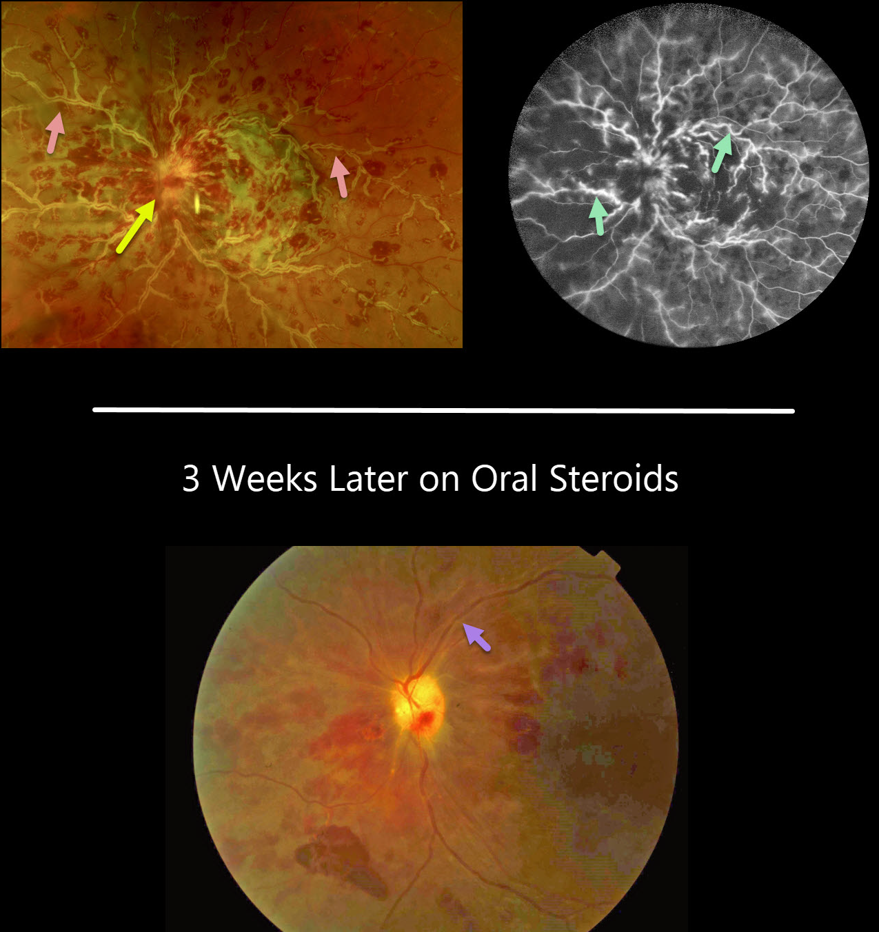
This 34YO African American female presented with several days of painless vision loss in her left eye. Vision was 20/30 OD and HM OS with an afferent defect OS. Anterior segments were normal.
There was mild disc edema OD (not shown) and florid disc edema OS with retinal vasculitis predominantly involving the veins.
On fluorescein angiography, the retinal veins in the left eye were diffusely stained.
On further review of systems, she had a new skin rash on her right forearm, but was otherwise asymptomatic. Testing revealed elevated angiotensin-converting enzyme and lysozyme levels, and a chest X-ray showed bilateral perihilar fullness.
With a working diagnosis of sarcoidosis, she is currently being managed with oral steroids and has been evaluated by both rheumatology and pulmonology.
While her retinal vasculitis has responded well to treatment, her vision remains HM three weeks later.
Learning Points:
Frosted branch angiitis is a retinal vasculitis that gets its name due to the involved vessels resembling the branches of a tree that are “frosted” with snow. Although these findings may be isolated, they are often considered a phenotype found in a multitude of inflammatory conditions, most commonly CMV retinitis.
The inflammation is usually highly responsive to steroids, and the visual prognosis is generally good, although some may develop secondary neovascularization in the anterior and posterior segments due to widespread ischemia.
FROSTED BRANCH ANGIITIS
Originally posted on @retina.rocks March 12, 2021
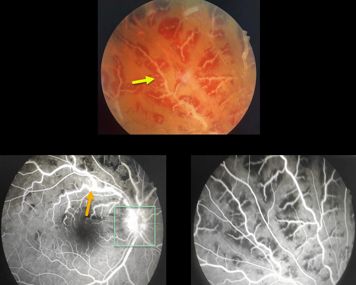
This 19yo presented with counting fingers (CF) vision OD and 20/20 OS.
There is a frosted appearance to the superior retinal veins with surrounding retinal hemorrhages. Fluorescein angiography (FA) shows some vascular staining and optic nerve leakage.
The patient was started on oral steroids, and one week later, the fundus findings had improved significantly (not pictured). Bloodwork came back negative.
Learning Points:
Frosted branch angiitis is a retinal vasculitis that gets its name due to the involved vessels resembling the branches of a tree that are “frosted” with snow.
Although these findings may be isolated, they are often considered a phenotype found in a multitude of inflammatory conditions, most commonly CMV retinitis.
The inflammation is usually highly responsive to steroids, and the visual prognosis is generally good, although some patients may develop secondary neovascularization in the anterior and posterior segments due to widespread ischemia.
SYSTEMIC LUPUS ERYTHEMATOSUS (SLE)
Originally posted on @retina.rocks December 29, 2020
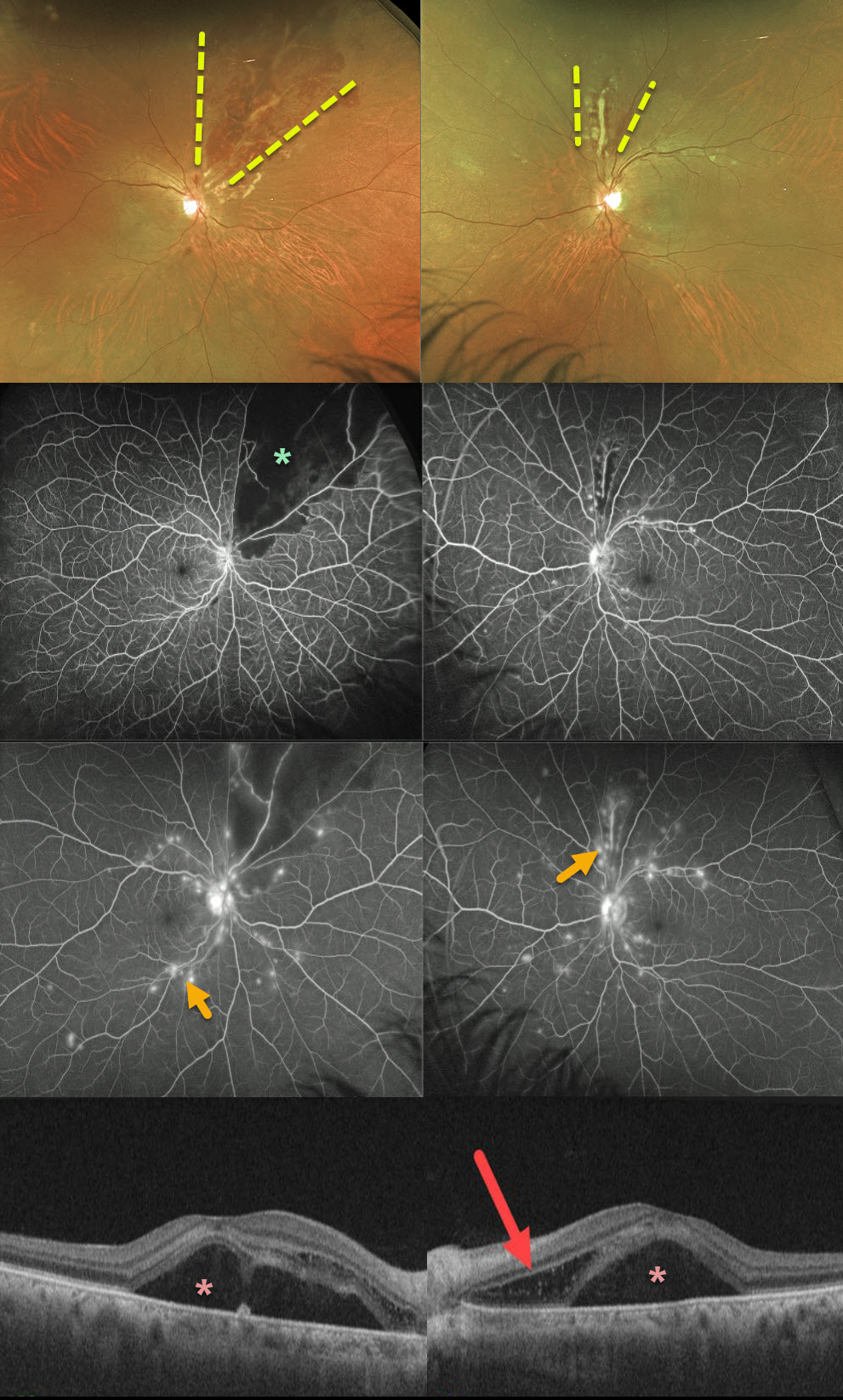
This 52YO female presented with severe bilateral vision loss (20/400 OD, counting fingers OS) following a several-week gastrointestinal illness with 60-pound weight loss.
She has bilateral areas of hemorrhagic retinal vasculitis, with areas of ischemia and leakage seen on fluorescein angiography. OCT shows macular subretinal fluid and a bacillary detachment in the left macula.
She had been hospitalized recently for hypertension and kidney failure and was diagnosed with a previously asymptomatic stroke. Extensive blood work was negative, and she denied symptoms of Behcet’s.
We suspected an underlying rheumatologic cause, and she was subsequently diagnosed with severe systemic lupus erythematosus (SLE) vasculitis.
She was started on high-dose oral prednisone followed by Cytoxan and hydroxychloroquine. Her ocular findings rapidly improved, and at her last examination 4 months later, vision had improved to 20/40 OD and 20/100 OS.
Learning Points:
SLE-associated retinopathy more typically appears as hypertensive retinopathy (bilateral nerve fiber layer infarcts and retinal hemorrhages), often in patients with cerebral vasculitis. Purtscher-like retinopathy can also be seen.


