

Fundus Albipunctatus
<< Back to Cases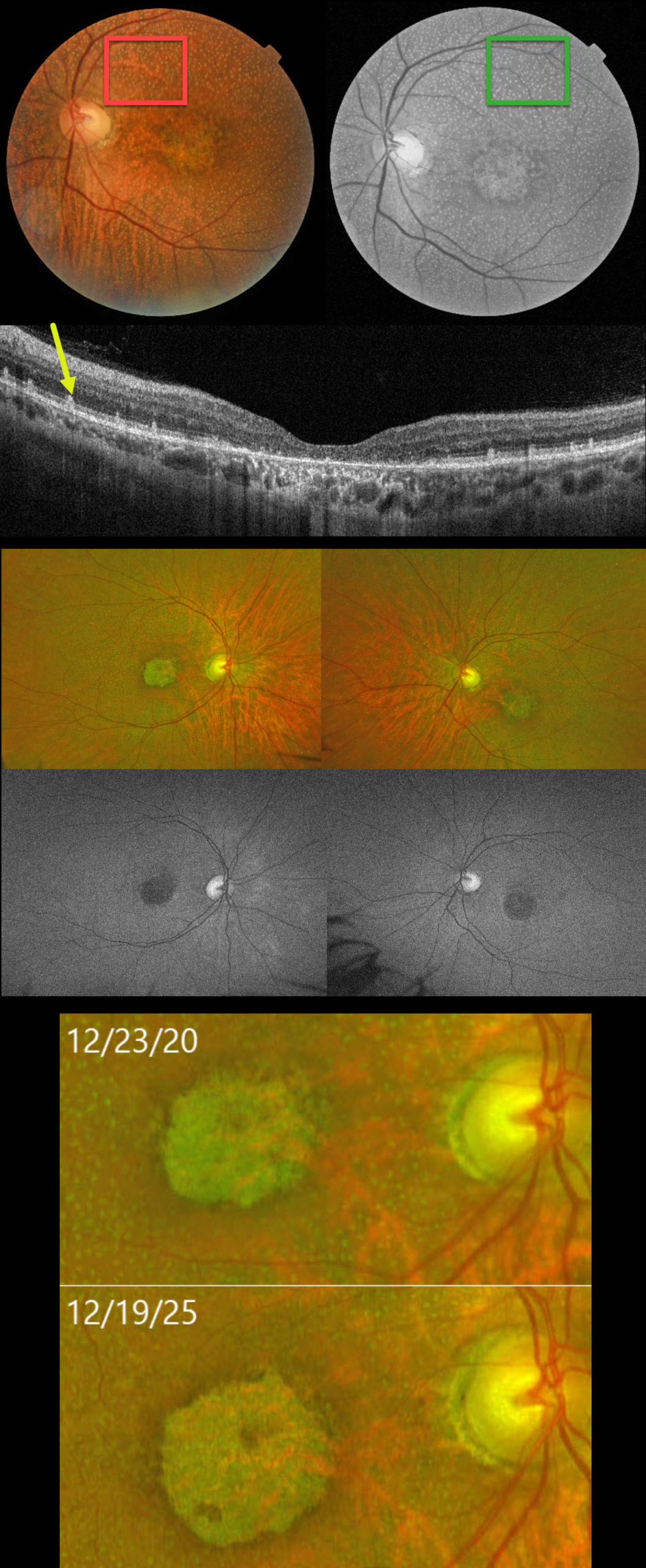
RDH5-ASSOCIATED DISEASE WITH MACULAR ATROPHY
João Pedro Marques
Originally posted on @retina.rocks February 17, 2026
This 58YO female was initially seen by us on 8/6/18 with a prior diagnosis of geographic atrophy and subretinal drusenoid deposits. She described a long-standing history of nyctalopia and poor vision. Vision was 20/640 OU.
Color photography shows innumerable subretinal yellow-white dots extending from the periphery of each macula into the midperiphery. These dots are most evident on red-free imaging. Bilateral foveal macular atrophy is noted. OCT scanning shows central outer retinal atrophy with more peripheral hyperreflective outer retinal deposits, corresponding to the funduscopic dots, which extend into the outer nuclear layer. Optos fundus autofluorescence (FAF) is hazy due to a decreased autofluorescent signal from each fundus. Genetic testing was homozygous for a class IV variant in the RDH5 gene.
When last examined on 12/31/25, vision was 20/800 OU. Her funduscopic findings were stable except for mild enlargement of the macular atrophy.
Learning Points:
Fundus albipunctatus (FA), an autosomal recessive form of congenital stationary night blindness, presents with scattered yellow-white dots that spare the central macula and extend into the midperipheries. Patients experience stable night blindness with normal central acuity. It is most commonly caused by homozygous mutations in the RDH5 gene, which encodes the retinol dehydrogenase protein. This protein is found in the RPE and helps recycle vitamin A in the visual cycle. The white dots are thought to contain 11-cis retinal precursors. This results in decreased retinoid-derived fluorophores, accounting for the diminished FAF in these eyes (Sergouniotis et al, Ophthalmology 2011;118:1161-1670).
A similar phenotype to FA, but with notable progression and a rod-cone dystrophy ERG, is retinitis punctata albescens (RPA). It can be associated with mutations in several genes, including RLBP1, RHO, RDS, RDH5, and RPE65. RDH5 can be associated with both FA and RPA. Some cases initially diagnosed as FA progress and develop macular atrophy (Bianco et al, AJO 2024;267:160-171), as in our patient. We therefore believe that RDH5-associated disease is a better term, as it encompasses the typical FA and RPA phenotypes associated with RDH5.
FUNDUS ALBIPUNCTATUS
Originally posted on @retina.rocks August 26, 2024
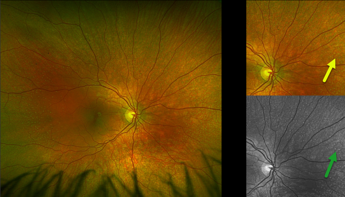
This healthy 10YO girl was referred for asymptomatic findings on retinal examination. There was no ocular family history. Vision was 20/40 OU.
Optos color RG imaging of the right eye shows innumerable subretinal yellow-white dots extending from outside each macula into the midperiphery. These dots are most evident in green-channel imaging. Identical findings were noted in the left eye (not shown). Genetic testing revealed double heterozygous RDH5 mutations.
Learning Points:
Fundus albipunctatus, an autosomal recessive form of congenital stationary night blindness, presents with scattered yellow-white dots that extend into the midperipheries, sparing the macula. Patients experience stable night blindness with normal central acuity. It is caused by a mutation in the RDH gene, which encodes the retinol dehydrogenase protein. This protein, found in the RPE helps recycle vitamin A in the visual cycle, and the white dots are thought to contain 11-cis retinal precursors. This results in decreased retinoid-derived fluorophores, accounting for the diminished FAF in these eyes (Sergouniotis et al, Ophthalmology 2011;118:1161-1670).
FUNDUS ALBIPUNCTATUS
Originally posted on @retina.rocks November 5, 2020
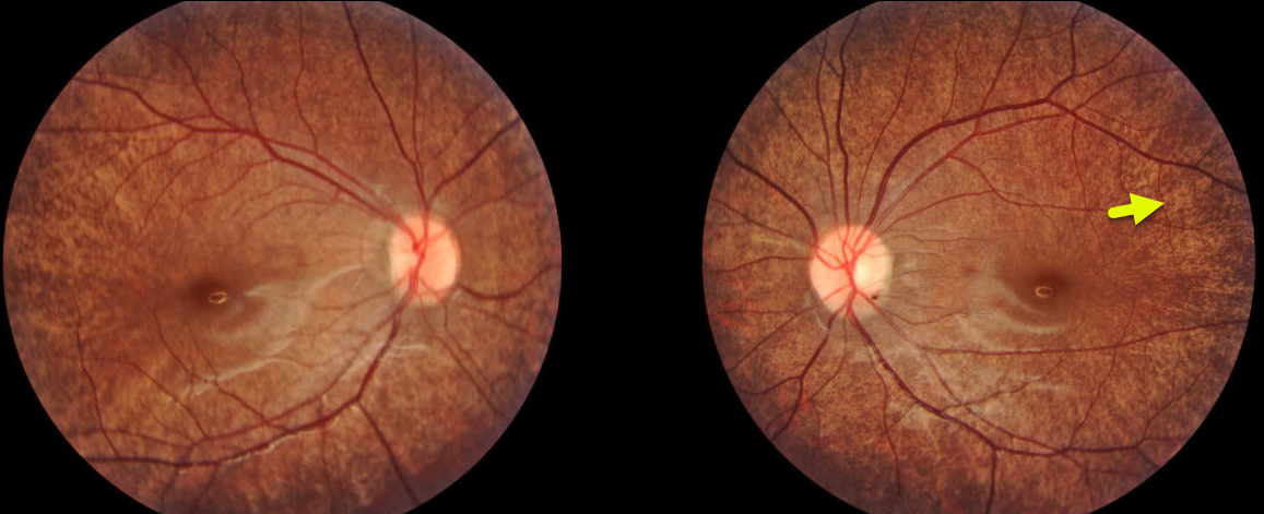
This patient has fundus albipunctatus, a rare autosomal recessive form of congenital stationary night blindness. The characteristic numerous small white-yellow specks spare the fovea.
Learning Points:
Fundus albipunctatus is caused by a mutation in the RDH5 gene, which codes for retinol dehydrogenase, which converts 11-cis retinol to 11-cis retinal within the RPE.
The optic disc, retinal vessels, visual acuity, visual fields, and color vision are usually normal. Electroretinograms (ERG) are typically depressed but normalize after prolonged dark adaptation, in contrast with retinitis punctata albescens.


