

16 Funny Pictures
<< Back to Cases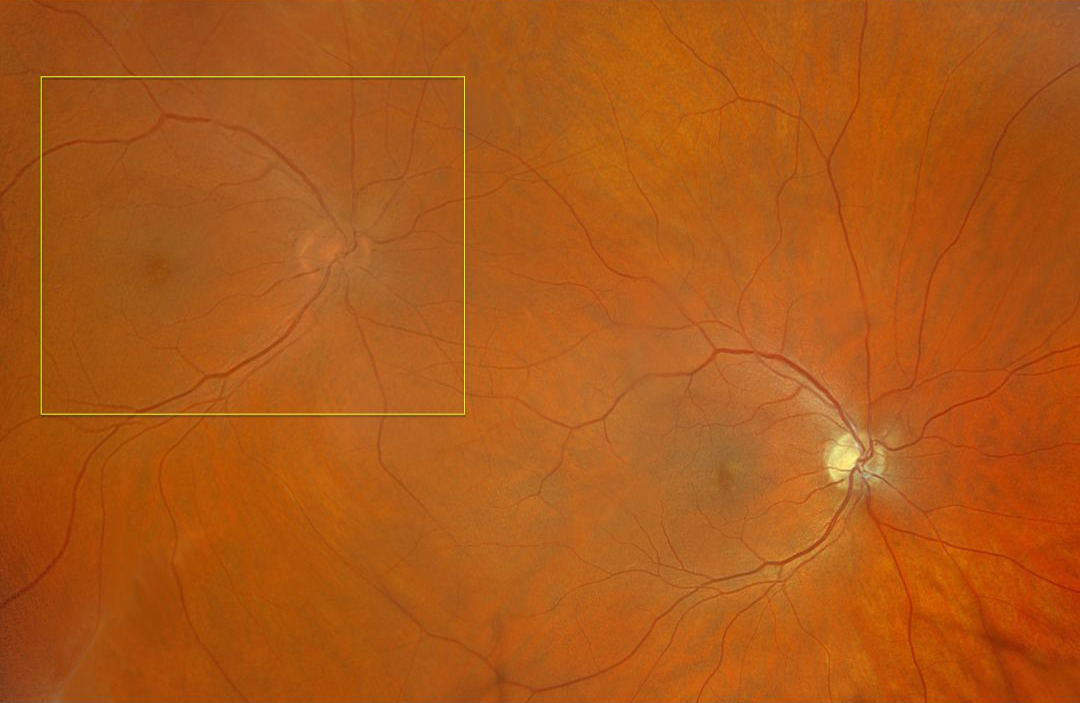
FULL-THICKNESS EYEWALL TRANSPLANT WITH INADVERTENT GRAFT INVERSION
Originally posted on @retina.rocks April 1, 2026
This healthy 43YO male fighter pilot developed a complex corneal laceration in his left eye. Despite multiple surgical procedures, he was left with an opaque cornea and an inability to fly due to the lack of normal binocular vision. Full-thickness eyewall transplantation from his normal left posterior segment into his right temporal posterior segment was therefore performed.
Intraoperatively, the dyslexic surgeon inadvertently inverted the allograft, resulting in the donor graft being inverted 180 degrees vertically. Optos color RGB imaging shows the donor nerve and macula in the right superotemporal midperiphery. Note that the disc should have been placed temporal to the donor macula, not nasal. Although surgically successful, this left the patient with monocular diplopia instead of the desired monocular binocular stereopsis. He remains grounded, and his case is still mired in litigation.
And happy April Fool’s Day!
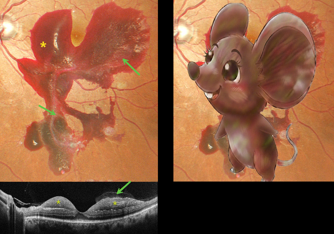
MOUSE MACULOPATHY AKA PROLIFERATIVE DIABETIC RETINOPATHY (PDR) WITH SUBHYALOID HEMORRHAGE
Originally posted on @retina.rocks February 24, 2026
This 48YO male with a 15-year history of poorly controlled type 2 diabetes presented with one day of blurred vision in his left eye. Vision was 20/20 bilaterally.
Pseudocolor SLO imaging shows a large macular hemorrhage, sparing the foveal center, which extends into the midperiphery. The hemorrhage is mostly subhyaloid with some breakthrough blood into the vitreous. The blood compartments are well visualized on OCT.
Learning Points:
The normal vitreous undergoes a decades-long process of orchestrated degeneration, including liquefaction and gradual separation from the retinal surface. The posterior vitreous detachment (PVD) begins in the perifoveal retina early in life, followed by a foveal PVD, then a peripheral PVD, and then finally a complete PVD when the vitreous separates from the optic nerve (Johnson AJO 2010;149:371-382). Although a diabetic subhyaloid hemorrhage can extend throughout the macula, our patient’s findings illustrate the normal increase in vitreofoveal adhesion.
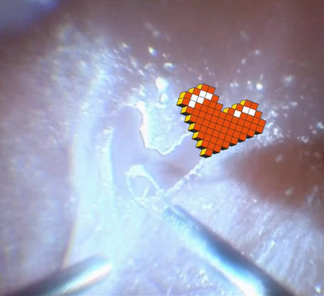
INTRAOPERATIVE HEART DURING TRIAMCINOLONE-ASSISTED MEMBRANE PEELING
The European VitreoRetinal Society (EVRS), Claudio Brancato and Gregorio Lo Giudice
Originally posted on @retina.rocks February 13, 2026
This 75YO male presented with hand motion vision in his right eye from a rhegmatogenous retinal detachment (RRD). Intraoperatively, triamcinolone was injected to stain the posterior hyaloid. During FINESSE Flex Loop-assisted membrane peeling, some love came back at us from the patient’s retina. The retina remained attached postoperatively, and vision improved to 20/50.
Learning Points:
Valentine’s Day traces its origins to a blend of ancient Roman customs, early Christian tradition, and the imaginative influence of medieval literature. What began as mid-February festivals gradually became associated with figures named Valentine, remembered for acts of compassion and steadfast conviction. Over time, poets such as Chaucer helped link the date with courtly love, giving rise to the exchange of affectionate notes and tokens. By the 18th and 19th centuries, these gestures evolved into the expensive cards, flowers, and symbols of romance that define the holiday today, transforming a complex historical tapestry into a widely celebrated expression of affection.
So, for you retinal surgeons out there, what could be a more romantic Valentine’s Day gift than offering to vitrectomize your loved one?
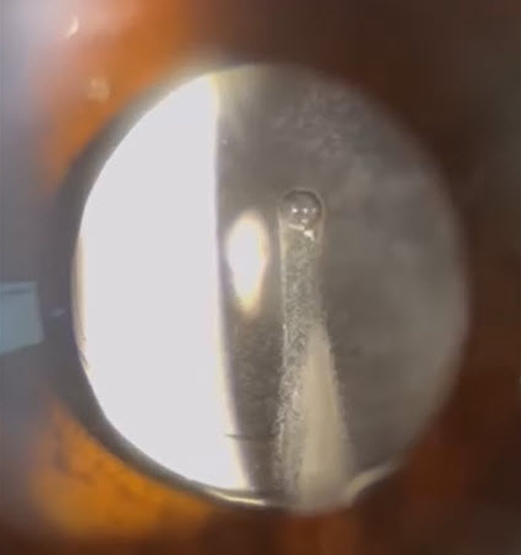
TRIAMCINOLONE-MOXIFLOXACIN (TriMoxi)
Adam Mueller, Benjamin Conkright, and Andrew Steele
Originally posted on @retina.rocks October 31, 2025
This 64YO female presented for her one-day post-dropless cataract surgery with some minor irritation and floaters. Vision without correction was 20/25+. Unbeknownst to our happy patient, a sinister ghoul was lurking behind her perfectly centered intraocular implant.
Our practice, along with many others, offers ‘no drop’ cataract surgery as a convenience to our patients. Instead of needing to take postoperative antibiotic and steroid drops, 0.2cc of TriMoxi (triamcinolone acetate 15mg/cc with moxifloxacin 1mg/cc) is injected into the anterior inferior vitreous at the end of the cataract surgery. Although this liquid usually remains well outside the visual axis, patients will often experience mild floaters perioperatively. Rarely, as in this patient, the liquid disperses throughout the vitreous cavity, causing significant transient floaters.
INTRAOCULAR FOREIGN BODY?
Originally posted on @retina.rocks April 1, 2025
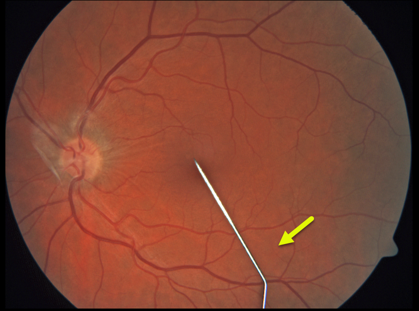
This healthy 24YO goth female presented with severe pain and a floater in her left eye immediately following a failed intraocular tattoo procedure earlier that day. Vision was 20/40.
Color photography shows a posteriorly dislocated intraocular tattoo needle lying on the inferior macular surface. Adherent vitreous is noted on the optic nerve. She was immediately taken to surgery for the removal of the metallic intraocular foreign body (IOFB).
Learning Points:
HAPPY APRIL FOOLS!!
Although this case is a fake-out from an old fundus camera utilizing a fixation stick that appears as a pseudo-IOFB, cosmetic tattooing is unfortunately a very real thing, including corneal, subconjunctival, and episcleral tattoos (https://eyewiki.org/Eye_Tattooing).
The procedure is performed by either the individual performing it on themselves or non-ophthalmic trained individuals and is obviously associated with numerous complications, including globe penetration, traumatic cataract, retinal detachment, endophthalmitis, and orbital cellulitis. Ink-related complications include dye migration into the anterior chamber, where it can coat the lens and posterior corneal surface and cause secondary
POSTERIOR VITREOUS DETACHMENT
Originally posted on @retina.rocks February 14, 2025
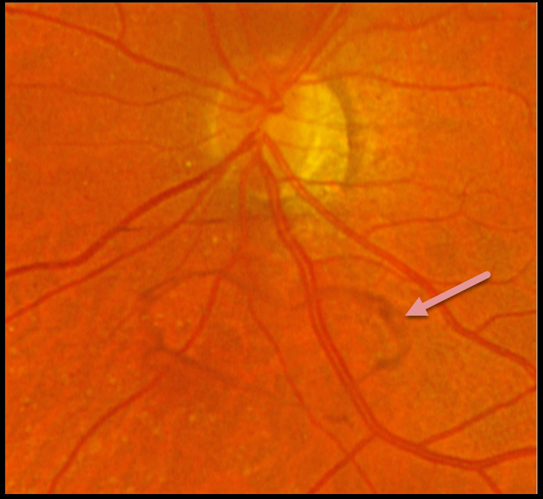
This 61YOF put a smile on our face by presenting with this asymptomatic heart-shaped Weiss ring just inferior to her left nerve.
HAPPY VALENTINE’S DAY!
MICKEY MOUSE MACULOPATHY
Originally posted on @retina.rocks November 13, 2024
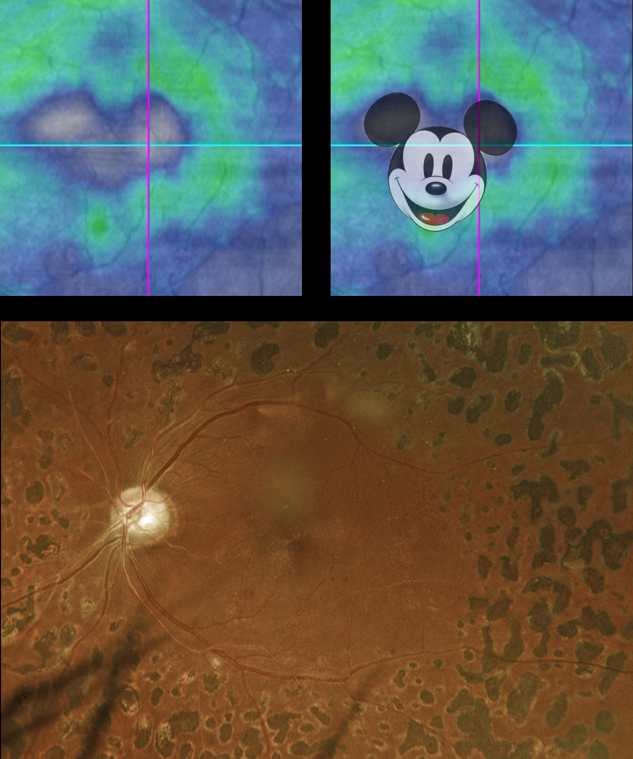
This 52YO diabetic male rodent has a history of proliferative diabetic retinopathy and full panretinal photocoagulation (PRP). Vision was stable at 20/30.
Optos color RGB imaging shows full PRP scarring. The macula is clinically ischemic with some vascular sheathing. Triton swept-source OCT shows nasal and central atrophy consistent with Mickey Mouse Maculopathy (MMM).
Created by Walt Disney and Ub Iwerks, Mickey Mouse celebrates his birthday on November 18th. This date commemorates his first appearance in the animated short film “Steamboat Willie” in 1928, famous for Mickey’s debut and for its synchronized sound.
On January 1, 2024, the famous mouse entered the public domain, allowing anyone to use this version of the character in their creative works without needing Disney’s permission or payment. However, Disney still retains the copyright on any subsequent variations of Mickey until they reach the 95-year mark.
HAPPY WORLD SMILE DAY
Pier Luigi Guerin
Originally posted on @retina.rocks October 4, 2024
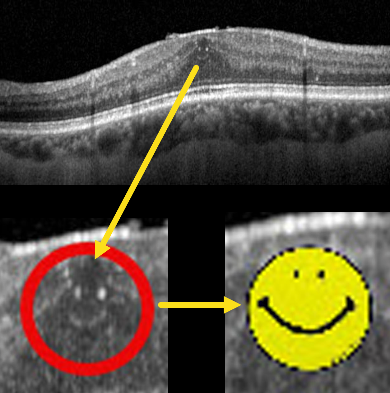
This 70YO male with type 2 diabetes presented with these asymptomatic OCT findings that made our day. Vision was 20/25.
World Smile Day is celebrated on the first Friday of October each year, a tradition that began in 1999. The day was created by Harvey Ball, the commercial artist who designed the iconic yellow smiley face. Ball was concerned that the over-commercialization of his symbol would dilute its original intent of spreading goodwill and cheer. To counteract this, he established World Smile Day to remind people of the power of a smile and to encourage acts of kindness around the world.
The day is marked by various activities that promote smiling and kindness. People are encouraged to perform acts of kindness and to smile more, spreading positivity and happiness. The Harvey Ball World Smile Foundation, established after Ball’s passing in 2001, continues to sponsor the day and organize events. These events range from community activities to global initiatives, all aimed at creating smiles and fostering a spirit of generosity and joy.
So, perform some random acts of kindness today and make the world a better place because you’re in it.
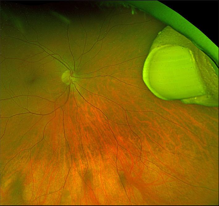
INTRAOCULAR THUMB
Originally posted on @retina.rocks April 1, 2024
This healthy 7YO boy was playing with his older brother while watching their favorite Three Stooges bit about Niagara Falls.
Horseplay literally turned into tragedy when they presented to the emergency room with the older brother’s hand impaled into his younger brother’s right eye.
Both were immediately taken to surgery, where the attacking brother’s thumb was amputated at the distal phalanx, then removed internally via an open sky pars plana vitrectomy approach. Six months later, following removal of the silicone oil, vision was 20/30.
And Happy April Fool’s Day!
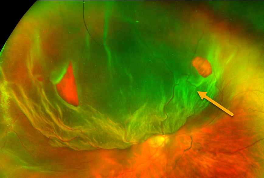
JACK O- LANTERN DETACHMENT
Sandeep Kumar
Originally posted on @retina.rocks October 31, 2023
This 45YO female presented with a superior Jack-O-Lantern macula-off retinal detachment in her right eye. The detachment was successfully repaired with vitrectomy and C3F8 tamponade.
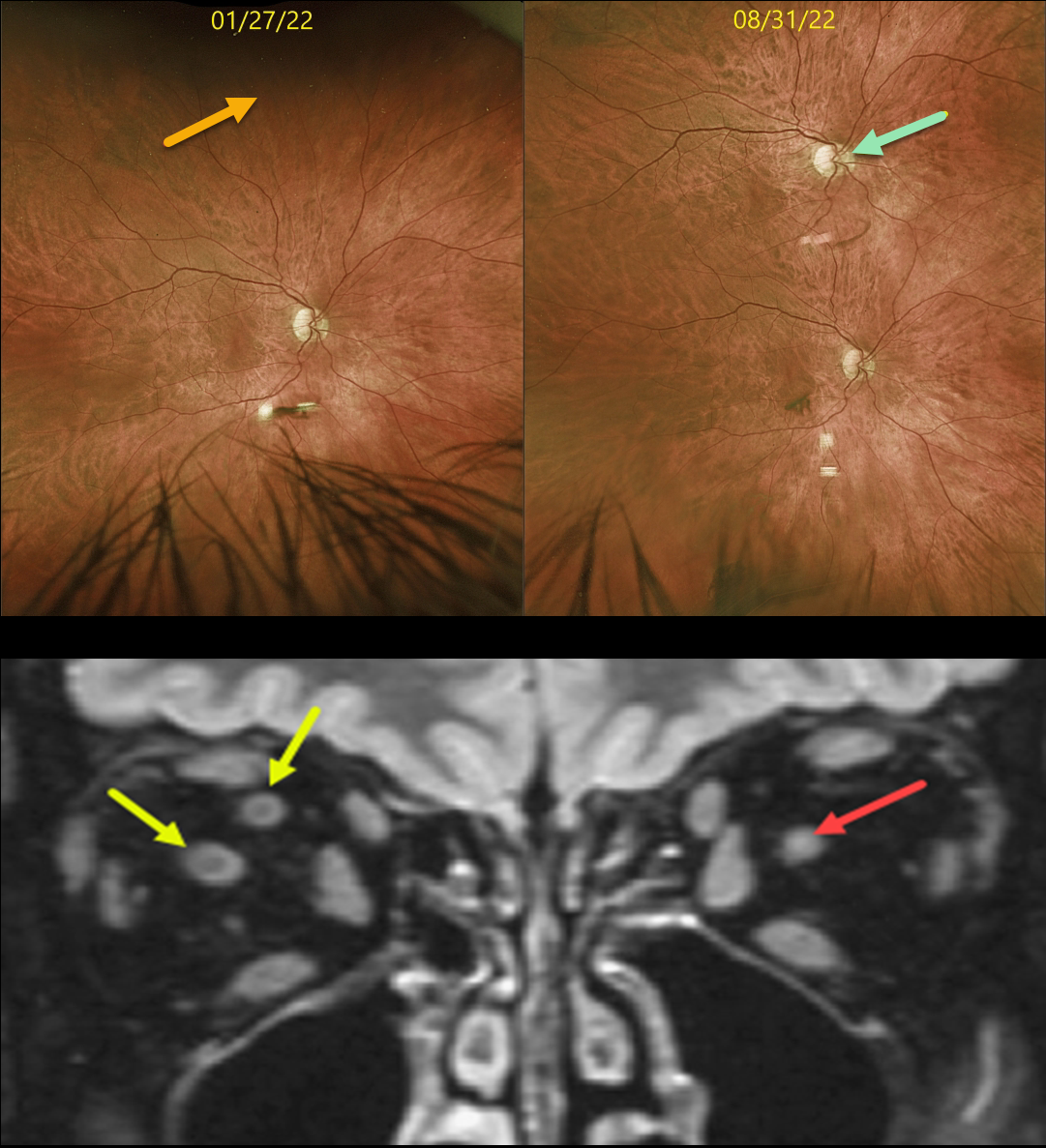
DUPLICATED OPTIC NERVE
Originally posted on @retina.rocks March 31, 2023
This 61YO diabetic male has been followed yearly for routine retinopathy screenings. On 1/27/22, vision was 20/20 with a normal right fundus. Note that the superior periphery is also normal.
On 8/31/22, he returned complaining of a new inferotemporal peripheral scotoma with monocular vertical diplopia in this eye. Vision was 20/20 centrally and 20/20 inferiorly. A new optic nerve and macula had developed in the interim. These findings were confirmed on MRI scanning, which revealed a single nerve OS and two nerves OD in the retro-orbital space.
Learning Points:
Duplication of the optic nerve is a non-heritable developmental abnormality with an extremely low prevalence of about 1 per 23.5 million live births, and is a rare cause of monocular diplopia. To the best of our knowledge, ours is the first such known de novo case.
AND HAPPY APRIL FOOL’S DAY 2023!
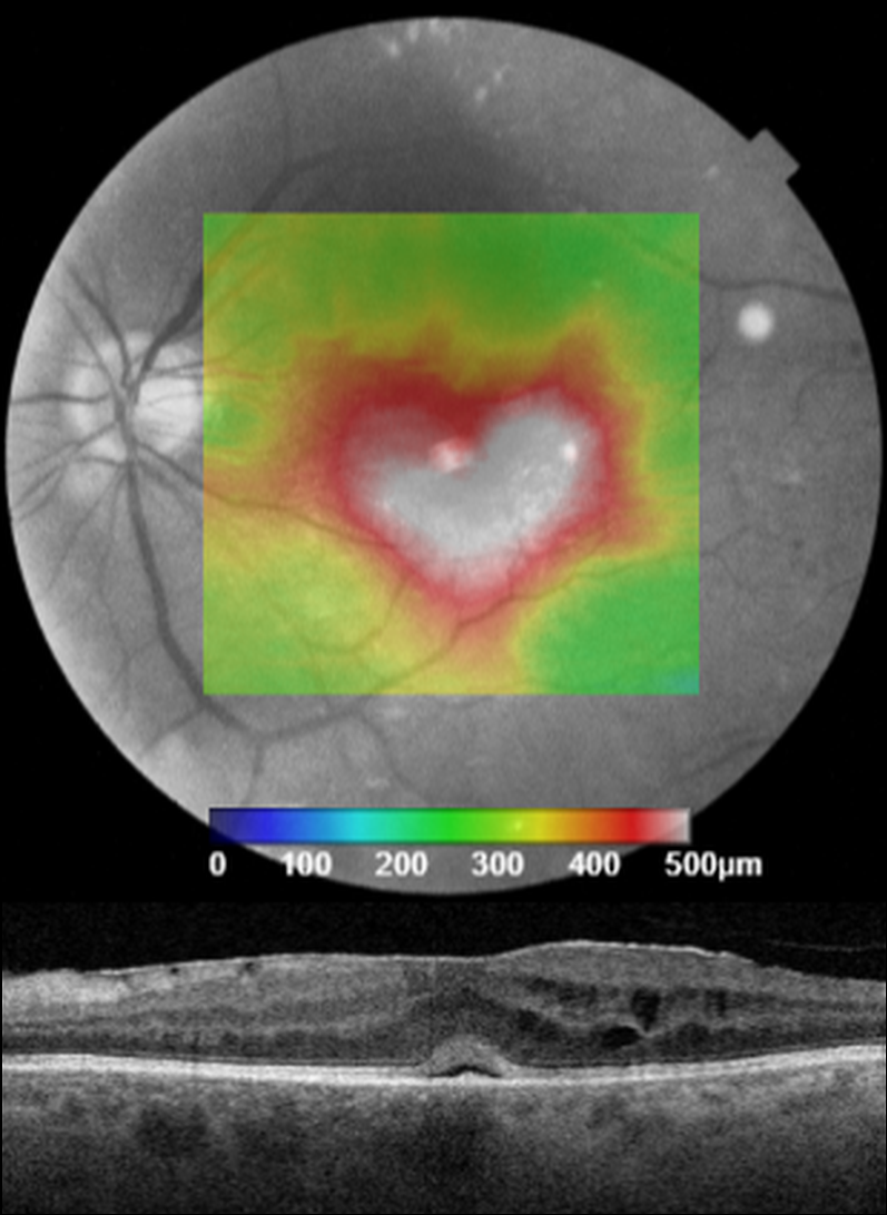
VALENTINE MACULAR EDEMA
Originally posted on @retina.rocks February 14, 2023
This patient presented with 20/200 vision from center-involved diabetic macular edema.
Unfortunately, we don’t have any further clinical information for this patient, but we still wish everyone a ‘heart-felt’ Happy Valentine’s Day!
WOLVERINE RETINOPATHY
Originally posted on @retina.rocks May 10, 2022
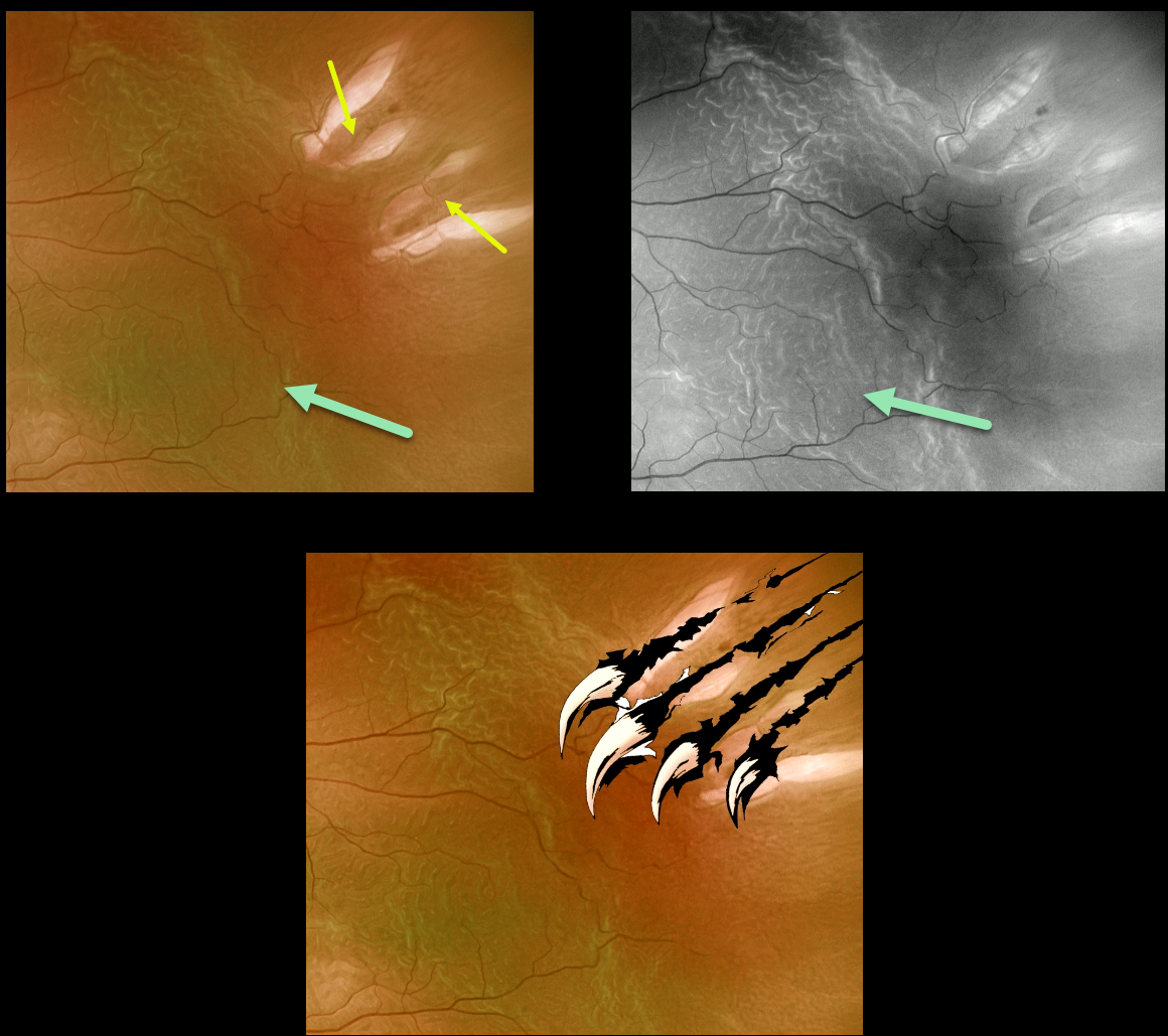
This 72YO male presented with a macula-off rhegmatogenous retinal detachment (RRD).
Optos imaging gives the illusion of four breaks, although in actuality, there are only two tractional tears. The hydrated outer retinal folds are best visualized on green channel imaging.
Vision was 20/60, and the retina was successfully reattached with emergent pars plana vitrectomy.
SEPHIROTH RETINOPATHY
Originally posted on @retina.rocks March 25, 2022
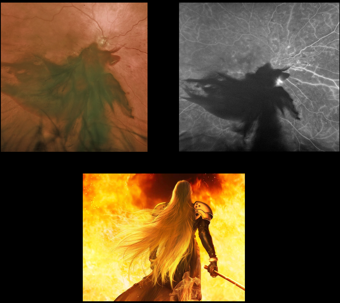
We all know it’s a large subhyaloid hemorrhage from proliferative diabetic retinopathy, but check your imagination and tell us what you see.
We see Sephiroth from Final Fantasy!
CONGENITAL HYPERTROPHY OF THE RPE (CHRPE)
Originally posted on @retina.rocks December 2, 2021
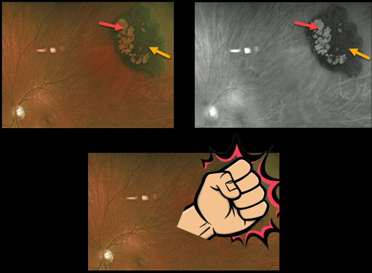
This 25YO female presented with a classic asymptomatic solitary congenital hypertrophy of the RPE (CHRPE). It almost resembles a fist!
The borders are well demarcated, with variable coal-black coloration and lacunae of depigmentation.
Learning Points:
Although CHRPE lesions can enlarge with time (see Shields et al, Ophthalmology 2003;110:1968-1973), they carry virtually no malignant potential. Histologic RPE hypertrophy has several clinical presentations.
The typical isolated CHPRE lesions (today’s post) and bear tracks are not associated with intestinal polyps or cancer, and thus require no systemic evaluation.
Only the multiple, small comet-shaped lesions are part of the familial polyposis spectrum.
OZURDEX GHOST
Originally posted on @retina.rocks November 26, 2021
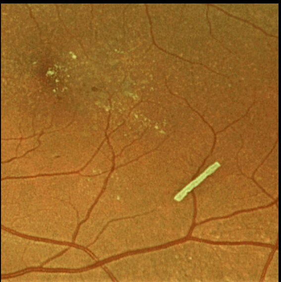
This 71YO female is getting Ozurdex intravitreal injections every 2 months to control her macular edema.
Learning Points:
The Ozurdex implant slowly releases the steroid and eventually totally absorbs, with the clinical effect usually lasting about 2 to 4 months.
The ghost-like remnants can persist for many months (see Kim et al, Retina 2020;40;2226-2231).
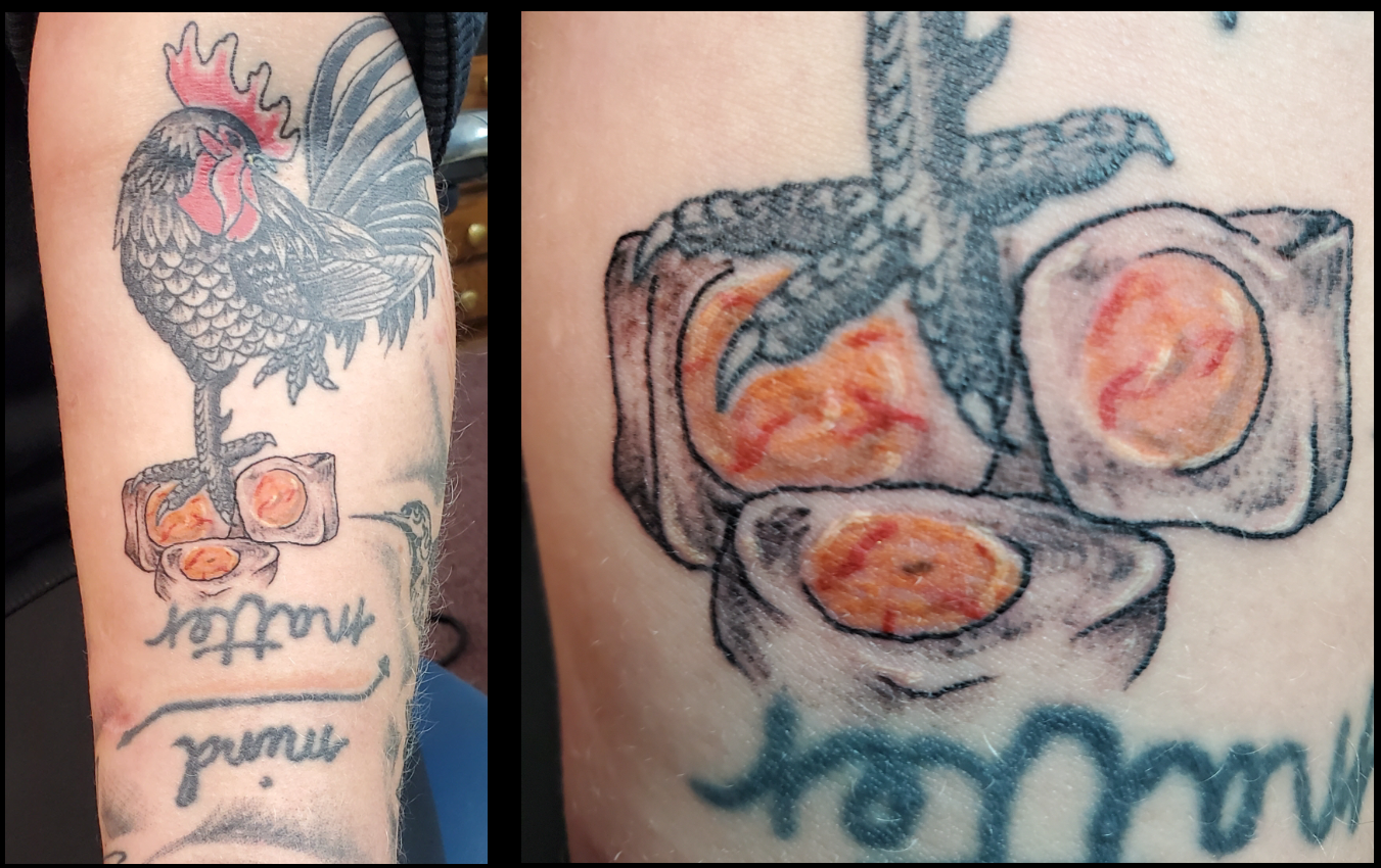
OUR FAVORITE PATIENT
Originally posted on @retina.rocks November 21, 2021
This 38YO female has always been our favorite patient, especially after she showed up for her recent examination with this one-day old Retina Rocks tattoo!
Send us your Retina Rocks tattoo, and as long as it is on a visible part of your body, we will send you a $5 Starbucks gift card!!
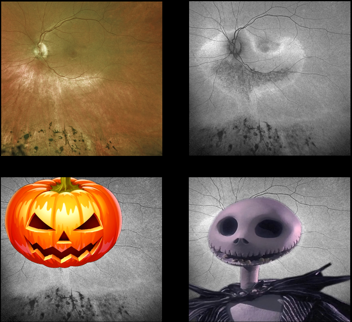
HALLOWEEN RETINOPATHY
Originally posted on @retina.rocks October 29, 2021
This is a patient with retinitis pigmentosa, but do you see Jack O’Lantern or Jack Skellington? Maybe something else spooky?
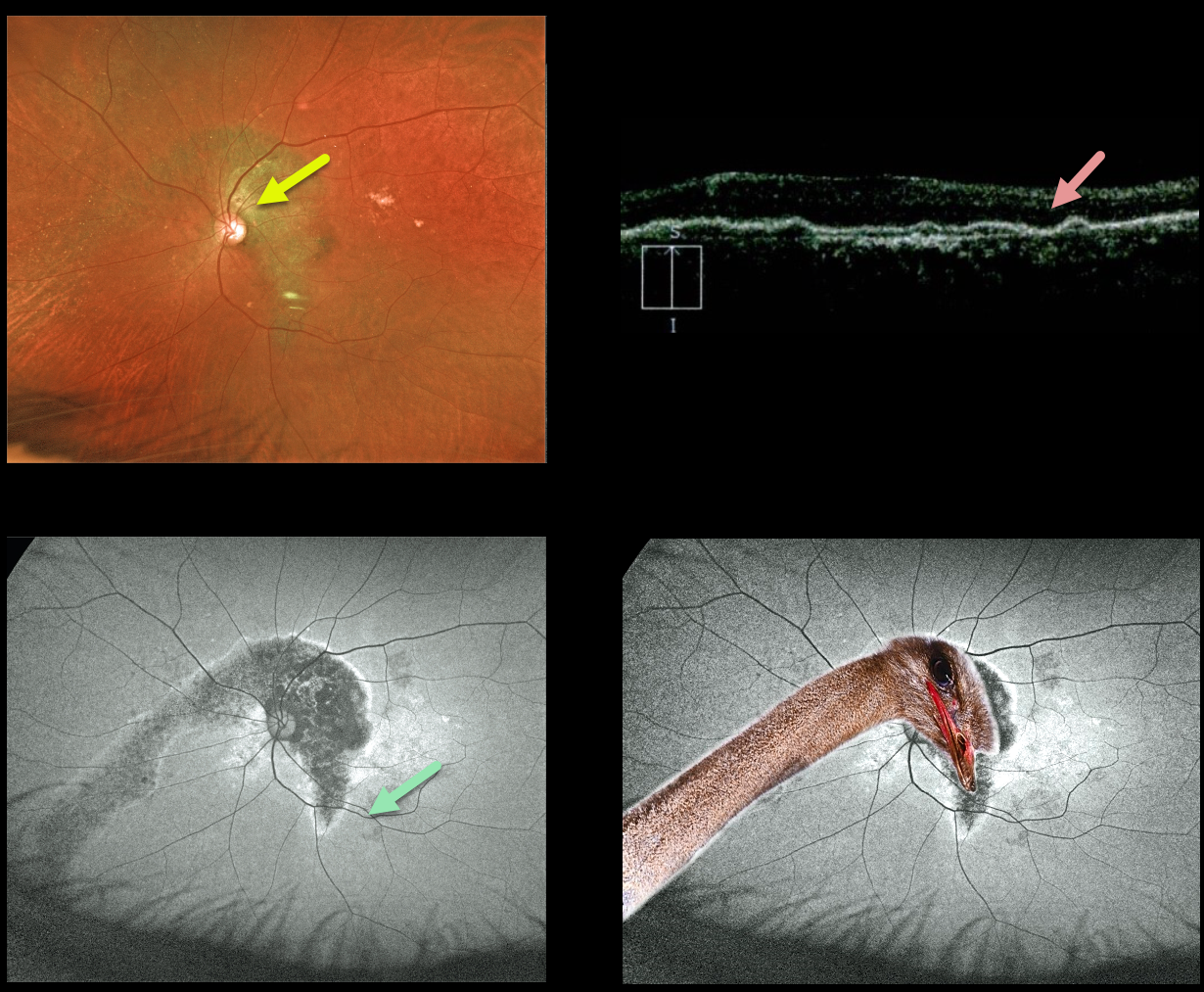
OSTRICH MACULOPATHY
Originally posted on @retina.rocks September 24, 2021
This 79YO male has counting-fingers vision in his left eye, likely due to complications of chronic central serous retinopathy.
Optos color imaging shows increased subretinal pigmentation extending around the nerve superiorly and into the macula and inferonasal periphery.
Fundus autofluorescence (FAF) more dramatically shows these changes as hypo-FAF. OCT scanning shows diffuse thinning of the overlying neurosensory retina.
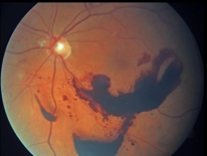
MERMAID MACULOPATHY
Originally posted on @retina.rocks September 8, 2021
This 55YO female with a history of proliferative diabetic retinopathy presented with a symptomatic preretinal hemorrhage. Vision was 20/50.
You don’t need to have an overly active imagination to see this rare finding of mermaid maculopathy.
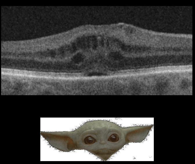
BABY YODA MACULOPATHY
Originally posted on @retina.rocks May 4, 2021
In a galaxy far, far away, this wife of Retina Rock’s founder had an unexpected visitor following her cataract surgery.
Grogu, otherwise known as Baby Yoda or The Child, decided to take refuge within her retina as cystoid macular edema (CME). Thankfully, for the mental health of both the patient and her retinal specialist husband, the CME resolved following topical steroids and non-steroidals.
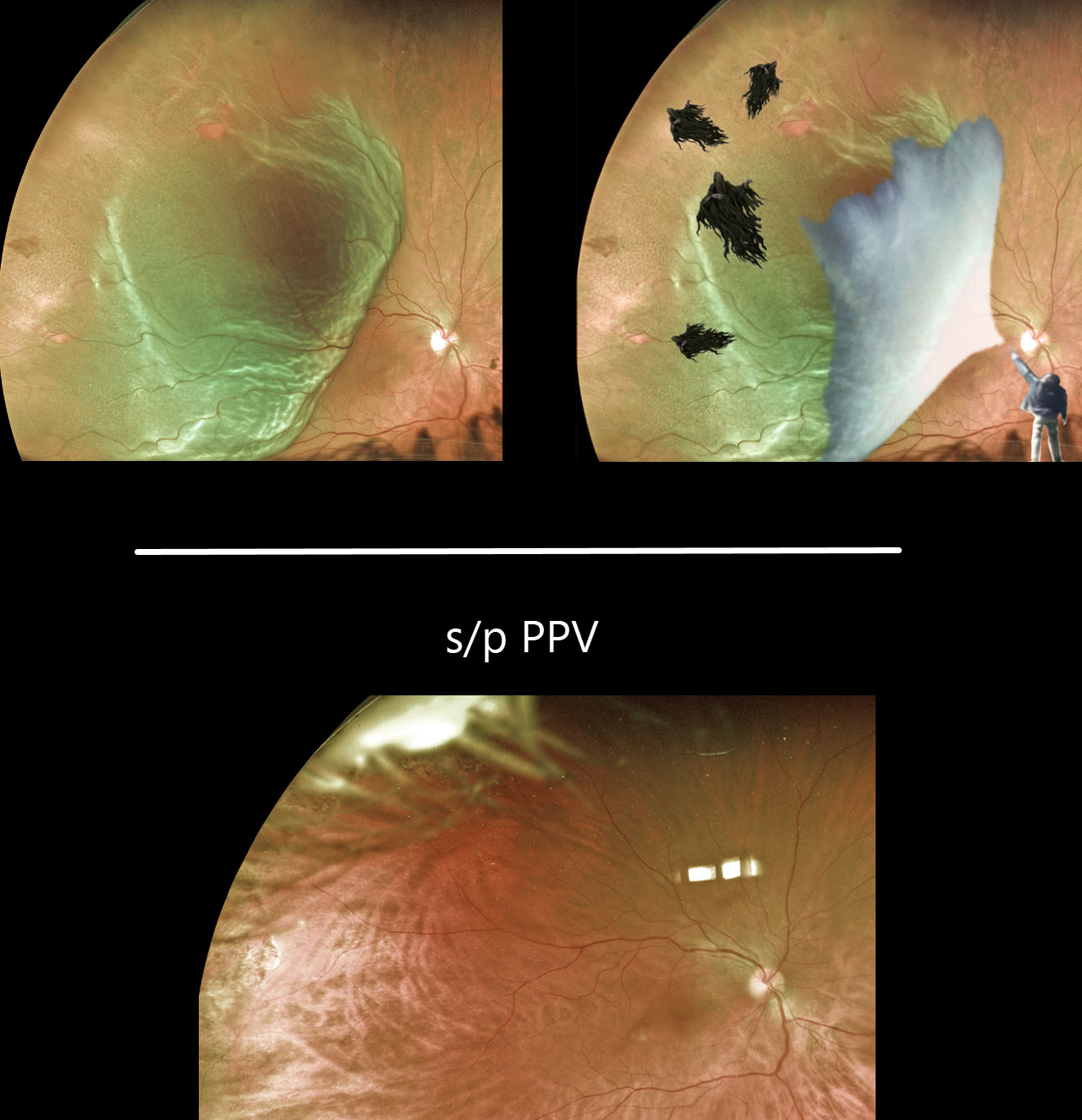
DEMENTOR DETACHMENT
Originally posted on @retina.rocks March 22, 2021
Multiple temporal retinal tears are visible in this superotemporal macula-on rhegmatogenous retinal detachment. Yet it appears to be held off like Harry Potter’s Dementors.
We happen to have a powerful wizard, too.
Just look at the beautifully attached retina after a successful pars plana vitrectomy!
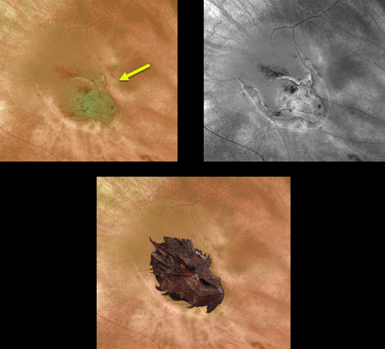
DRAGON RETINOPATHY
Originally posted on @retina.rocks February 1, 2021
The fiery dragon decided to make an appearance in the back of this patient’s eye. This dangerous predator caused its human prey to experience large black floaters and flashing lights.
Fortunately, the beast, aka retinal tear, was vanquished with an in-office barrier laser.
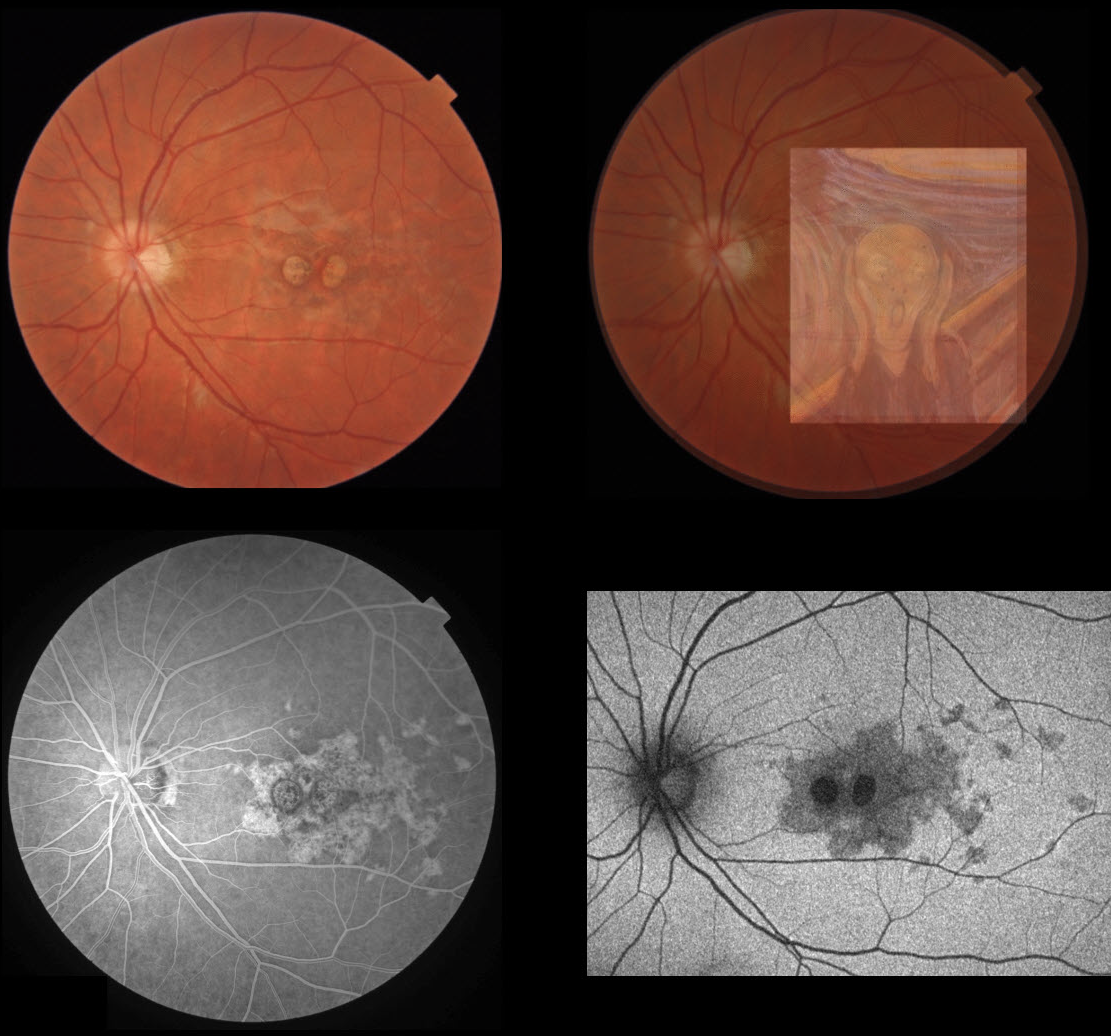
SCREAM MACULOPATHY
Originally posted on @retina.rocks January 1, 2021
This 18YO patient presented in August 2019 (over 6 months before the COVID epidemic rocked our world) with these unusual idiopathic asymptomatic macular findings.
But isn’t it prescient that his macula eerily revealed a strikingly natural rendition of Edvard Munch’s 1893 ‘The Scream of Nature’?
If only we could have recognized what his macula was literally screaming out to us, we could have alerted the authorities and maybe things would have turned out differently for all of us.
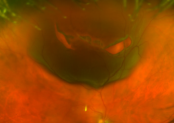
HALLOWEEN RETINOPATHY
Originally posted on @retina.rocks October 30, 2020
This patient presented with a jack-o’-lantern retinal detachment 2 weeks before Halloween.
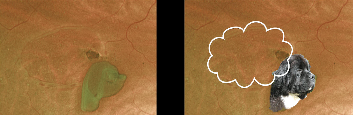
DOGGONE HOLE
Originally posted on @retina.rocks September 19, 2020
What an interesting operculated retinal break. What do you think this dog is thinking?
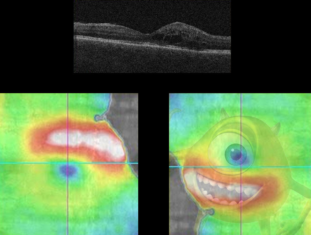
MIKE WAZOWSKI MACULOPATHY
Originally posted on @retina.rocks August 28, 2020
Here we see a patient with the ever-elusive Mike Wazowski Maculopathy (for you Baby Boomers, he stars in Monsters, Inc.).
The OCT image, rotated 180 degrees, details a classic Mike Wazowski pattern of edema.
In reality, the patient has some non-central diabetic macular edema.
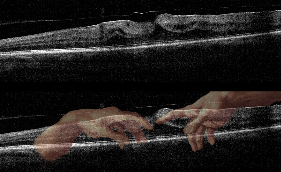
MICHELANGELO MACULOPATHY
Originally posted on @retina.rocks May 1, 2020
This patient has an epiretinal membrane with some vitreomacular traction.
This case was submitted by Dr. Jennifer Reynolds. The “artistic” mind of the submitting doctor, noticed the hand of the Creator in this scan!
INTRAOCULAR FOREIGN BODY
Originally posted on @retina.rocks April 1, 2020
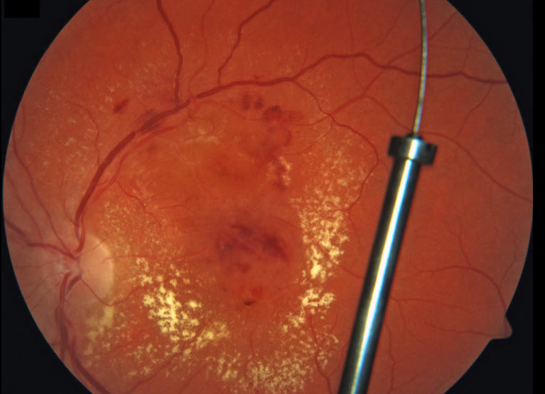
An automobile manufacturing worker from our local Ford plant presented with an acute injury to his left eye while installing a catalytic converter. A large metallic intraocular foreign body (IOFB) shot through the pars plana in a self-sealing wound and lodged in the posterior vitreous without any direct retinal injury. Since the material was made primarily of inert platinum, the IOFB was left in place and the patient has remained remarkably stable for 3 years.
We hypothesize that the platinum exhibited powerful anti-fungal and anti-bacterial effects helping to prevent infection. The diffuse diabetic macular edema with surrounding lipid also spontaneously resolved, sparking interest in intravitreal platinum as a more durable treatment for macular leakage. The results of the NIH-sponsored Phase 3 MORON Study, Might IntraOcular Platinum ResOlve Edema Better ThaN Really Expensive Anti-VEGF Agents, are eagerly anticipated in late 2020..
Yes, of course it’s APRIL FOOLS!
This patient has diabetic retinopathy with macular edema. The “IOFB” is really a broken fundus camera fixation stick projected onto the posterior pole image.
HUMMINGBIRD MACULOPATHY
Originally posted on @retina.rocks February 28, 2020
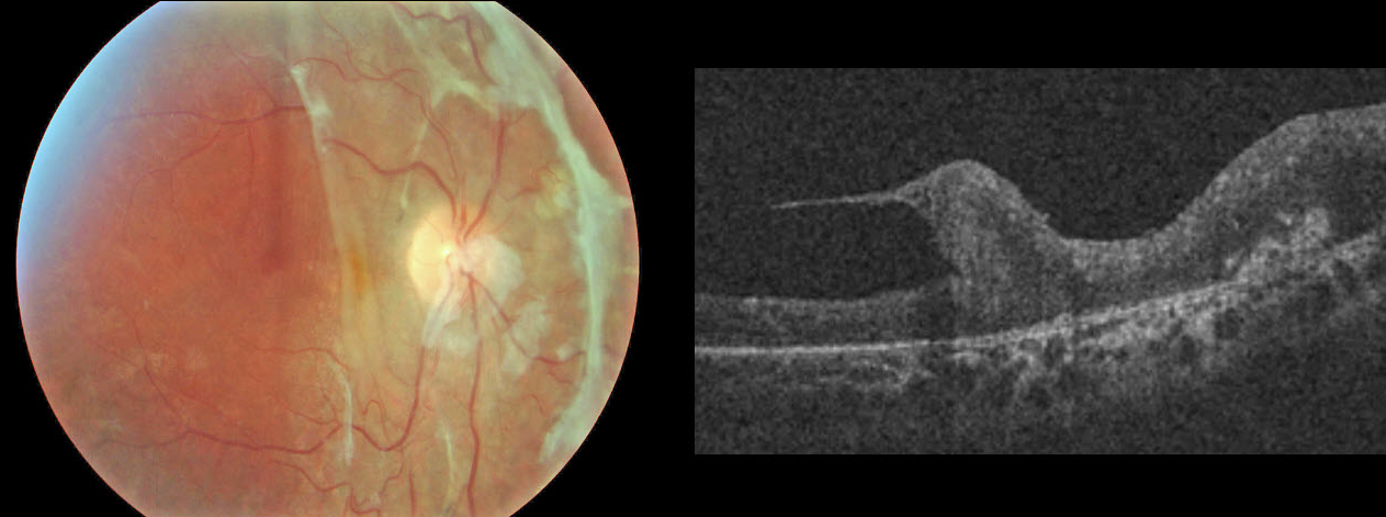
This rare condition was discovered after a diabetic TRD repair. Thankfully, no birds or retinas were harmed in this process.
VALENTINE RETINOPATHY
Originally posted on @retina.rocks February 14, 2020
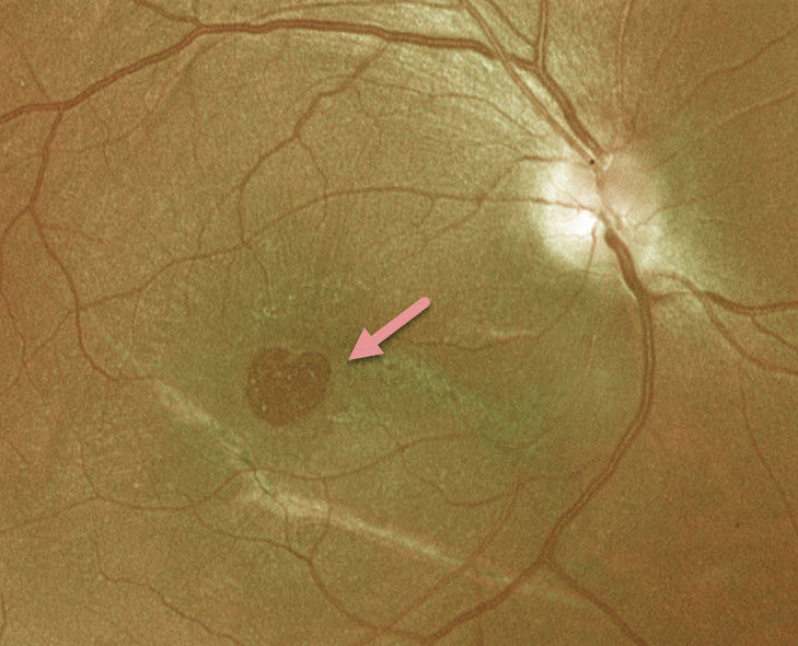
This patient has a heart-shaped macular hole just in time for Valentine’s Day!
VOLCANIC MACULOPATHY
Originally posted on @retina.rocks January 20, 2020
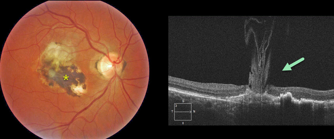
The OCT scan shows unusual vitreous strands adhering to an inactive histoplasmosis scar resembling an erupting volcano.


