

Hematologic Malignancies (Leukemia, Multiple Myeloma) and Hyperviscosity
<< Back to Cases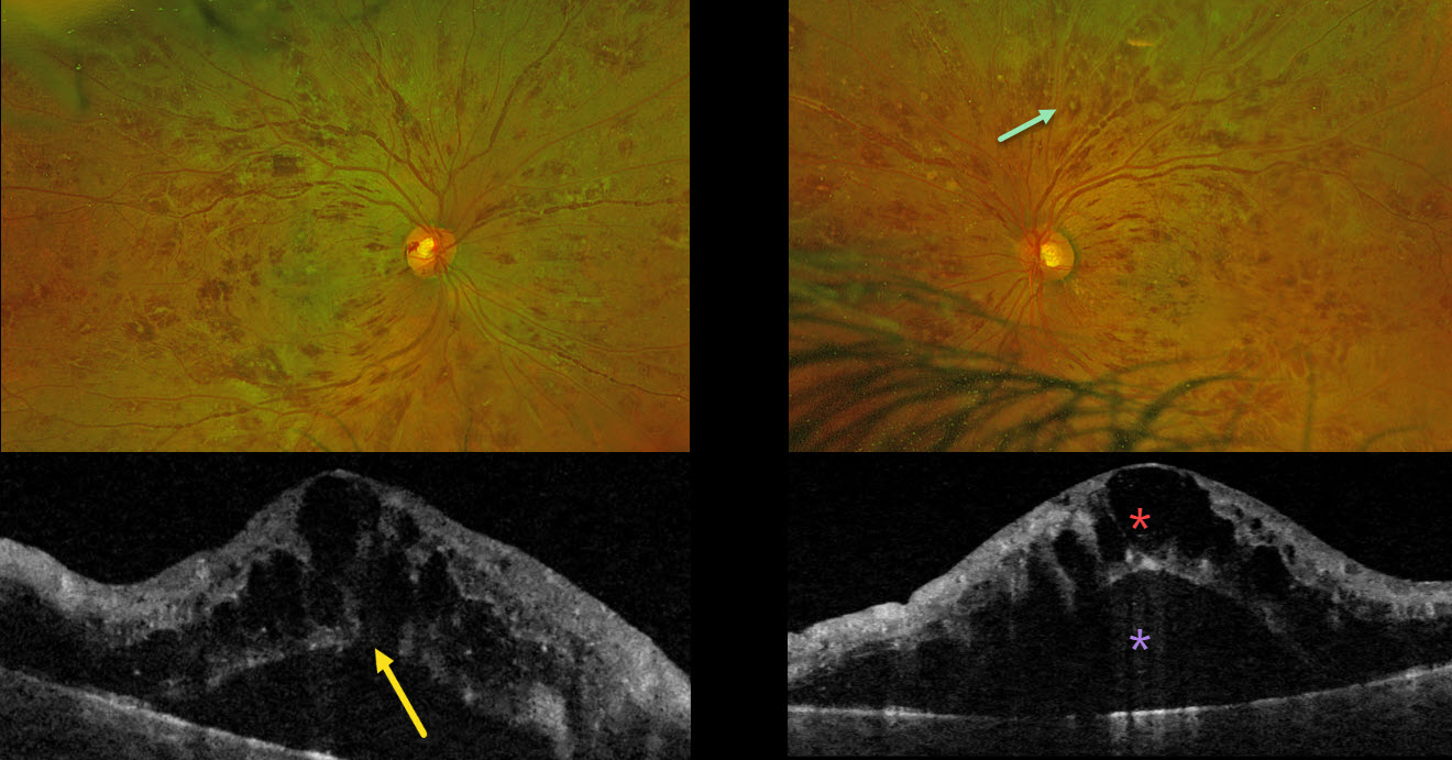
LEUKEMIA WITH PSEUDO-BILATERAL CRVO
Mattie Adams
Originally posted on @retina.rocks July 28, 2025
This previously healthy 38YO male presented with 1 month of bilateral blurriness. Vision was 20/60 OD and 20/80 OS.
Optos color RG imaging shows severe, bilateral, mostly white-centered retinal hemorrhages extending from the maculae into the retinal peripheries. Triton swept-source OCT shows marked bilateral cystic foveal swelling with subretinal fluid. An outer macular hole is probably present OD. His white blood cell count was markedly elevated at 206,400. Subsequent emergent oncologic workup led to the diagnosis of chronic myelogenous leukemia.
Learning Points:
The term “Roth spot” was named after Dr. Moritz Roth, who, in 1872, noted white-centered hemorrhages in patients with bacterial endocarditis. He felt that these white spots were septic emboli that originated from an infected cardiac valve. However, similar lesions can be seen in many other disorders, including leukemia, hyperviscosity, anemia, diabetes, and hypertension (Duane et al Ophthalmology 1980;87:66-69). The white material may represent a variety of causes, including fibrin. So, these lesions are best called white-centered hemorrhages, unless one is specifically referring to those found associated with bacterial endocarditis.
In our patient’s case, the severity of the retinal hemorrhages gave an appearance resembling bilateral central retinal vein occlusions (CRVO). However, CRVO hemorrhages are located predominantly in the nerve fiber layer (our patients’ hemorrhages were deep and white-centered), and the optic nerve is usually swollen with this degree of hemorrhage (our patients’ nerves were flat).
LEUKEMIA
Hailey Dunlow
Originally posted on @retina.rocks May 12, 2025
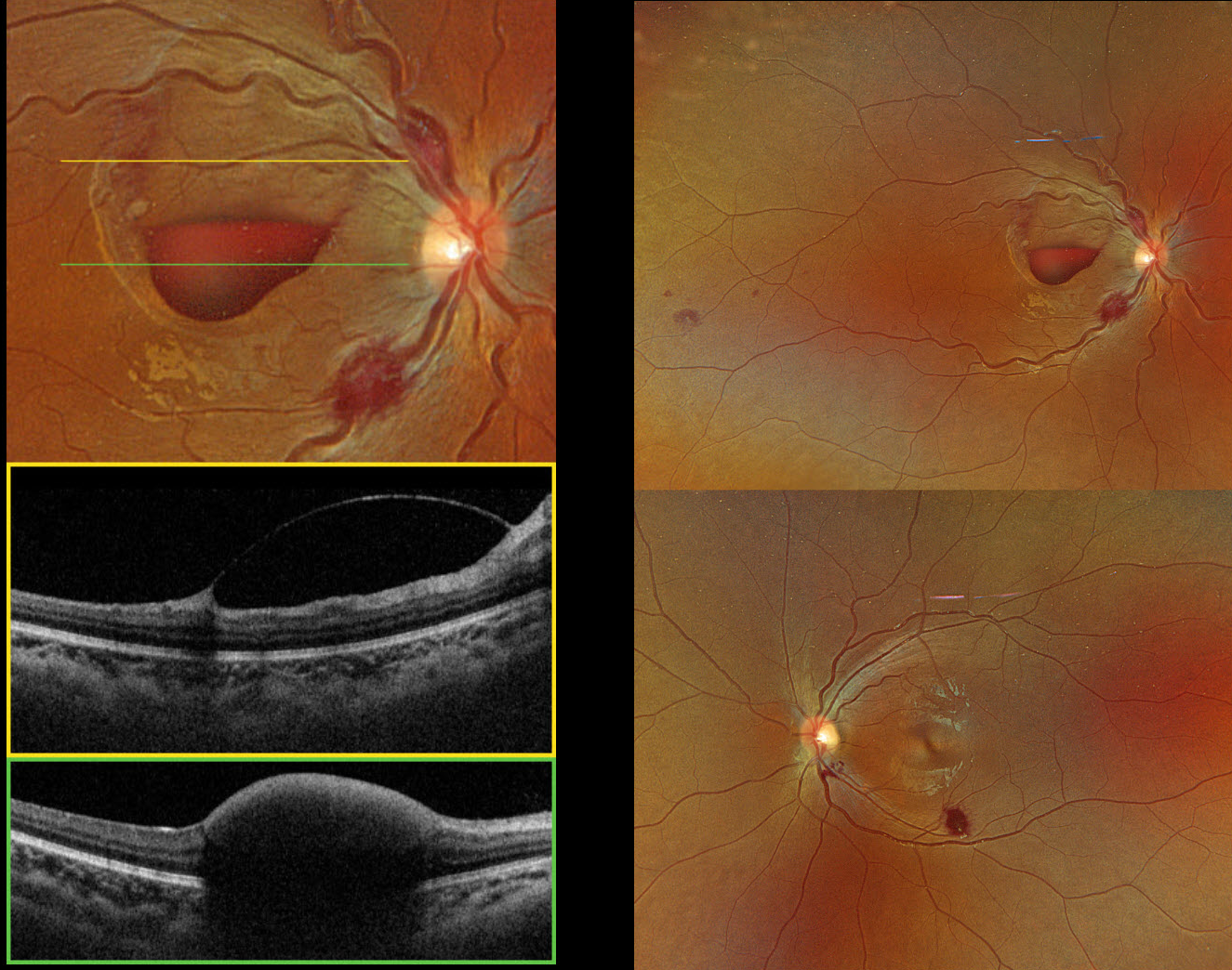
This previously healthy 14YO female presented with two weeks of vision loss in her right eye. Vision was hand motion OD and 20/25 OS.
Optos color RGB imaging shows bilateral inner and deep retinal hemorrhages, along with an inferior macular sub-internal limiting membrane (ILM) hemorrhage OD. Triton swept-source OCT confirms the location of the macular blood OD.
She was immediately sent to the emergency room. Complete blood count revealed pancytopenia with HgB 3.8, WBC 1.4, and platelets 54, and the patient was subsequently diagnosed with acute myelogenous leukemia. She was immediately lost to follow-up.
Learning Points:
Ocular leukemic manifestations are primarily caused by accompanying hematologic abnormalities, including hyperviscosity, anemia, and thrombocytopenia (Soman et al, Ophthalmology Retina 2018;2:17-23). Posterior segment findings include retinal hemorrhages (sub-ILM, nerve fiber layer, or deeper, white-centered), cotton-wool spots, dilated retinal veins, and serous retinal detachment. Leukemic infiltration of the optic nerve, retina, and choroid can also occur. Sub-ILM hemorrhages are usually self-limiting and resolve within several months of treatment of the underlying disease (Ophthalmology Retina 2018;2:494-501).
MULTIPLE MYELOMA
Originally posted on @retina.rocks May 5, 2025
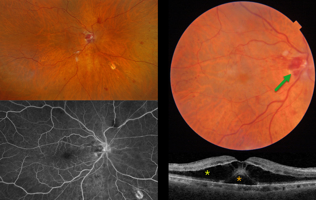
This 81YO male presented with two days of vision loss OD. He has a history of diet-controlled type 2 diabetes and a 5-year history of multiple myeloma. He was examined by us 2 weeks earlier for a routine visit for inactive herpes simplex iridocyclitis. Vision was 20/40 on that visit with a normal OCT (not shown), but had decreased to counting fingers at the current visit.
Triton color imaging shows a faint, round serous macular detachment with scattered small inner retinal and nerve hemorrhages. Swept-source OCT shows subfoveal fluid with overlying cystic edema in the outer nuclear layer. Optos color RGB imaging shows scattered retinal hemorrhages throughout the right posterior pole, with a normal left fundus. Fluorescein angiography shows no leakage.
Learning Points:
Monoclonal gammopathies (paraproteinemia) result from the production of monoclonal gamma-globulins in the blood, including multiple myeloma (IgG or IgA) and Waldenstrom macroglobulinemia (IgM). Retinal findings are often caused by hyperviscosity (Rajagopal et al, Surv Ophthalmology 2016;61:236-247) and include intraretinal or deep retinal hemorrhages, dilated vessels, or vascular occlusions.
A unique retinopathy found in these disorders is termed paraproteinemic maculopathy (Mansour et al, Ophthalmology 2014;121:1925-1932). Somewhat simulating optic pit maculopathy, patients develop serous macular detachments with overlying cystic outer retinal edema and occasionally outer macular holes. There is no angiographic leakage. The macular fluid, which is usually refractory to laser photocoagulation, intravitreal injections, and photodynamic therapy, is treated primarily by decreasing blood immunoglobulin levels.
Unfortunately, our patient died 2 months after our examination.
LEUKEMIA
Kanwaljeet Harjot Madan
Originally posted on @retina.rocks August 28, 2024
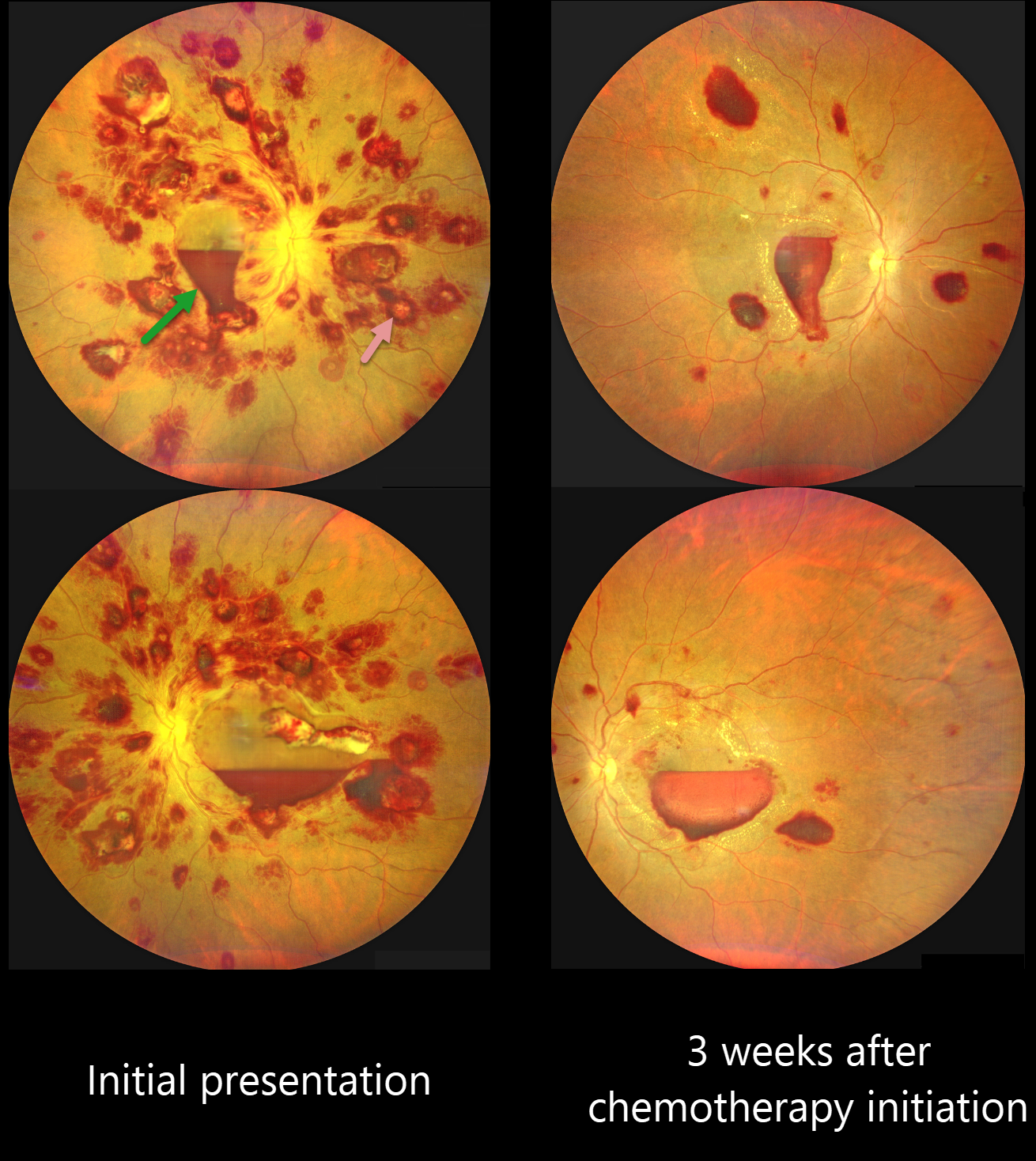
This 42YO previously healthy male presented with one week of bilateral decreased vision. Vision was 20/100 OD and 20/200 OS.
Color photography shows an extensive bilateral nerve fiber layer and deep white-centered retinal hemorrhages. Layered macular sub-internal limiting membrane (ILM) hemorrhages are noted as well, with some yellow devitalized blood in the temporal left macula. Hematologic workup revealed elevated blast cells, severe anemia (HgB 5.4) and thrombocytopenia.
He was diagnosed with acute myeloid leukemia and referred to oncology. He started chemotherapy, and 3 weeks later, his vision improved to 20/70 OU. The HgB was 8.2 with decreased blast cells. The bilateral retinal hemorrhages were markedly decreased.
This case was submitted by Kanwaljeet Harjot Madan.
Learning Points:
The term “Roth spot” was named after Dr. Moritz Roth, who, in 1872, noted white-centered hemorrhages in patients with bacterial endocarditis. He felt that these white spots were septic emboli that originated from an infected cardiac valve. However, similar lesions can be seen in many other disorders, including leukemia, hyperviscosity, anemia, diabetes, and hypertension (Duane et al Ophthalmology 1980;87:66-69). The white material may represent a variety of causes, including fibrin. So, these lesions are best called white-centered hemorrhages, unless one is specifically referring to those found associated with bacterial endocarditis.
Ocular leukemic manifestations are primarily caused by accompanying hematologic abnormalities, including hyperviscosity, anemia, and thrombocytopenia (Soman et al, Ophthalmology Retina 2018;2:17-23). Posterior segment findings include retinal hemorrhages (sub-ILM, nerve fiber layer or deeper, white-centered), cotton-wool spots, dilated retinal veins, and serous retinal detachment. Leukemic infiltration of the optic nerve, retina, and choroid can also occur. The sub-ILM hemorrhages are usually self-limiting and resolve within several months of treating the underlying disease (Ophthalmology Retina 2018;2:494-501).
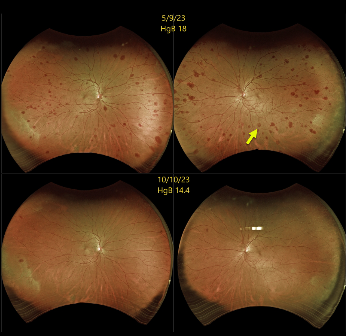
SECONDARY POLYCYTHEMIA
Originally posted on @retina.rocks December 18, 2023
This 51YO female was referred for asymptomatic retinal hemorrhages in each eye. She had a history of pulmonary hypertension. Vision was 20/20 bilaterally.
Optos color RG imaging shows scattered retinal hemorrhages in all quadrants, most of which are deep with white centers. A complete blood count revealed a hemoglobin of 18.0 g/dl, and she was immediately referred for a hematology consult.
Subsequent hematologic workup was negative, and she was felt to have secondary polycythemia from her pulmonary hypertension. She has since been treated with serial phlebotomies.
When last examined 5 months later, the retinal hemorrhages were markedly decreased, and her hemoglobin had decreased to 14.4 g/dl.
Learning Points:
The term “Roth spot” was named after Dr. Moritz Roth, who, in 1872, noted white-centered hemorrhages in patients with bacterial endocarditis. He felt that these white spots were septic emboli that originated from an infected cardiac valve.
However, similar lesions can be seen in many other disorders, including leukemia, hyperviscosity, anemia, diabetes, and hypertension (Duane et al Ophthalmology 1980;87:66-69). The white material may represent a variety of causes, including fibrin. So these lesions are best called white-centered hemorrhages, unless one is specifically referring to those found associated with bacterial endocarditis.
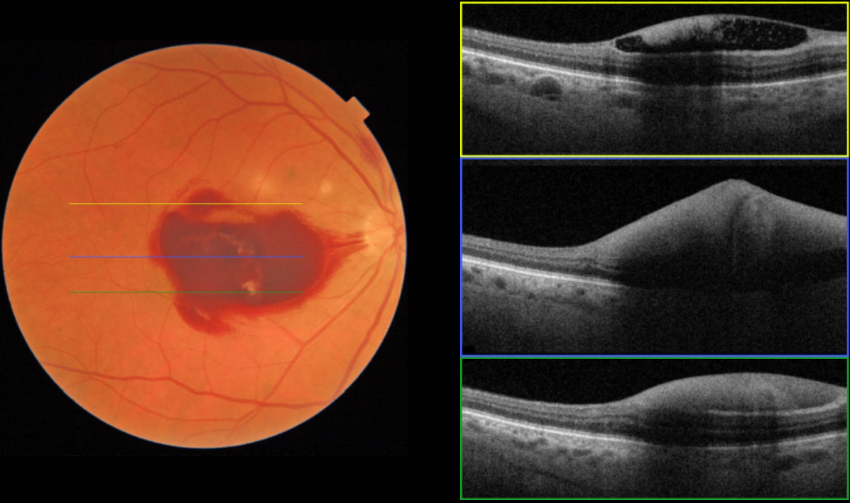
LEUKEMIA
Originally posted on @retina.rocks August 7, 2023
This 63YO male, who is undergoing treatment for leukemia, presented with one day of acute vision loss in his right eye. He was recently discharged from the hospital for chemotherapy-induced pancytopenia. His CBC showed: WBC 0.5, HgB 5.6, and Platelets 2. Vision was counting fingers OD.
Fundus imaging shows sub-internal membrane (ILM) hemorrhage. The OCT scans confirm hemorrhage in the sub-ILM space. We expect our patient’s macular hemorrhage to resolve as his blood counts normalize
Learning Points:
Ocular leukemic manifestations are primarily caused by accompanying hematologic abnormalities, including hyperviscosity, anemia, and thrombocytopenia (Soman et al, Ophthalmology Retina 2018;2:17-23).
Posterior segment findings include retinal hemorrhages (sub-ILM, nerve fiber layer, or deeper, white-centered), cotton-wool spots, dilated retinal veins, and serous retinal detachment. Leukemic infiltration of the optic nerve, retina, and choroid can also occur.
The sub-ILM hemorrhages are usually self-limiting and resolve within several months of treating the underlying disease (Ophthalmology Retina 2018;2:494-501).
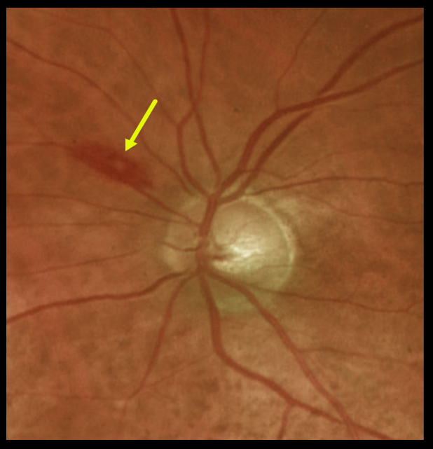
LYMPHOMA WITH WHITE-CENTERED RETINAL HEMORRHAGE
Originally posted on @retina.rocks November 10, 2022
This 84YO female was found to have a white-centered retinal hemorrhage superonasal to the optic nerve disc.
Complete blood count revealed a white blood count of 19,000 and a hemoglobin of 7.9. She was subsequently diagnosed with atypical diffuse large B-cell lymphoma.
Learning Points:
The term “Roth spot” was named after Dr. Moritz Roth, who, in 1872, noted white-centered hemorrhages in patients with bacterial endocarditis.
He felt that these white spots were septic emboli that originated from an infected cardiac valve. However, similar lesions can be seen in many other disorders, including leukemia, hyperviscosity, anemia, diabetes, and hypertension.
The white material may represent a variety of causes, including fibrin. These lesions are best called white-centered hemorrhages, unless one is specifically referring to a hemorrhage in a patient with bacterial endocarditis.
For a review of white-centered retinal hemorrhages, see Duane et al Ophthalmology 1980;87:66-69.
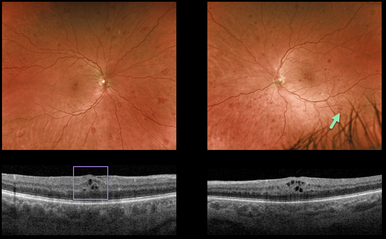
LEUKEMIA
Originally posted on @retina.rocks October 28, 2022
This 50YO male presented with bilateral asymptomatic retinal hemorrhages. He gave a 5-year history of well-controlled type 2 diabetes with a recent A1C of 6.3%, and was previously diagnosed with only mild nonproliferative diabetic retinopathy.
Optos imaging shows scattered retinal hemorrhages in all quadrants, most of which are deep with white centers. Mild bilateral macular edema is seen on OCT scanning.
The deeper, round retinal hemorrhages were felt to be atypical for diabetes. We therefore ordered a complete blood count (CBC), which revealed a white blood cell count over 80,000 u/L and polychromatic red blood cells.
Immediate referral to oncology revealed undiagnosed chronic myelogenous leukemia, and treatment was initiated.
Learning Points:
The term “Roth spot” was named after Dr. Moritz Roth, who, in 1872, noted white-centered hemorrhages in patients with bacterial endocarditis. He felt that these white spots were septic emboli that originated from an infected cardiac valve.
However, similar lesions can be seen in many other disorders, including leukemia, hyperviscosity, anemia, diabetes, and hypertension.
The white material may represent a variety of causes, including fibrin. So these lesions are best-called white-centered hemorrhages, unless one is specifically referring to a patient with bacterial endocarditis. See Duane et al Ophthalmology 1980;;87:66-69.
HYPERVISCOSITY WITH MONOCLONAL GAMMOPATHY
Originally posted on @retina.rocks July 8, 2021
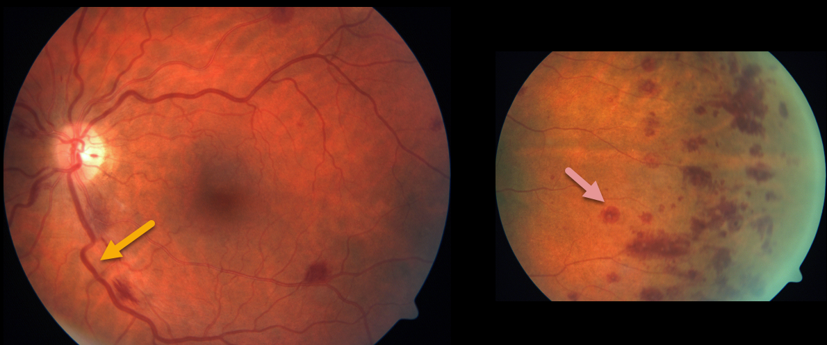
This patient with known monoclonal gammopathy presented with bilateral engorged retinal vessels and scattered deep, white-centered retinal hemorrhages. These are most prominent in the temporal periphery.
Learning Points:
The term “Roth spot” was named after Dr. Moritz Roth, who, in 1872, noted white-centered hemorrhages in patients with bacterial endocarditis. He felt that these white spots were septic emboli that originated from an infected cardiac valve.
However, similar lesions can be seen in many other disorders, including leukemia, hyperviscosity, anemia, diabetes, and hypertension. The white material may represent a variety of causes, including fibrin.
So these lesions are best called white-centered hemorrhages, unless one is specifically referring to a lesion in a patient with bacterial endocarditis. See Duane et al Ophthalmology 1980;87;66-69.
EXUDATIVE RETINAL DETACHMENT WITH OUTER MACULAR HOLE
Originally posted on @retina.rocks June 25, 2021
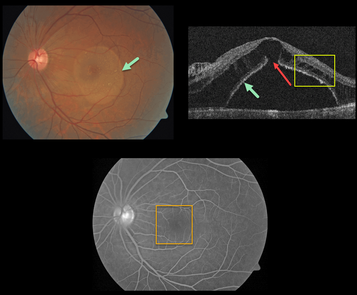
This patient presented with a sharply-circumscribed macular serous detachment, cystic outer retinal edema, and an outer macular hole. Fluorescein angiography is essentially normal without leakage.
An optic pit was absent clinically and on OCT imaging. Serum protein electrophoresis was normal, and a systemic cancer workup was negative.
Learning Points:
Serous detachment can be an uncommon ocular manifestation of paraproteinemia (see Mansour et al, Ophthalmology 2014;121;1925-1932). These detachments, which resemble those seen in optic pit maculopathy, are characterized by a well-defined serous detachment without angiographic leakage. A central outer macular hole is often present.
The fluid is likely due to an osmotic gradient generated by subretinal immunoglobulins that passively migrate into the subretinal space.
We do not know the cause of our patient’s findings, given his lack of an optic pit and a negative systemic workup.
LEUKEMIA
Originally posted on @retina.rocks February 22, 2021
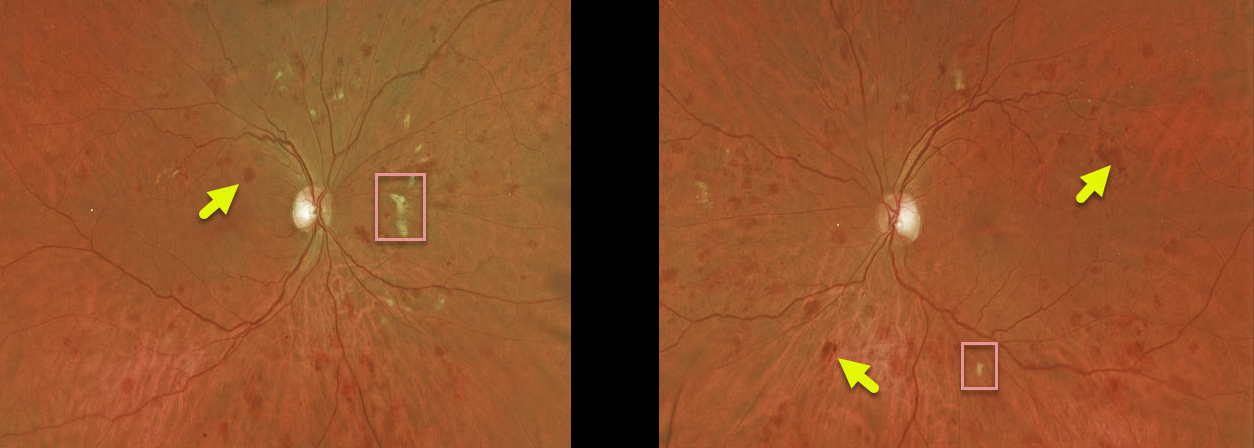
This patient with acute myeloid leukemia (AML) was referred by their oncologist for an eye examination following a failed allogeneic stem cell transplant due to graft versus host disease. Vision was 20/30 OD and 20/25 OS. His WBC was 14.7 (with 32% marrow myeloblasts), hemoglobin was 8.2, and platelet count was 23,000.
This patient has large, deep blot hemorrhages in the outer plexiform layer as well as scattered cotton-wool spots, indicative of leukemic retinopathy.
Learning Points:
These deeper hemorrhages would be unusual for the more common disorders seen in clinical practice (including diabetic retinopathy, retinal vein occlusions, and hypertensive retinopathy) and are usually indicative of blood dyscrasias, including anemia and hematologic malignancies. These deeper hemorrhages are also seen peripherally in ocular ischemia.
MULTIPLE MYELOMA
Originally posted on @retina.rocks January 26, 2021
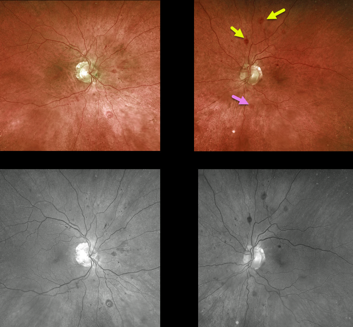
This 76YO male presented with normal vision and asymptomatic bilateral retinal hemorrhages following a recent diagnosis of multiple myeloma.
There are a variety of retinal hemorrhages seen on Optos and green-channel images, including inner retinal white-centered hemorrhages and deep blot hemorrhages in the outer plexiform layer.
Learning Points:
White-centered hemorrhages or “roth spots” can be seen in many disorders, including leukemia, hyperviscosity, anemia, diabetes, and hypertension. The white material may represent a variety of causes, including fibrin.
The term “Roth spots” should thus be reserved only for patients with bacterial endocarditis. See Duane et al Ophthalmology 1980;87;66-69.
The deeper hemorrhages would be unusual for the more common disorders seen in clinical practice (including diabetic retinopathy, retinal vein occlusions, and hypertensive retinopathy), and are usually indicative of blood dyscrasias, including anemia and hematologic malignancies. These deeper hemorrhages are also seen peripherally in ocular ischemia.
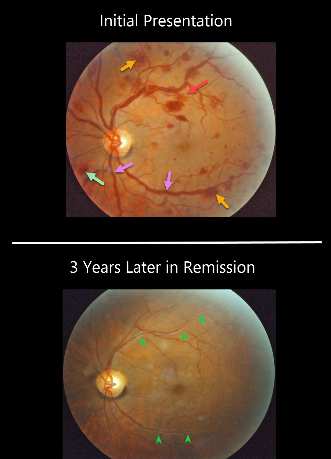
MULTIPLE MYELOMA
Originally posted on @retina.rocks August 6, 2020
This patient presented with bilateral multiple flame-shaped and white-centered retinal hemorrhages. There is also at least one deeper, round outer plexiform hemorrhage just inferonasal to the nerve. The retinal veins are somewhat dilated and tortuous, consistent with hyperviscosity. This patient was subsequently diagnosed with multiple myeloma.
Three years later, the patient was in remission after receiving treatment. Although the vascular caliber has normalized, vascular sheathing remained due to prior damage to the vessel walls.
Learning Points:
White-centered hemorrhages or “roth spots” can be seen in many disorders, including leukemia, hyperviscosity, anemia, diabetes, and hypertension. The white material may represent a variety of causes, including fibrin.
The term “Roth spot” was originally named after Dr. Moritz Roth, who, in 1872, noted white-centered hemorrhages in patients with bacterial endocarditis. He felt that these white spots were septic emboli that originated from an infected cardiac valve.
This term should thus be reserved only for patients with bacterial endocarditis. See Duane et al Ophthalmology 1980;87;66-69.


