

Henle Fiber Layer Hemorrhages
<< Back to Cases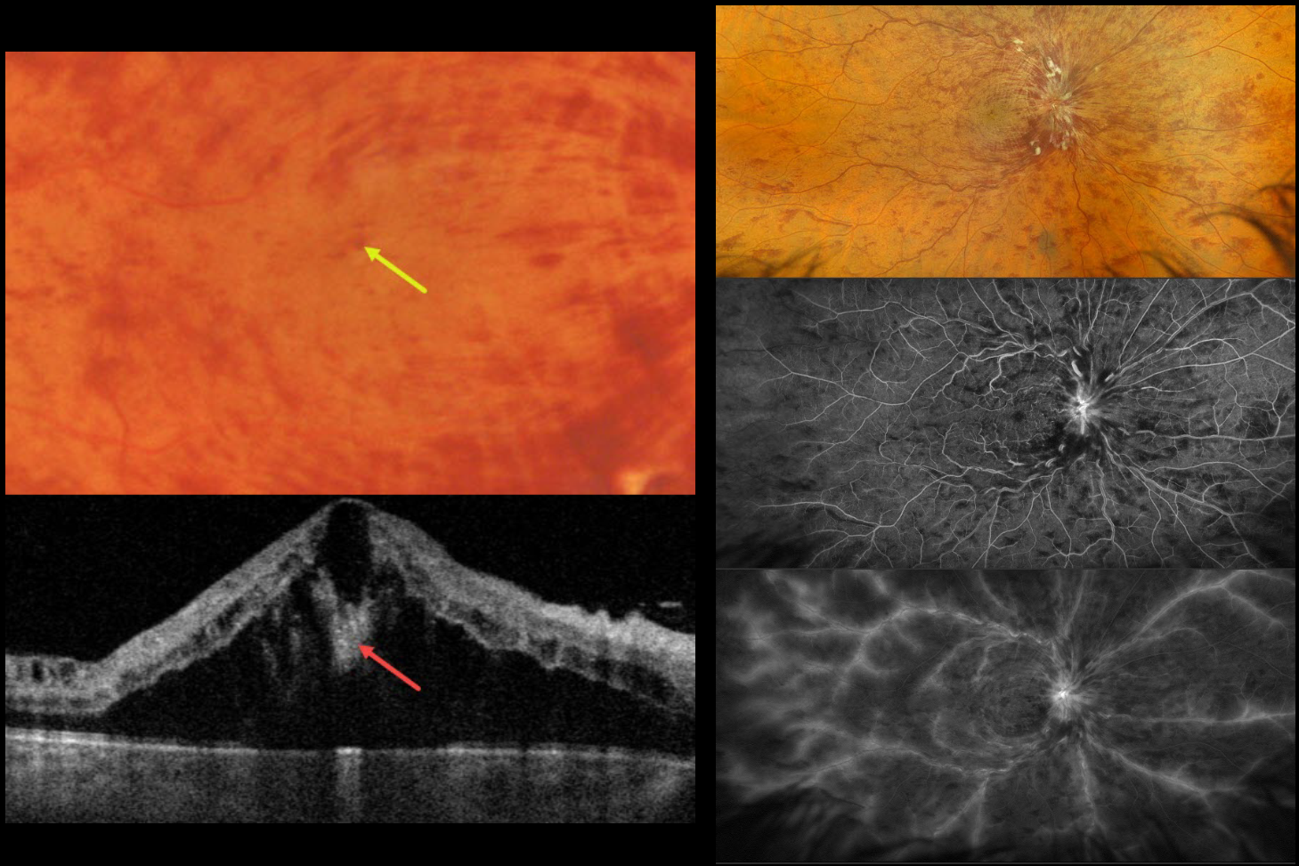
CRVO WITH CENTRAL BOUQUET HEMORRHAGE
Originally posted on @retina.rocks June 26, 2026
This healthy 52YO male presented with recent severe vision loss in his right eye. Vision was 20/400 OD and 20/20 in his normal OS.
Color photography and Optos RGB imaging show a central retinal vein occlusion (CRVO) with marked retinal hemorrhages and macular edema. Inner blood-filled retinal cysts are noted on funduscopy and swept-source OCT. On fluorescein angiography, the occlusion is fairly well perfused with late leakage from the nerve, macula, and retinal veins. Monthly anti-VEGF therapy was started.
Learning Points:
Cicinelli et al recently reported that central bouquet hemorrhages in retinal vein occlusion are associated with poor visual outcomes due to localized effects of elevated venous pressure and macular ischemia (IOVS 2025;66(12):62).
Prior to the introduction of intravitreal injections in the early 2000s, there were essentially no treatments for CRVO-related macular edema. Anti-VEGF injections are now generally the initial treatment of choice, with about 50-60% of eyes gaining at least 3 lines of vision. Intravitreal steroids, usually Ozurdex, are more often used as secondary treatment. Macular grid laser photocoagulation is not performed because it has been shown to confer no visual benefit in the Central Vein Occlusion Study Group (Ophthalmology 1995;102:1425-1433).
CENTRAL BOUQUET HEMORRHAGE
Evgenii Chernov
Originally posted on @retina.rocks May 13, 2025
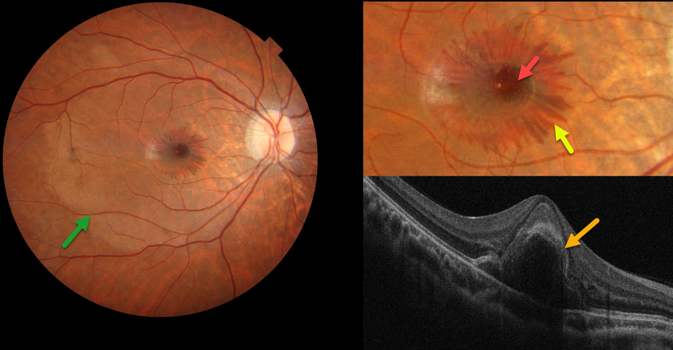
This 24YO male woke with distorted vision in his right eye the day he presented to our clinic. Over the next 2 hours, a central spot appeared in his vision. He recalled an active tennis game the previous day but denied any direct ocular trauma. Vision was 20/200 OD and 20/25 in his normal OS.
Color photography shows central foveal blood, surrounding radiating outer plexiform (OPL) blood, and a deep vs subretinal whitish discoloration of the macula. On OCT, the central blood is located in the outer retina.
Learning Points:
Radiating deep macular hemorrhages are seen in multiple conditions, including Valsalva and trauma. Their unique radiating appearance is caused by blood accumulating in Henle’s layer. Kon Graversen et al described hemorrhagic unilateral retinopathy, an idiopathic disorder with a similar appearance (Retina 2014;34:483-489).
Ramthohul et al recently described a central bouquet hemorrhage with a round intraretinal blood spot surrounded by these OPL hemorrhages (Retina 2024;44:551-557). Various etiologies included neovascular AMD, myopic lacquer cracks, macular telangiectasia, and trauma associated with angioid streaks. Despite the macular changes consistent with commotio retinae, our patient adamantly denied ocular trauma.
NEOVASCULAR AMD WITH HENLE LAYER HEMORRHAGE
Originally posted on @retina.rocks July 29, 2024
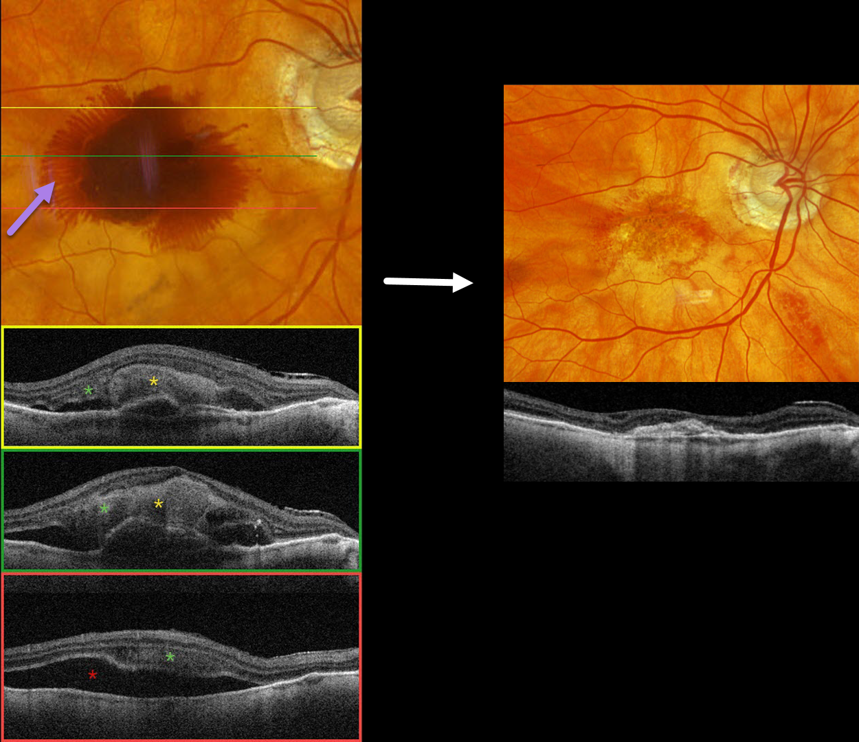
This 97YO male has a history of counting-finger vision in his left eye due to foveal macular atrophy. On 2/29/24, vision was 20/40 in his non-exudative right eye. He presented on 3/12/24 with sudden vision loss OD and new hand motions vision.
Optos color RGB imaging shows fairly dense central subretinal blood, with surrounding radiating blood in the deeper Henle’s layer (outer plexiform layer). Triton swept-source OCT shows variable thick subretinal blood and more inferior subretinal fluid. Henle’s blood is seen as a diffuse hyperreflective obscuration of the outer retinal layers. Vitrectomy with subretinal TPA was performed two days later. Two months later, vision was counting fingers, and the macula was completely free of fluid and blood.
Learning Points:
Radiating deep macular hemorrhages are seen in multiple conditions, including Valsalva and trauma. We have not previously seen these hemorrhages in neovascular AMD. Their unique radiating appearance results from blood accumulating in Henle’s layer. Kon Graversen et al described hemorrhagic unilateral retinopathy, an idiopathic disorder with a similar appearance (Retina 2014;34:483-489).
HEMORRHAGIC UNILATERAL RETINOPATHY
Asma Samsudeen and Ashish Sharma
Originally posted on @retina.rocks May 25, 2023
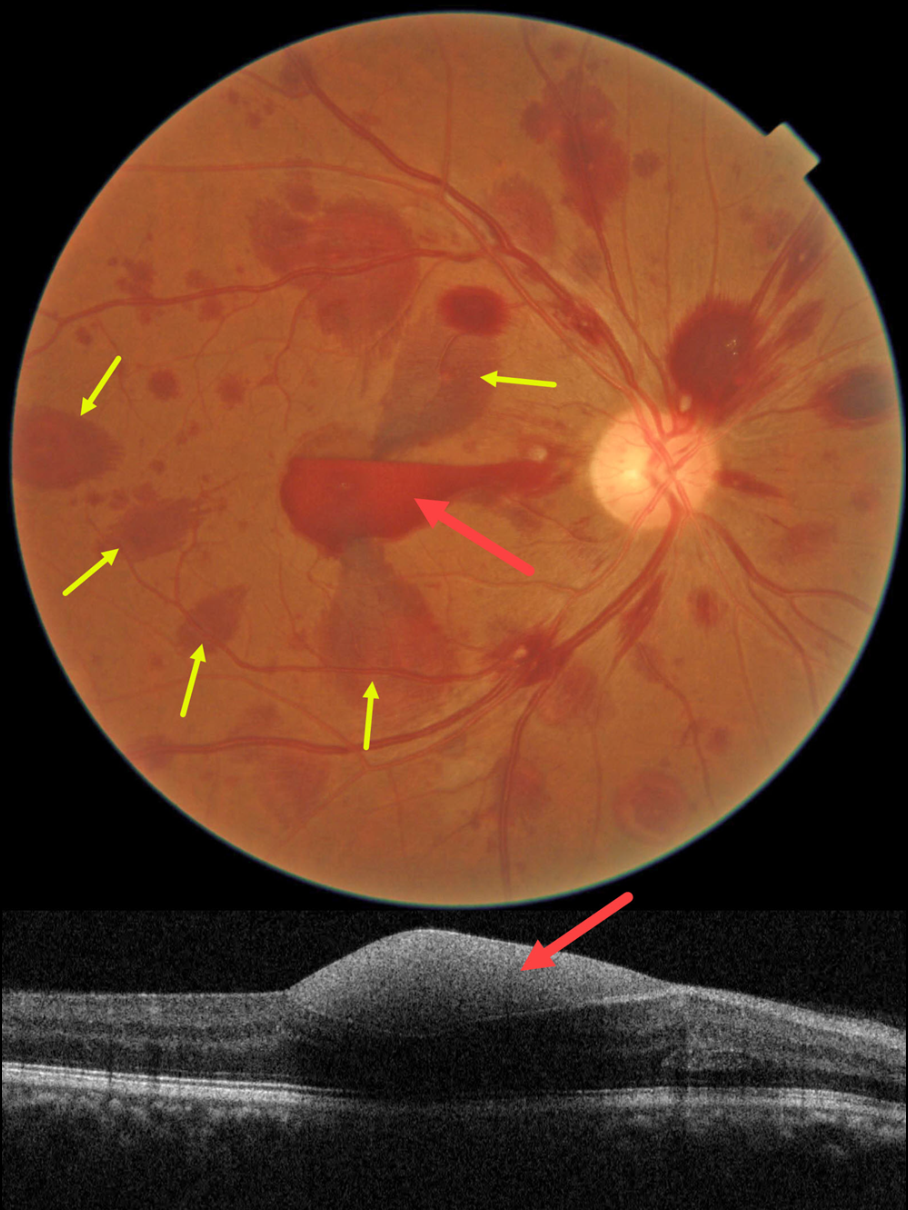
This healthy 52YO male noticed acute vision loss in his right eye two days earlier. He denied Valsalva or trauma. Vision was counting fingers in his right eye and 20/20 in his normal left eye. He has a history of untreated hypertension, and his BP in the office was 180/110.
Color imaging shows multifocal, mostly deep, retinal hemorrhages scattered throughout the right posterior pole. Some of these hemorrhages are radiating outwards from the macular center in Henle’s layer. A subhyaloid vs sub-ILM hemorrhage is also noted.
Learning Points:
Named by Kon Graversen et al (Retina 2014;34:483-489), hemorrhagic unilateral retinopathy presents with unilateral, deep intraretinal hemorrhages and sudden vision loss ranging from 20/20 to hand motion.
There are no associated systemic conditions, nor a history of trauma or Valsalva maneuver. It is most common in females. Vision usually recovers completely within 6 months.
We do not believe our patient’s findings were caused by his hypertension since they were unilateral and predominated by deeper retinal findings. Observation was recommended.
ANEMIA
Originally posted on @retina.rocks February 28, 2023
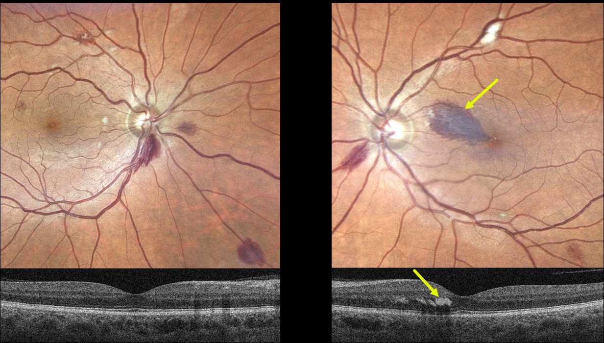
This 55YO male presented with a one-week history of a paracentral scotoma in his left eye. Vision was 20/40 OD and 20/60 OS. He gave a history of aplastic anemia, and his hemoglobin was 6.2 when he developed these symptoms. Blood transfusion brought his hemoglobin to over 8. He told us that his blood pressure runs low to normal.
Optos imaging shows scattered bilateral deep and superficial retinal hemorrhages with a few cotton wool spots. A tear-drop-shaped, larger hemorrhage is noted in the superonasal left macula. This hemorrhage localizes to the outer plexiform layer on swept-source OCT.
Learning Points:
The central retinal artery supplies the inner two-thirds of the retina, while the choroid supplies the outer third (the photoreceptor layers), the RPE, and the choroid. The outer plexiform layer (OPL) is the ‘end of the line’ for the inner retinal circulation.
In severe anemia, the more proximal inner retina removes whatever oxygen is available, leaving the deeper layers relatively ischemic. This can cause dysfunction of the retinal vascular endothelial tight junctions, which allows red blood cells to enter and stain the OPL, appearing clinically as deep retinal blot hemorrhages.
Deep blot OPL hemorrhages also appear for similar reasons in the temporal retinal periphery in ocular ischemia.
We advised observation, and expect his findings and symptoms to improve as long as his anemia is controlled.
HENLE LAYER HEMORRHAGE
Originally posted on @retina.rocks April 18, 2022
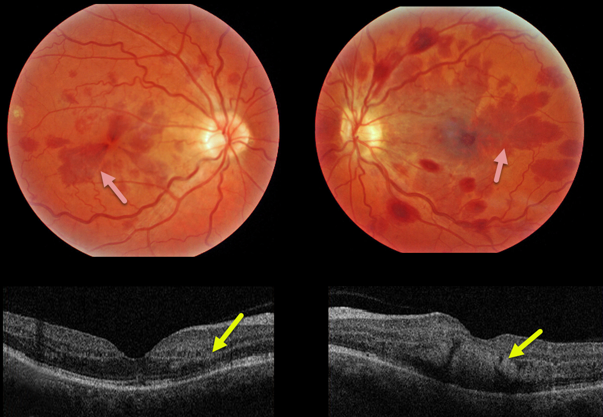
This 47YO female presented with bilateral vision loss. She had a seizure 10 days prior, losing consciousness and hitting her head. Upon awakening, she had reduced vision in her right eye and complete central loss in her left. Vision was 20/60 OD and counting fingers at 3 feet OS. Head CT scanning was normal.
Triton color imaging shows deep radiating wedge-shaped retinal hemorrhages centered in each macula. Swept-source OCT shows that these hemorrhages are located primarily in the outer plexiform layer (OPL).
Learning Points:
Radiating deep macular hemorrhages are seen in multiple conditions, including Valsalva (likely this case) and trauma. Their unique radiating appearance is caused by blood accumulating in Henle’s layer.
Kon Graversen et al described hemorrhagic unilateral retinopathy, an idiopathic disorder with a similar appearance (Retina 2014;34:483-489).
VALSALVA RETINOPATHY
Originally posted on @retina.rocks March 3, 2022
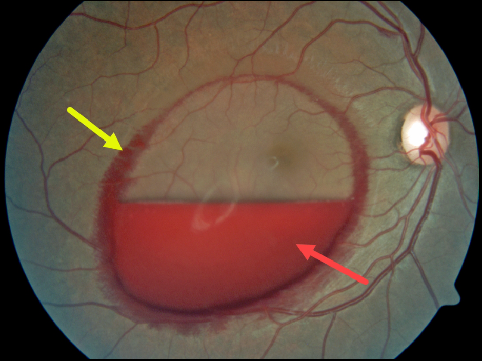
This patient presented with an acute layered sub-internal limiting membrane (ILM) hemorrhage due to a Valsalva maneuver. There is also a rim of blood along the circumference of the ILM detachment, which is most likely located in Henle’s layer, given its radiating deep retinal pattern.
Learning Points:
Sub-ILM hemorrhages most commonly follow a Valsalva maneuver or trauma, but can also happen spontaneously, as with anemia.
These hemorrhages virtually always resolve without sequelae, although the blood can be drained into the inferior vitreous by creating a small opening in the ILM using either a thermal or Nd:YAG laser.
HEMORRHAGIC UNILATERAL RETINOPATHY
Originally posted on @retina.rocks October 26, 2021
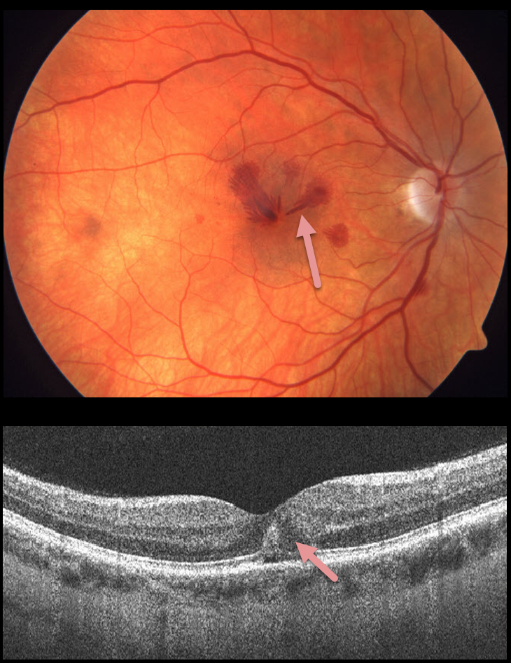
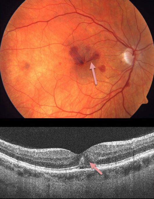
This 41YO female presented with acute unilateral vision loss in her right eye. Vision was 20/50 OD and 20/20 OS. The exam revealed unilateral radiating deep Henle layer retinal hemorrhages with no identifiable systemic or ocular etiologies.
Learning Points:
Named by Kon Graversen et al (Retina 2014;34:483-489), hemorrhagic unilateral retinopathy presents with unilateral, deep intraretinal hemorrhages and sudden loss of vision ranging from 20/20 to hand motion.
There are no associated systemic conditions, nor a history of trauma or Valsalva maneuver. It is most common in females. Vision usually recovers completely within 6 months.
HEMORRHAGIC UNILATERAL RETINOPATHY
Originally posted on @retina.rocks December 21, 2019
This 41YO female presented with acute unilateral vision loss in her right eye. Vision was 20/50 OD and 20/20 OS. Exam revealed unilateral radiating deep Henle layer retinal hemorrhages with no identifiable systemic or ocular etiologies.
Learning Points:
Named by Kon Graversen et al (Retina 2014;34:483-489), hemorrhagic unilateral retinopathy presents with unilateral, deep, intraretinal hemorrhages with sudden reduced vision ranging from 20/20 to hand motion.
There are no associated systemic conditions, nor history of trauma or Valsalva maneuver. It is most common in females. Vision usually recovers completely within 6 months.


