

High Myopia Atrophic
<< Back to Cases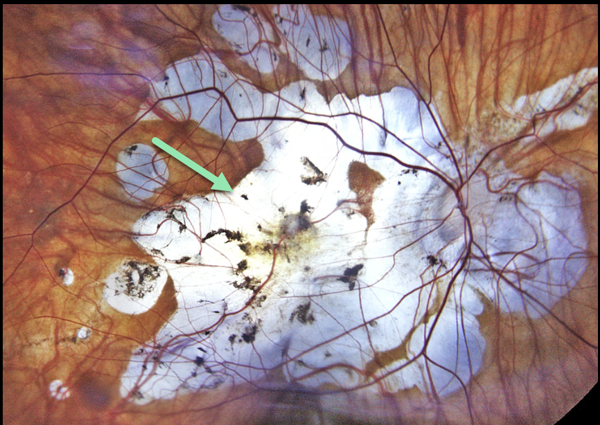
AMBLYOPIC MYOPIC STAPHYLOMA WITH MATURE CATARACT
Liubov Sitaruk
Originally posted on @retina.rocks August 11, 2025
This 64YO female has a lifetime history of anisometropic amblyopia with a myopic posterior staphyloma OD and presented with a mature cataract in this eye. Vision was light perception. She was clearly informed that surgery would likely not restore much vision. The goal was to remove the dense opacity and maximize any remaining visual potential.
Postoperative color imaging of the posterior pole shows severe peripapillary and myopic atrophy. Vision was 20/250.
Our patient reminds us that amblyopia is not an absolute contraindication for cataract surgery. Preoperative counseling is critical so that the patient understands the surgical goals. In general, the adage “under promise and over deliver” is a good rule for any healthcare provider to live by.
LACQUER CRACKS
Originally posted on @retina.rocks March 17, 2025
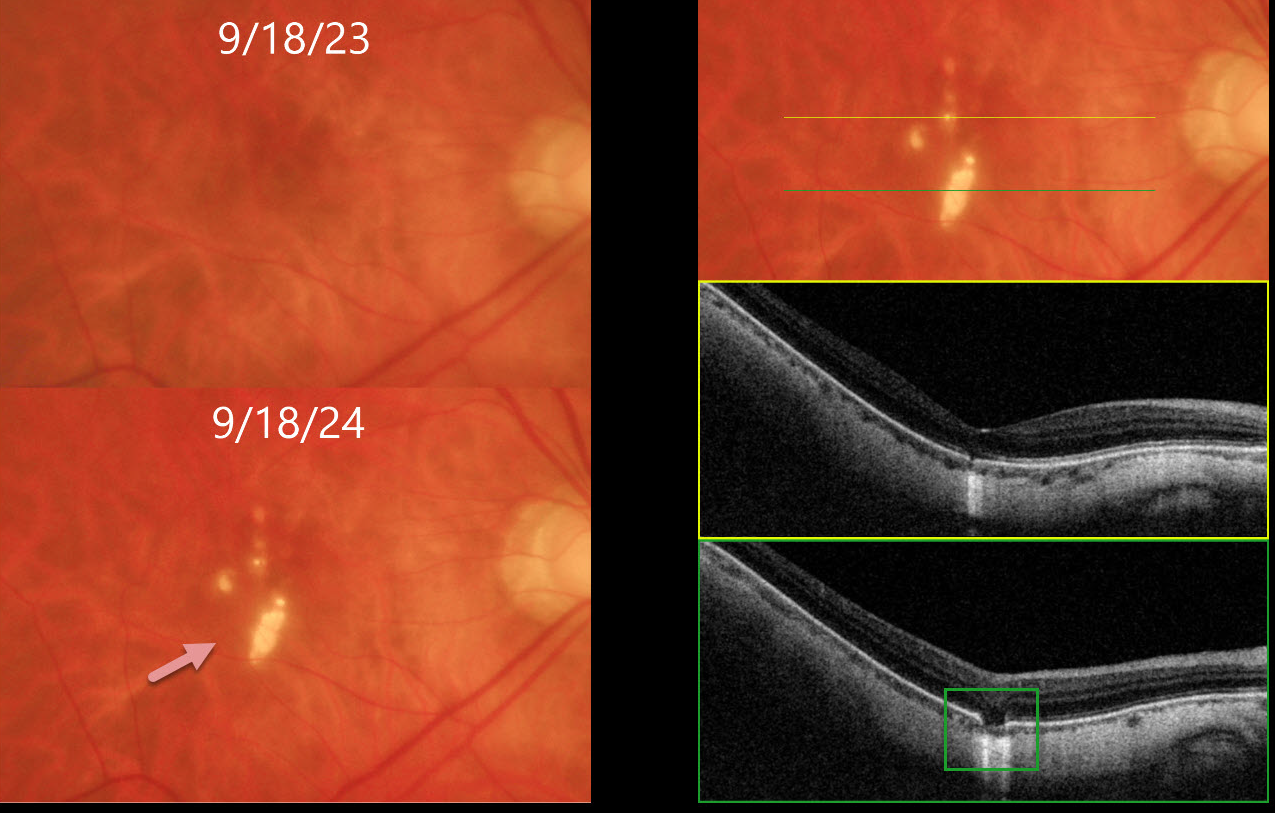
This 54YO highly myopic female is being followed with PRN Avastin injections in her left eye for myopic macular neovascularization. She was asymptomatic, and vision was stable at 20/50 OD and 20/60 OS.
Optos color RGB imaging of her right macula shows numerous lacquer cracks. Triton swept-source OCT shows outer retinal and RPE loss with increased choroidal transmission. Remarkably, her macula was normal one year earlier. Continued observation was recommended.
Learning Points:
Lacquer cracks are a common finding in patients with high myopia. They consist of breaks in Bruch’s membrane due to the outpouching/stretching of the pathologically myopic eye and increase the risk for macular neovascularization. Acutely, they are often associated with avascular subretinal blood, which resolves with observation.
MYOPIC PERIVASCULAR CHORIORETINAL ATROPHY
Yuenpang Cheung
Originally posted on @retina.rocks February 25, 2025
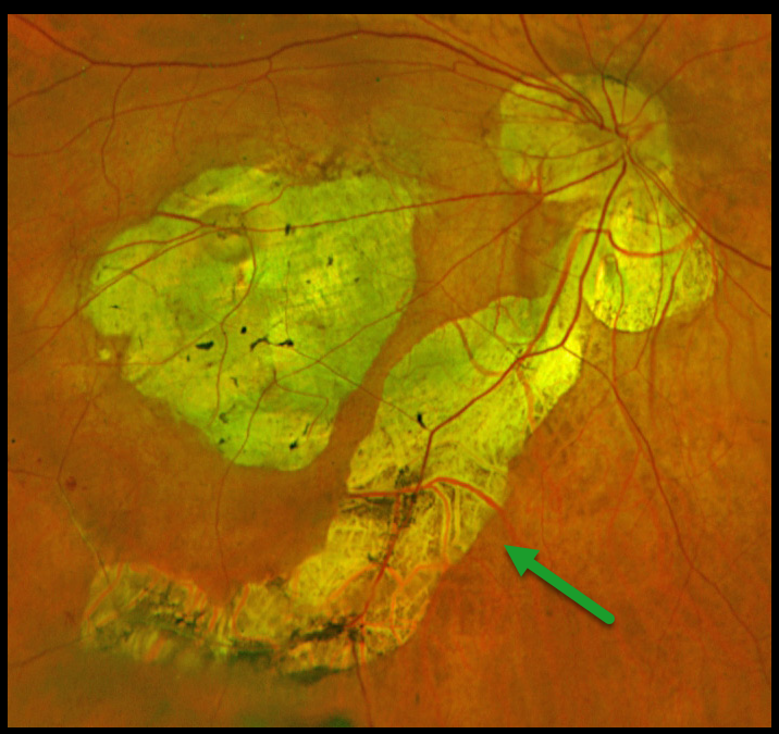
Optos color RG imaging of this highly myopic patient shows severe myopic degeneration extending from the central macula inferiorly, along with peripapillary atrophy that extends beneath the entire inferotemporal arcade.
Learning Points:
Perivascular chorioretinal atrophy is a newly described finding in highly myopic eyes (Benahmed et al, AJO 2025;271:498-506). The atrophy occurs along the first few bifurcations of the major retinal vessels and always develops in eyes with a posterior staphyloma. An anterior scleral protrusion along the retinal vessel is seen in 74% of eyes, suggesting that anomalous scleral curvature may cause this unusual atrophy.
OPTIC NERVE CONUS + PERIPAPILLARY RETINAL DETACHMENT
Originally posted on @retina.rocks March 13, 2024
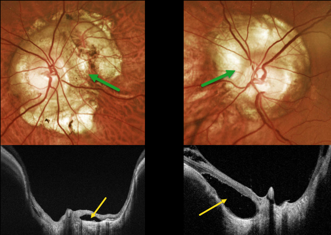
This 42YO male with high myopia was referred for asymptomatic optic nerve changes. Vision was 20/30 OD and 20/25 OS.
Optos color RG imaging shows bilateral peripapillary atrophy. Clinically, this scarring is markedly excavated from the surrounding retinal plane. Triton swept-source OCT confirms peripapillary staphyloma. Localized retinal detachments are noted bilaterally, particularly within the nasal aspect of atrophy around the left nerve. Within these detachments, there is variable loss of RPE, absence of chorioid, and variable retinal disorganization.
Learning Points:
An optic nerve conus, most commonly found in high myopes, is a staphylomatous-like depression of the optic nerve and surrounding tissue.
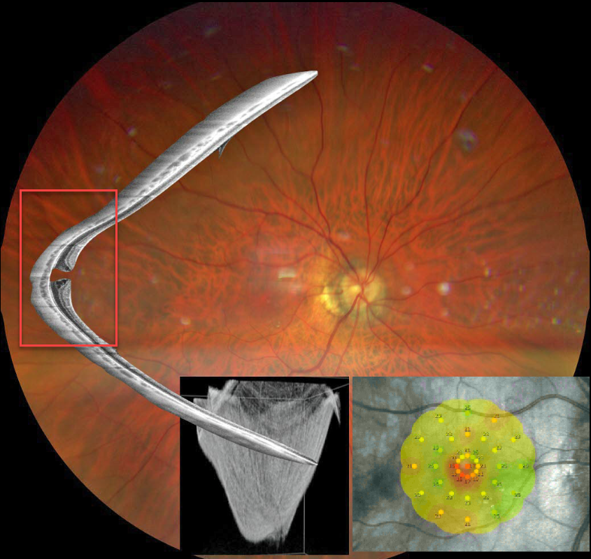
STAPHYLOMA + MACULAR HOLE
Barbara Parolini and Veronika Matello
Originally posted on @retina.rocks February 13, 2024
This Eyecare Clinic (in Brescia, Italy) case was submitted by Barbara Parolini, Veronika Matello, Giulia Freschi, and Roberta Penzani.
This 48YO highly myopic male presented for a sudden drop in vision. Vision was 20/100 OD and 20/20 in OS.
Color photography shows a blunted macular reflex. Widefield OCT shows a conical-shaped staphyloma of the entire posterior pole with a full-thickness macular hole. Microperimetry shows a decreased but not absent foveal sensitivity.
According to the Myopic Traction Maculopathy Staging System (Parolini et al, Eur J Ophthalmol 2021;31:1299-1312), this eye is in Stage 1c. The left eye showed a similar staphyloma but without a macular hole (not shown). The patient declined surgery.
Learning Points:
A macular staphyloma consists of a concave posterior bowing of the eye wall in patients with high myopia. These myopic patients also have a markedly thinned choroid throughout the posterior pole.
Due to stretching of the posterior retinal structures, these eyes are at increased risk of numerous findings, including macular schisis/traction and macular neovascularization.
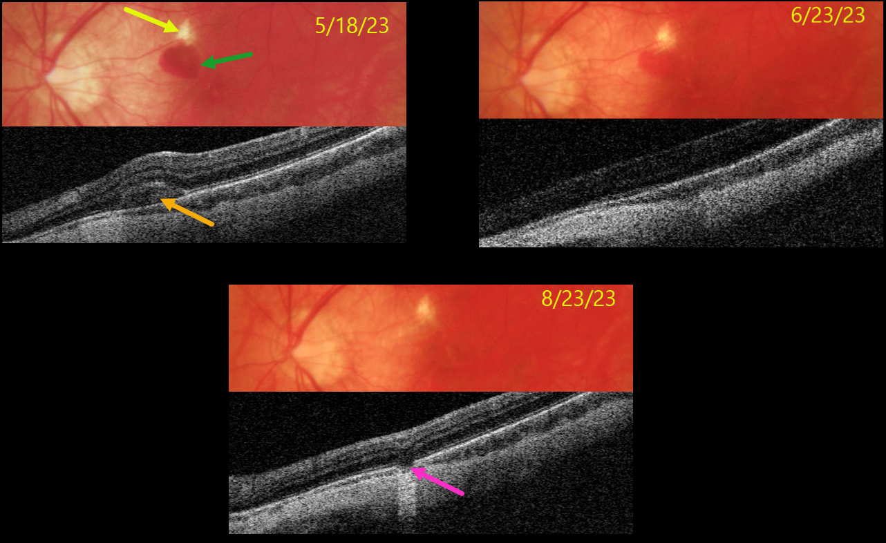
LACQUER CRACK
Originally posted on @retina.rocks November 13, 2023
This 33YO highly myopic male presented on 5/18/23 with vision loss in his left eye. Vision was 20/40.
Color photography shows a superior macular lacquer crack just superior to a round subretinal hemorrhage. The blood is hyperreflective on Triton swept-source OCT. The blood gradually absorbed with observation.
When last examined on 8/23/23, the blood had completely resolved, and vision remained at 20/40, but with resolved symptoms. OCT scanning through the lacquer crack shows a defect in the outer retinal bands and RPE.
Learning Points:
Lacquer cracks are a common finding in patients with high myopia. They consist of breaks in Bruch’s membrane due to the outpouching/stretching of the pathologically myopic eye and increase the risk for macular neovascularization. In some cases, similar to this patient, self-limiting small subretinal avascular hemorrhages occur that do not require anti-VEGF therapy.
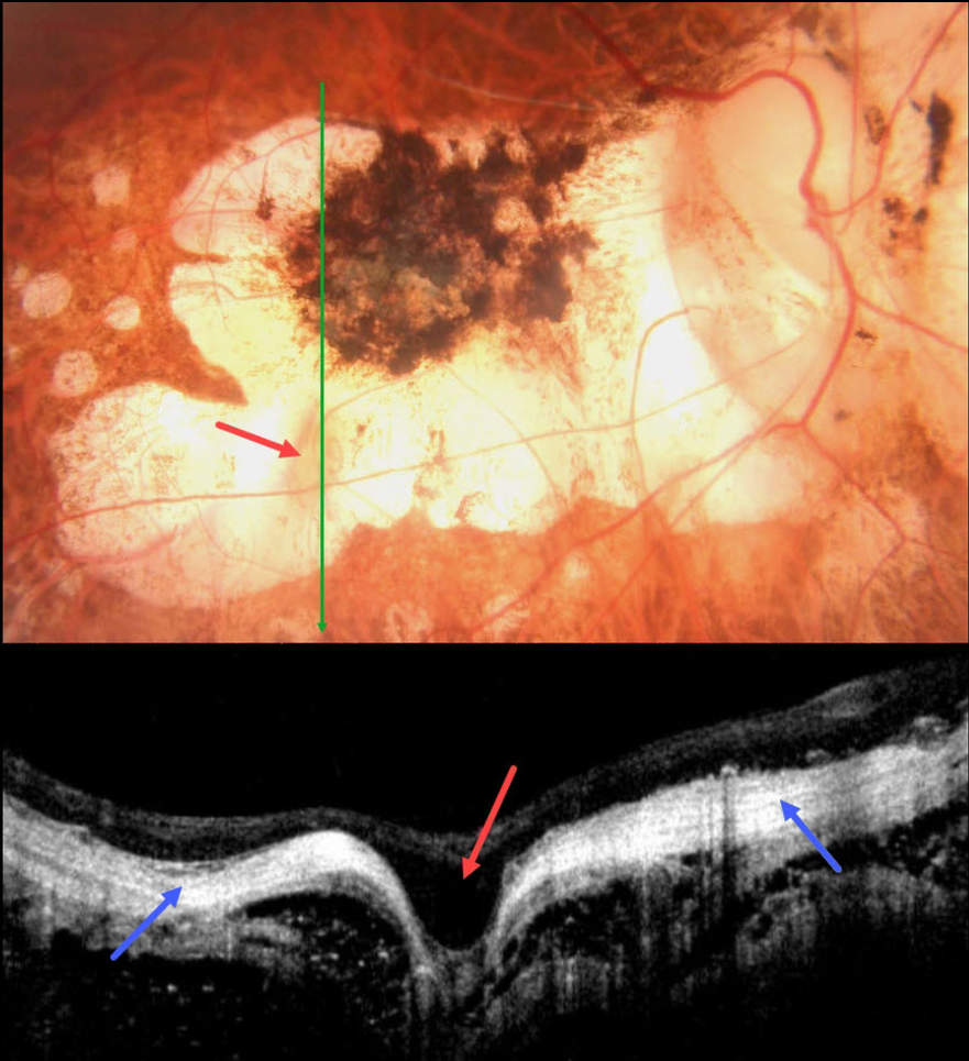
HIGH MYOPIA
Asma Samsudeen and Ashish Sharma
Originally posted on @retina.rocks September 14, 2023
This 58YO male with a history of degenerative myopia presented with 6 months of vision loss in his right eye. He was blind in his left eye following vitrectomy surgery 7 years earlier. Vision was counting fingers OD.
Color photography shows severe variably pigmented atrophic myopic degeneration. An area of orange coloration is seen within the larger area of atrophy inferiorly.
On OCT, this area appears as a focal staphyloma. The overlying retina is draped across the staphyloma. The choroid is so thin as to be invisible on OCT, with a hyperreflective underlying sclera. The orbital tissue is well seen posterior to the sclera.
Learning Points:
Degenerative myopia is a major cause of worldwide visual impairment. Due to the stretching of the posterior retinal structures, there is an increased risk for numerous findings, including atrophic retinal and choroidal thinning, posterior staphyloma, macular schisis/traction, and macular neovascularization (see Ohno-Matsui et al for a recent review, Invest Ophthalmol Vis Sci 2021;62(5);5)..
Initially described by Freund et al (Arch Ophthalmol 2003;121:197-204), intrachoroidal cavitations are most commonly found in high myopes as a peripapillary yellow-orange subretinal lesion. It is difficult to determine whether our patient has a cavitation above the staphyloma.
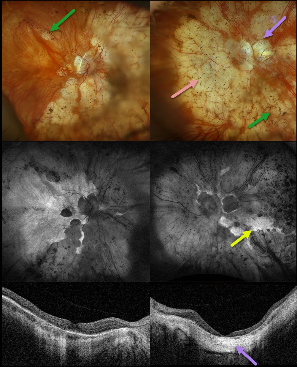
RETINITIS PIGMENTOSA + MYOPIC DEGENERATION
Originally posted on @retina.rocks August 30, 2023
This 78YO female has been followed by us for several years for retinitis pigmentosa (RP) and degenerative myopia. Vision is stable at 20/30 OD and hand motion OS. Multimodal imaging shows a fascinating fusion of findings for these two disorders.
Optos color RGB imaging shows typical RP findings of bone spicules (intraretinal pigment migration) extending from outside the arcades into the peripheries, superimposed on severe degenerative myopic changes.
Fundus autofluorescence (FAF) shows hypo-FAF within the areas of myopic atrophy, with hyper-FAF more peripherally from the RP-induced outer retinal atrophy.
A macular staphyloma is also seen in the left eye and confirmed on swept-source OCT. The degenerative myopic changes just spare the right macular center, which has no staphyloma.
Learning Points:
Genetic testing showed pathogenic heterozygous mutations for OCA2 and RPGR. Fortunately, the RP spares each macula. Her myopic degeneration has been stable, and we are hopeful that her central vision will be preserved in her right eye.
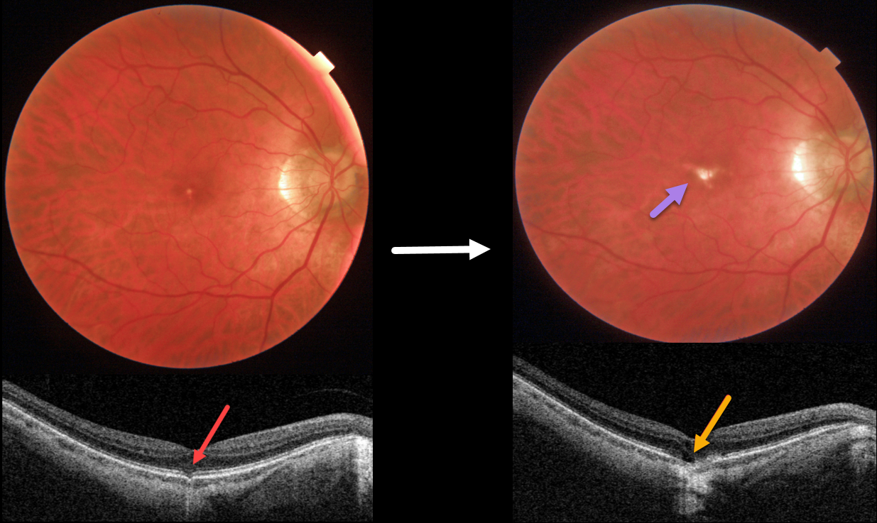
OUTER FOVEAL MICRODEFECT
Originally posted on @retina.rocks April 13, 2023
This 58YO male was referred for an asymptomatic foveal lesion in his right eye. There was a history of high myopia prior to LASIK surgery years earlier. Vision was 20/40 OD.
Color photography shows a small atrophic foveal scar. OCT shows a focal EZ defect with a possible tiny choroidal excavation. He returned about 2 years later for a routine follow-up visit.
Although he was binocularly without a new complaint, his vision had decreased to 20/200. There was now a definite, much enlarged foveal lacquer crack. OCT scanning shows a new outer foveal microdefect.
Learning Points:
Outer foveal microdefects (Cohen et al, Ophthalmology Retina 2021;5:553-561) have been described in numerous conditions, including macular telangiectasia, tamoxifen use, ABCA4 disorders (cone-rod dystrophies, Stargardt disease, and fundus flavimaculatus), phototoxicity, trauma, and vitreomacular traction disorders.
We are not aware of a prior report showing these lesions in association with myopic lacquer cracks. These defects, caused by focal loss of the outer retinal layers, leave an optically empty space that can be mistaken for subretinal fluid but is more of an optical gap within the outer retina.
Focal choroidal excavation (FCE), originally described by Jampol et al in 2006, is likely part of the pachychoroid spectrum since it is usually associated with a thickened choroid and pachyvessels. The FCE can be observed, although symptomatic manifestations, including active central serous retinopathy or macular neovascularization, often require treatment. An excellent review is by Verma et al, Br J Ophthalmol 2021;105:1043-1048.
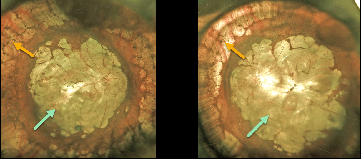
HIGH MYOPIA WITH STAPHYLOMA
Originally posted on @retina.rocks April 4, 2023
This 72YOM has a lifelong history of severe degenerative myopia. Vision was hand motion bilaterally.
Optos color imaging of both eyes shows confluent myopic posterior staphylomas and chorioretinal atrophy extending into each retinal midperiphery. Encircling cobblestone degeneration is seen more anteriorly.
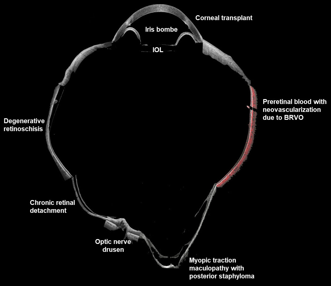
WIDEFIELD OCT
Veronika Matello and Barbara Parolini
Originally posted on @retina.rocks April 1, 2023
This composite widefield OCT image combines multiple pathologies to create a quite sick virtual eye. All the scans were taken with the Canon Xephilio S1.
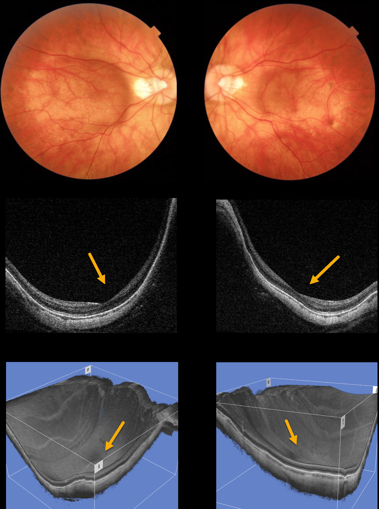
HIGH MYOPIA
Originally posted on @retina.rocks January 6, 2023
This 42YO male has a history of high myopia, of -14 diopters OD and -12.5 diopters OS. Vision is 20/60 OU.
Color photographs show bilateral macular staphyloma. Swept-source OCT B-scans and 3-D reconstruction more dramatically show the macular staphylomas.
Learning Points:
A macular staphyloma is a concave posterior bowing of the sclera in patients with high myopia. These myopic patients also have a markedly thinned choroid throughout the posterior pole.
Due to stretching of the posterior retinal structures, these patients are at increased risk of numerous findings, including macular schisis/traction and macular neovascularization (MNV).
MACULAR HOLE
Veronika Matello and Barbara Parolini
Originally posted on @retina.rocks October 25, 2022
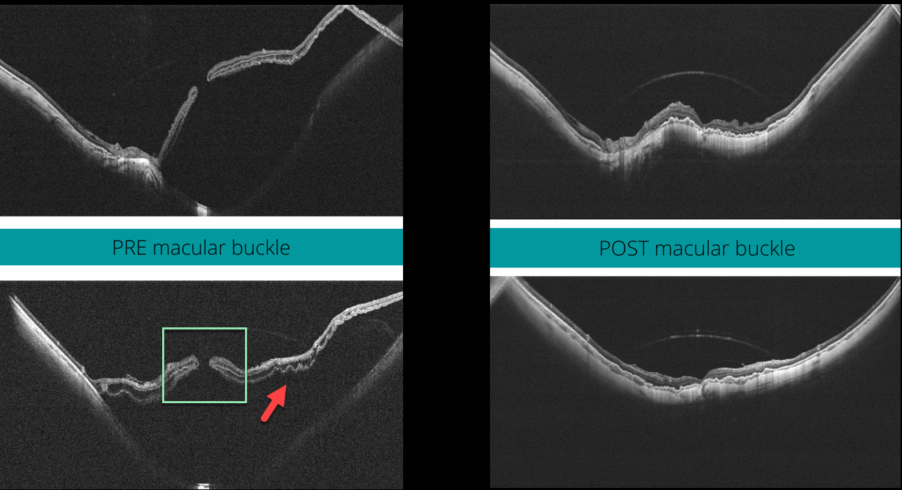
This 58YO highly myopic female presented with 20/400 vision in her left eye. The axial length was 32.45 mm, and her refractive error prior to LASIK surgery was 13D.
Widefield OCT scanning (Canon Xephilio SQ) shows a large macular retinal detachment with (or associated to) a full-thickness macular hole, defined as stage 4c according to the Myopic Traction Maculopathy Staging System (Parolini et al, Eur J Ophthalmol 2021;31:1299-1312).
Pars plana vitrectomy with an ILM flap and macular buckling was performed. One month postoperatively, the retina was flat, and the macular hole closed. Vision was 20/80.
Learning Points:
In our experience, these detachments are totally tractional, with the macular hole developing before or after the retina detaches. The macular buckle treats both the macular schisis and detachment, whereas the vitrectomy with ILM peeling closes the macular hole.
HIGH MYOPIA WITH AVASCULAR RETINAL HEMORRHAGES AND LACQUER CRACK FORMATION
Originally posted on @retina.rocks January 31, 2022
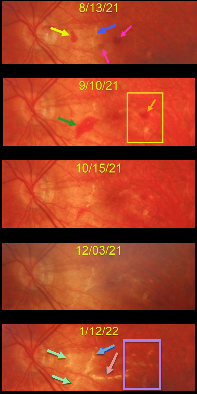
This 23YO high myopic female presented on 8/13/21 with some symptomatic central and paracentral scotomas. Vision was 20/80.
There were a few round subretinal hemorrhages near the central macula, along with an asymptomatic nasal macular hemorrhage. A few small lacquer cracks were noted.
She was then monitored monthly with an unusual rapid succession of new and resolving hemorrhages with subsequent lacquer crack development.
On 9/10/21, the prior hemorrhages were resolving, but a new area of subretinal blood was noted along the inferior edge of the previously noted lacquer crack. There was also a new hemorrhage in the distal macula. These hemorrhages all resolved by the 12/3/21 exam.
When last seen on 1/12/22, in the areas of prior subretinal blood, there are new lacquer cracks, enlargement of the originally noted lacquer crack, and a large new horizontal lacquer crack inferior to the fovea. In the region of the distal temporal macular hemorrhage noted on 9/10/21, there are numerous new lacquer cracks. Vision was 20/60, and we continue to monitor her for macular neovascularization (MNV).
Learning Points:
Lacquer cracks are a common finding in patients with high myopia. They consist of breaks in Bruch’s membrane due to the outpouching/stretching of the pathologically myopic eye and increase the risk for MNV.
In some cases, similar to this patient, they can have small avascular subretinal hemorrhages, not associated with neovascularization, and are monitored. These are best confirmed with fluorescein angiography. In cases with an active membrane, they are treated with anti-VEGF injections.
HIGH MYOPIA
Originally posted on @retina.rocks August 19, 2021
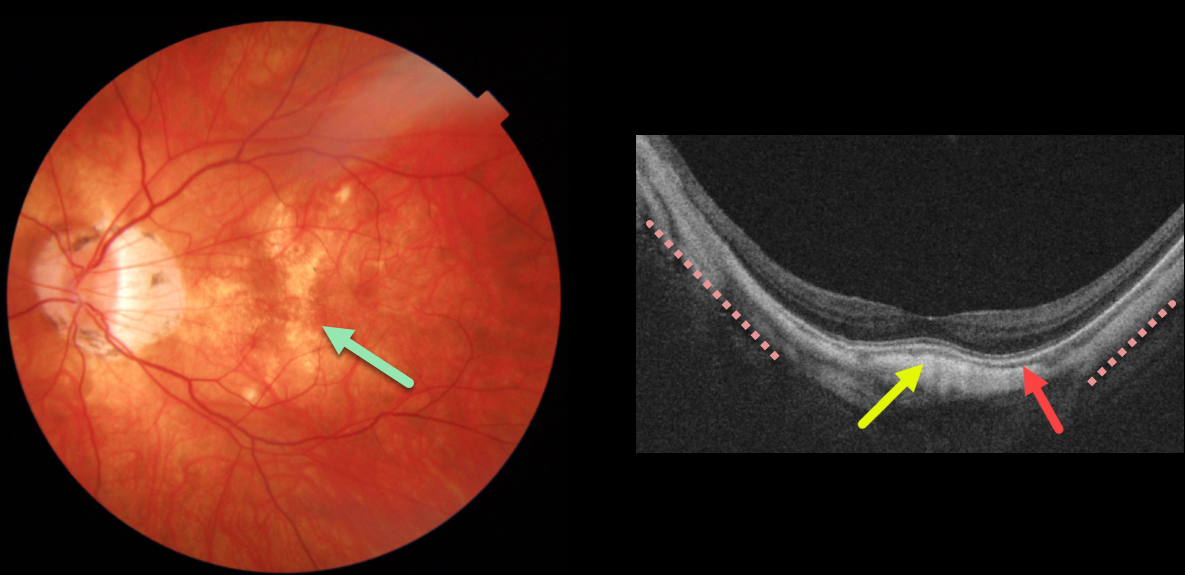
This high myope has extensive dry myopic atrophic pigmentary changes noted on Triton color imaging.
Triton swept-source OCT shows typical myopic concave posterior bowing of the eye wall, except more centrally, where there is a convex elevation beneath the fovea. The choroid is also markedly thinned throughout the posterior pole.
Learning Points:
Originally described in 2008 by Gaucher et al (AJO 2008;145:909-914), a dome-shaped macula is a macular convexity found in eyes with high myopia, often associated with a posterior staphyloma. It can be associated with avascular subretinal fluid and macular neovascularization.
See Kumar et al for an excellent recent review of this entity (Surv Ophthalmology 2021;66:560-571).
HIGH MYOPIA
Originally posted on @retina.rocks July 27, 2020
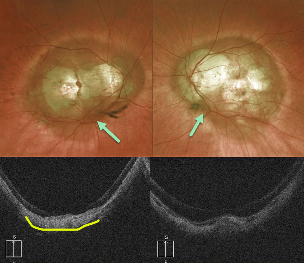
Our highly myopic patient has a fairly symmetrical bilateral posterior staphyloma that encircles each optic nerve and macula. Vision is counting fingers OD and 20/50 OS.
OCT demonstrates steep scleral curvature and outpouching.
Learning Points:
Posterior staphyloma is a weakening, thinning, and outpouching of the sclera most commonly seen in degenerative myopia.
See Moriyama et al Ophthalmology 2011;118;1626-1637 for some amazing 3D MRI reconstructions of myopic staphylomas.
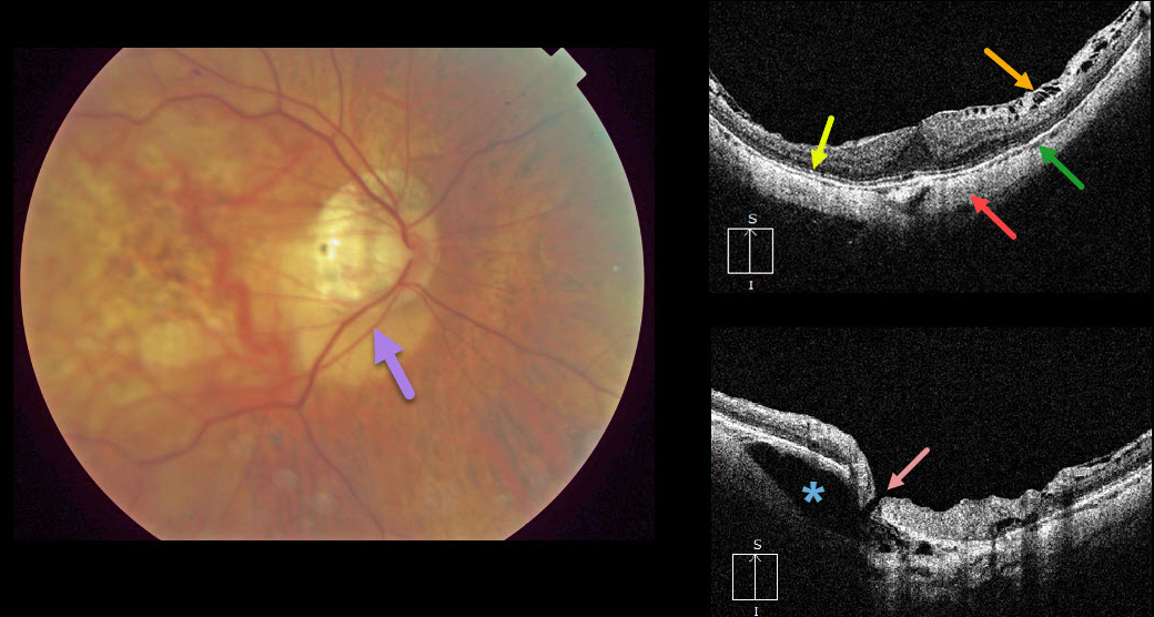
HIGH MYOPIA & INTRACHOROIDAL CAVITATION
Originally posted on @retina.rocks July 9, 2020
Peripapillary intrachoroidal cavitation is a relatively rare abnormality most commonly found in high myopia. Clinically, these subretinal lesions have an orange coloration and are usually located inferior to the optic nerve.
OCT scanning of the central macula shows a variety of typical myopic findings, including posterior bowing of the sclera, variable attenuation of the ellipsoid zone, markedly thinned choroid, and a variable epiretinal membrane with inner retinal schisis most prominent superiorly.
OCT scanning centered over the optic nerve reveals the intrachoroidal cavitation. There also seems to be a tiny full-thickness retinal defect that communicates directly with the cavitation.
Learning Points:
To learn more about intrachoroidal cavitations and high myopia, see Spaide et al Retina 2012;32;1037-1044.
See Ikuno Retina 2017;37;2347-2351 for a nice overview of macular findings in high myopia.


