

High Myopia MNV
<< Back to Cases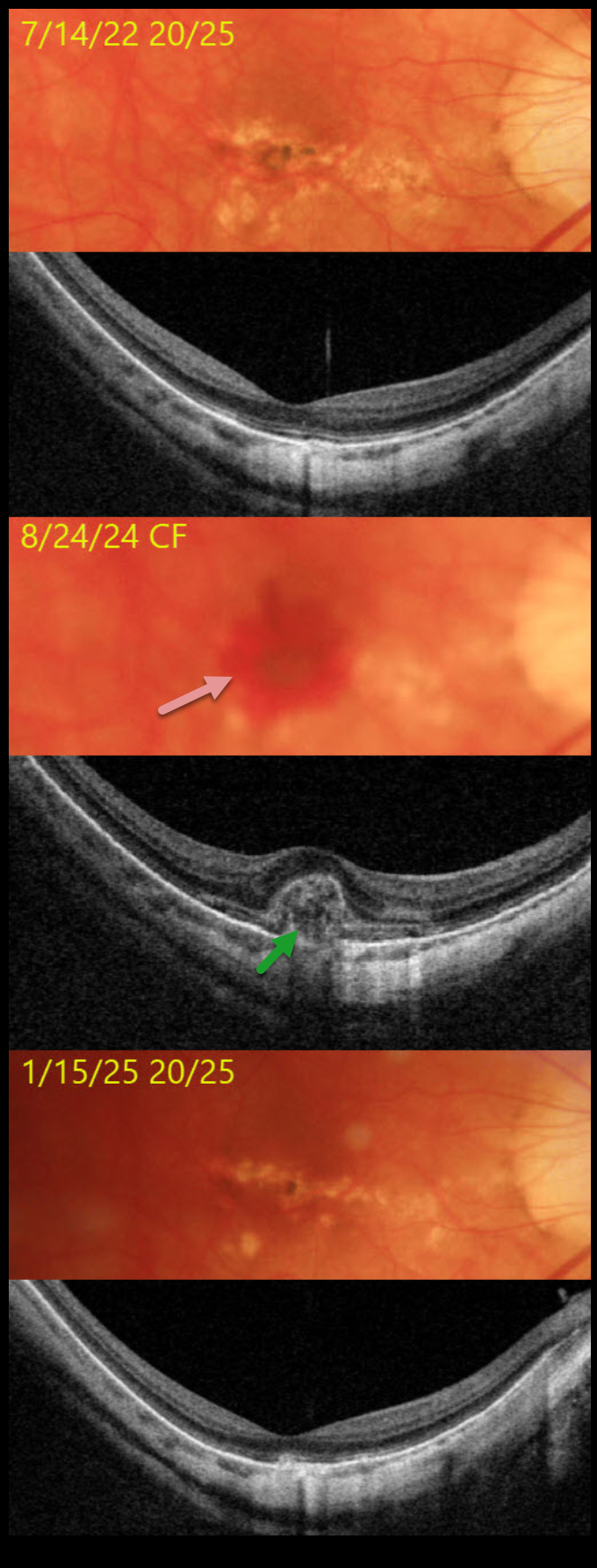
MYOPIC MACULAR NEOVASCULARIZATION
Originally posted on @retina.rocks August 6, 2025
This 53YO highly myopic male was seen on 7/14/22 for a routine exam with 20/25 vision. Triton color imaging shows dry myopic degeneration, macular pigment loss, and clumping. Triton swept-source OCT shows variable loss of the outer retinal bands and diffuse choroidal thinning.
He returned on 8/24/24 with vision loss from a counting-fingers submacular hemorrhage. OCT shows hyperreflective subretinal blood elevating and extending into the outer retina. Following monthly intravitreal Avastin injections, vision improved to 20/25 on 1/15/25. All blood resolved, and the OCT mostly returned to its pre-blood appearance.
Learning Points:
In our experience, myopic macular neovascularization (MNV) presents quite differently from neovascular AMD. Significant subretinal blood (as in our patient), subretinal fluid, or intraretinal fluid are rarely seen. Often, an active MNV will present as a new dot of subretinal blood or pigment. Occasionally, there will just be symptomatic vision loss or metamorphopsia without clinical or OCT findings. Anti-VEGF therapy is effective, and these lesions can often be managed with PRN therapy, which is what we recommended for our patient (Sakata et al, Retina 2023;43:1863-1871).
MYOPIC MACULAR NEOVASCULARIZATION
Originally posted on @retina.rocks January 1, 2024
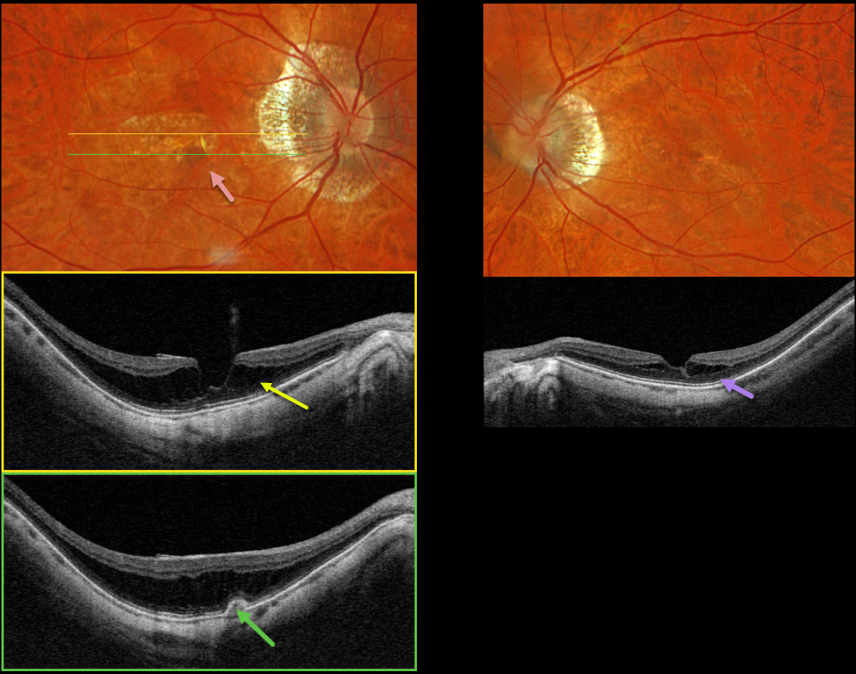
This 72YO female recently moved to our area and self-referred herself to establish new retinal care. One year earlier, she received a series of five monthly anti-VEGF injections OD for myopic macular neovascularization (MNV), and felt that her vision was stable since the last injection about 6 months earlier. Vision was 20/60 OD and 20/40 OS.
Optos color RGB imaging OD shows myopic peripapillary atrophy, central macular pigment loss, and a pigmented subretinal lesion.
Triton swept-source OCT through the macular center shows a shallow staphylomatous posterior bowing of the sclera with overlying outer macular schisis. The choroid is diffusely thinned. Scanning the pigmented lesion reveals a small type 1 MNV (below the RPE) without fluid. Imaging of her left eye shows much milder, central schisis in the outer plexiform layer. Observation was recommended.
Learning Points:
In our experience, myopic MNV presents quite differently from those with neovascular AMD. Significant subretinal blood, subretinal fluid or intraretinal fluid are rarely seen.
Often, an active MNV will present as a new dot of subretinal blood or pigment. Occasionally, there will just be symptomatic vision loss or metamorphopsia without clinical or OCT findings.
Anti-VEGF therapy is effective, and these lesions can often be managed with PRN therapy (Sakata et al, Retina 2023;43:1863-1871) as in our case.
MYOPIC MACULAR NEOVASCULARIZATION
Originally posted on @retina.rocks September 22, 2022
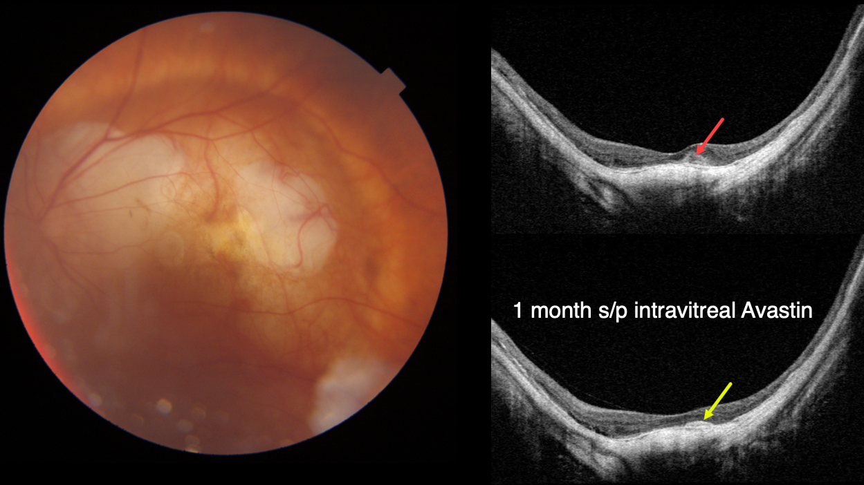
This 92YO female presented with new vision loss of 20/200 in her left eye. She has a history of high myopia, with hand motion vision OD due to a macular staphyloma. Baseline vision in her left eye is 20/40.
Fundus photography shows peripapillary and mostly superior macular atrophy.
Swept-source OCT shows a posterior staphyloma with a dome appearance to the central macula. A new small hyperreflective macular neovascularization (MNV) is noted.
One month following an intravitreal Avastin injection, vision improved to 20/40 with shrinkage of the MNV.
Learning Points:
In our experience, myopic MNV presents quite differently from those found in neovascular AMD.
Significant subretinal blood, subretinal fluid or intraretinal fluid are rarely seen. Often an active MNV will present as a new dot of subretinal blood or pigment.
Occasionally there will just be symptomatic vision loss or metamorphopsia without clinical or OCT findings.
Anti-VEGF therapy is effective, and these lesions can often be managed with PRN therapy.
HIGH MYOPIA
Originally posted on @retina.rocks August 11, 2022
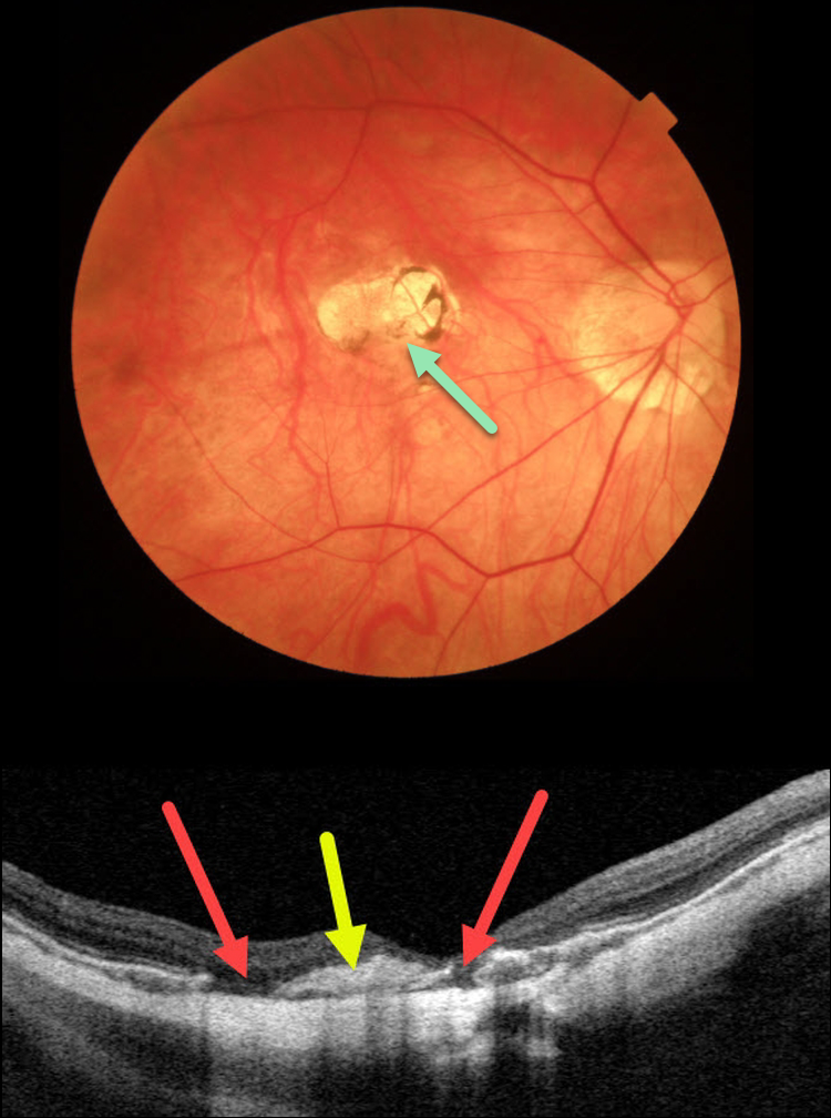
This 48YO female has a history of a previously treated myopic macular neovascularization (MNV) in her right eye, which has remained stable with observation for years. Triton color imaging shows an inactive atrophic foveal scar.
Swept source OCT shows an inactive hyperreflective central MNV with adjacent RPE defects. The choroid is severely thinned, consistent with her high myopia.
Learning Points:
An RPE aperture is a rare, recently described OCT finding most commonly seen in non-exudative AMD. Different from an RPE tear, these round RPE discontinuities typically develop from an avascular RPE detachment that remodels over time, resulting in atrophy of the RPE and outer retina.
This remodeling may result from regression of drusenoid material or damage to the RPE due to separation from the choriocapillaris (Querques G et al. Retina. 2016;36:S65-S72).
Some hypothesize that these lesions may represent small RPE micro-tears from underlying type 1 MNV causing increased hydrostatic pressure, leading to damage/atrophy of the RPE.
RPE apertures can also be found in patients with acquired vitelliform lesions (Falavarjani K et al, Canadian J Ophthalmol 2022;57:e91-e94).
We are not aware of a previously described case caused by myopic MNV.
MYOPIC MACULAR NEOVASCULARIZATION
Originally posted on @retina.rocks July 26, 2022
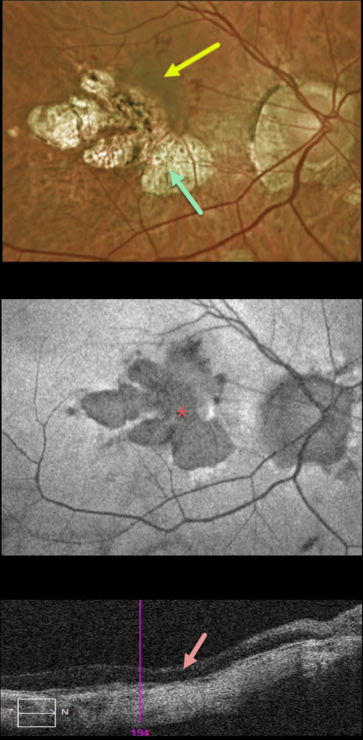
This 51YO female received prior intravitreal injections in her right eye about 5 years earlier, but was subsequently lost to follow-up until she was referred to our office. She felt that her vision had been stable and poor in each eye for years. Vision was counting fingers OD and 20/200 OS.
Optos color imaging of her right eye shows myopic peripapillary and macular scarring extending through the macular center. A pigmented macular neovascularization (MNV) with some dots of subretinal blood extends superonasally from the atrophy. Fundus autofluorescence (FAF) shows areas of hypo-FAF corresponding to the areas of clinical atrophy. Triton swept-source OCT shows central retinal atrophy.
Given the foveal atrophy, severe vision loss, and absence of acute symptoms, we recommended careful observation. We will consider anti-VEGF therapy if the MNV causes increasing exudation.
MYOPIC MACULAR NEOVASCULARIZATION
Originally posted on @retina.rocks February 24, 2021
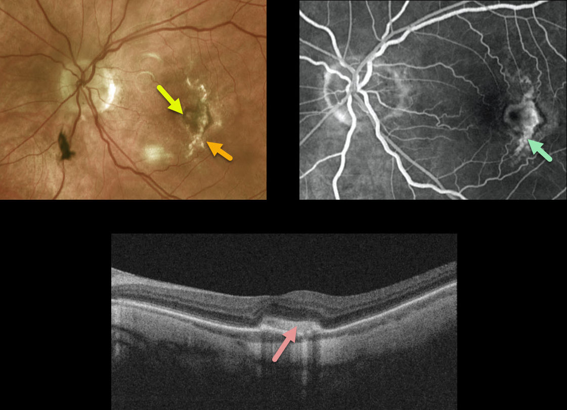
This 69YO female came in for a routine exam with a few months of vision loss in her left eye.
There is a new small area of subfoveal pigment adjacent to an old curvilinear scar more temporally. Although this temporal scar appeared to represent a choroidal rupture, she denied any prior trauma.
Axial length is 27.30 mm in this eye, nearly 1 mm greater than her fellow eye, indicating that this scar is likely a large and unusual lacquer crack.
Fluorescein angiography (FA) shows staining of the temporal scar and subfoveal pigment. An OCT B-scan through the subfoveal pigment shows a type 2 macular neovascularization (MNV) without fluid.
Learning Points:
Myopic MNV often presents as a pigmented subretinal lesion, often with minimal to no fluid due to the underlying thinned choroid.
In our experience, the presence or absence of new blood or new visual symptoms is more useful than the OCT, which usually shows no fluid, or FA, which often shows no leakage. Anti-VEGF injections were therefore recommended.


