

HIV
<< Back to Cases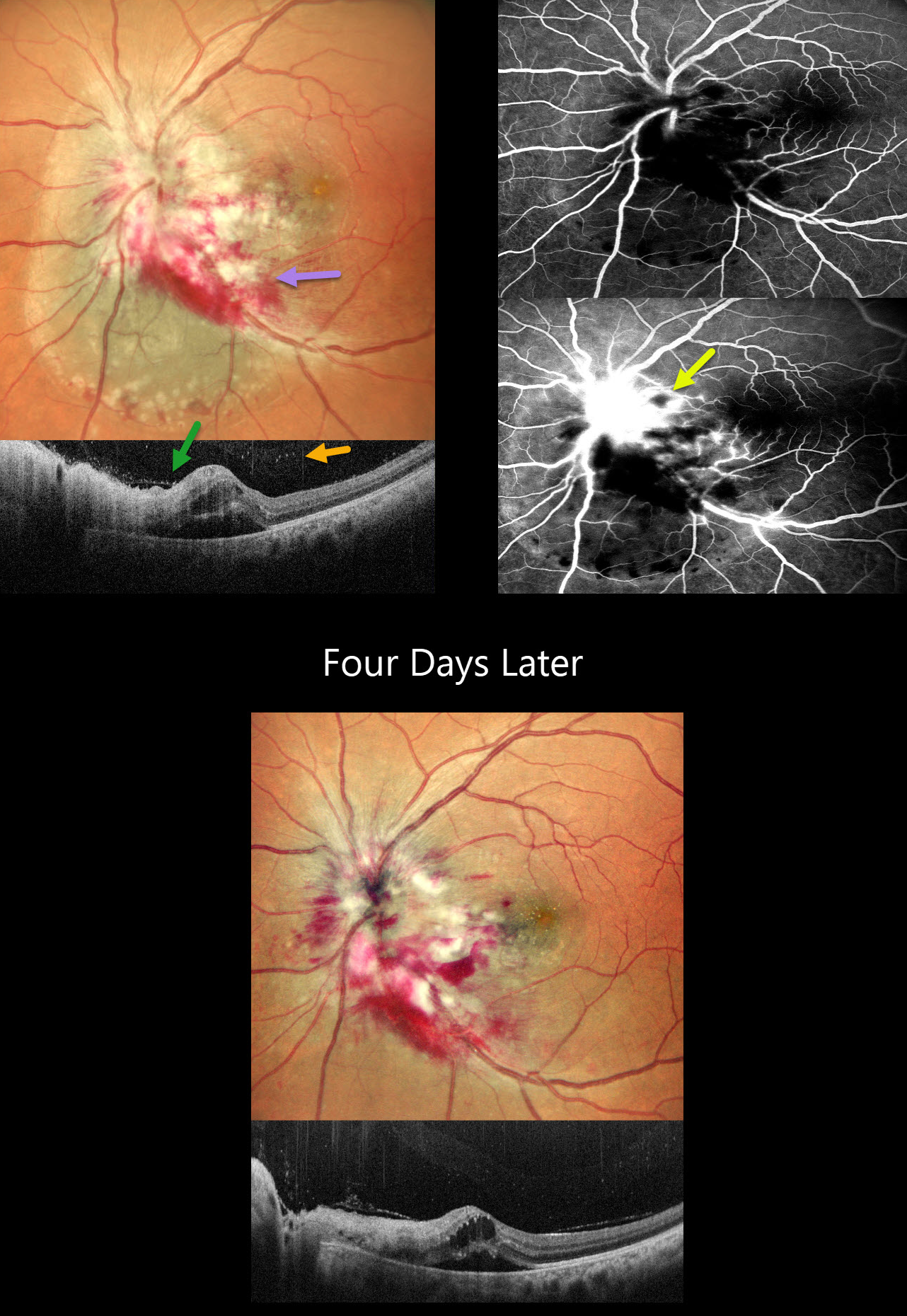
CMV PAPILLITIS AND RETINITIS
Akansha Sharma and Manish Nagpal
Originally posted on @retina.rocks September 18, 2025
This 42YO female with a known history of HIV infection presented with 1 month of vision loss in her left eye. Her last CD4 count was 38 despite being compliant with HAART therapy. Her husband was HIV positive. Vision was 20/20 in her normal OD and 20/400 in OS.
Multicolor SLO imaging shows active cytomegalovirus (CMV) retinitis involving the nerve, which extends nasally and inferotemporally. Subretinal fluid is noted clinically and on OCT. OCT also shows full-thickness disorganization and hyperreflectivity, along with overlying vitreous cells. Fluorescein angiography shows early blockage with late disc and retinal leakage.
Oral valganciclovir and biweekly intravitreal injections were started. Four days later, vision improved to 20/90 with improvement in the papilledema and retinitis.
Learning Points:
CMV retinitis develops as a reactivation of latent CMV in immunosuppressed individuals. Before effective antiretroviral treatment emerged in the mid to late 1990’s, CMV retinitis developed in up to 40% of HIV/AIDS patients, often within the last 6 months of life. Treatment includes a combination of systemic and intravitreal medications, including ganciclovir, foscarnet, and cidofovir. Rhegmatogenous retinal detachment is a common late sequela and requires vitrectomy with silicone oil.
CMV RETINITIS
Rohan Jain and Manish Nagpal
Originally posted on @retina.rocks February 6, 2025
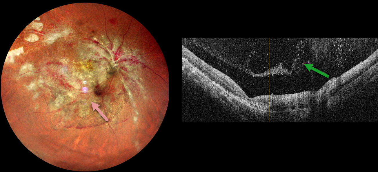
This 34YO male with a known history of HIV infection presented with 1 month of vision loss in his right eye. He was treated with a six-month course of systemic anti-tuberculous therapy for pulmonary tuberculosis two years earlier. One month earlier, he was diagnosed with HIV AIDS with a CD4 count of 43 and was started on systemic anti-retroviral therapy. Vision was counting fingers OD and 20/20 in his normal OS.
Multicolor imaging shows variably active cytomegalovirus (CMV) retinitis throughout the right posterior pole, including patches of necrotic white retina and retinal hemorrhages. The fundus is ischemic with sheathed arterioles. OCT scanning shows a vitreous separation with extensive vitreous cells.
Learning Points:
CMV retinitis develops as a reactivation of latent CMV in immunosuppressed individuals. Before effective antiretroviral treatment emerged in the mid to late 1990’s, CMV retinitis developed in up to 40% of HIV/AIDS patients, often within the last 6 months of life. Treatment includes a combination of intravenous and intravitreal medications, including ganciclovir, foscarnet, and cidofovir. Rhegmatogenous retinal detachment is a common late sequela and requires vitrectomy with silicone oil.
Our patient’s funduscopic findings are classic for CMV retinitis. However, patients with this degree of retinitis will usually have bilateral findings, and our patient’s left fundus was normal. These immunosuppressed patients also cannot mount an adequate immune response and therefore usually have a quiet vitreous, unlike our patient. It is therefore possible that there may also be an underlying tuberculous component. Unfortunately, our patient was immediately lost to follow-up before we could start intravitreal and systemic CMV therapy.
CMV RETINITIS WITH RETINAL DETACHMENT
Originally posted on @retina.rocks September 2, 2024
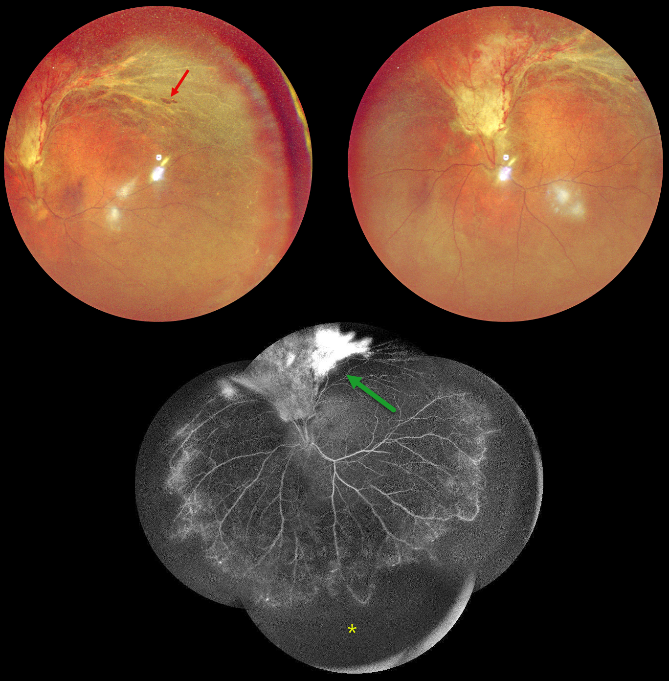
This 48YO HIV positive male on highly active antiretroviral therapy (HAART) presented with 2 months of vision loss in his left eye. His last CD4 count was 81. Vision was 20/20 OD and counting fingers OS.
Multicolor imaging shows active cytomegalovirus (CMV) retinitis with retinal detachment extending superiorly from the optic nerve into the superotemporal midperiphery. A few atrophic retinal breaks are noted. Fluorescein angiography of his left eye shows variable leakage within the area of retinitis superiorly and severe encircling peripheral ischemia. Vitrectomy with silicone oil was scheduled to repair the detachment in his left eye.
Learning Points:
CMV retinitis develops as a reactivation of latent CMV in immunosuppressed individuals. Before effective antiretroviral treatment emerged in the mid to late 1990’s, CMV retinitis developed in up to 40% of HIV/AIDS patients, often within the last 6 months of life. Treatment includes a combination of intravenous and intravitreal medications, including ganciclovir, foscarnet and cidofovir. Rhegmatogenous retinal detachment is a common late sequela and requires vitrectomy with silicone oil.
CMV RETINITIS
Originally posted on @retina.rocks October 23, 2020
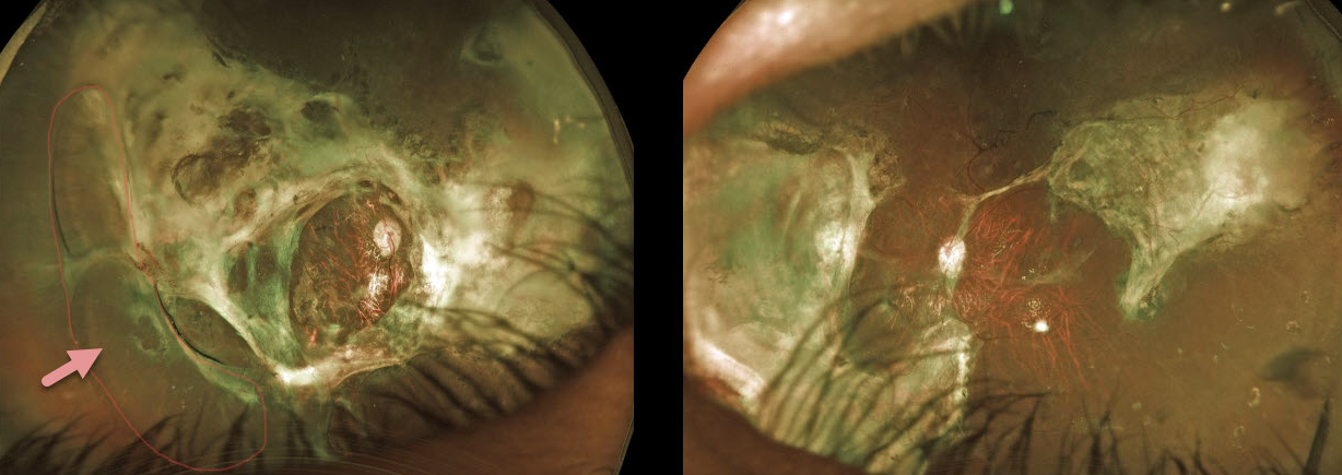
This 29YO male presented to us 6 years earlier with bilateral cytomegalovirus (CMV) retinal detachments. At that time, he was severely ill from untreated HIV/AIDS.
He underwent bilateral vitrectomy surgery with silicone oil. Since then, he has done extremely well systemically, with normal CD4 counts and undetectable viral loads.
Despite severe bilateral chorioretinal scarring and a partial peripheral detachment under oil OD, vision is 20/400 bilaterally, and he is functioning well.
We have no plans to remove the oil, as his retinal appearances have remained stable since surgery.
Learning Points:
Cytomegalovirus (CMV) retinitis develops as a reactivation of latent CMV in immunosuppressed individuals. Before effective antiretroviral treatment emerged in the mid to late 1990’s (highly active antiretroviral therapy, HAART), CMV retinitis developed in up to 40% of HIV/AIDS patients, often within the last 6 months of life.
Treatment includes a combination of intravenous and intravitreal medications, including ganciclovir, foscarnet, and cidofovir.
Rhegmatogenous retinal detachment is a common late sequela of CMV infection and may require vitrectomy for repair.
HIV RETINOPATHY
Originally posted on @retina.rocks September 24, 2020
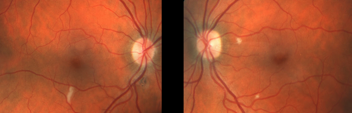
Our patient was HIV positive with a CD4 count of 9.
There is an isolated nerve fiber layer infarct (cotton wool spot, CWS) in the right eye and two CWS in the left eye.
Learning Points:
Cotton-wool spots are nonspecific and are found in many disorders, including hypertensive and diabetic retinopathy. They can also be seen with HIV/AIDS when the CD4 count is under 200.
The etiology of CWS in HIV/AIDS is not known, although they do not contain virus particles.
PNEUMOCYSTIS CHOROIDITIS
Originally posted on @retina.rocks September 11, 2020
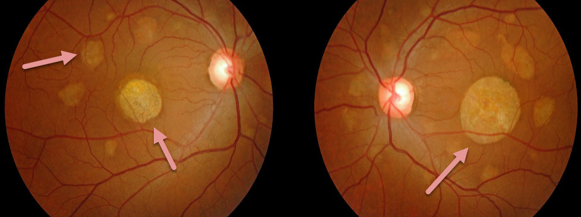
This patient had a known history of treated HIV infection and presented with asymptomatic bilateral irregularly round patches of choroidal and RPE pigmentary loss.
Our patient’s findings show what the lesions of pneumocystis choroiditis look like upon resolution.
Learning Points:
In the early days of the HIV/AIDS epidemic, pneumocystis pneumonia was one of the most common opportunistic infections. Aerosolized pentamidine was therefore given as prophylaxis. The medication, however, did not reach the entire lungs, and patients often had septic pneumocystis emboli from an incompletely treated pneumonia.
Pneumocystis choroiditis was a rare manifestation of this systemic infection, where patients developed multifocal, flat, bilateral choroidal lesions that were usually asymptomatic and had a striking absence of inflammation.
Systemic prophylaxis was subsequently recommended, which better treated the underlying pneumonia and prevented systemic infection.
Highly active antiretroviral therapy (HAART), introduced in the late 1990’s, restores the health of the underlying immune system and has turned this type of finding into something for the history books.
CMV RETINITIS
Originally posted on @retina.rocks June 8, 2020
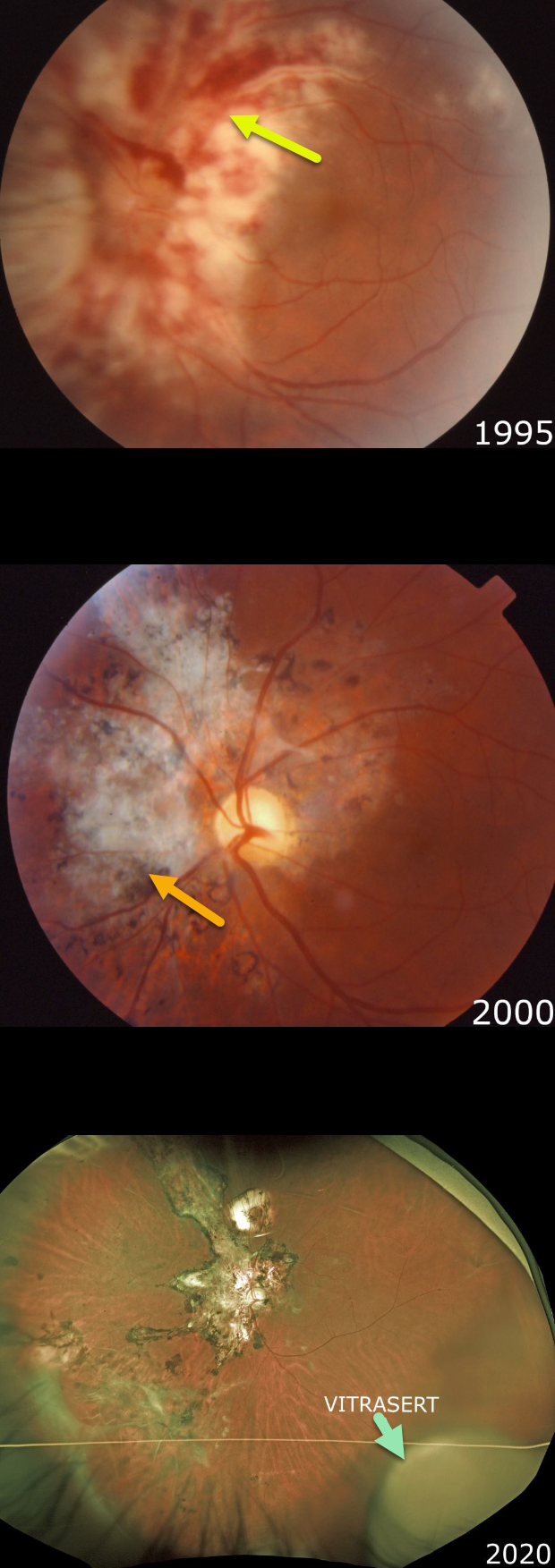
Our patient presented in 1995 with classic active CMV retinitis involving the peripapillary and macular regions of his left eye. We immediately referred him to the University of Kentucky for implantation of a then-investigational sustained-release ganciclovir implant, Vitrasert.
Note the hemorrhagic, whitish retinal necrosis, which often follows a vascular distribution. The retinitis spreads outwards, leaving behind a thinned necrotic retina and RPE.
Following Vitrasert implantation, the retinitis resolved with secondary scarring. He was then started on highly active antiretroviral therapy (HAART).
In 2007, a rhegmatogenous retinal detachment, a common late sequela of CMV infection, was successfully repaired with vitrectomy (not pictured).
As of 2020, he continues to do well with an undetectable viral load and a normal CD4 count. Vision is 20/20 in his normal right eye and 20/400 OS. The ganciclovir Vitrasert implant remains visible in the inferotemporal periphery.
Learning Points:
Cytomegalovirus (CMV) retinitis develops as a reactivation of latent CMV in immunosuppressed individuals. Before effective antiretroviral treatment emerged in the mid to late 1990’s, CMV retinitis developed in up to 40% of HIV/AIDS patients, often within the last 6 months of life.
Treatment includes a combination of intravenous and intravitreal medications, including ganciclovir, foscarnet, and cidofovir.
Vitrasert was approved by the FDA in 1996 and discontinued in 2013 due to patent expiration. The demise of Vitrasert is directly linked to HAART.
As occurred in our patient, pharmacologic restoration of immune function allows the patient’s own immune system to control CMV.
CMV RETINITIS
Originally posted on @retina.rocks March 12, 2020
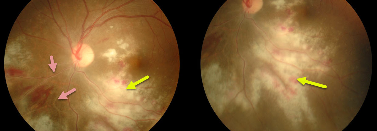
Our patient shows typical CMV retinitis with active fluffy white hemorrhagic retinitis and vasculitis.
This patient is at high risk for severe vision loss due to both the optic nerve and macular center being threatened.
Immediate intravitreal antiviral injections followed by systemic treatment were recommended.
Learning Points:
Cytomegalovirus retinitis (CMV) is a usually devastating infection most commonly seen in end-stage HIV/AIDS.
Fortunately, this is now rarely seen due to the advent of highly active antiretroviral therapy (HAART) in the mid-1990’s.
CMV retinitis is characterized by confluent hemorrhagic necrosis often following a vascular distribution. Retinal detachments develop in about three-quarters of eyes, especially with more extensive areas of retinitis.


