

HRVO
<< Back to Cases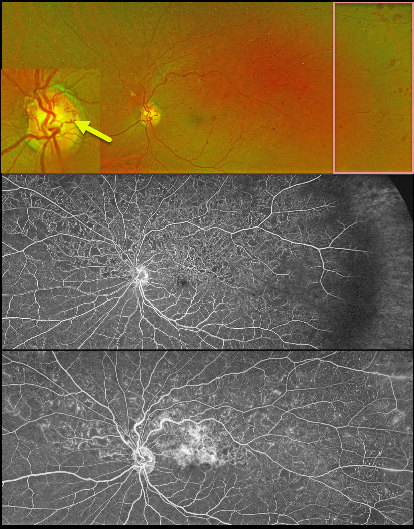
HRVO
Originally posted on @retina.rocks November 8, 2023
This 63YO male was referred for relatively asymptomatic retinal findings in his left eye. Vision was 20/60.
Optos color RG imaging shows a superior hemiretinal vein occlusion (HRVO) with mild hemorrhages, mostly in the temporal periphery. The occlusion is well-perfused without retinal neovascularization on fluorescein angiography. Prominent collateral vessels are noted on the optic disc superiorly. Observation was recommended.
Learning Points:
Branch retinal vein occlusions are virtually always caused by a retinal artery compressing a retinal vein within its common adventitial sheath. Some people are born with an anomalous two-trunked central retinal vein that independently drains the superior and inferior retinal hemispheres.
HRVO, similar to central retinal vein occlusion, is caused by compression of the retinal vein posterior to the lamina cribrosa (see Hayreh, Arch Ophthalmol 1980;98:1600-1609).
Collateral vessels are dilated, pre-existing vessels that bypass an occlusion. These are seen in the posterior pole most commonly following retinal venous occlusions, but can also occur on the optic disc with optic nerve tumors. The endothelial tight junctions are intact, so they don’t leak on angiography. This helps differentiate them from neovascularization, which does leak.
HRVO
Originally posted on @retina.rocks December 1, 2022
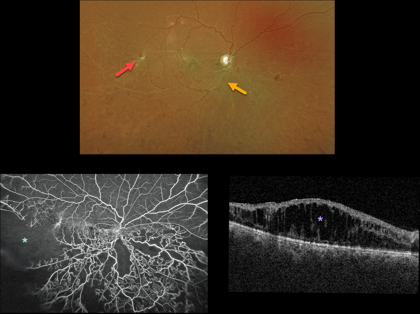
This 60YO male presented with hand motion vision in his right eye from an inferior hemiretinal vein occlusion (HRVO).
Optos color imaging shows white and sheathed retinal vessels inferiorly and extensive temporal collateral vessels.
Fluorescein angiography confirms the marked clinical ischemia. There was no neovascularization. OCT scanning shows marked diffuse macular edema.
The patient refused anti-VEGF therapy, and actually got up and walked out of the exam room before we could finish our discussion regarding why treatment was needed!
Learning Points:
Branch retinal vein occlusions are virtually always caused by a retinal artery compressing a retinal vein within its common adventitial sheath. Some people are born with an anomalous two-trunked central retinal vein that independently drains the superior and inferior retinal hemispheres.
HRVO, similar to central retinal vein occlusion, is caused by compression of the retinal vein posterior to the lamina cribrosa (see Hayreh, Arch Ophthalmol 1980;98:1600-1609).
HRVO
Originally posted on @retina.rocks June 14, 2022
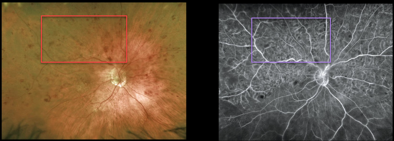
This 88YO female presented with counting fingers vision in her right eye from a symptomatic superior hemiretinal vein occlusion (HRVO).
Optos imaging shows moderate superior hemispheric inner retinal hemorrhages, and fluorescein angiography shows a well-perfused occlusion. OCT scanning showed moderate central macular edema (not shown). Anti-VEGF therapy was started.
Learning Points:
Branch retinal vein occlusions are virtually always caused by a retinal artery compressing a retinal vein within its common adventitial sheath. Some people are born with an anomalous two-trunked central retinal vein that independently drains the superior and inferior retinal hemispheres.
HRVO, similar to central retinal vein occlusion, is caused by compression of the retinal vein posterior to the lamina cribrosa (see Hayreh, Arch Ophthalmol 1980;98:1600-1609).
HRVO
Originally posted on @retina.rocks January 13, 2022
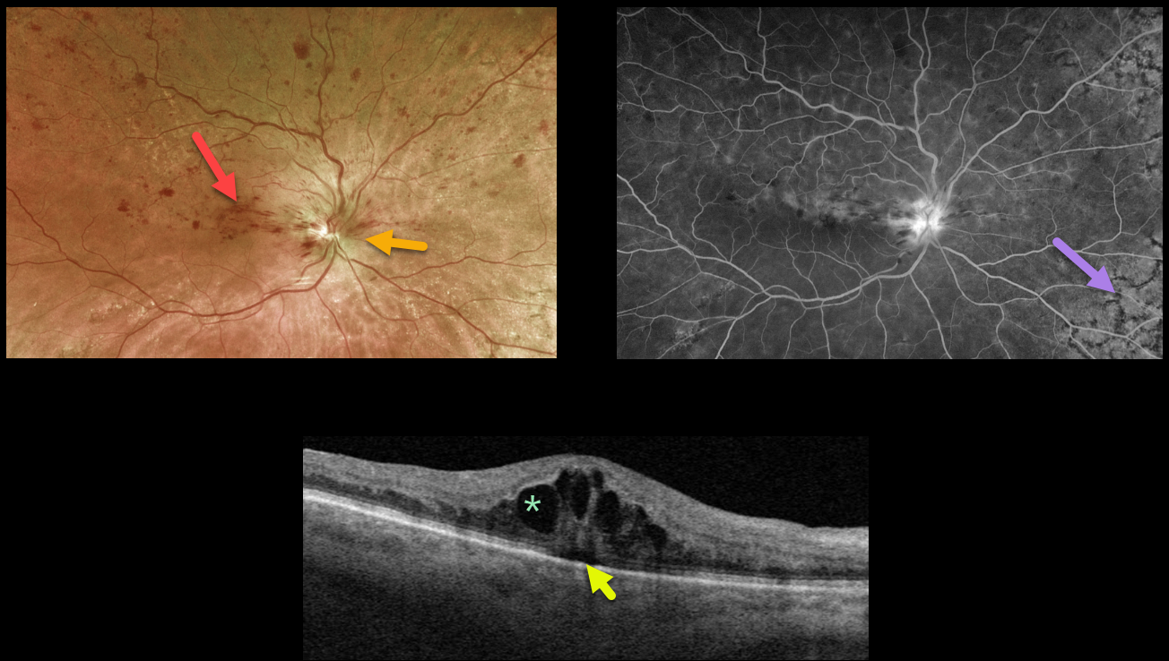
This 81YO female presented with 20/60 vision from a symptomatic superior hemiretinal vein occlusion (HRVO). Optos imaging shows foveal and superior inner retinal hemorrhages with a swollen optic nerve, and fluorescein angiography shows a well-perfused occlusion.
Swept-source OCT confirms moderate cystic macular edema with a trace amount of underlying subretinal fluid.
Age-related peripheral reticular degeneration of the RPE is best seen angiographically as an incidental finding.
The patient is receiving ongoing anti-VEGF injections to control her macular edema. Intravitreal injections (anti-VEGF or steroids) are the treatment of choice, although grid laser may help decrease the treatment burden.
Learning Points:
Branch retinal vein occlusions are virtually always caused by a retinal artery compressing a retinal vein within its common adventitial sheath.
Some people are born with an anomalous two-trunked central retinal vein that independently drains the superior and inferior retinal hemispheres.
HRVO, similar to central retinal vein occlusion, is caused by compression of the retinal vein posterior to the lamina cribrosa (see Hayreh, Arch Ophthalmol 1980;98:1600-1609).
HRVO
Originally posted on @retina.rocks July 5, 2021
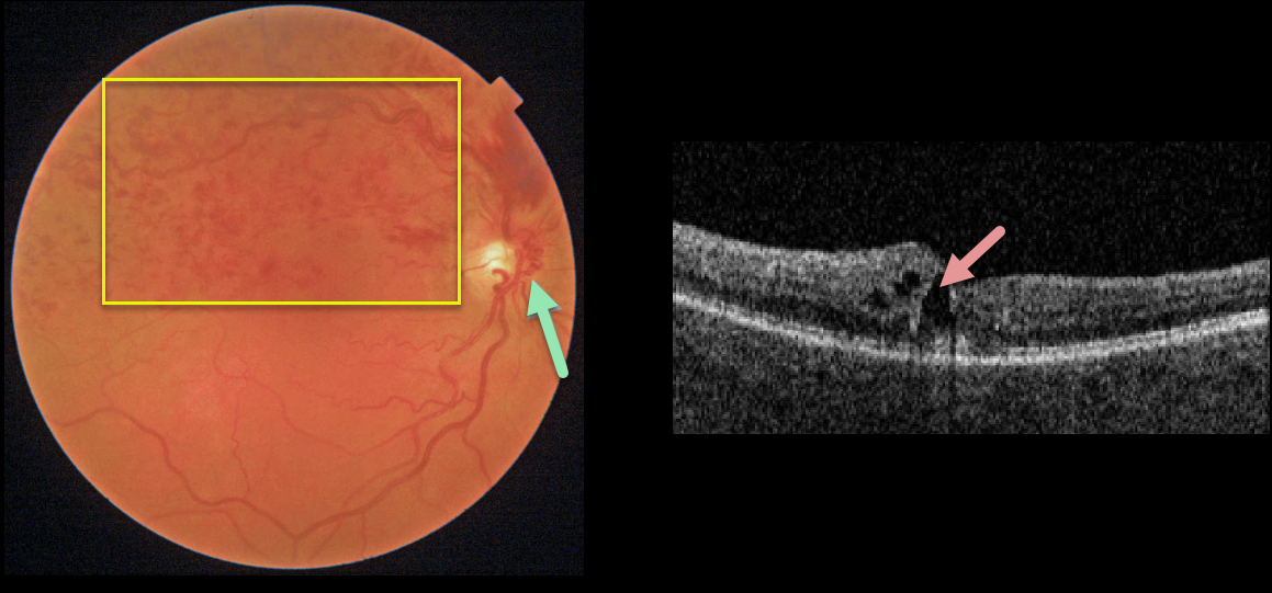
This 62YO female presented with 20/30 vision from a symptomatic superior hemiretinal vein occlusion (HRVO). Fundus imaging shows moderate retinal hemorrhages in the superior retinal hemisphere and a prominent collateral vessel along the nasal aspect of the optic nerve. There is superior macular edema on OCT.
Learning Points:
Branch retinal vein occlusions (BRVO) are virtually always caused by a retinal artery compressing a retinal vein within its common adventitial sheath. Some people are born with an anomalous two-trunked central retinal vein that independently drains the superior and inferior retinal hemispheres.
HRVO, similar to central retinal vein occlusion, is caused by compression of the retinal vein posterior to the lamina cribrosa (see Hayreh, Arch Ophthalmol 1980;98:1600-1609).
Collateral vessels are dilated, pre-existing vessels that bypass an occlusion. These are most commonly seen in the posterior pole following retinal venous occlusions, but can also occur on the optic disc with optic nerve meningiomas.
The endothelial tight junctions are intact, so there’s no leakage on angiography. This helps differentiate them from neovascularization, which does leak.
HRVO
Originally posted on @retina.rocks March 2, 2021
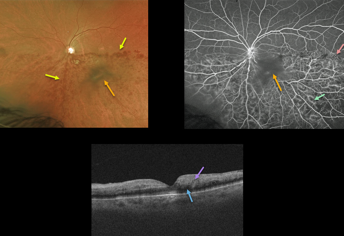
This 78YO female presented with diffuse intraretinal hemorrhages in the inferior half of her posterior pole from an inferior hemiretinal vein occlusion. She had a long history of uncontrolled hypertension, although her blood pressure had not been acutely elevated.
Fluorescein angiography shows good perfusion, some areas of vascular staining/leakage, and some telangiectasia at the border of the occlusion temporally. The large central blob in both the Optos image and the angiogram is clinically noted as a dense vitreous condensation, not blood.
OCT shows no macular edema with mild outer retinal shadowing due to the hyperreflective inner retina. Since there is no edema, we are currently observing the patient and are hoping the vision will improve as the retinal blood absorbs.
Learning Points:
Over time, patients can develop retinal atrophy from ischemic BRVO. In addition, collateral vessels are a common finding seen in venous occlusions.
HEMI-RETINAL VEIN LCCUSION
Originally posted on @retina.rocks January 21, 2020
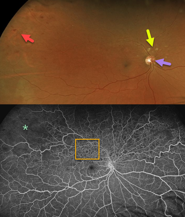
This patient has a superior hemi-reinal vein occlusion with superotemporal intraretinal hemorrhages and cotton wool spots superior to the optic nerve.
The fluorescein angiogram (FA) shows extensive retinal vascular telangiectasia and peripheral ischemia, indicating that this patient is at risk for developing retinal
neovascularization.
Learning Points:
Collateral vessels of the optic nerve can develop with retinal vein occlusions. Prominent optic nerve collateral vessels are seen in the fundus photo. These don’t leak angiographically proving that they’re not neovascular.


