

Hydroxychloroquine (Plaquenil) & Bull’s Eye Maculopathy
<< Back to Cases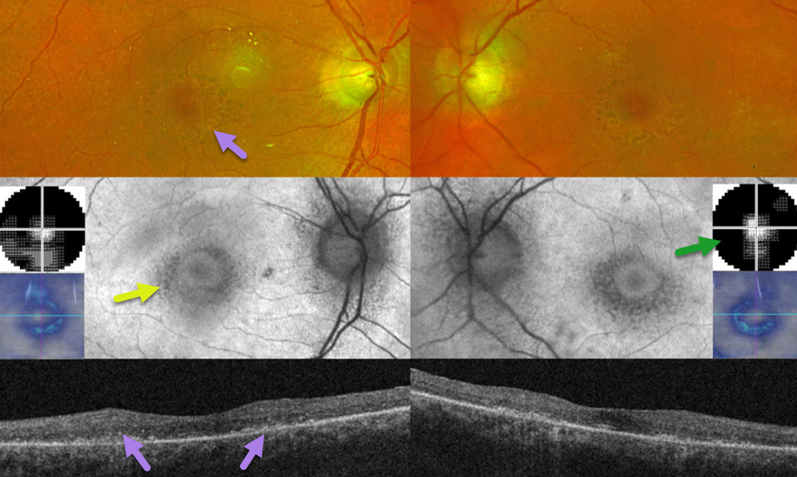
HYDROXYCHLOROQUINE TOXICITY
Originally posted on @retina.rocks April 14, 2026
This 78YO female was on Plaquenil (hydroxychloroquine) for 7 years for her “arthritis.” After noticing progressive reading difficulties, she decided to stop this medicine about 3 months earlier. She was screened annually by her local eye doctor, who referred her for possible toxicity. Vision was 20/40 OD and 20/30 OS.
Optos color imaging and OCT show classic findings for severe hydroxychloroquine toxicity, including an oval area of foveal pigment loss/mottling and outer retinal atrophy. These areas of pigment loss hypo-autofluoresce. 10-2 visual fields reveal small islands of preserved central vision.
Learning Points:
Current screening guidelines should make Plaquenil toxicity a thing of the past. The American Academy of Ophthalmology updated its screening guidelines in 2025 (Marmor et al, Ophthalmology 2026;133:439-450).
A baseline screening examination with fundus photography, spectral-domain or swept-source OCT, and fundus autofluorescence (FAF) should be performed as soon as treatment is started to note preexisting conditions and for future comparison. Annual screening is recommended but may be deferred during the first 5 years if there are no significant risk factors, ie, high daily dosage, renal disease, concomitant drugs such as tamoxifen, macular disease, and older age.
The recommended daily dosage remains unchanged at <=5.0 mg/kg/day real weight, with the dosage under 400 mg/day for severely obese individuals. The primary screening tools are OCT with widefield FAF. Secondary confirmatory tests include 24-2C visual field testing (which screens the parafoveal and pericentral regions simultaneously) and multifocal ERG. The pericentral field needs to be evaluated, as Asians typically have more eccentric macular and midperipheral involvement.
HYDROXYCHLOROQUINE TOXICITY
The European VitreoRetina Society (EVRS) and Debolina Deb
Originally posted on @retina.rocks October 31, 2025
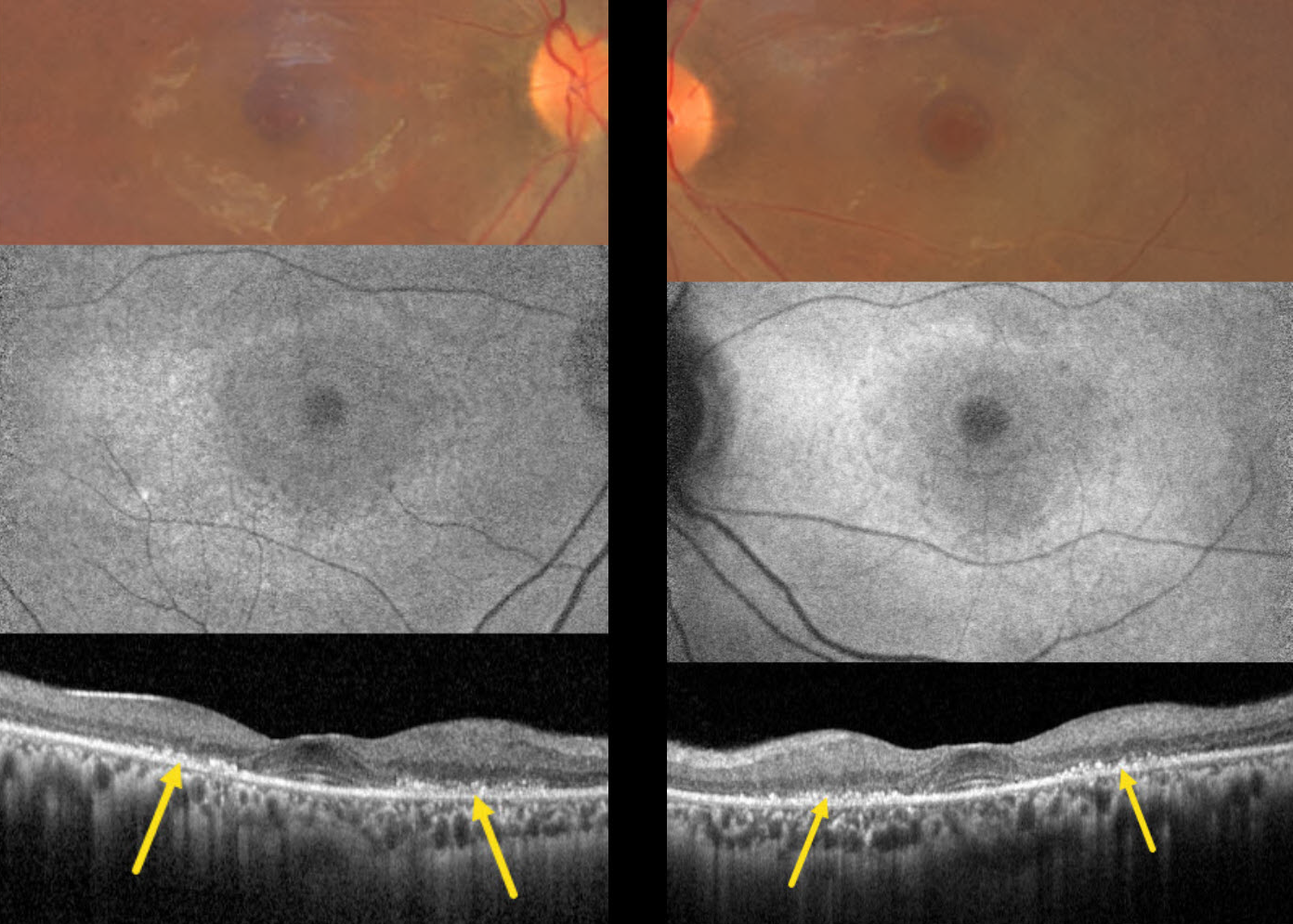
This 28YO Indian female presented with several months of seeing occasional black spots. She has systemic lupus erythematosus, which has been treated with hydroxychloroquine (HCQ) for the past 5 years. Her calculated cumulative dose of HCQ was 365 grams. She denied having prior baseline or screening examinations for HCQ toxicity. Vision was 20/30 OU.
Color photography shows bilateral perifoveal pigmentary changes. Fundus autofluorescence (FAF) shows a rim of parafoveal hyper-FAF. OCT scanning shows paracentral outer retinal thinning, with disorganized clumps of hyperreflective material overlying thinned RPE.
Learning Points:
Current screening guidelines should make hydroxychloroquine toxicity, as found in our patient, a thing of the past. Last revised in 2016, the American Academy of Ophthalmology (AAO) recommends that a baseline exam be performed before starting the medication, with annual screenings beginning at least 5 years after initiating treatment, unless major risk factors are present. However, in practice, patients are usually screened yearly once they are placed on this medication.
OCT is performed annually to assess outer retinal findings, including ellipsoid zone loss. Humphrey 10-2 visual field testing (24-2 for Asians, since their macular involvement is usually more peripheral) is also needed annually, as about 10% of patients will have field loss despite normal examinations and OCT testing (Ahn et al., AJO 2017;184:11-18).
Plaquenil dosing should also be based on real, not ideal, weight to better predict the optimal dose. See Marmor et al Ophthalmology 2016;123:1386-1394 for the full screening guidelines. Despite the most recent AAO guidelines being almost 10 years old, a recent publication found that over one-third of patients are still overdosed (Moussa et al, Ophthalmology Retina 2025;9:814-817).
Toxicity still developed in our patient despite not falling into any of the high-risk categories (cumulative HCQ dose < 1000 grams, young age, not obese). We have written a letter to her rheumatologist to immediately stop HCQ treatment.
MACULAR HOLE WITH PSEUDO BULLS-EYE MACULOPATHY
Originally posted on @retina.rocks June 24, 2024
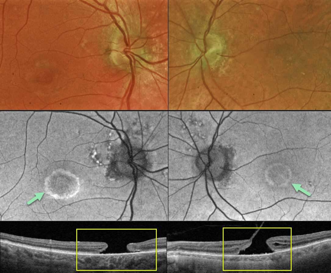
This 83YO male presented with a history of chronic bilateral vision loss. Vision was 20/150 OD and 20/200 OS.
Optos color RG imaging shows a bull’s eye pattern of bilateral pigmentary changes along with a large macular hole OU. Fundus autofluorescence (FAF) shows the bull’s-eye lesions as hyper-FAF. Triton swept-source OCT confirms bilateral macular holes.
Our patient has bilateral chronic macular holes with secondary RPE depigmentation, resulting in a bull’s-eye appearance. Due to the chronicity of the holes, observation was recommended.
Learning Points:
Bull’s eye maculopathy is characterized by a rim of subretinal pigment loss and outer retinal atrophy, most commonly found in inherited retinal diseases (usually ABCA4 disorders) and hydroxychloroquine/chloroquine toxicity.
HYDROXYCHLOROQUINE TOXICITY + PACHYCHOROID
Originally posted on @retina.rocks March 11, 2024
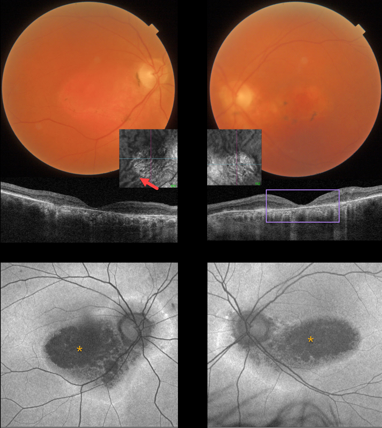
This 67YO female has a known history of Plaquenil toxicity. Her bilateral 20/200 vision and macular findings have been stable since discontinuing this medication 30 years ago.
Triton color imaging and swept-source OCT show classic findings for severe hydroxychloroquine toxicity, including an oval area of foveal pigment loss and outer retinal atrophy.
Most patients will have preserved central foveal pigment giving a bulls-eye appearance, although our patient has loss of outer retina and RPE throughout. Interestingly, choroidal en face imaging shows bilateral dilated choroidal pachyvessels.
On fundus autofluorescence (FAF), the central maculas are hypo-FAF, although an atypical hyper-FAF ring extends around each optic nerve.
Learning Points:
Current screening guidelines should make Plaquenil toxicity, as found in our patient, a thing of the past. Last revised in 2016, the American Academy of Ophthalmology (AAO) recommends that a baseline exam be performed before starting the medication, with annual screenings beginning at least 5 years after initiating treatment, unless major risk factors are present. However, in practice, patients are usually screened yearly once they are placed on this medication.
Optical coherence tomography (OCT) is performed annually to assess outer retinal findings, including ellipsoid zone loss. Humphrey 10-2 visual field testing (24-2 for Asians, since their macular involvement is usually more peripheral) is also needed annually, as about 10% of patients will have field loss despite normal examinations and OCT testing.
Plaquenil dosing should also be based on real, not ideal, weight to better predict the optimal dose. See Marmor et al Ophthalmology 2016;123:1386-1394 for the full screening guidelines.
HYDROXYCHLOROQUINE TOXICITY
Originally posted on @retina.rocks February 9, 2023
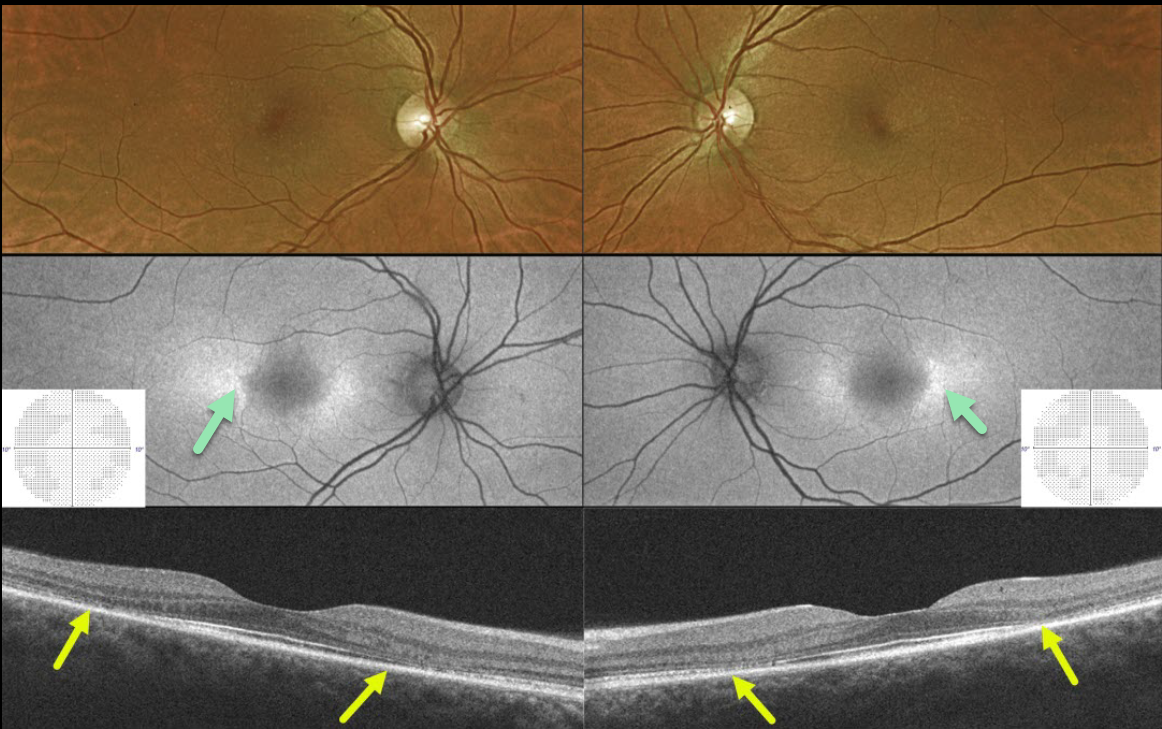
This 43YO female presented without visual complaint and 20/25 vision bilaterally. She gave a 27-year history of Plaquenil use for lupus.
Optos color imaging shows subtle pigment loss surrounding each fovea. Fundus autofluorescence (FAF) shows more obvious changes with perifoveal hyper-FAF nasally and temporally. 10-2 visual fields were normal.
Swept-source OCT B-scan imaging shows classic pericentral outer retinal atrophy.
Learning Points:
Current screening guidelines should make Plaquenil toxicity a thing of the past. Last revised in 2016, the American Academy of Ophthalmology (AAO) recommends that a baseline exam be performed before starting the medication, with annual screenings beginning at least 5 years after initiating treatment, unless major risk factors are present.
However, in practice, patients are usually screened yearly once they are placed on this medication.
Optical coherence tomography (OCT) is performed annually to assess outer retinal findings, including ellipsoid zone loss.
Our case emphasizes the importance of using multiple testing modalities to detect toxicity at the earliest possible stage.
HYDROXYCHLOROQUINE TOXICITY
Will Gibson
Originally posted on @retina.rocks November 30, 2022
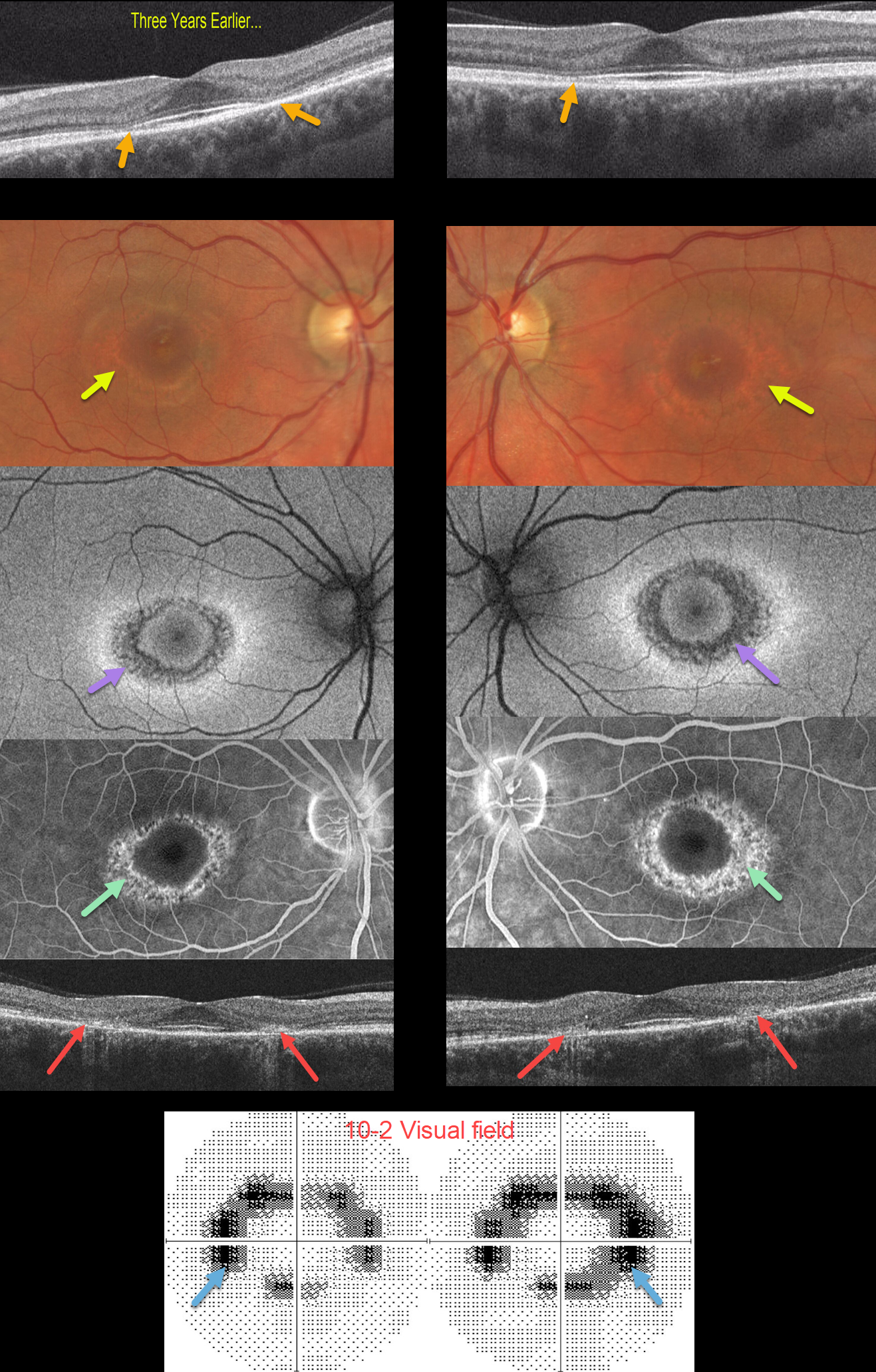
This 43YO female was seen in 2019 by a general ophthalmologist at an outside practice with somewhat subtle paracentral outer retinal atrophy. Vision was 20/20 without symptoms. She was taking Plaquenil for 8 years for rheumatoid arthritis. Unfortunately, these OCT findings for early Plaquenil toxicity were missed.
She was subsequently lost to followup until she presented 3 years later. Although vision remained at 20/20 OU, she now complained of donut-shaped scotomas bilaterally. There were now dramatic and classic findings for Plaquenil toxicity captured on multimodal imaging.
Optos color imaging shows a bullseye pattern of pigmentary loss encircling each fovea. Fundus autofluorescence shows hypo-FAF and fluorescein angiography shows window defects corresponding to these bullseye changes.
OCT scanning shows symmetrical paracentral outer retinal and RPE atrophy. Finally, 10-2 visual fields show a ring of bilateral paracentral visual field loss.
Learning Points:
Current screening guidelines should make Plaquenil toxicity a thing of the past. Last revised in 2016, the American Academy of Ophthalmology (AAO) recommends that a baseline exam be performed before starting the medication with annual screenings beginning at least 5 years after initiating treatment, unless major risk factors are present. However, in practicality, patients are usually screened yearly once they are placed on this medication.
Optical coherence tomography (OCT) is performed annually, looking for outer retinal findings, including ellipsoid zone loss. Humphrey 10-2 visual field testing (24-2 for Asians since their macular involvement is usually more peripheral) is needed annually as well, since about 10% of patients will have field loss despite normal examinations and OCT testing.
Plaquenil dosing should also be based on real, not ideal, weight to better predict the optimal dose. See Marmor et al Ophthalmology 2016;123:1386-1394 for the full screening guidelines.
HYDROXYCHLOROQUINE TOXICITY
Originally posted on @retina.rocks August 19, 2022
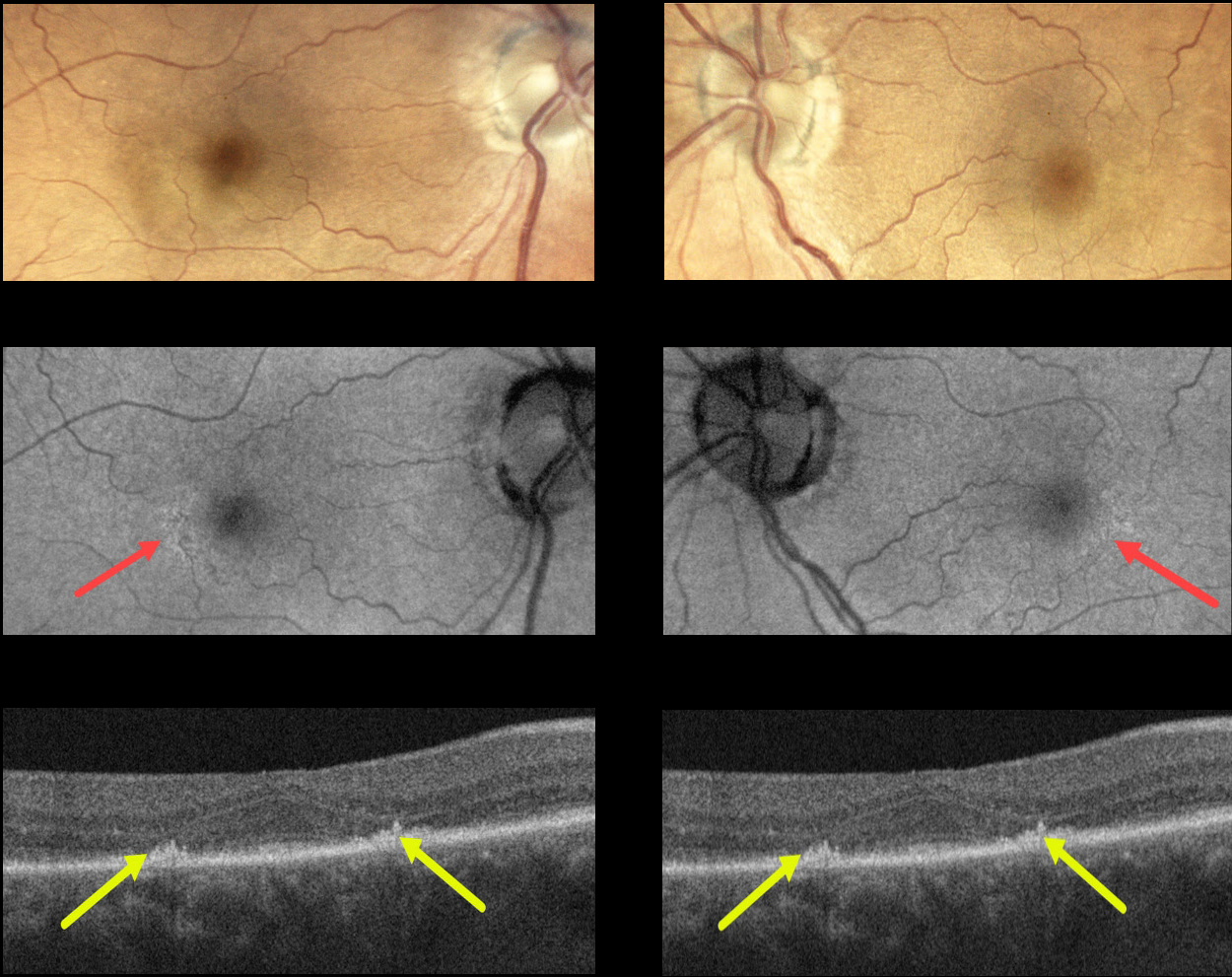
This 73YO female presented with bilateral hydroxychloroquine (Plaquenil) macular toxicity despite being treated for only 5 years. Vision was 20/40 OD and 20/30 OS.
Color photography shows very subtle pigmentary changes encircling each fovea. Fundus autofluorescence (FAF) shows subtle changes as well. The findings are most evident with OCT, which shows symmetrical paracentral outer retinal atrophy.
Learning Points:
Current screening guidelines should make Plaquenil toxicity a thing of the past. Last revised in 2016, the American Academy of Ophthalmology (AAO) recommends that a baseline exam be performed before starting the medication with annual screenings beginning at least 5 years after initiating treatment, unless major risk factors are present.
However, in practicality, patients are usually screened yearly once they are placed on this medication. Optical coherence tomography (OCT) is performed annually, looking for outer retinal findings, including ellipsoid zone loss.
Humphrey 10-2 visual field testing (24-2 for Asians since their macular involvement is usually more peripheral) is needed annually as well, since about 10% of patients will have field loss despite normal examinations and OCT testing.
Plaquenil dosing should also be based on real, not ideal, weight to better predict the optimal dose. See Marmor et al Ophthalmology 2016;123;1386-1394 for the full screening guidelines.
Our patient’s case is somewhat unique in that macular toxicity developed after only 4 years of therapy. This reinforces that screening and treatment guidelines must always be individualized.
HYDROXYCHLOROQUINE TOXICITY
Originally posted on @retina.rocks January 17, 2022
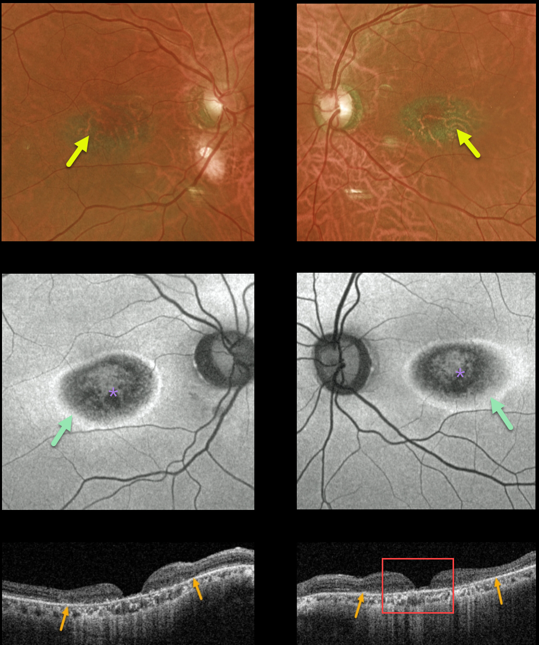
This 77YO female had a long prior history of Plaquenil (hydroxychloroquine) use for lupus. Although it was discontinued 10 years ago, vision was 20/200 OU with classic findings for Plaquenil toxicity.
Optos color imaging shows a hyperpigmented bull’s-eye pattern of pigmentary changes surrounding each macular center. Fundus autofluorescence (FAF) shows pericentral hypo-FAF from RPE damage, with a surrounding narrow ring of hyper-FAF.
Triton swept-source OCT shows outer retinal thinning with more severe near-total central neurosensory retinal thinning with pseudo-macular holes. To further confuse the OCT findings, she also gave a history of macular hole surgery in one of her eyes!
Learning Points:
Current screening guidelines should make Plaquenil toxicity a thing of the past. Last revised in 2016, the American Academy of Ophthalmology (AAO) recommends that a baseline exam be performed before starting the medication, with annual screenings beginning at least 5 years after initiating treatment, unless major risk factors are present.
However, in practice, patients are usually screened yearly once they are placed on this medication. Optical coherence tomography (OCT) is performed annually to assess outer retinal findings, including ellipsoid zone loss.
Humphrey 10-2 visual field testing (24-2 for Asians, since their macular involvement is usually more peripheral) is also needed annually, as about 10% of patients will have field loss despite normal examinations and OCT testing.
Plaquenil dosing should also be based on real, not ideal, weight to better predict the optimal dose. See Marmor et al Ophthalmology 2016;123;1386-1394 for the full screening guidelines.
HYDROXYCHLOROQUINE TOXICITY
Originally posted on @retina.rocks May 25, 2021
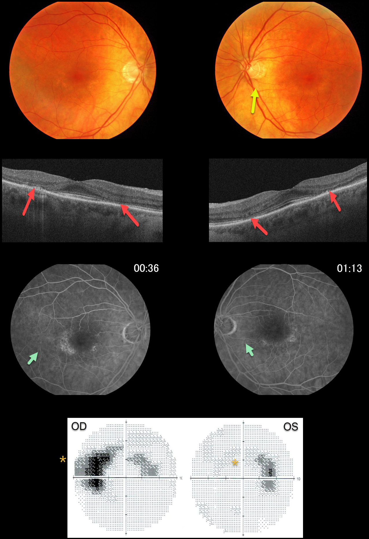
This 72YO female gave a 30-year history of Plaquenil (hydroxychloroquine) use for lupus. Vision was 20/50 OD and 20/20 OS.
There are subtle yet classic findings for Plaquenil toxicity, including a bull’s-eye pattern of pericentral outer retinal atrophy and pigment loss. This is seen clinically as loss of the pericentral orange RPE pigment, on swept-source OCT as thinning of the pericentral photoreceptor elements, and on fluorescein angiography as hyperfluorescent window defects. 10-2 visual fields show bilateral paracentral scotomas.
In the pre-OCT era, patients were followed until clinical or angiographic pigmentary changes developed, at which point the medication was discontinued. Unfortunately, this patient was not properly screened and developed irreversible changes.
Learning Points:
Current screening guidelines should make Plaquenil toxicity a disease of the past
Optical coherence tomography (OCT) is performed annually to assess outer retinal findings, including ellipsoid zone loss.
Humphrey 10-2 visual field testing (24-2 for Asians, since their macular involvement is usually more peripheral) is also needed annually, as about 10% of patients will have field loss despite normal examinations and OCT testing. Plaquenil dosing should also be based on real, not ideal, weight to better predict the optimal dose.
See Marmor et al, Ophthalmology 2016;123:1386-1394 for the full screening guidelines.
HYDROXYCHLOROQUINE TOXICITY
Originally posted on @retina.rocks May 20, 2020
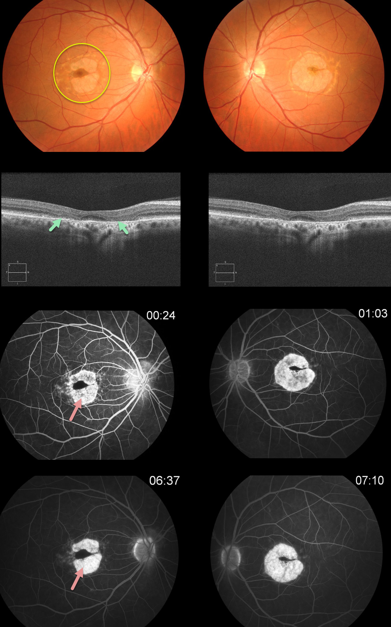
This patient has classic findings for Plaquenil (hydroxychloroquine) toxicity, including a bull’s-eye pattern of pericentral RPE and outer retinal atrophy. This is seen clinically as loss of the orange RPE pigment, on OCT as loss of the photoreceptor elements and RPE, and on fluorescein angiography as hyperfluorescent window defects.
In the pre-OCT era, patients were followed until clinical or angiographic pigmentary changes developed, at which point the medication was discontinued. Unfortunately, this patient was not properly screened and developed irreversible changes.
Learning Points:
Current screening guidelines should make Plaquenil toxicity a disease of the past
Optical coherence tomography (OCT) is performed annually to assess outer retinal findings, including ellipsoid zone loss.
Humphrey 10-2 visual field testing (24-2 for Asians, since their macular involvement is usually more peripheral) is also needed annually, as about 10% of patients will have field loss despite normal examinations and OCT testing. Plaquenil dosing should also be based on real, not ideal, weight to better predict the optimal dose.
See Marmor et al, Ophthalmology 2016;123:1386-1394 for the full screening guidelines.


