

Hypertensive (HTN) Retinopathy and Choroidopathy
<< Back to Cases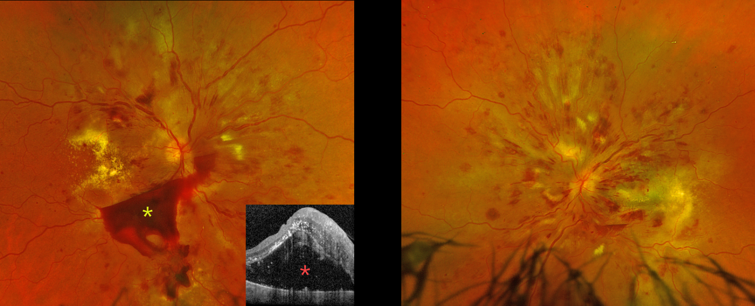
HYPERTENSIVE RETINOPATHY
Originally posted on @retina.rocks September 2, 2026
This 42YO undomiciled female presented with several months of bilateral vision loss. She has a complex medical history of hypertension, type 2 diabetes (last A1c 9.9), end-stage renal disease on dialysis, chronic obstructive pulmonary disorder, and bipolar disorder. The day prior to presenting to our clinic, she was seen at an outside emergency department for intractable vomiting in the setting of 3 consecutively missed dialysis sessions. Her blood pressure in the ED was 199/105 after self-stopping blood pressure medications because of vomiting. She had left the ED against medical advice. Vision was counting fingers OD and hand motion OS.
Optos color RG imaging OD shows diffuse intraretinal hemorrhages, multiple subhyaloid hemorrhages, cotton-wool spots (CWS), and extensive intraretinal and subretinal exudation. OCT shows marked exudative subretinal fluid with intraretinal hyperreflectivity and thickened inner retinal layers. Similar findings are noted OS.
Learning Points:
Malignant hypertension, defined as blood pressure above 180 systolic and/or 120 diastolic, is a life-threatening medical emergency. Eye doctors are often uniquely positioned to diagnose this condition (Shukla et al, Indian J Ophthalmol 2025;73:1552-1555). Patients will often present with bilateral optic nerve swelling, flame-shaped retinal hemorrhages, CWS, and, with more chronic disease, lipid precipitates in the nasal macular outer plexiform layer.
Our patient’s funduscopic findings were more likely hypertensive than from proliferative diabetic retinopathy. She was instructed to return to the emergency room for blood pressure control, and a poor visual prognosis was discussed.
HYPERTENSIVE RETINOPATHY WITH SUB-ILM HEMORRHAGE
Abhishek Karra and Ravindra Karra
Originally posted on @retina.rocks August 31, 2026
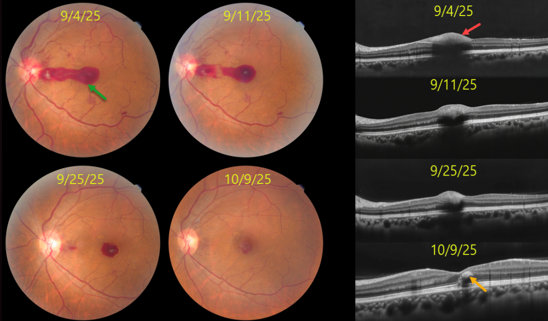
This 44YO female presented with sudden vision loss OS with uncontrolled hypertension (180/110 mmHg). Vision was 20/20 OD and 20/200 OS.
Color photography shows a pre-retinal hemorrhage extending from the temporal edge of the disc through the central macula, disc and peripapillary inner retinal hemorrhages, and blot and nerve fiber layer hemorrhages. The pre-retinal blood localizes to the sub-internal limiting membrane (ILM) space on OCT. The retinal findings normalized over the next 4-5 weeks with visual improvement to 20/20. OCT at the final visit shows a small amount of mid-retinal foveal blood.
Learning Points:
Malignant hypertension, defined as blood pressure above 180 systolic and/or 120 diastolic, is a life-threatening medical emergency. Eye doctors are often uniquely positioned to diagnose this condition (Shukla et al, Indian J Ophthalmol 2025;73:1552-1555). Patients will often present with bilateral optic nerve swelling, flame-shaped retinal hemorrhages, CWS, and, with more chronic disease, lipid precipitates in the nasal macular outer plexiform layer.
Sub-ILM hemorrhage, to the best of our knowledge, has not been previously described in hypertensive retinopathy. It is usually associated with retinal arterial macroaneurysm, Valsalva retinopathy, trauma, and blood dyscrasias. In our patient’s case, we hypothesize that the sub-ILM likely originated from the disc and dissected temporally across the macula.
HYPERTENSIVE RETINOPATHY AND CHOROIDOPATHY
Originally posted on @retina.rocks February 18, 2026
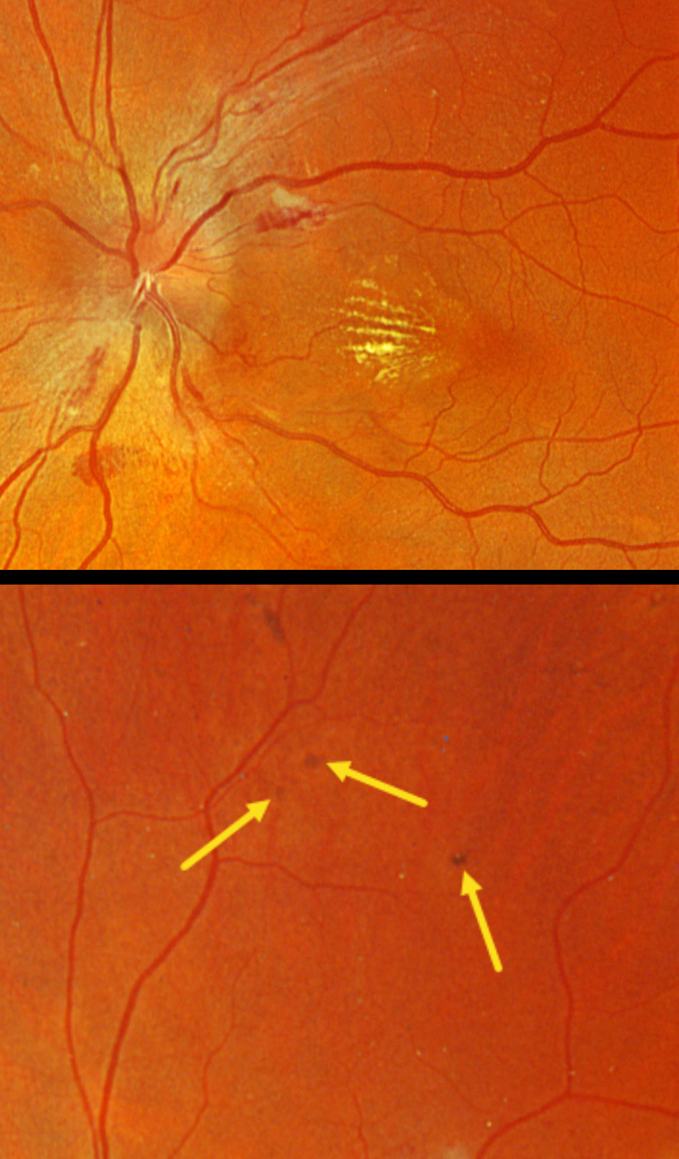
This 46YO male presented to his local optometrist 1 week earlier with a few days of bilateral vision loss. His doctor diagnosed malignant hypertension due to retinal findings and a blood pressure of about 280/160. He was hospitalized for a few days and presented to us for further evaluation. Vision was 20/30 OD and 20/50 OS.
Optos color RGB imaging shows optic nerve edema, peripapillary hemorrhages, and a nasal macular lipid star. Several small pigmented Elschnig spots are noted superiorly OS. He was immediately lost to follow-up.
Learning Points:
Malignant hypertension, defined as blood pressure above 180 systolic and/or 120 diastolic, is a life-threatening medical emergency. Eye doctors are in a unique position to often diagnose this condition. Patients will often present with bilateral optic nerve swelling, flame-shaped retinal hemorrhages, and, with more chronic disease, lipid precipitates in the nasal macular outer plexiform layer. Focal (Elschnig spots) or linear (Siegrist streaks) choroidal pigmentary changes develop from hypertensive choroidopathy.
HYPERTENSIVE RETINOPATHY
Shraddha Raj Shrivastava and Manish Nagpal
Originally posted on @retina.rocks December 11, 2025
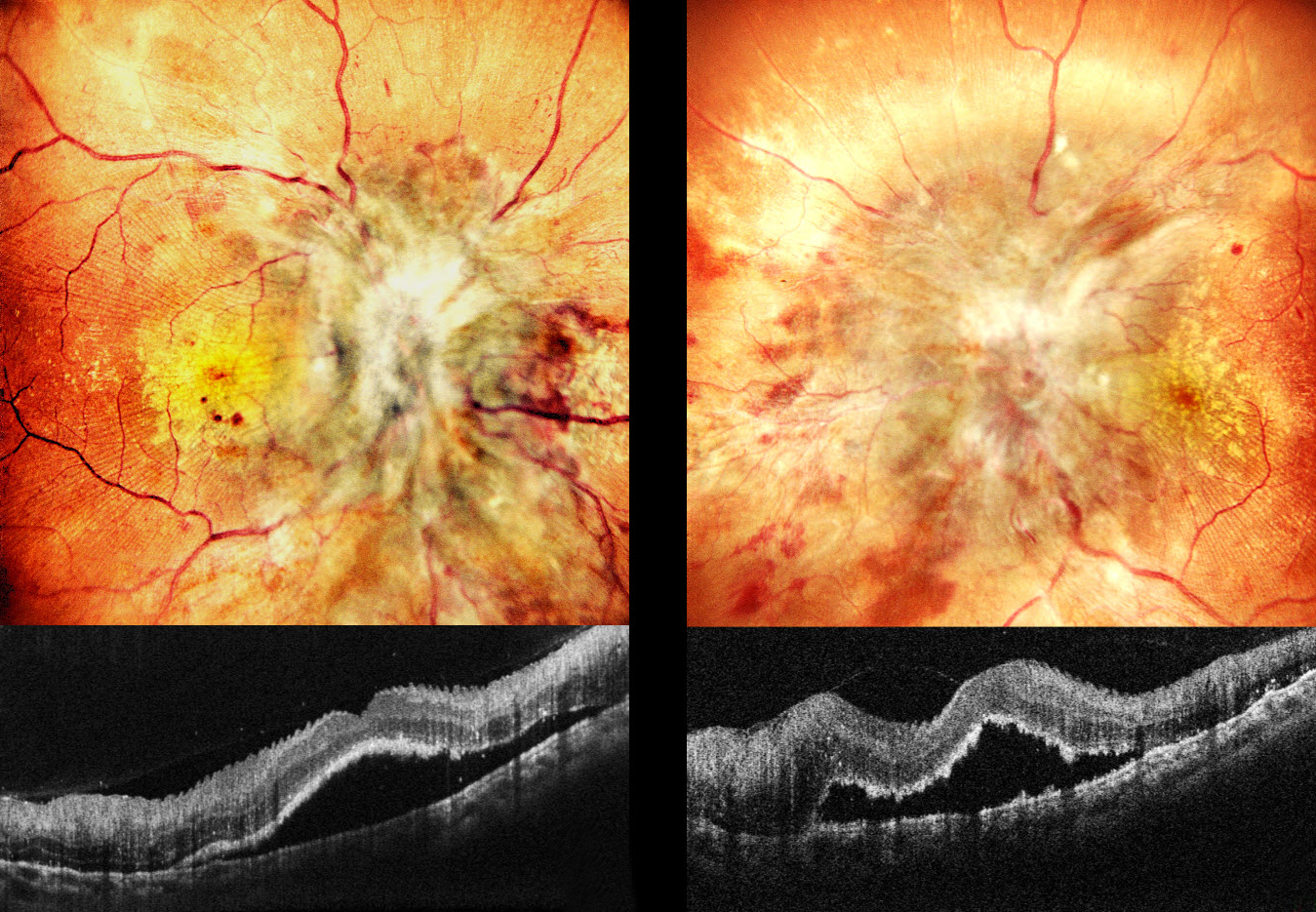
This 20YO male has a known history of chronic kidney disease. Despite hemodialysis, he’s had multiple episodes of malignant hypertension over the prior 2 months. He presented with 1 month of severe vision loss of counting fingers at 2 meters OU.
Pseudocolor SLO imaging shows profound bilateral disc edema with more peripheral nerve fiber layer infarcts (cotton-wool spots, CWS), retinal hemorrhages, and macular lipid. Fine inner retinal radiating striae are noted temporally. OCT scanning shows subfoveal fluid with hyperreflective ragged outer segments. Some small dots of outer retinal hyperreflectivity likely represent lipid.
Learning Points:
Malignant hypertension, defined as blood pressure above 180 systolic and/or 120 diastolic, is a life-threatening medical emergency. Eye doctors are often uniquely positioned to diagnose this condition (Shukla et al., Indian J Ophthalmol 2025;73:1552-1555). Patients will often present with bilateral optic nerve swelling, flame-shaped retinal hemorrhages, CWS, and, with more chronic disease, lipid precipitates in the nasal macular outer plexiform layer.
Posterior segment findings include exudative retinopathy, choroidopathy, and neuropathy. Retinopathy, due to breakdown of the inner retinal blood-retinal barrier, results in retinal hemorrhages and CWS. Choroidopathy, caused by choroidal fibrinoid necrosis, acutely creates exudative retinal detachment and, upon resolution, can leave behind generalized coarse, focal (Elschnig spots) or linear (Siegrist streaks) pigmentary changes. Papilledema is defined as bilateral disc swelling due to raised intracranial pressure. The nerve findings in malignant hypertension are likely due more to localized ischemic changes than to elevated intracranial pressure (Kishi et al, Arch Ophthalmology 1985:1198-1206).
HYPERTENSIVE RETINOPATHY
Ayushi Gupta and Vishal Agrawal
Originally posted on @retina.rocks October 27, 2025
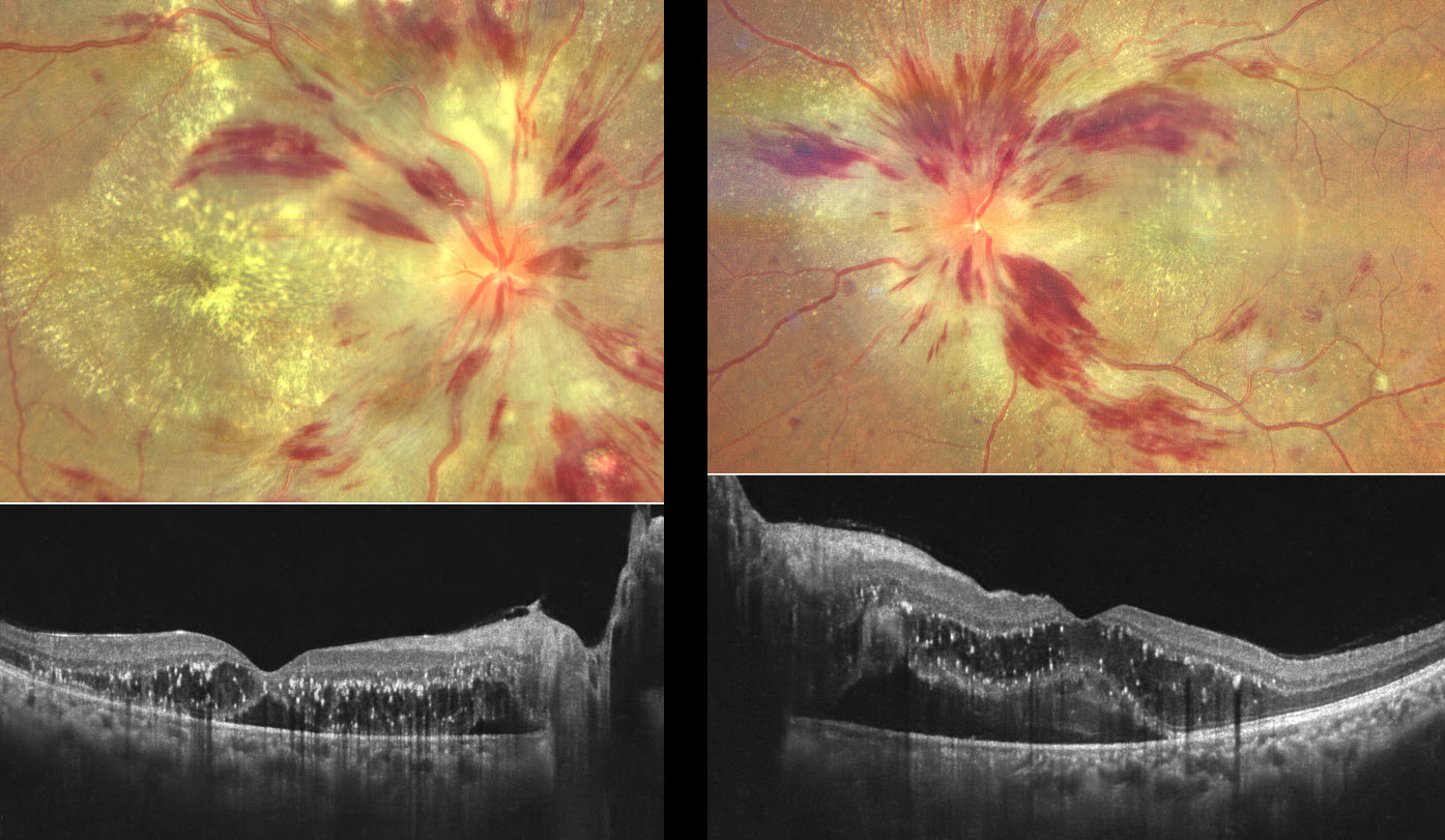
This previously healthy 40YO female presented with 2 months of painless bilateral vision loss. She had occasional headaches, which she self-medicated with painkillers. Vision was counting fingers OD and 20/120 OS.
Color photography in her right eye shows a mildly swollen nerve, extensive hemorrhages in the nerve fiber layer, nerve fiber layer infarcts (cotton-wool spots), and a macular lipid star. OCT scanning shows outer retinal fluid with hyperreflective lipid flecks and foveal subretinal fluid. Similar findings are present in her left eye.
Her blood pressure in our office was 166/122 mmHg. She was immediately referred for emergent medical care.
Learning Points:
Malignant hypertension, defined as blood pressure above 180 systolic and/or 120 diastolic, is a life-threatening medical emergency. Eye doctors are in a unique position to often diagnose this condition. Patients will often present with bilateral optic nerve swelling, flame-shaped retinal hemorrhages, and, with more chronic disease, lipid precipitates in the nasal macular outer plexiform layer. Since the macular fluid emanates from the optic nerve, the macular lipid and thickening are virtually always worse nasally and are an important clinical clue for this diagnosis.
HYPERTENSIVE RETINOPATHY FROM METHAMPHETAMINE
Originally posted on @retina.rocks May 22, 2025
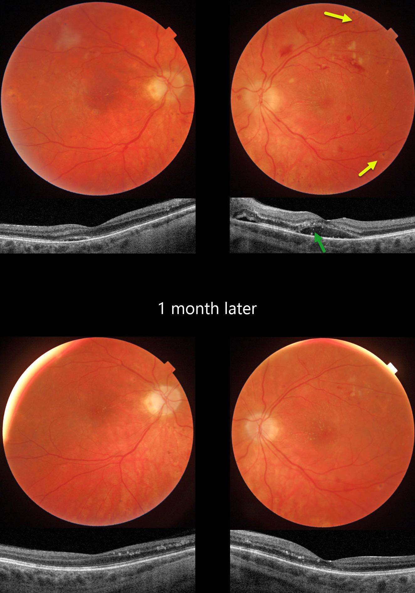
This 21YO female presented with bilateral vision loss. She was admitted to the hospital 2 weeks earlier for malignant hypertension (HTN), likely from methamphetamine abuse. Blood pressure at that time was 292/208. Vision was hand motion OD and 20/80 OS.
Triton color photography of her left eye shows a somewhat pale and swollen nerve, mostly inner retinal hemorrhages, and resolving nerve fiber layer infarcts (cotton-wool spots, CWS). Faint lipid exudates radiate nasally and superiorly from the macular center. Multifocal small hyperpigmented choroidal lesions are noted outside the arcades. Swept-source OCT shows subretinal fluid with hyperreflective lipid in the subretinal space and outer nuclear layer. Similar but much milder findings are noted in her right eye.
When examined 1 month later, vision remained at hand motion OD and improved to 20/40 OS with improved funduscopic and OCT findings.
Learning Points:
Malignant hypertension, defined as blood pressure above 180 systolic and/or 120 diastolic, is a life-threatening medical emergency. Eye doctors are in a unique position to often diagnose this condition. Patients will often present with bilateral optic nerve swelling, flame-shaped retinal hemorrhages, CWS, and, with more chronic disease, lipid precipitates in the nasal macular outer plexiform layer.
Acute hypertensive choroidopathy outside the setting of pre-eclampsia, in our experience, is quite rare. Independent of the disc and neurosensory retinal findings, patients present with localized multifocal serous retinal detachments (de Venecia and Jampol, Arch Ophthalmol 1984;102:68-73). With blood pressure control, these resolve often with minimal funduscopic changes. Focal (Elschnig spots) or linear (Siegrist streaks) choroidal pigmentary changes may result from more severe disease.
HYPERTENSIVE RETINOPATHY AND CHOROIDOPATHY
Yuenpang Cheung
Originally posted on @retina.rocks December 25, 2024
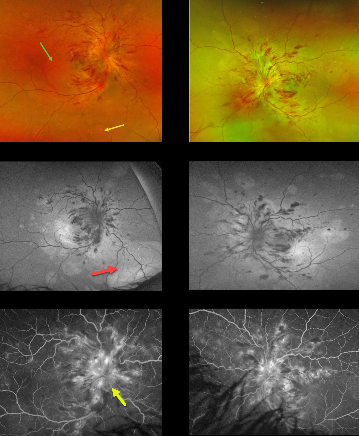
This 21YO female was admitted to the hospital 2 weeks earlier for malignant hypertension. She underwent prior failed renal transplantation due to tubulocystic renal disease and was currently on dialysis. Vision was counting fingers bilaterally.
Optos color RG imaging shows marked bilateral papilledema with mostly inner retinal hemorrhages radiating into the midperipheries. Multifocal blisters of exudative serous retinal detachments are scattered throughout each posterior pole, including the macula and midperiphery. These exudative detachments are best imaged on fundus autofluorescence (FAF), where they are hyper-FAF. Fluorescein angiography shows marked optic nerve leakage and scattered more peripheral subretinal leakage.
Learning Points:
Malignant hypertension, defined as blood pressure above 180 systolic and/or 120 diastolic, is a life-threatening medical emergency. Eye doctors are in a unique position to often diagnose this condition. Patients will often present with bilateral optic nerve swelling, flame-shaped retinal hemorrhages, and, with more chronic disease, lipid precipitates in the nasal macular outer plexiform layer.
Acute hypertensive choroidopathy outside the setting of pre-eclampsia, in our experience, is quite rare. Independent of the disc and neurosensory retinal findings, patients present with localized multifocal serous retinal detachments (de Venecia and Jampol, Arch Ophthalmol 1984;102:68-73). With blood pressure control, these resolve often with minimal funduscopic changes. Focal (Elschnig spots) or linear (Siegrist streaks) choroidal pigmentary changes may result from more severe disease. The acute findings are thought to result from choroidal arterial constriction with secondary choriocapillaris necrosis (Hayreh et al, Ophthalmology 1986;93:1383-1400). This causes ischemic injury to the overlying RPE with a breakdown of the outer blood-retinal barrier and exudative subretinal fluid.
HYPERTENSIVE RETINOPATHY
Originally posted on @retina.rocks February 20, 2024
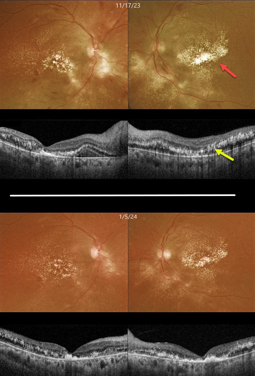
This 43YO female was recently discharged from the hospital for severe hypertension. She told us she was hospitalized for about a month and a half, with treatment including a medically induced coma. Vision was 20/200 OU.
Color imaging shows bilateral retinal lipid extending from the maculas into the midperipheries. Swept source OCT shows bilateral central outer retinal thinning and scattered hyperreflective lipid extending from the outer plexiform layer to above the RPE.
When she returned 6 weeks later, vision remained at 20/200 OU, and the macular lipid was slowly regressing.
Learning Points:
Malignant hypertension, defined as blood pressure above 180 systolic and/or 120 diastolic, is a life-threatening medical emergency. Eye doctors are in a unique position to often diagnose this condition.
Patients will often present with bilateral optic nerve swelling, flame-shaped retinal hemorrhages, and, with more chronic disease, lipid precipitates in the nasal macular outer plexiform layer.
Since the macular fluid emanates from the optic nerve, macular lipid and thickening are virtually always worse nasally, which is an important clinical clue for this diagnosis.
HYPERTENSIVE RETINOPATHY
Originally posted on @retina.rocks July 3, 2023
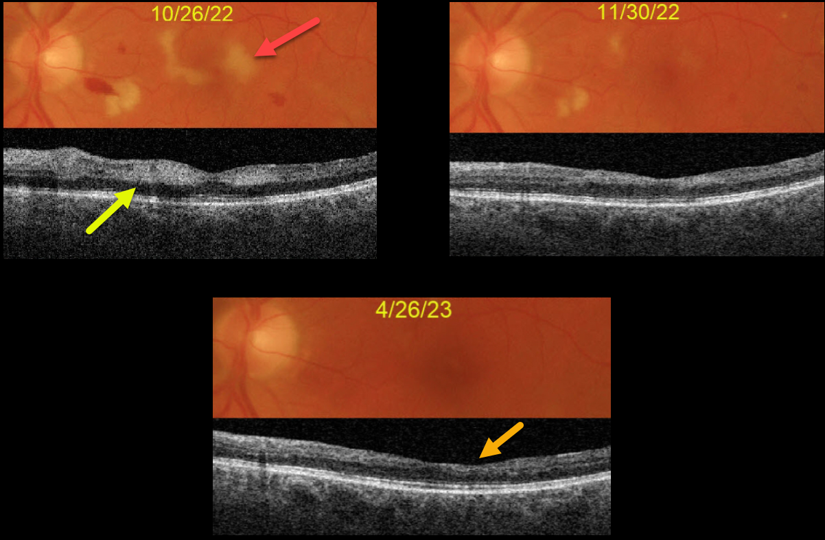
This 63YO male complained of vision loss in his left eye following left carotid endarterectomy. His blood pressure was markedly elevated perioperatively, but he reported feeling better. Blood pressure was 175/101 when seen by his doctor one week earlier. Vision was 20/25 OD and 20/200 OS.
Hypertensive retinopathy was noted bilaterally, with scattered retinal hemorrhages and nerve fiber layer infarcts (cotton-wool spots, CWS).
Color imaging of the left eye shows CWS virtually involving the macular center. Swept source OCT shows inner retinal hyperreflectivity from these lesions.
Over the following months, both his blood pressure and macular appearance normalized. When last seen on 4/26/23, vision was 20/40, although he was still quite symptomatic from paracentral scotomas due to inner retinal atrophy from the resolved CWS.
HYPERTENSIVE RETINOPATHY
Originally posted on @retina.rocks November 24, 2022
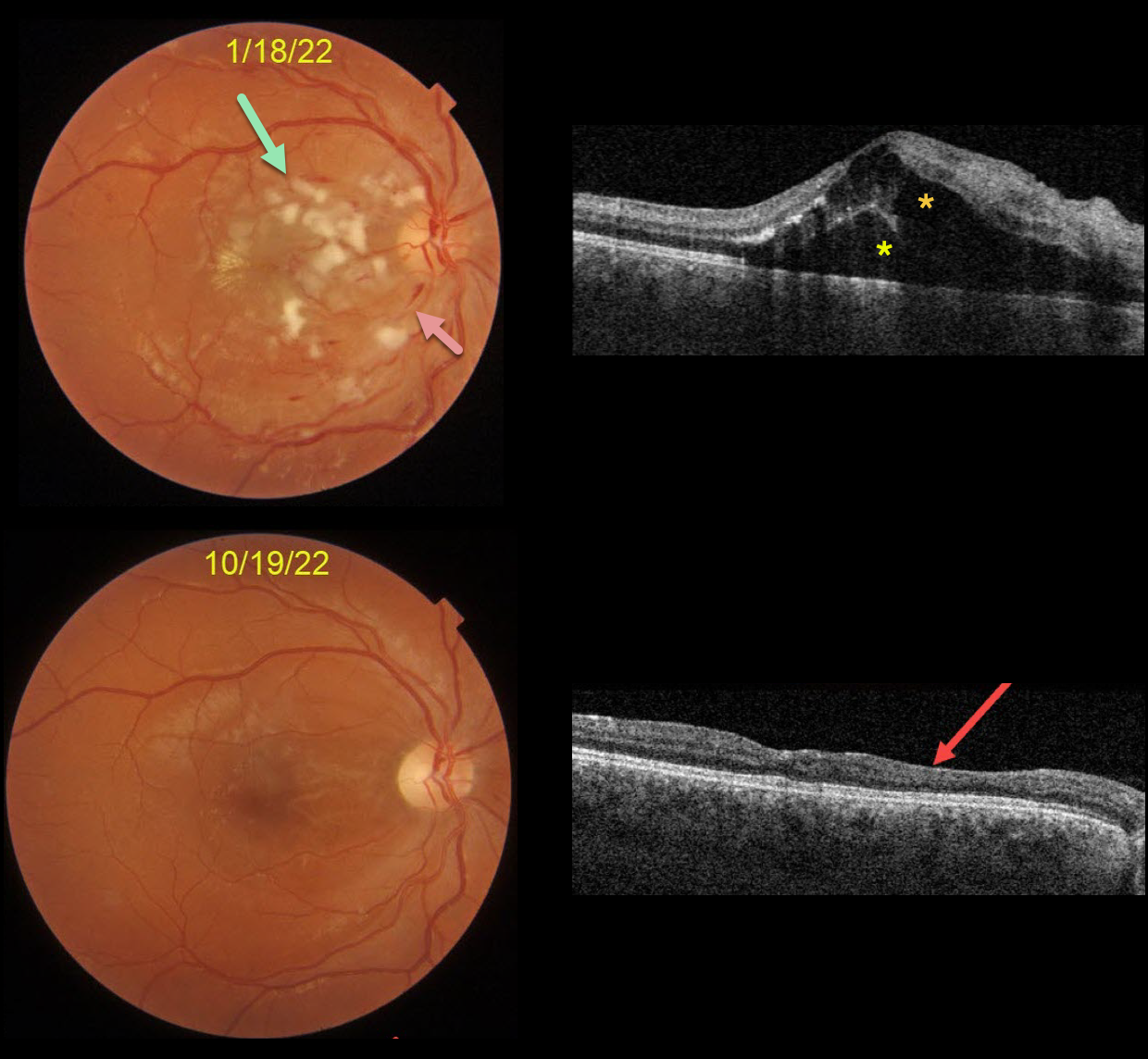
This is a follow-up from our 2/4/22 post of a patient with malignant hypertension.
This 22YO male presented on 1/18/22 with subacute vision loss of 20/200 OD and 20/50 OS. Blood pressure was 169/135. Color imaging shows nerve fiber layer infarcts (cotton-wool spots) and mild inner retinal hemorrhages.
OCT scanning shows mostly outer macular edema emanating from the nerve, with foveal subretinal fluid.
Nine months later, the macular and OCT findings have mostly normalized. OCT scanning shows thinning with disorganization of retinal inner layers (DRIL) nasally OD.
Learning Points:
Malignant hypertension, defined as blood pressure above 180 systolic and/or 120 diastolic, is a life-threatening medical emergency. Eye doctors are in a unique position to often diagnose this condition.
Patients will often present with bilateral optic nerve swelling, flame-shaped retinal hemorrhages, and, with more chronic disease, lipid precipitates in the nasal macular outer plexiform layer.
Since the macular fluid emanates from the optic nerve, the macular thickening is always worse nasally (as in our case), and is an important clinical clue for this diagnosis.
HYPERTENSIVE RETINOPATHY vs AION
Originally posted on @retina.rocks August 16, 2022
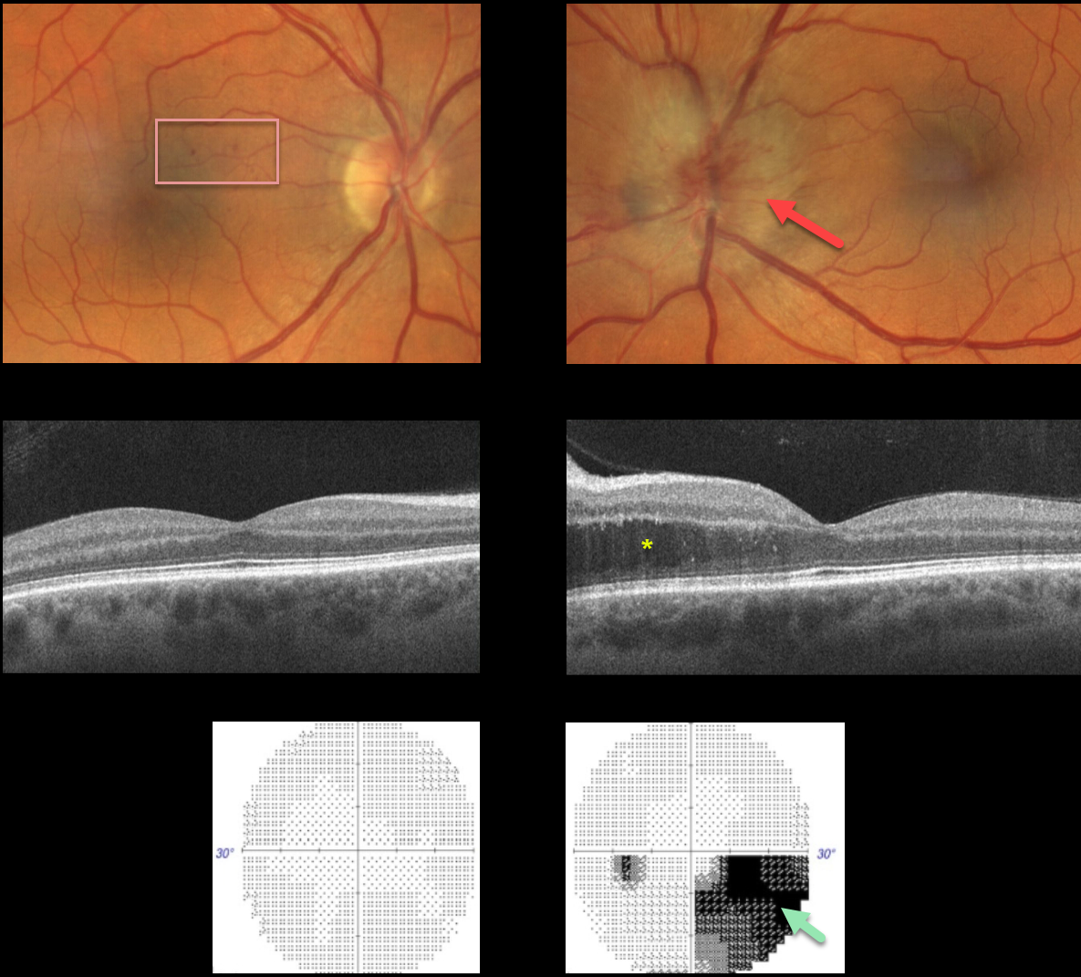
This 57YO male presented with unilateral optic nerve swelling in his left eye and a few faint nasal macular retinal hemorrhages in his right eye. Vision was 20/40 OD and 20/25 OS.
OCT was normal OD and thickened in the nasal outer nuclear layer OS. Visual fields were normal OD and inferiorly depressed, especially inferonasally, OS.
Blood pressure was 190/110, and he was immediately referred to the emergency room.
Learning Points:
Malignant hypertension, defined as blood pressure above 180 systolic and/or 120 diastolic, is a life-threatening medical emergency. Eye doctors are in a unique position to often diagnose this condition.
Patients will often present with bilateral optic nerve swelling, flame-shaped retinal hemorrhages, and, with more chronic disease, lipid precipitates in the nasal macular outer plexiform layer. However, rarely, the ocular findings may be asymmetric or even unilateral.
Since the macular fluid emanates from the optic nerve, the macular thickening is always worse nasally (as in our case), and is an important clinical clue for this diagnosis.
Our patient may also have had an isolated anterior ischemic optic neuropathy in his left eye and mild hypertensive retinopathy in his right eye. The partial altitudinal deffect is more common with this diagnosis than hypertension.
HYPERTENSIVE RETINOPATHY
Originally posted on @retina.rocks July 11, 2022
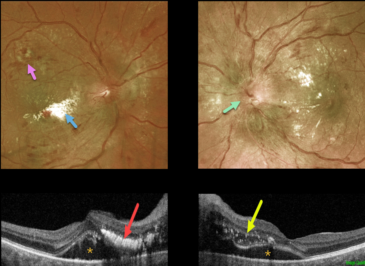
This 44YO male patient presented with a 2-week history of bilateral vision loss. He denied any past medical history.
Vision was 20/150 OD and 20/50 OS. Optos imaging shows bilateral optic nerve swelling, flame-shaped retinal hemorrhages, and macular lipid.
Swept-source OCT shows bilateral subretinal fluid extending from the nerve into the macular centers. A plaque of outer retinal hyperreflective lipid is noted in the nasal right macula, with hyperreflective dots of suspended lipid within the outer retinal edema in his left eye.
Blood pressure was 242/136. He was immediately sent to the emergency room for treatment of his malignant hypertension.
Learning Points:
Malignant hypertension, defined as blood pressure above 180 systolic and/or 120 diastolic, is a life-threatening medical emergency. Eye doctors are in a unique position to often diagnose this condition.
Patients will often present with bilateral optic nerve swelling, flame-shaped retinal hemorrhages, and, with more chronic disease, lipid precipitates in the nasal macular outer plexiform layer. Since macular fluid originates from the optic nerve, macular thickening is always worse nasally (as in our case) and is an important clinical clue for this diagnosis.
HYPERTENSIVE RETINOPATHY
Originally posted on @retina.rocks February 4, 2022
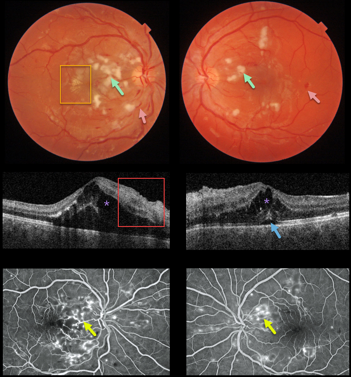
This 22YO male presented with subacute vision loss of 20/200 OD and 20/50 OS. Triton color imaging shows diffuse bilateral cotton wool spots with intraretinal hemorrhages, along with an early macular star temporal to the right fovea.
Swept-source OCT shows bilateral intraretinal thickening (mostly in the outer nuclear layer) and hyperreflective inner retinal layers, along with subfoveal fluid.
Optos fluorescein angiography shows bilateral telangiectasia and staining, mostly around the nerve fiber layer infarcts.
Blood pressure was 169/135. He was immediately sent to the nearest emergency room.
Learning Points:
Malignant hypertension, defined as blood pressure above 180 systolic and/or 120 diastolic, is a life-threatening medical emergency. Eye doctors are in a unique position to often diagnose this condition.
Patients will often present with bilateral optic nerve swelling, flame-shaped retinal hemorrhages, and, with more chronic disease, lipid precipitates in the nasal macular outer plexiform layer.
Since the macular fluid originates from the optic nerve, macular thickening is always worse nasally and is an important clinical clue for this diagnosis.
PDR + HYPERTENSIVE RETINOPATHY
Originally posted on @retina.rocks November 30, 2021
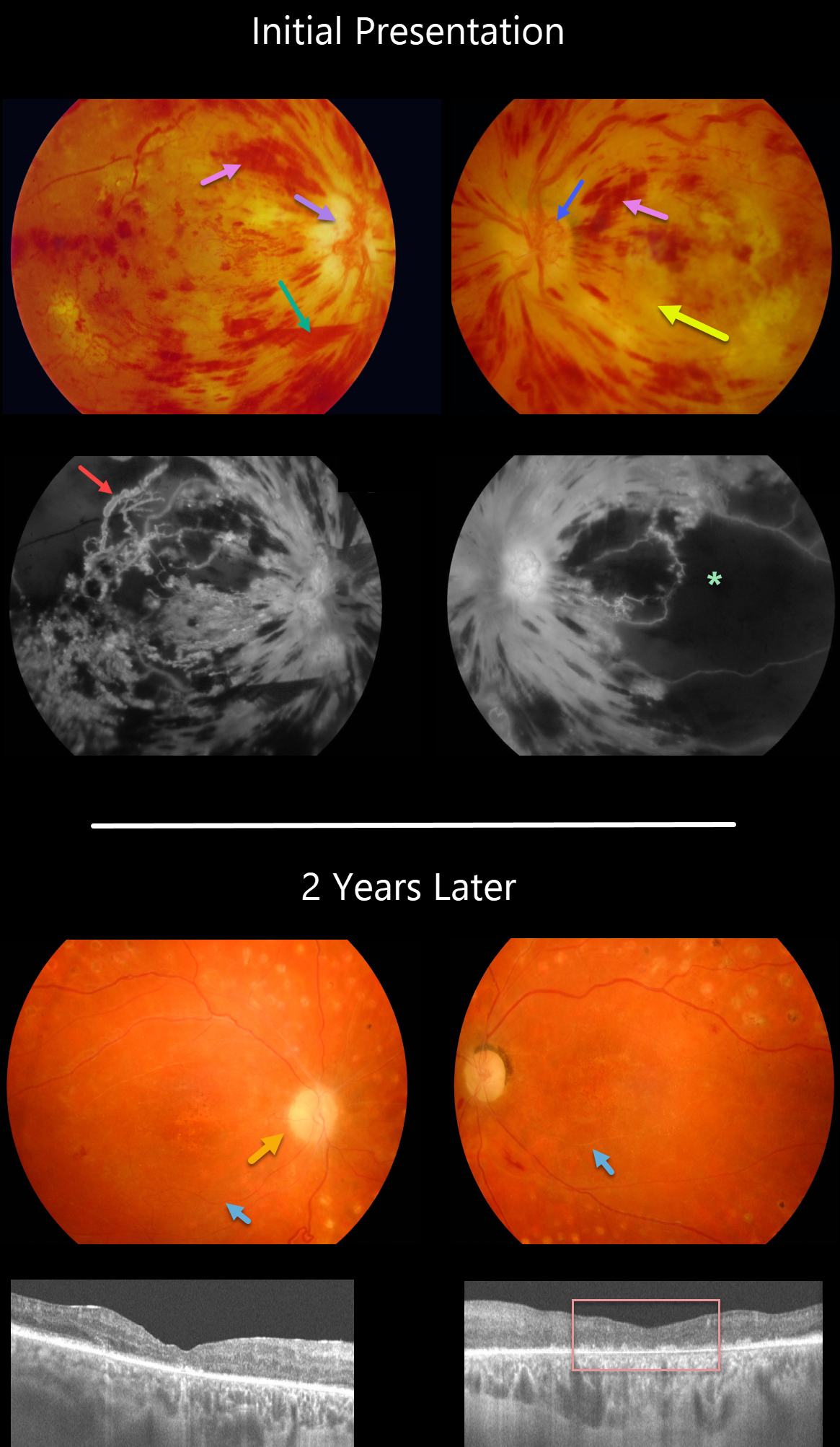
This 30YO female presented in 2008 with acute severe bilateral vision loss. She had a history of well-controlled type 1 diabetes.
There were severe bilateral retinal hemorrhages with white, ischemic maculas. A small preretinal hemorrhage was present just inferior to the right nerve. The right optic nerve was swollen. Disc neovascularization was noted in her left eye.
Fluorescein angiography showed severe macular ischemia, especially in her left eye, with near total obliteration of the normal macular vascular anatomy. Prominent staining venous beading was present in the right superotemporal macula.
Her blood pressure was severely elevated. Following emergent hypertensive control, bilateral anti-VEGF therapy was given, followed by panretinal photocoagulation.
Two years later, vision returned to 20/40 OD and 20/400 OS. The optic nerves were pale, the retinal vessels were narrowed and sheathed, and the foveas had dry pigmentary changes.
OCT scanning showed thinned, atrophic maculae with disorganized inner retinal layers (DRIL).
Learning Points:
Malignant hypertension, defined as blood pressure above 180 systolic and/or 120 diastolic, is a life-threatening medical emergency. Eye doctors are in a unique position to often diagnose this condition. Patients will often present with bilateral optic nerve swelling, flame-shaped retinal hemorrhages, and, with more chronic disease, lipid precipitates in the nasal macular outer plexiform layer.
Hypertensive retinopathy can have a similar appearance to diabetic retinopathy, and patients (like this one) can even present with signs of both.
Over time, patients with hypertensive retinopathy will develop near complete regression of all findings following systemic blood pressure treatment and control.
HYPERTENSIVE RETINOPATHY
Originally posted on @retina.rocks October 18, 2021
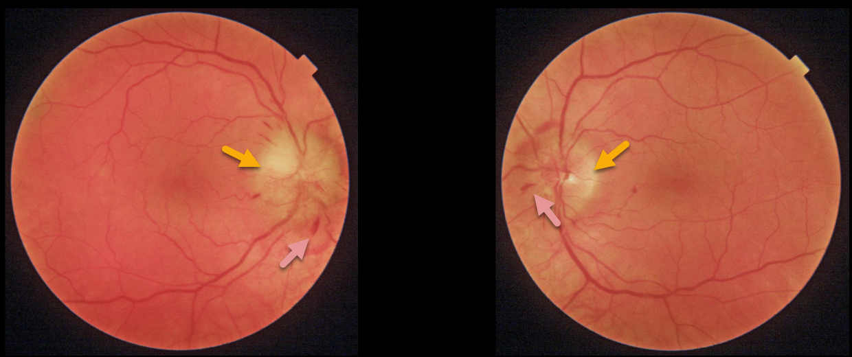
This 56YO male presented with two weeks of bilateral fuzzy vision. He gave a history of well-controlled hypertension and type 2 diabetes. Vision was 20/30 OD and 20/25 OS. Both optic nerves are swollen, and there are scattered intraretinal hemorrhages.
Blood pressure was 210/130. He was immediately sent to the nearest emergency room.
Learning Points:
Malignant hypertension, defined as blood pressure above 180 systolic and/or 120 diastolic, is a life-threatening medical emergency. Eye doctors are in a unique position to often diagnose this condition. Patients will often present with bilateral optic nerve swelling, flame-shaped retinal hemorrhages, and, with more chronic disease, lipid precipitated in the nasal macular outer plexiform layer.
HYPERTENSIVE RETINOPATHY
Originally posted on @retina.rocks March 9, 2021
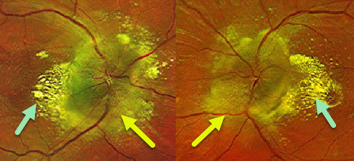
This 24YO male presented with these fundus findings. Both optic nerves were severely and diffusely swollen with a lipid star in the nasal macula. Blood pressure in the office was 224/145.
Learning Points:
The nasal macular lipid star originated from the optic nerve, where fluid and lipoproteins leaked into the surrounding tissues.
When the fluid is absorbed, the lipid precipitates out in Henle’s layer. This is also seen in other causes of optic nerve swelling or inflammation, including neuroretinitis.
The lipid supranasal to the right optic nerve is a bit unusual.
HYPERTENSION + PROLIFERATIVE DIABETIC RETINOPATHY (PDR)
Originally posted on @retina.rocks February 2, 2021
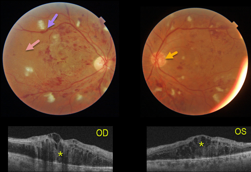
This 41YO male presented after a recent hospitalization for severe hypertension with vision of counting fingers OD and 20/60 OS.
The bilateral nerve fiber layer infarcts (cotton-wool spots) are more characteristic of hypertension than of diabetes.
Severe retinal hemorrhages and disc neovascularization are noted in each eye. The right eye, in particular, is severely ischemic, with Triton imaging showing a featureless, ischemic retina and white vessels, most pronounced in the temporal retina.
The venous sausaging is a clinical sign of severe ischemia, and when present, virtually always shortly precedes or is indicative of concurrent proliferative disease. Triton swept-source OCT shows severe bilateral center-involving diabetic macular edema.
He subsequently received multiple bilateral anti-VEGF injections followed by panretinal photocoagulation, with marked improvement of the central edema.
HYPERTENSIVE RETINOPATHY
Originally posted on @retina.rocks December 22, 2020
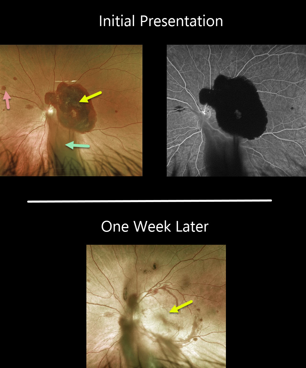
This 40YO male presented with sudden vision loss in his left eye. Vision was 20/200 OS and 20/20 in his normal right eye.
Fundus photo shows a large subhyaloid hemorrhage covering the macula, vitreous hemorrhage extending inferiorly, and some scattered deep retinal hemorrhages. Fluorescein angiography shows no leakage or source for the blood.
Blood pressure was 235/128. He was immediately sent to the ER for further management.
One week later, vision spontaneously improved to 20/30, the subhyaloid hemorrhage almost completely resolved, and the deep retinal hemorrhages were fading.
Learning Points:
The subhyaloid blood and deep retinal hemorrhages, along with the unilateral presentation, are all quite unusual for hypertensive retinopathy. We are not sure if these hemorrhages were related to his hypertension, but thankfully, they are resolving now that his blood pressure has normalized.
HYPERTENSIVE RETINOPATHY
Originally posted on @retina.rocks September 7, 2020
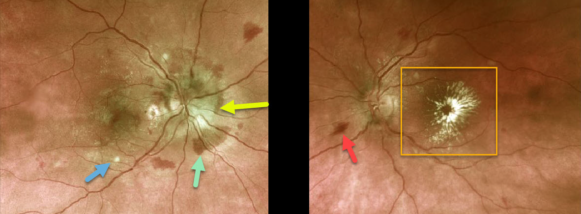
This previously healthy 36 YO female presented with recent vision loss of 20/200 bilaterally.
The right optic nerve is moderately swollen with surrounding mostly deep retinal hemorrhages, faint scattered macular lipid, and a few nerve fiber layer infarcts (cotton wool spots). The left nerve is flat with a few surrounding inner flame-shaped retinal hemorrhages and a prominent foveal lipid star.
Blood pressure was 239/159, and she was sent immediately to the emergency room for malignant hypertension.
Learning Points:
Her presentation was a bit unusual, in that systemic problems usually cause symmetric ocular findings. Her blood pressure was likely elevated for weeks to months since the macular lipid is a late finding.
We are not sure why the left nerve is not swollen, but it certainly was in the past. The lipid star is a telltale sign of prior optic nerve swelling, in which fluid and lipoproteins leaked into the surrounding tissues. When the fluid is absorbed, the lipid precipitates out in Henle’s layer. This is also seen in other causes of optic nerve swelling or inflammation, including neuroretinitis.
The deep retinal hemorrhages in her right eye are also unusual, since these are usually found in the nerve fiber layer with hypertension.
HYPERTENSIVE RETINOPATHY
Originally posted on @retina.rocks April 10, 2020
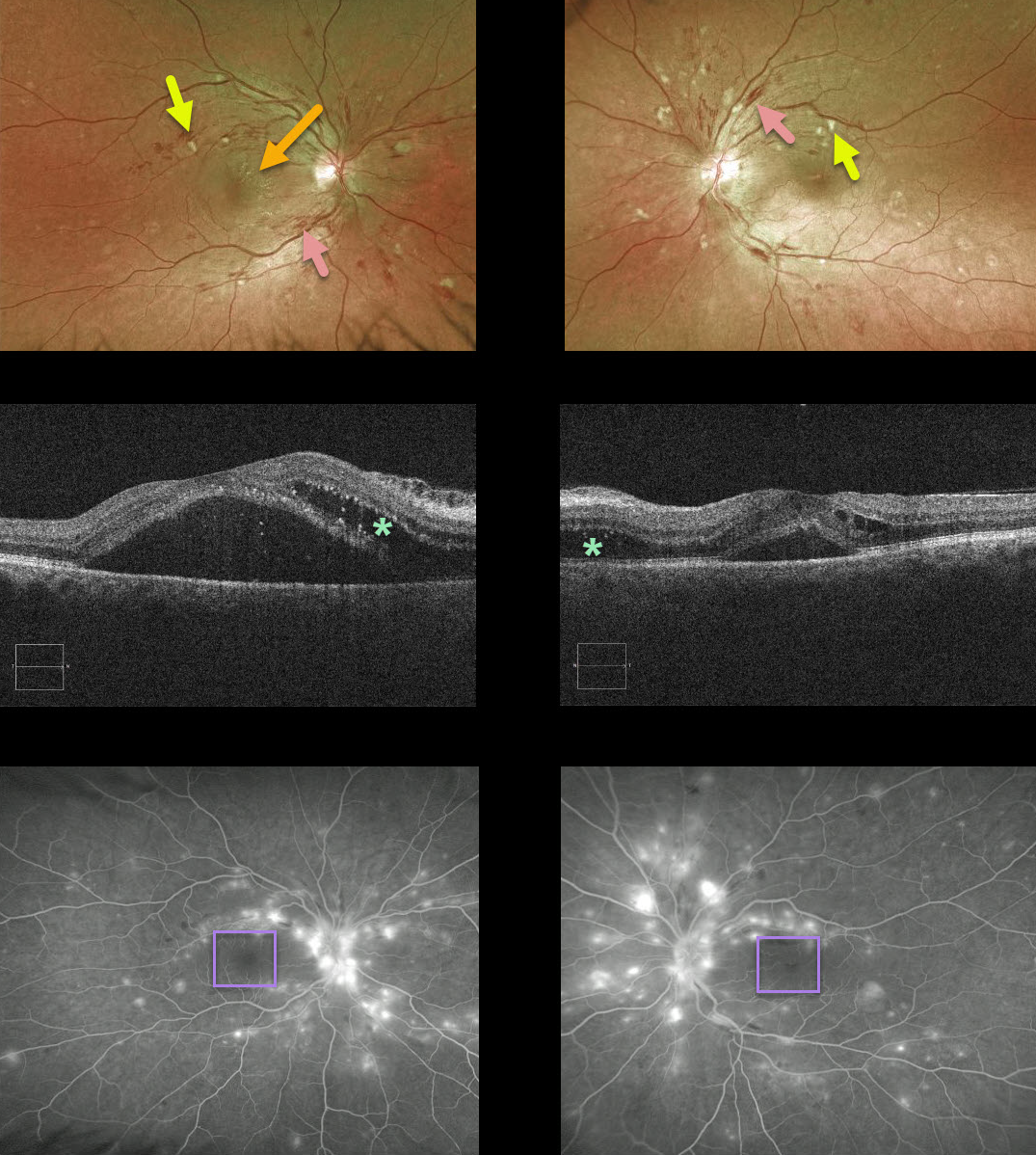
This patient presented with bilateral inner-retinal flame-shaped hemorrhages and nerve fiber layer infarcts. There was no past ocular or past medical history. In the office, her blood sugar was normal but her blood pressure was 160/92.
In this patient’s OCT, the macular fluid is coming mostly from the nerves; note the early nasal macular star OD and the bilateral outer retinal fluid on OCT that is most prominent nasally.
Angiography also shows a striking absence of macular leakage that should have been present if diabetes were the cause.
Although her blood pressure was not that severely elevated, we suspected it was likely much higher in the near past. She had no health coverage and no primary care doctor, so we sent her to the ER for further treatment and management.
Learning Points:
Hypertensive retinopathy can have a similar appearance to diabetic retinopathy. Clues to hypertensive cause include clinical, angiographic, and OCT signs.
Over time, patients with hypertensive retinopathy will develop near complete regression of all findings following systemic blood pressure treatment and control.


