

Hypotony
<< Back to Cases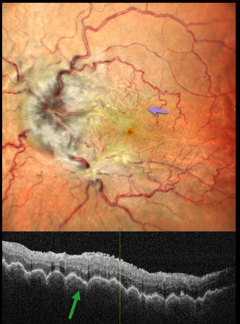
HYPOTONY MACULOPATHY
Rohan Jain and Manish Nagpal
Originally posted on @retina.rocks May 26, 2025
This 44YO male presented with 2 months of decreased vision in his left eye. A retinal detachment repair was done 9 months earlier with vitrectomy and silicone oil, followed by oil removal 3 months later. Vision was 20/20 in his normal OD and 20/90 OS. The intraocular pressure (IOP) was 10 mmHg OD and 3 mmHg OS. Pseudophakodonesis of the IOL was present OS.
Pseudocolor SLO imaging shows a moderately swollen nerve, tortuous retinal vessels, and extensive, irregularly oriented chorioretinal folds throughout the posterior pole. OCT shows a thickened choroid with overlying undulating chorioretinal folds.
We advised IOL explantation with vitrectomy and silicone oil, but he was immediately lost to follow-up.
Learning Points:
Hypotony maculopathy most commonly develops following surgery (including glaucoma filtration surgery and wound leaks) and trauma. Findings include low IOP, chorioretinal folds, and occasionally optic disc edema. Findings usually promptly resolve once the IOP normalizes.
HYPOTONY MACULOPATHY
Originally posted on @retina.rocks May 13, 2024
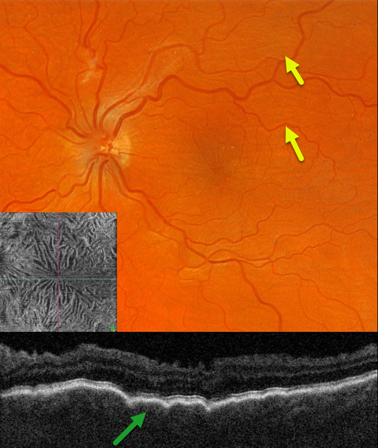
This 39YO male with a history of pigmentary glaucoma underwent goniotomy surgery by an outside practice in his left eye 3 weeks earlier. This eye has been hypotonus since, and vision was 20/150.
Optos color RG imaging shows faint diffuse chorioretinal folds throughout the posterior pole. Triton swept-source OCT B-scan shows a thickened choroid with overlying folds, and an irregularly rippled inner retina. En face OCT through the internal limiting membrane provides a unique perspective on inner retinal distortion.
Learning Points:
Hypotony maculopathy is characterized by low IOP and chorioretinal folds, most commonly following glaucoma filtration surgery. Optic nerve edema can also be seen due to decreased axoplasmic transport.
We expect our patient’s findings to improve once his intraocular pressure normalizes.
HYPOTONY
Originally posted on @retina.rocks February 29, 2024
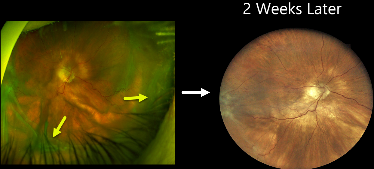
This 84YO female received an intravitreal Eylea HD injection in her right eye on 12/28/23 for macular edema from a branch retinal vein occlusion. She presented on 1/18/24, complaining of waking several days earlier with severe painless vision loss in this eye. She denied trauma or Valsalva. Vision was counting fingers, and intraocular pressure was 1 mmHg. There were no apparent wound leaks or conjunctival blebs.
Optos color RG imaging shows severe, diffuse, irregular scleral folds without choroidal detachment.
Treatment was initially conservative with topical atropine, prednisolone acetate, and oral steroids. However, findings were stable, and IOP remained at 1 mmHg after 3 days. The patient then underwent ultrasound biomicroscopy (UBM) to assess for a suspected posterior cyclodialysis cleft, followed by cryotherapy.
One week later, we planned on injecting an expandable C3F8 gas bubble in hopes of normalizing the eye pressure. When she presented on 2/1/24 for this procedure, her vision remained at counting fingers, but her IOP had increased to 2 mmHg. Fundus examination was nearly normal with almost complete resolution of the scleral folds.
Learning Points:
Although the definitive etiology of our patient’s hypotony remains unclear, our working theory is either a posterior cyclodialysis cleft or a very slow-leaking injection wound. As described by Lee et al. (Am J Ophthalmol Case Rep. 2021 Sep; 23: 101134), incomplete penetration into the vitreous cavity, early injection, or continued injection on needle withdrawal could lead to the formation of a posterior cyclodialysis cleft during intravitreal injections. No cleft was visible on gonioscopy, but a strong suspicion of a posterior cleft was raised on UBM. Our other differential included a slow-leaking scleral wound from a prior injection. No leak was visible at initial examination, but with an IOP of 1 mmHg, a spontaneous leak would not be expected. Both these theories are confounded by a history of severe (-15D) myopia prior to cataract surgery, with possible thinned sclera predisposing her to the hypotonus scleral folds.
CHOROIDAL DETACHMENT
Originally posted on @retina.rocks August 15, 2022
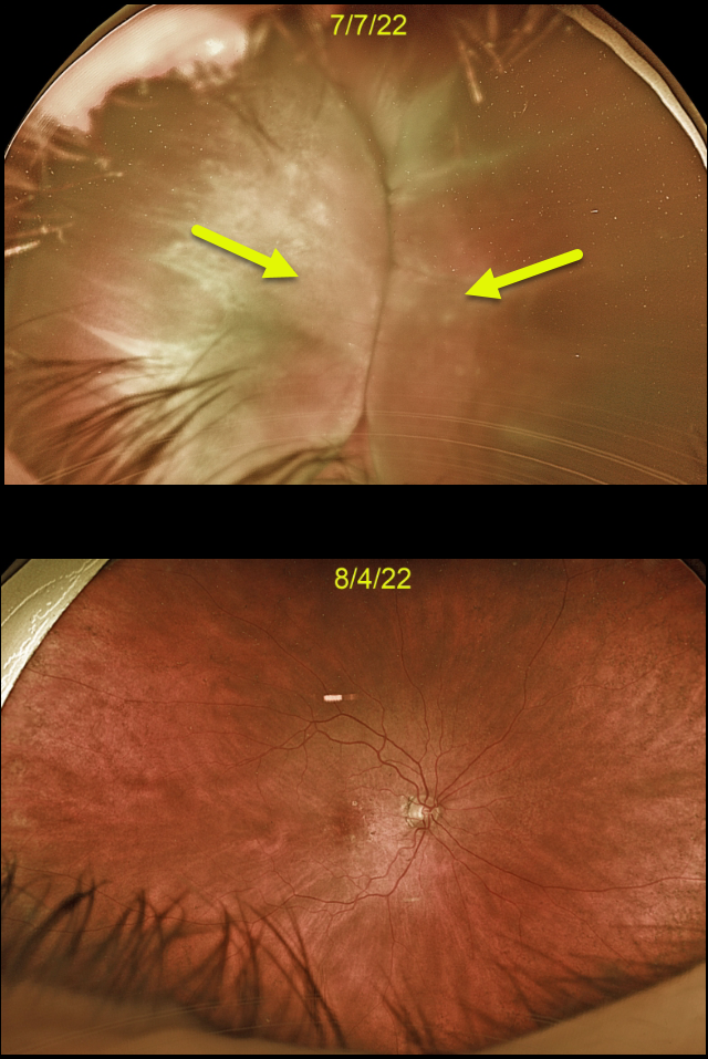
This 88YO female developed kissing choroidal detachments immediately following Ahmed glaucoma valve placement.
The choroidal detachment spontaneously resolved after 1 month once the intraocular pressure normalized.
Learning Points:
Choroidal detachment is relatively common immediately following glaucoma surgery.
Two mechanisms are believed to be at play: hypotony allows fluid to accumulate in the suprachoroidal space and inflammation increases the choroidal permeability. The detached ciliary body may also produce less aqueous, thus creating a self-perpetuating cycle.
Often, observation is sufficient, but topical steroids and cycloplegics can be helpful to aid in resolution.
CHOROIDAL DETACHMENT
Originally posted on @retina.rocks July 14, 2022
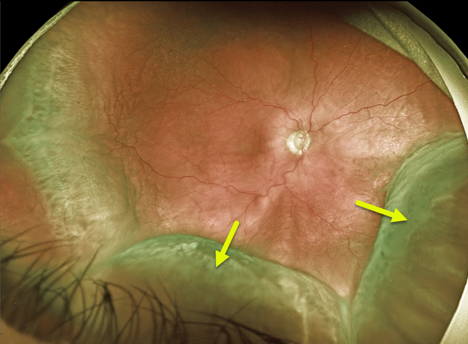
This 68YO male patient presented with peripheral choroidal detachments following placement of a Baerveldt shunt 1 week earlier. Vision was counting fingers, and pressure was 1 mmHg. All wounds were secure, and the anterior chamber was formed.
Learning Points:
Choroidal detachment is relatively common immediately following glaucoma surgery. Two mechanisms are believed to be at play:
1) hypotony allows fluid to accumulate in the suprachoroidal space, and 2) inflammation increases the choroidal permeability. The detached ciliary body may also produce less aqueous, thus creating a self-perpetuating cycle.
Often, observation is sufficient, but topical steroids and cycloplegics can be helpful in promoting resolution. We are following this patient closely and expect the choroidals to resolve as postoperative pressure increases.
HYPOTONY MACULOPATHY
Originally posted on @retina.rocks April 12, 2022
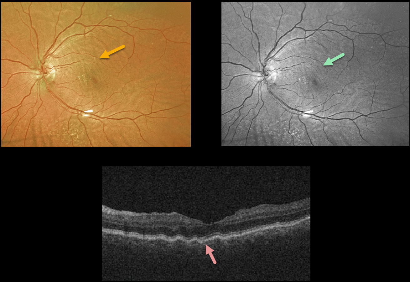
This 60YO female developed hypotony in her left eye following a Baerveldt implant.
Chorioretinal folds are seen on the Optos color photo and are more pronounced in the green channel. OCT shows the unilateral radiating left macular folds.
Although pressures were initially almost zero for about 2 months postoperatively, they have since risen to about 10 with persistence of the chorioretinal folds.
Vision is 20/30, and we continue to follow her.
HYPOTONY MACULOPATHY
Originally posted on @retina.rocks September 1, 2020
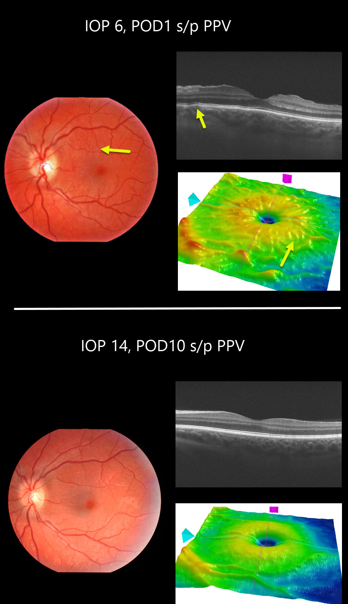
This patient developed hypotony maculopathy one day following vitrectomy for vitreous floaters. Intraocular pressure was 6mmHg and all sclerotomy sites appeared tight. The retinal vessels are somewhat engorged and dilated.
Chorioretinal folds are seen in the fundus photo, OCT B-scan and thickness map.
These findings completely resolved 9 days later when his IOP recovered spontaneously to 14mmHg.
Learning Points:
Hypotony maculopathy is characterized by low IOP and chorioretinal folds, most commonly following glaucoma filtration surgery. Optic nerve edema can also be seen due to decreased axoplasmic transport.


