

Idiopathic Central Serous Chorioretinopathy (ICSC)
<< Back to Cases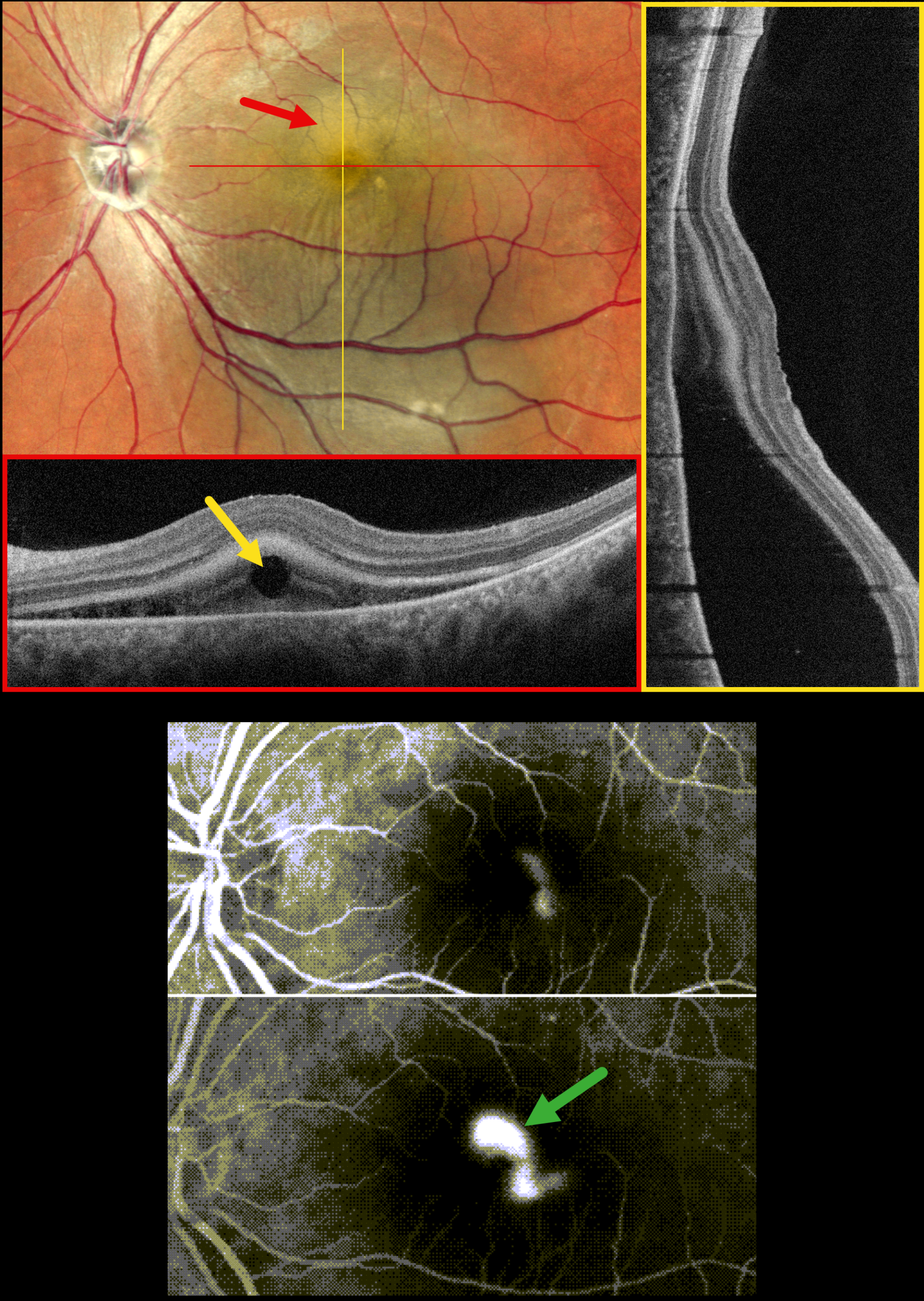
FIBRINOUS IDIOPATHIC CENTRAL SEROUS CHORIORETINOPATHY
Rohan Jain and Manish Nagpal
Originally posted on @retina.rocks June 22, 2026
This 37YO male presented with one month of decreased vision in his left eye. There was no history of steroid or testosterone use. Vision was 20/20 in his normal OD and 20/40 OS.
MultiColor SLO imaging shows a large macular serous detachment extending into the inferior midperiphery, associated with fibrinous subretinal fluid. A horizontal OCT B-scan shows hyperreflective subretinal fibrin with a round hyporeflective lesion. A vertical B-scan shows the serous detachment extending inferiorly. Fluorescein angiography shows a smokestack leak.
Learning Points:
In idiopathic central serous chorioretinopathy (ICSC), fluorescein dye diffuses through a physiological break in the RPE and then pools within the subretinal space. The clearing within our patient’s inferotemporal subretinal fibrin (as evidenced by the hyporeflective round lesion on OCT) is likely caused by serous fluid entering the subretinal space and ‘washing away’ some of the fibrin.
Most patients with ICSC have a single, unilateral leak. Blot leaks are most common, with only 10% of cases exhibiting a smokestack. The subretinal fibrin noted in our patient is more commonly found in steroid-induced disease, although our patient denied steroid exposure.
PRIOR CHRONIC ICSC WITH NEW TRACTIONAL MACULAR HOLE-RRD WITH POSTOPERATIVE MENISCUS MICROPYON
Originally posted on @retina.rocks October 29, 2025
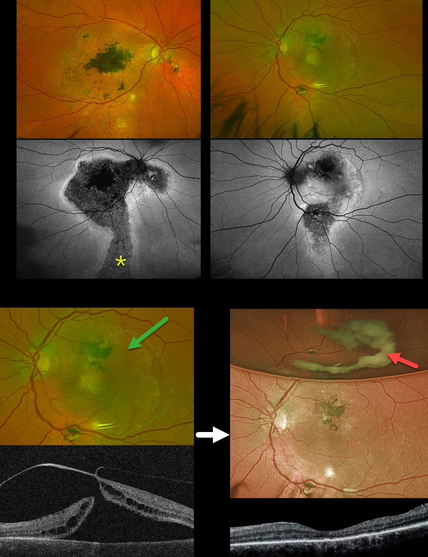
This 58YO male was referred for a retinal detachment in his left eye. He has a long history of idiopathic central serous chorioretinopathy (ICSC). He was previously treated elsewhere for neovascular AMD OD. For many years, he has been receiving intravitreal Avastin OS every 1-2 months. Vision was counting fingers OD and 20/400 OS.
Optos color RG imaging shows bilateral macular scarring with confluent hyperpigmented foveal changes and intraretinal pigment migration, especially in his right eye. Inferior hypopigmented gutters are most obvious on fundus autofluorescence (FAF) as hypo-FAF with surrounding hyper-FAF margins. A rhegmatogenous retinal detachment (RRD) occupies the entire left macula. Triton swept-source OCT shows a full-thickness macula hole, active vitreoretinal traction on the temporal edge of the hole, and a macular detachment.
The tractional macular hole with secondary RRD in our patient’s left eye is completely unrelated to his poorly treated ICSC. One month following vitrectomy, vision improved to 20/100. The retina was completely attached, and the macular hole was closed. A meniscus micropyon is adherent to the inferior meniscus of the gas bubble.
Learning Points:
Photodynamic therapy (PDT) is currently the standard of care for ICSC with symptomatic acute or chronic subretinal fluid. Unfortunately, Visudyne is rarely available outside the US, which has led to the adoption of unproven therapies, including mineralocorticoid receptor antagonists (eplerenone), beta blockers, and intravitreal anti-VEGF agents. Recent meta-analyses and systematic reviews confirm that these alternative therapies are ineffective (Felipe et al Int J Retina Vitreous 2022;8;34 and Kim et al Ophthalmology 2025;132:343-353). Despite the clear lack of efficacy in the literature, specialists both within and outside the US continue to use these drugs (Vankatesh et al Int J Retina Vitreous 2023;9;61). Our patient’s macular findings are a testament to what can happen when proven therapies are ignored.
Russell et al recently reported a unique ophthalmoscopic sign, a meniscus micropyon, that follows PPV with gas tamponade for all causes (Retina 2024;44:1766-1776). Features they noted that distinguish this sterile micropyon from post-PPV fibrin include a delayed appearance, hyperautofluorescence, and absence of anterior chamber or vitreous fibrinous strands/sheets. Vitreous biopsy in two of their cases revealed predominantly white blood cells. The micropyon appearance was associated with epiretinal proliferation (80%). Recurrent RRD from proliferative vitreoretinopathy (PVR) developed 33% of eyes, 12% re-detached without frank PVR, and 18% developed worsening epiretinal membranes.
Our patient’s meniscus micropyon disappeared as the bubble resolved. Three months postoperatively, vision was 20/60, and the retina remained completely reattached without PVR.
PACHYCHOROID NEVOASCULARIZATION
Originally posted on @retina.rocks March 31, 2025
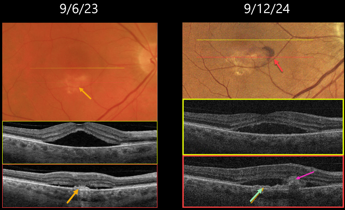
This 64YO male presented on 9/6/23 with several weeks of vision loss in his right eye. Vision was 20/40 OD.
Triton color imaging shows a faint round foveal serous detachment. Swept-source OCT confirms a classic idiopathic central serous chorioretinopathy (ICSC) foveal detachment. There is a small hyperreflective placoid elevation of the RPE along the inferior edge of the detachment corresponding to a yellow-white area of subretinal pigmentation. Due to significant symptoms, he elected for early photodynamic therapy (PDT), which successfully resolved the serous fluid two months later with 20/30 vision (not shown).
He did well until he returned on 9/12/24 with recurrent blurred vision OD, which had decreased slightly to 20/40. There is new subretinal blood within the region of the persistent placoid RPE elevation inferiorly. OCT shows recurrent serous foveal fluid centrally. There is an enlargement of the shallow inferior RPE elevation adjacent to the subretinal blood. Monthly intravitreal Avastin therapy was started.
Learning Points:
Initially described by Pang and Freund, pachychoroid neovasculopathy is a variant of type 1 (below the RPE) macular neovascularization (MNV) found as part of the pachychoroid spectrum, including ICSC and pachychoroid pigment epitheliopathy (Retina 2015;35:1-9). About one-third of chronic ICSC eyes with flat irregular RPE detachments will harbor an MNV on OCT angiography (Bousquet et al, Retina 2018;38:629-639), which is probably a form of pachychoroid neovascularization (Hwang et al, Retina 2020;40:1724-1733).
In retrospect, our patient most likely had a pachychoroid neovascularization, which initially responded to PDT. When he returned one year later with a definite active MNV, we elected to start anti-VEGF monotherapy. He was unfortunately immediately lost to follow-up.
FIBRINOUS IDIOPATHIC CENTRAL SEROUS CHORIORETINOPATHY
Ayushi Gupta and Vishal Agrawal
Originally posted on @retina.rocks February 24, 2025
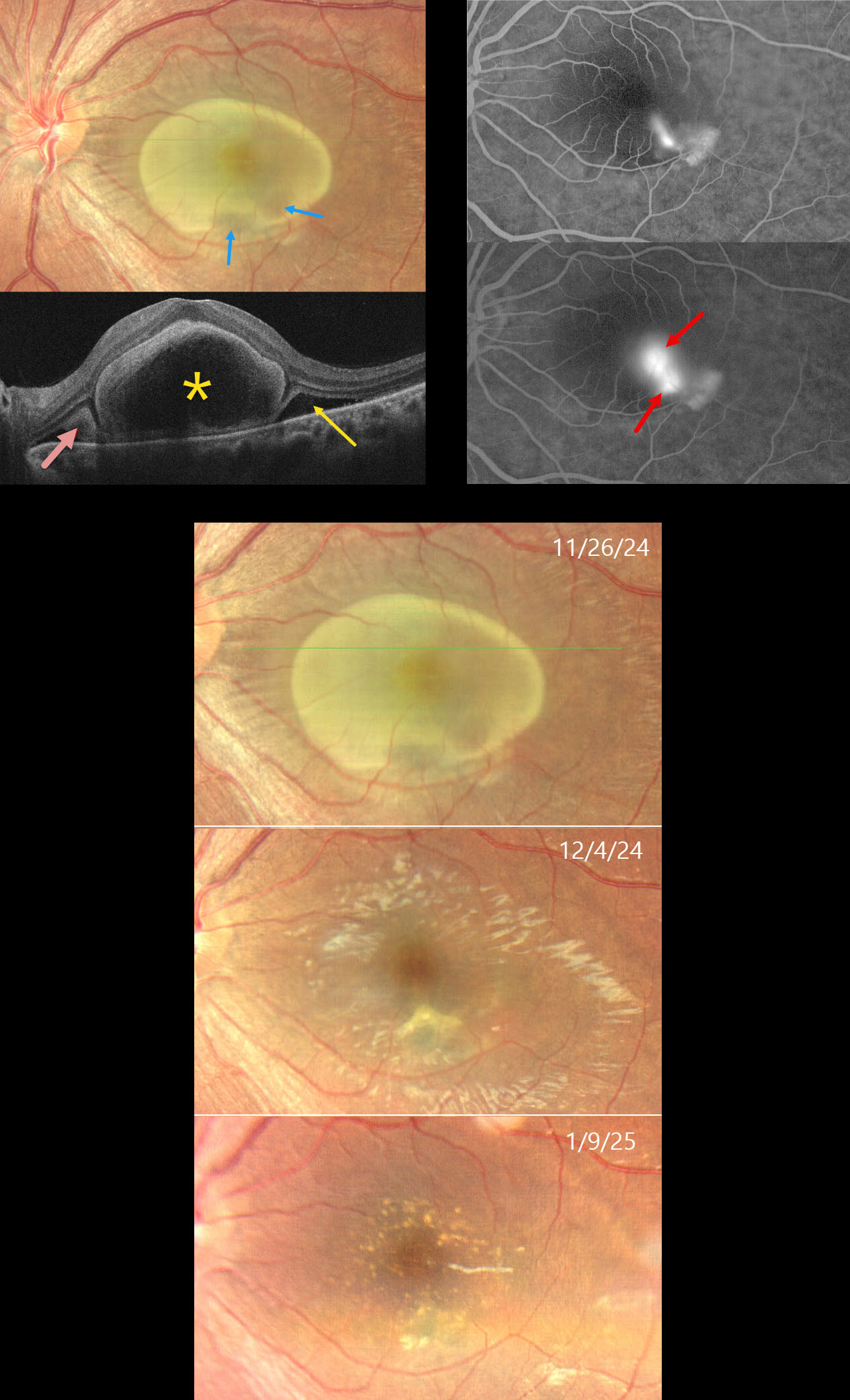
This 31YO female complained of decreased vision in her left eye for 6 weeks. She was diagnosed elsewhere with cysticercosis and referred to us for further evaluation. Vision was 20/20 in her normal OD and 20/200 OS.
Color photography shows a yellow oval lesion occupying nearly the entire macula. OCT scanning shows a large subfoveal bacillary layer detachment (BALAD). Hyperreflective fibrin likely lines the inner border of the BALAD. Subretinal fibrin and subretinal fluid (SRF) are noted on either edge of the BALAD. Fluorescein angiography shows either a smokestack or two independent blot leaks with staining inferotemporally. The yellow foveal lesion is relatively depigmented within the region of the angiographic leakage. The lesion and fluid spontaneously resolved over about 6 weeks, with residual drusen-like changes. Vision at the final visit was 20/20.
Learning Points:
In idiopathic central serous chorioretinopathy (ICSC), fluorescein dye diffuses through a physiological break in the RPE and then pools within the subretinal space. The clearing within our patient’s inferotemporal subretinal fibrin (as evidenced by the hyporeflective temporal SRF on OCT) is likely caused by serous fluid entering the subretinal space and ‘washing away’ some of the fibrin.
Most patients with ICSC have a single, unilateral leak. Blot leaks are most common, with only 10% of cases exhibiting a smokestack. The multifocal leaks and subretinal fibrin noted in our patient are more commonly found in steroid-induced disease, although our patient denied steroid exposure. Bacillary layer (cone and rod inner and outer segments) detachment appears as a unique dome-shaped collection of intraretinal fluid from photoreceptor splitting. These detachments can be observed in an increasing number of conditions, most classically Vogt-Koyanagi-Harada disease (Cicinelli et al, Ophthalmology Retina 2020;4:454-456). BALAD is also a biomarker for severe ICSC with steroid use (Casella et al, Int J Retina Vitreous 2024;10;96).
CENTRAL SEROUS CHORIORETINOPATHY WITH EXUDATIVE RD AND COMCOMITANT TB
Anand Temkar, Navneet Mehrotra, and Manish Nagpal
Originally posted on @retina.rocks November 7, 2024
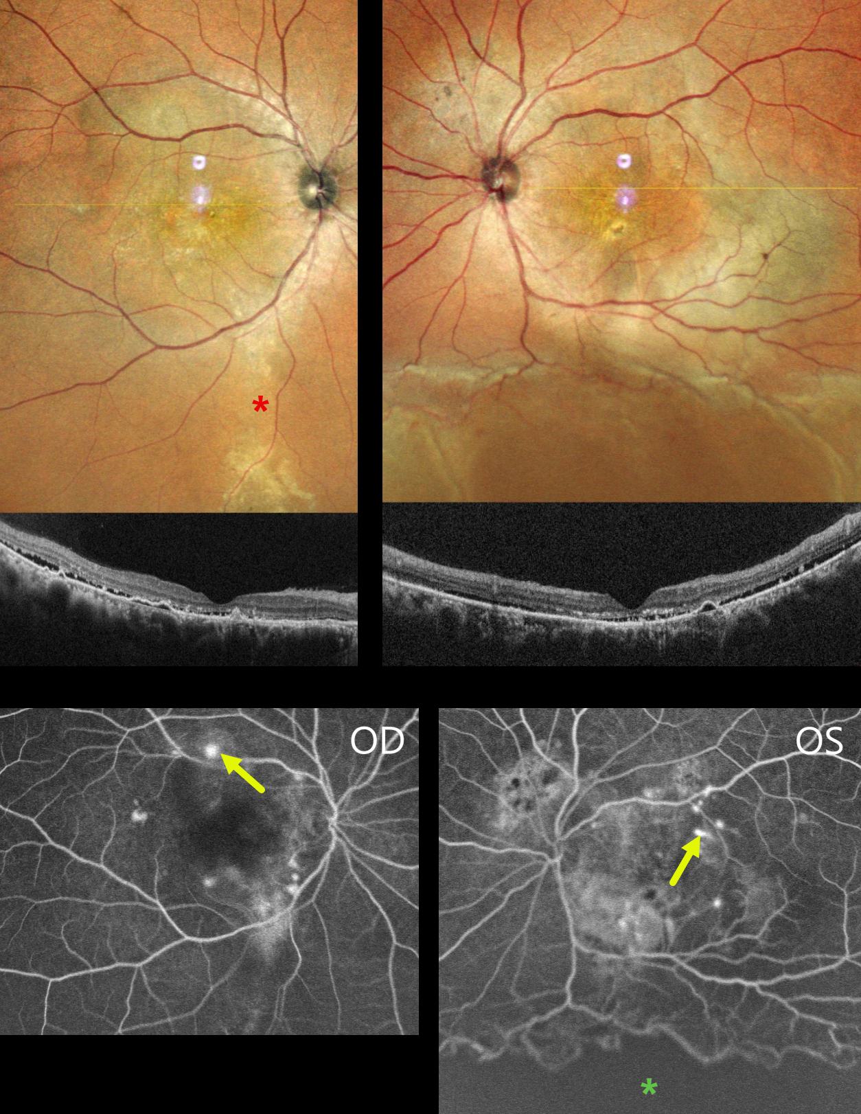
This 39YO male presented with 4 months of bilateral vision loss. He was diagnosed elsewhere with chronic idiopathic central serous chorioretinopathy (ICSC) and was placed on azathioprine and spironolactone 3 months earlier. There was no history of steroid use or injections. Vision was 20/120 OD and 20/200 OS.
MultiColor imaging shows macular pigmentary changes that extend in a gutter inferiorly OD. Diffuse macular pigmentary changes are noted in his left eye that communicate with an inferior exudative retinal detachment. OCT scanning shows shallow subretinal fluid with hyperreflective shed outer segments, outer retinal hyperreflective foci, and RPE detachments. Fluorescein angiography shows bilateral multifocal subretinal blot leakage. A broad area of ischemia is present overlying the inferior detachment of the OS.
Mantoux skin testing (15x18mm) and QuantiFERON-TB Gold testing were positive. Chest CT showed fibrotic strands in the right lung. He was referred for systemic antituberculosis treatment. A week later, his local ophthalmologist reported that his vision and retinal findings improved, but he was then lost to follow-up.
Learning Points:
Our patient presented with severe bilateral idiopathic central serous chorioretinopathy (ISCS) with multifocal leaks in each eye and a secondary exudative detachment in his left eye. There are rare case reports of TB occurring in patients with ICSC, and these are likely coincidental findings (Khan et al, BMJ Case Rep 2017:bcr-2016-216471).
CILIORETINAL ARTERY + CENTRAL SEROUS CHORIORETINOPATHY
Mayelinne García, Luis Montero and Alberto Ruiz
Originally posted on @retina.rocks September 24, 2024
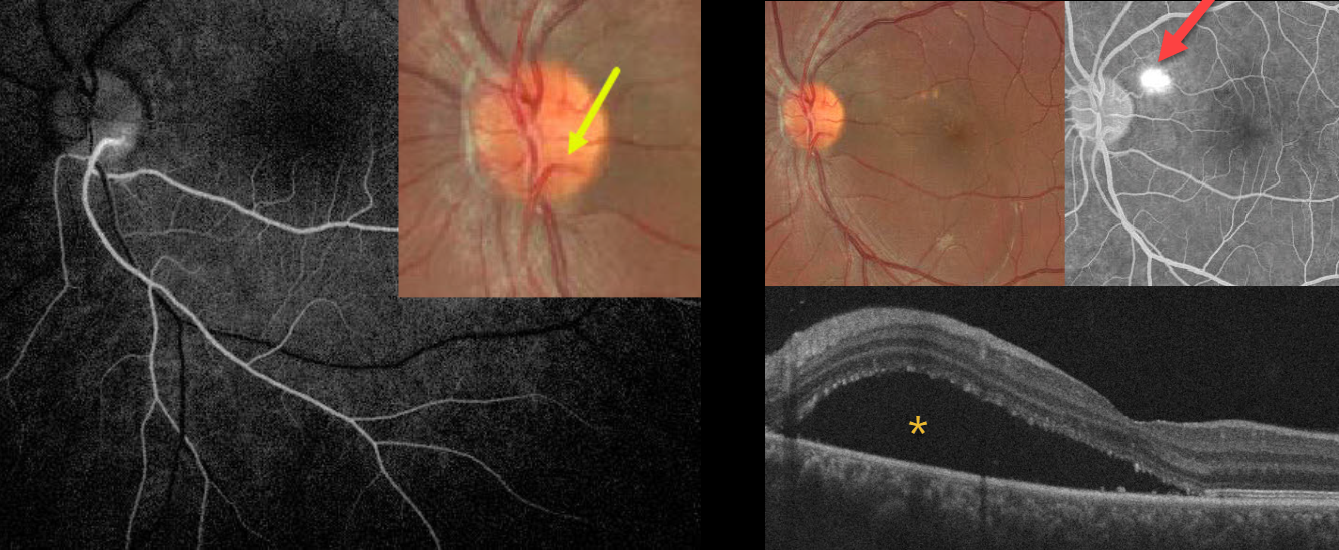
This healthy 25YO male presented with 3 weeks of vision loss in his left eye from active idiopathic central serous chorioretinopathy (ICSC). Vision was 20/20 in his normal OD and 20/110 OS.
A cilioretinal vessel originates within the inferotemporal aspect of the nerve and arches inferiorly. Fluorescein angiography reveals that this cilioretinal artery supplies the entire inferotemporal retina. Color imaging of the macula shows a large serous detachment with some drusen-like changes. This detachment is confirmed on OCT scanning. A blot leak is seen angiographically. Due to the patient’s symptoms, thermal laser was applied to the angiographic leak.
Learning Points:
The inner two-thirds of the neurosensory retina are supplied by the central retinal artery, with the choroid supplying the RPE and photoreceptor layer. Cilioretinal arteries, which arise from the posterior choroidal circulation, are present in up to 50% of individuals and are bilateral in about 25% (Schneider et al, Acta Ophthalmologica 2021;99:e310-e318).
IDIOPATHIC CENTRAL SEROUS CHORIORETINOPATHY WITH LOCULATED OUTER CHOROIDAL FLUID
Originally posted on @retina.rocks June 25, 2024
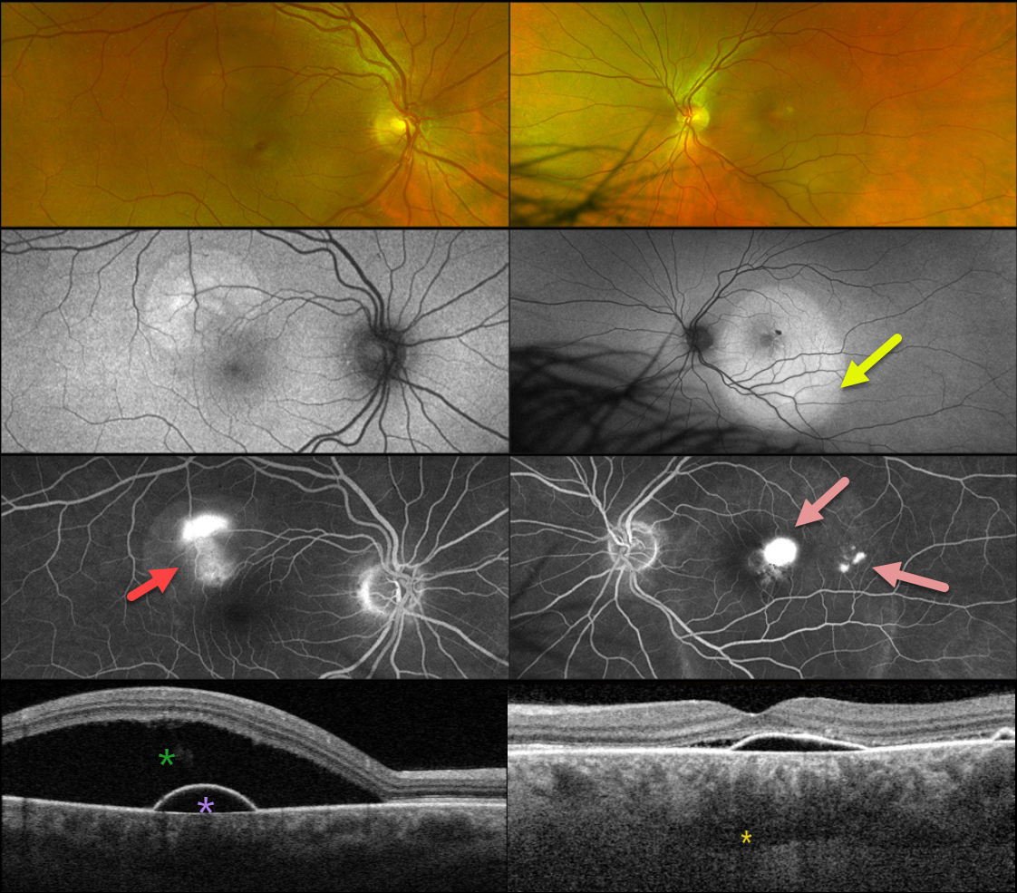
This healthy 48YO male presented with 1-2 weeks of decreased vision. There was no past ocular history. Vision was 20/25 OD and 20/70 OS.
Optos color RG imaging shows a superotemporal serous detachment OD with a larger serous detachment throughout the left macula. Triton swept-source OCT shows a shallow RPE detachment with some nasal subretinal fluid. Loculated outer choroidal fluid is seen. A more dramatic serous detachment with an underlying RPE detachment is noted OD.
On fundus autofluorescence, these serous detachments show hyper-FAF. Fluorescein angiography shows a smokestack leak OD with multifocal blot leaks OS.
We will initially observe our patient and recommend photodynamic therapy if the fluid fails to resolve over the coming months.
Learning Points:
Initially described by Spaide and Ryan (AJO 2015;160:1211-1216), loculated outer choroidal fluid is found in about two-thirds of patients with idiopathic central serous chorioretinopathy (ICSC). It is best appreciated with swept-source or enhanced depth imaging OCT.
This fluid most commonly interdigitates with the larger outer choroidal vessels, as in our patient, but can rarely appear in the suprachoroidal space. It is more common with a thicker choroid and sclera (AJO 2022;235:197-203).
IDIOPATHIC CENTRAL SEROUS CHORIORETINOPATHY WITH INCIDENTAL OPTIC NERVE PIT
Originally posted on @retina.rocks May 30, 2024
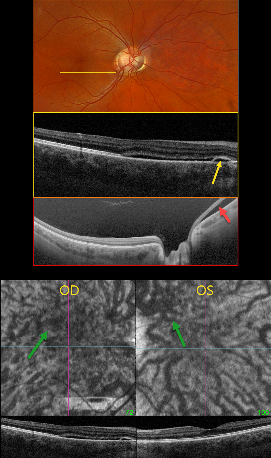
This healthy pregnant 32YO female presented with recent mild central vision loss in her right eye. Vision was 20/40 OD and 20/30 OS.
Optos color RGB imaging shows an anomalous right optic disc with an optic pit both nasally and temporally. Triton swept-source OCT shows both pits and subretinal fluid extending from the nerve nasally. OCT through the macula shows shallow nasal subretinal fluid extending into the macular center, along with a tiny nasal RPE detachment. This fluid does not communicate with the nerve or temporal pit. Optos Silverstone swept-source OCT through the posterior pole shows the subretinal fluid nasal to the nerve. Macular en face and B-scans show a thickened choroid with anomalous, thickened, and tortuous vessels.
Our patient’s symptoms are from idiopathic central serous retinopathy (ICSC), most likely related to her pregnancy. The macular fluid does not communicate with the temporal disc pit, and her macular OCT findings are classic for central serous. Fluorescein angiography was not performed due to her pregnancy. Although she has optic pit ‘maculopathy,’ this is in the nasal retina from her temporal disc pit. Observation was recommended.
Learning Points:
An optic nerve pit is a rare (about 0.1% prevalence) congenital anomaly thought to be caused by incomplete closure of the optic fissure during gestation. Pits are most commonly located within or along the inferior-temporal disc margin and usually appear as a gray depression. While most optic pits are asymptomatic, they can cause vision loss by forcing fluid into the inner and outer retina, and ultimately into the subretinal space (optic pit maculopathy). The source of the fluid remains a topic of ongoing debate, with the fluid originating either from the vitreous or the subarachnoid space.
POST-COVID FIBRINOUS CENTRAL SEROUS CHORIORETINOPATHY
Originally posted on @retina.rocks January 31, 2024
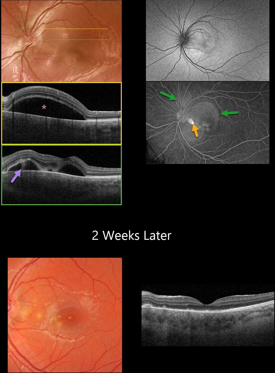
This 12YO boy presented with a few days of painless vision loss in his left eye. He had COVID one week earlier. There was no prior medical history. He was on no systemic medications. Vision was 20/30 in his normal right eye and 20/50 in his left eye.
Optos color RG imaging of his left eye shows a large macular serous detachment. Triton swept-source OCT confirms the serous detachment along with subretinal fibrin.
Fundus autofluorescence (FAF) shows faint peripapillary and macular hyper-FAF, and fluorescein angiography shows pooling beneath a nasal macular RPE detachment and larger areas of pooling within the multifocal serous detachments.
When he returned following 2 weeks of observation, the fluid and BALAD completely regressed with secondary drusen-like changes. Variable outer retinal atrophy is seen on OCT.
Learning Points:
Prior case reports of idiopathic central serous chorioretinopathy (ICSC) following COVID have been reported, although these have developed in patients who were also treated with systemic corticosteroids.
Fuganti et al reported 2 patients, both treated with systemic steroids, with post-COVID ICSC with BALAD (AJO Case Reports 2022;28;101690). We suspect our patient’s findings may have been COVID-related, given the atypical presentation and rapid resolution of the BALAD and serous fluid.
CHOROIDAL NEVUS MISDIAGNOSED AS CENTRAL SEROUS
Emma Oreskovic and Natasa Draca
Originally posted on @retina.rocks September 7, 2023
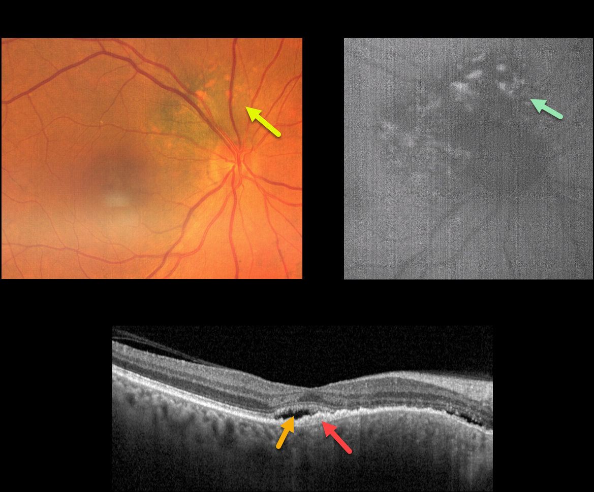
This 71YO female was referred for further treatment of chronic idiopathic central serous chorioretinopathy (ICSC) of her right eye, for which she had previously received unsuccessful anti-VEGF therapy at another institution. Vision was 20/100 OD and 20/20 in her normal left eye.
Fundus photography shows a variably pigmented, minimally thickened peripapillary choroidal nevus that extends from about 7-1 o’clock around the optic nerve margin. Overlying orange pigmentation is noted, which variably hyper-autofluoresces.
OCT scanning shows shallow foveal subretinal fluid with a subretinal layer of thickened hyperreflective material extending nasally overlying the thickened nevus. Beneath this is a very shallow, irregular elevation of the RPE.
Learning Points:
Clinical characteristics of choroidal nevi at risk of progressing to uveal malignant melanoma (MM) were initially described by Shields et al. (Ophthalmology 1995;102:1351-1361).
The most recent version of this famous acronym (Shields et al, Retina 2019;39:1840-1851) is To Find Small Ocular Melanoma Doing Imaging (TFSOM-DIM), which stands for To (Thickness >2mm) Find (subretinal Fluid) Small (Symptoms, vision <= 20/50) Ocular (Orange pigment) Melanoma (Melanoma hollow on ultrasonography) DIM (DIaMeter > 5mm).
ICSC and pachychoroid disorders, similar to syphilis, are one of the ‘great imitators,’ including uveal MM masquerading as ICSC (Negretti et al, Ophthalmology Retina 2023;7:171-177).
In our patient, on the central macular OCT B-scan, one sees a thickened choroid, subretinal fluid, and hyperreflective shed outer segments/lipofuscin, all of which are suggestive of ICSC.
This case emphasizes the importance of looking beyond the OCT, using both the clinical exam and multimodal imaging, for every patient, including those with chronic ICSC, to arrive at the correct diagnosis and treatment.
Since our patient has several high-risk characteristics (fluid, symptoms, and orange pigment), she was referred to an ocular oncologist for further management.
FIBRINOUS IDIOPATHIC CENTRAL SEROUS CHORIORETINOPATHY
Mattie Adams
Originally posted on @retina.rocks April 20, 2023
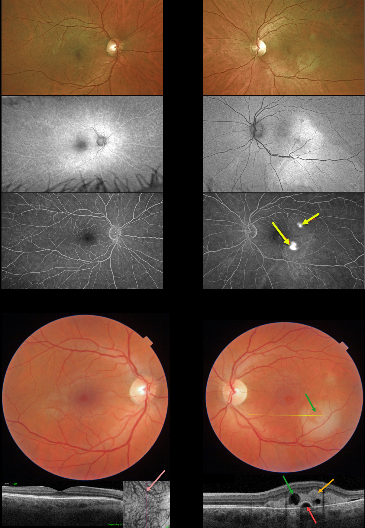
This 42YO male presented with mild symptoms of decreased color saturation and 20/40 vision in his left eye. The right eye was normal with 20/20 vision.
Color imaging shows creamy white subretinal fibrin in the inferotemporal left macula with a smaller area of fibrin superotemporally. Swept-source OCT through the larger inferior lesion shows hyperreflective, subretinal fibrin extending into the outer retinal layers. There is a small underlying retinal pigment epithelial detachment (PED), with a small overlying hyporeflective space corresponding funduscopically to an area devoid of fibrin.
Although the right macula appears normal funduscopically and on B-scan OCT, en face imaging shows somewhat dilated choroidal vessels.
Optos fundus autofluorescence (FAF) shows a hyper-FAF gutter extending from both fibrinous lesions. Fluorescein angiography shows two hyperfluorescent blot leaks.
Learning Points:
In idiopathic central serous chorioretinopathy (ICSC), fluorescein dye diffuses through a physiologic break in the RPE and then pools within the subretinal space. As in this case, the subretinal leak is almost always associated with a PED.
The clearing within the inferotemporal subretinal fibrin, which corresponds to the hyporeflective space within the fibrin on OCT, is caused by serous fluid entering the subretinal space and ‘washing away’ some of the fibrin.
Most patients with ICSC have a single, unilateral leak. Blot leaks are most common, with only 10% of cases exhibiting a smokestack leak. The multifocal leaks and subretinal fibrin noted in our patient are more commonly found in steroid-induced disease (our patient denied steroid exposure). Due to the complete lack of symptoms, initial observation was recommended.
PACHYCHOROID SPECTRUM
Originally posted on @retina.rocks November 22, 2022
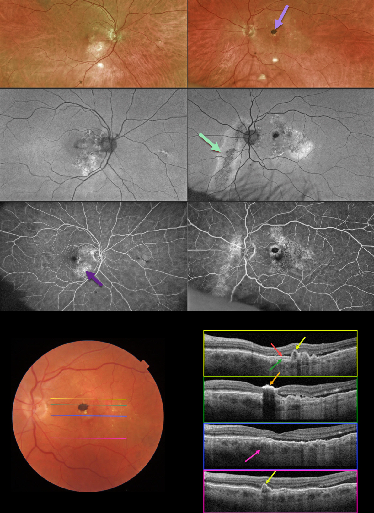
This 78YO male was referred for asymptomatic fundoscopic changes. Vision was 20/25 OD and 20/30 OS. He had a history of laser treatment in his left eye decades earlier.
Optos imaging shows bilateral areas of pigmentary loss in each macula, extending inferiorly. A hyperpigmented laser scar is noted in the left superior macula.
Fundus autofluorescence (FAF) shows variable hypo- and hyper-FAF, along with a prominent gutter extending from the left nerve inferonasally.
Fundus fluorescein angiography (FFA) shows window defects within these areas, with blockage from the hyperpigmented laser scar OS.
Swept-source OCT shows numerous findings. Scanning superior to the laser scar shows several choroidal polyps. A shallow elevation of the RPE exposes the underlying Bruch’s membrane. Scanning through the laser scar shows intense hyperreflectivity with posterior shadowing. Scanning through the macular center shows temporal subretinal fluid, along with a thickened choroid. Finally, scanning in the inferior macula shows an additional polyp.
Learning Points:
This case has an overwhelming amount of features from the pachychoroid spectrum, which includes idiopathic central serous retinopathy (ICSC), pachychoroid neovasculopathy, polypoidal choroidal vasculopathy, pachychoroid pigment epitheliopathy, peripapillary pachychoroid syndrome, and choroidal excavation.
The thermal laser treatment many years ago may have been for pachychoroid neovasculopathy, a recently described phenotype (see Pang and Freund, Retina 2015;35:1-9) found in the pachychoroid spectrum. These lesions are particularly common in patients with ICSC, and often remain silent as a flat, irregular RPE elevation on OCT without exudative complications.
Our patient likely had further progression of their pachychoroid disease, resulting in multiple polyps, which are found in polypoidal choroidal vasculopathy (PCV). PCV is a variant of type 1 macular neovascularization, also in the pachychoroid spectrum. PCV is likely distinct from age-related macular degeneration, occurring in a different population (more common in African Americans and Asians), with a relative lack of drusen, and the presence of a thicker choroid.
In addition to the findings above, the patient had a large gutter in his left eye, a common finding in ICSC. Gutters form from pathologies with chronic leakage. Conditions that form gutters other than ICSC include choroidal hemangiomas, choroidal nevi, and uveal melanoma.
For more information on the pachychoroid spectrum, see Cheung et al, Eye 2019;33:14-33 for a great pachychoroid disease review.
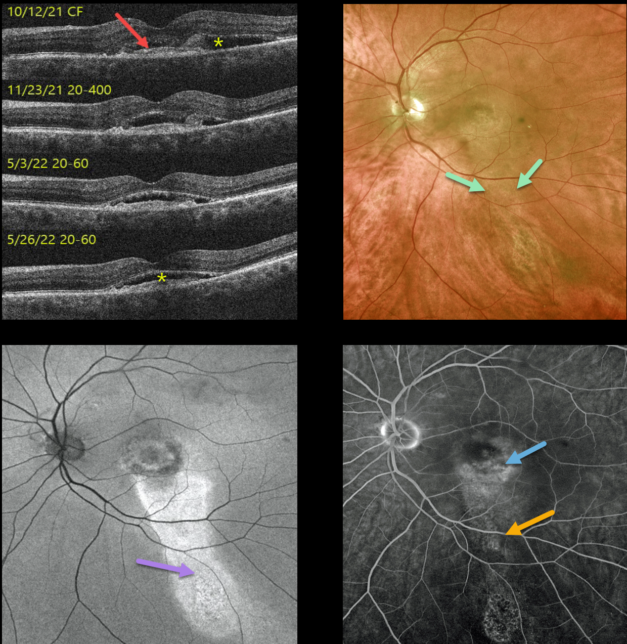
PACHYCHOROID NEOVASCULOPATHY WITH GUTTER
Originally posted on @retina.rocks July 1, 2022
This 69YO male presented with asymptomatic vision loss in his left eye. Vision was 20/30 OD and counting fingers OS.
A small central scar with fluid was present in the left macula (initial photo not shown). OCT scanning shows a shallowly elevated type 1 macular neovascularization with associated subretinal fluid. An intravitreal Avastin injection was given that day, and 1 month later, the fluid had improved. Vision was 20/400. A second intravitreal Avastin was given.
He was then lost to follow-up for about 6 months while he was out of state receiving intravitreal Eylea every 6 weeks. When he returned, his vision was 20/60 with shallow residual subretinal fluid. Optos color imaging shows central macular pigmentary changes with a gutter of inferior pigment loss. Fundus autofluorescence (FAF) more dramatically shows the hyper-FAF inferior gutter. Fluorescein angiography shows mild staining of the foveal MNV with an inferior gutter of window defects.
Due to an upcoming trip to Alaska and Canada, he requested trying photodynamic therapy (PDT) in an attempt to minimize the treatment burden. Several weeks after PDT, vision remained at 20/60, and the subretinal fluid showed slight improvement on OCT. We will see him again when he returns from his travels.
Learning Points:
Pachychoroid neovasculopathy is a recently described phenotype (see Pang and Freund, Retina 2015;35:1-9) found in the pachychoroid spectrum, which includes idiopathic central serous retinopathy (ICSC), pachychoroid neovasculopathy, polypoidal choroidal vasculopathy, pachychoroid pigment epitheliopathy, peripapillary pachychoroid syndrome, and choroidal excavation.
These lesions are particularly common in patients with ICSC and often present as a flat, irregular RPE elevation on OCT without exudative complications. The absence of drusen in our patient’s fellow eye, the thickened choroid, and gutter all point to the diagnosis of pachychoroid neovasculopathy with prior ICSC.
See Cheung et al, Eye 2019;33:14-33 for a great pachychoroid disease review.
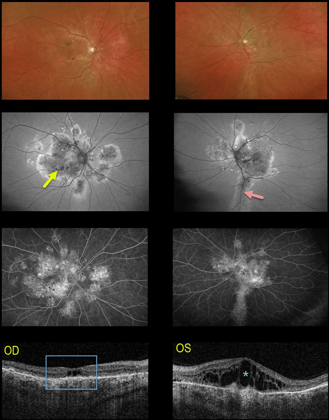
IDIOPATHIC CENTRAL SEROUS CHORIORETINOPATHY
Originally posted on @retina.rocks May 25, 2022
This 64YO male received prior photodynamic therapy in one of his eyes for idiopathic central serous chorioretinopathy (ICCS) and was subsequently lost to follow-up for 10-15 years. He presented with 20/400 vision bilaterally.
Multimodal imaging shows classic findings for chronic ICSC. Optos color imaging shows macular and peripapillary pigmentary changes with a suggestion of an inferior gutter OS.
Fundus autofluorescence better shows the bilateral pigmentary changes and inferior gutter OS, and fluorescein angiography shows variable chorioretinal staining.
Triton swept-source OCT shows bilateral thickened choroid, dry central cavitation-like changes OD, and marked cystoid macular edema OS.
Photodynamic therapy was performed for the left eye, although we are not certain this will improve his vision, given the chronicity of these findings.
Learning Points:
Macular edema is an uncommon finding with typical ICSC, likely due at least in part to the external limiting membrane acting as a barrier to subretinal fluid migrating into the retina. Eyes with chronic ICSC, however, can develop retinal edema as in this case.
Gutters are commonly seen in pathology with chronic subretinal leakage, including central serous retinopathy, choroidal hemangioma, choroidal nevus, and uveal melanoma. These pigmentary changes are usually best imaged with FAF.
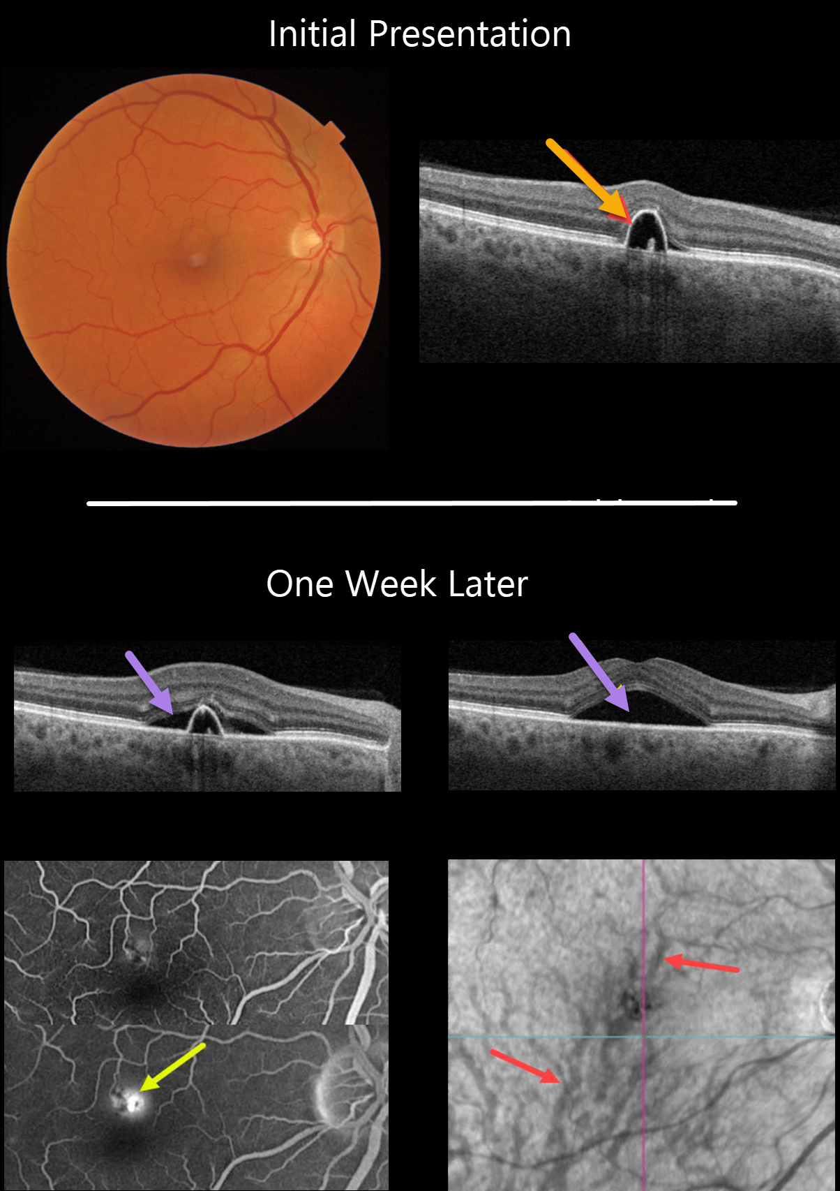
IDIOPATHIC CENTRAL SEROUS CHORIORETINOPATHY
Originally posted on @retina.rocks February 16, 2022
This 55YO male presented on 1/24/22 with a small symptomatic pigment epithelial detachment (PED) in the superior right fovea. Vision was 20/30.
One week later, he was subjectively worse, although vision remained at 20/30. There was a new small blister of foveal subretinal fluid, best captured on swept-source OCT.
Optos fluorescein angiography shows a typical blot leak along the dome of the PED. En face OCT shows a choroidal venous anastomosis.
Learning Points:
The choroidal venous system, as with the neurosensory retinal vasculature, usually respects the horizontal meridian. With pachychoroid disorders, these anastomoses develop, and in this patient’s case, the inferior vortex system drains nearly the entire macula.
For a wonderful review highlighting the concept of venous overload choroidopathy, see Spaide et al, Progress Retinal Eye Research 2022;86;100973.
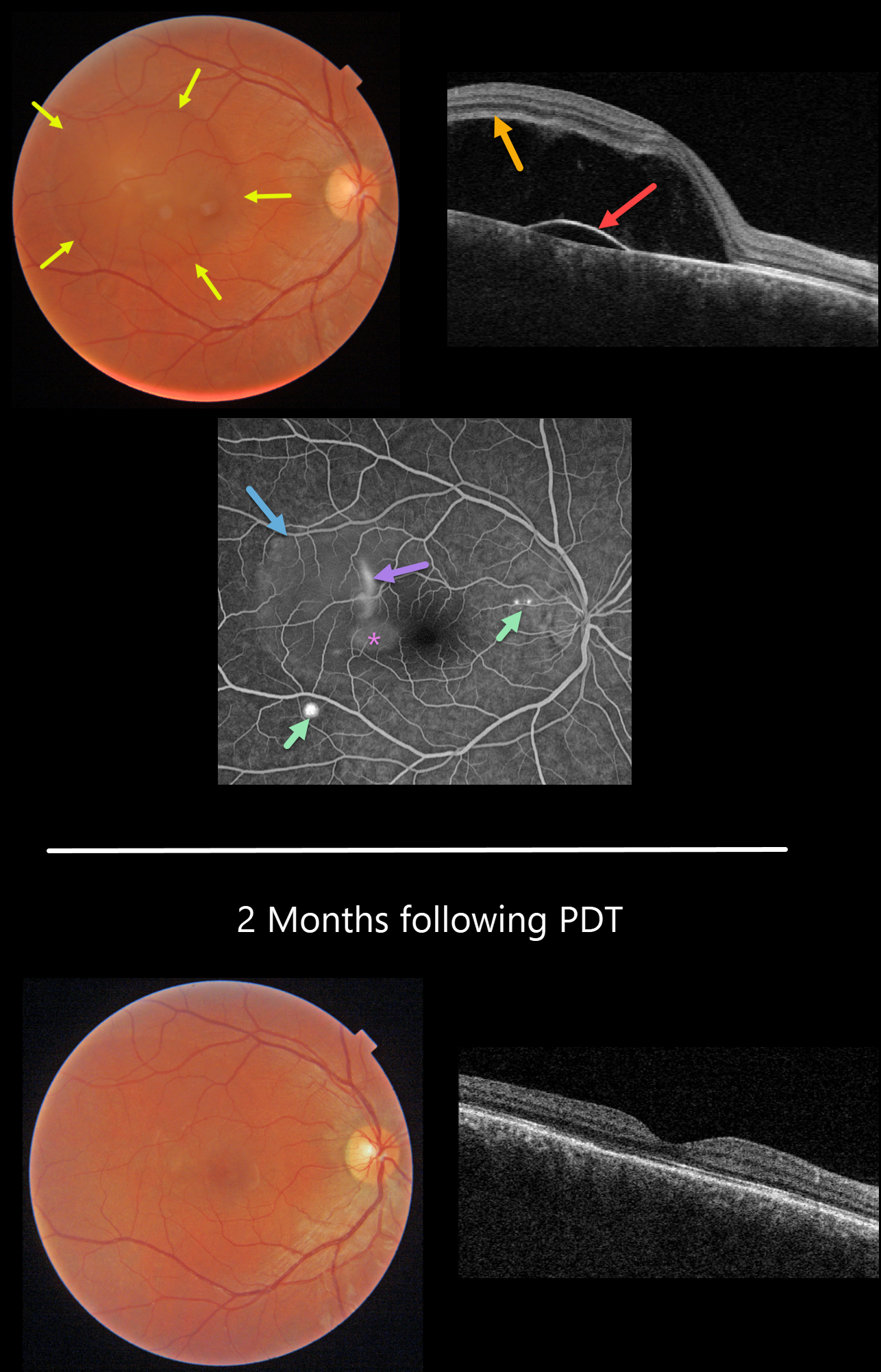
IDIOPATHIC CENTRAL SEROUS CHORIORETINOPATHY
Originally posted on @retina.rocks January 12, 2022
This 33YO male presented with recent central blurred vision in his right eye. He is a powerlifter and self-injects testosterone with possible anabolic steroids. Vision was 20/70 OD.
Triton color fundus photography shows a large serous macular detachment. Triton swept-source OCT shows a large serous neurosensory retinal detachment with an underlying shallow RPE detachment.
Optos fluorescein angiography shows classic findings for idiopathic central serous chorioretinopathy (ICSC), including peripheral blot leaks and a more central smokestack leak. Fluorescein leaks through a physiologic break in the RPE and accumulates in the subretinal space, giving rise to the smokestack, which is found in only 10% of cases. The fluorescein dye then diffuses and pools within the subretinal space, highlighting the extent of the serous detachment. Fluorescein also pools beneath the PED. In this case, subretinal pooling is almost always associated with an RPE detachment.
Most cases of central serous chorioretinal atrophy have a single unilateral leak. Multifocal cases are commonly associated with systemic steroids.
Our patient underwent PDT therapy, and vision returned to 20/20 two months later with dramatic resolution of the subretinal fluid and RPE detachment. Since he is asymptomatic with good vision in this eye, we continue to observe. He is trying to lower the amount of injected testosterone to lower the risk of recurrent ICSC.
IDIOPATHIC CENTRAL SEROUS CHORIORETINOPATHY
Originally posted on @retina.rocks November 1, 2021
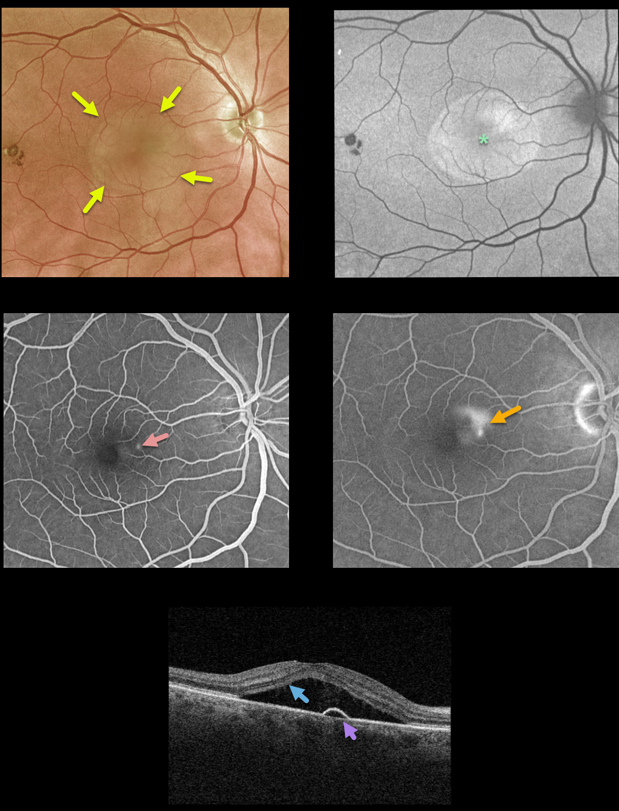
This 61YO male presented with classic findings for idiopathic central serous chorioretinopathy (ICSC). There is a large serous macular detachment that is diffusely hyperautofluorescent.
Fluorescein angiography shows a small dot of early hyperfluorescence with a late smokestack leak.
Triton swept-source OCT shows the serous detachment with a small underlying RPE detachment near the area of angiographic leakage.
Learning Points:
‘Smokestack’ leakage, which only occurs in about 10% of active ICSC cases, is due to a physiologic break in the retinal pigment epithelium (RPE), allowing the fluorescein to leak through the RPE and rise into the subretinal space.
Most ICSC serous detachments spontaneously resolve within 3 months and do not require treatment. Patients who can’t wait 3 months (e.g., pilots or retinal surgeons) or those with persistent symptomatic detachments are best treated with photodynamic therapy.
OSTRICH MACULOPATHY
Originally posted on @retina.rocks September 24, 2021
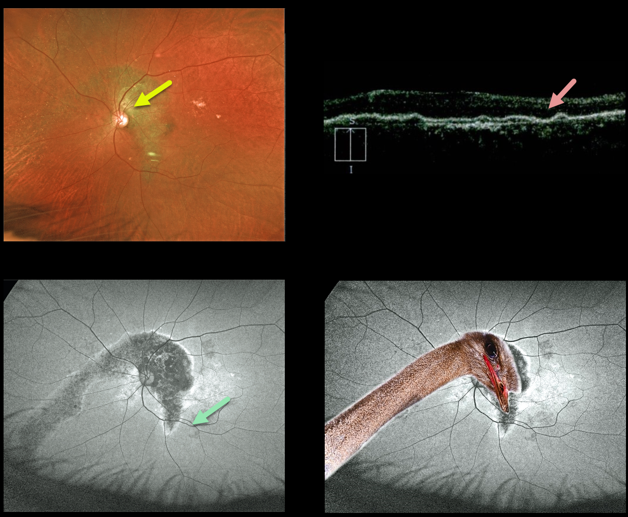
This 79YO male has counting-fingers vision in his left eye, likely due to complications of chronic central serous retinopathy.
Optos color imaging shows increased subretinal pigmentation extending around the nerve superiorly and into the macula and inferonasal periphery.
Fundus autofluorescence (FAF) more dramatically shows these changes as hypo-FAF. OCT scanning shows diffuse thinning of the overlying neurosensory retina.
IDIOPATHIC CENTRAL SEROUS CHORIORETINOPATHY
Originally posted on @retina.rocks December 14, 2020
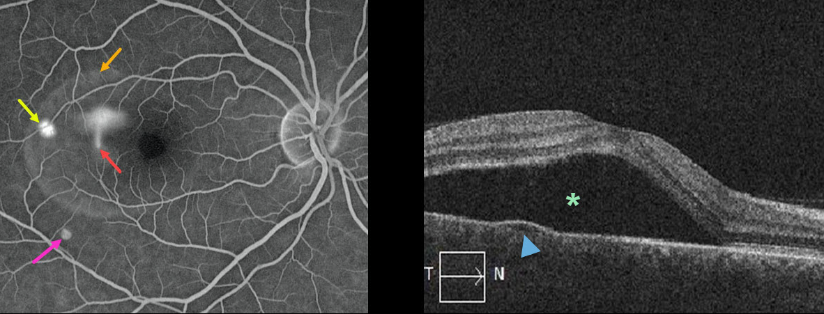
This 44YO male presented with 20/60 vision in his right eye. The left eye was normal with 20/20 vision.
His fluorescein angiogram (FA) shows classic findings for idiopathic central serous chorioretinopathy (ICSC). There is the more common blot leak, as well as the classic “smokestack” leak, which occurs in only about 10% of cases.
The FA also demonstrates the extent of the serous detachment; there is also a tiny RPE detachment without leakage in the distal inferior macula.
The OCT shows both the serous detachment and the small pigment epithelial detachment.
Learning Points:
Fluorescein leaks through a physiologic break in the retinal pigment epithelium (RPE) and rises in the subretinal space, giving the smokestack.
The fluorescein dye then diffuses and pools within the subretinal space, highlighting the extent of the serous detachment.
Most ICSC serous detachments spontaneously resolve within 3 months and do not require treatment. Patients who can’t wait the 3 months (i.e., pilots or surgeons), or those with persistent symptomatic detachments, are best treated with photodynamic therapy.
IDIOPATHIC CENTRAL SEROUS CHORIORETINOPATHY
Originally posted on @retina.rocks February 5, 2020
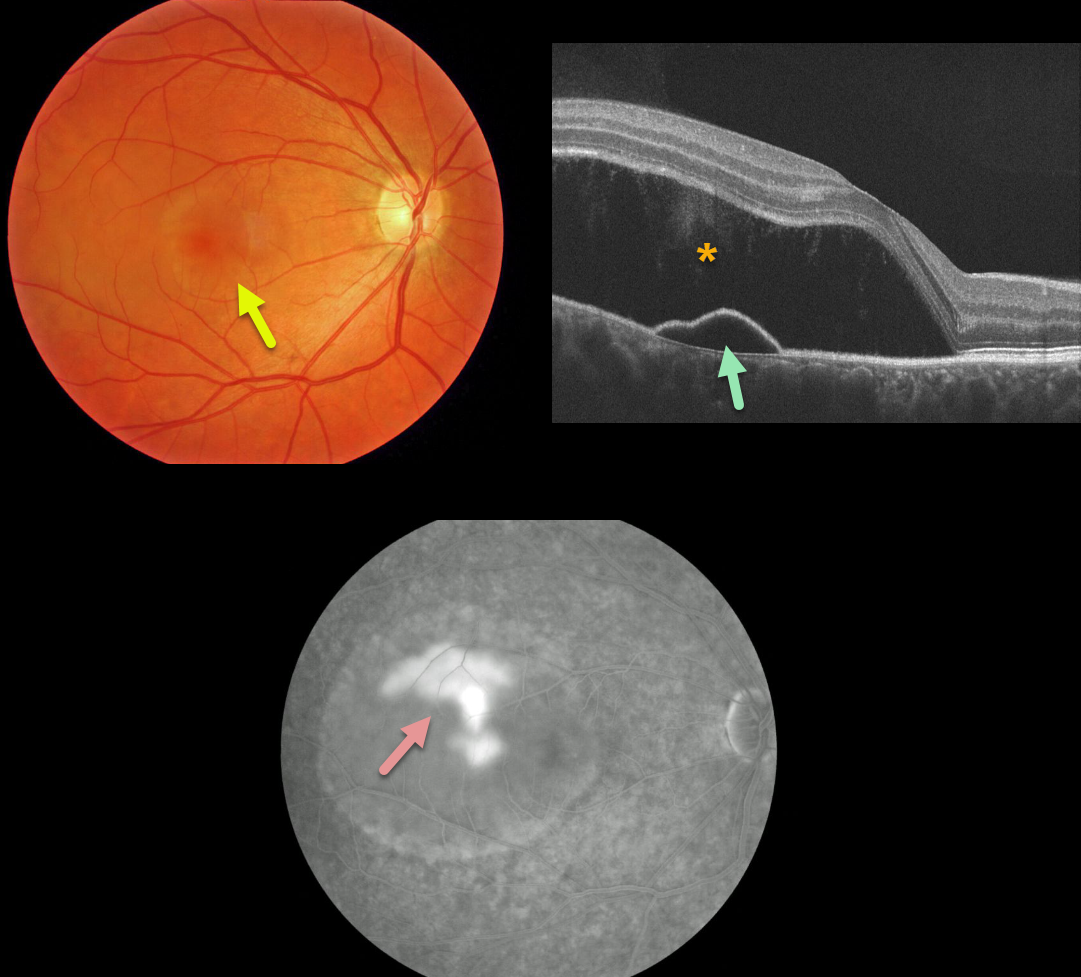
This patient has a large serous retinal detachment from idiopathic central serous chororetinopathy (ICSC).
OCT demonstrates the serous retinal detachment and a pigment epithelial detachment.
The late phase fluorescein angiography hows classic “smokestack” leakage.
Learning Points:
ICSC is part of a larger spectrum of pachychoroid diseases. Pachychoroid, meaning “thick” choroid, is secondary to large dilated Haller vessels.
Increased hydrostatic pressure from these dilated vessels can lead to RPE dysfunction, causing serous detachments and macular neovascularization.
The “smokestack” leakage pattern on fluorescen angiography is actually seen in only 10% of eyes with active ICSC.
Some cases resolve on their own, but in chronic cases, photodynamic therapy (PDT) is the gold standard for treatment.


