

Idiopathic Chorioretinal Folds, Retinal Folds and RPE Folds
<< Back to Cases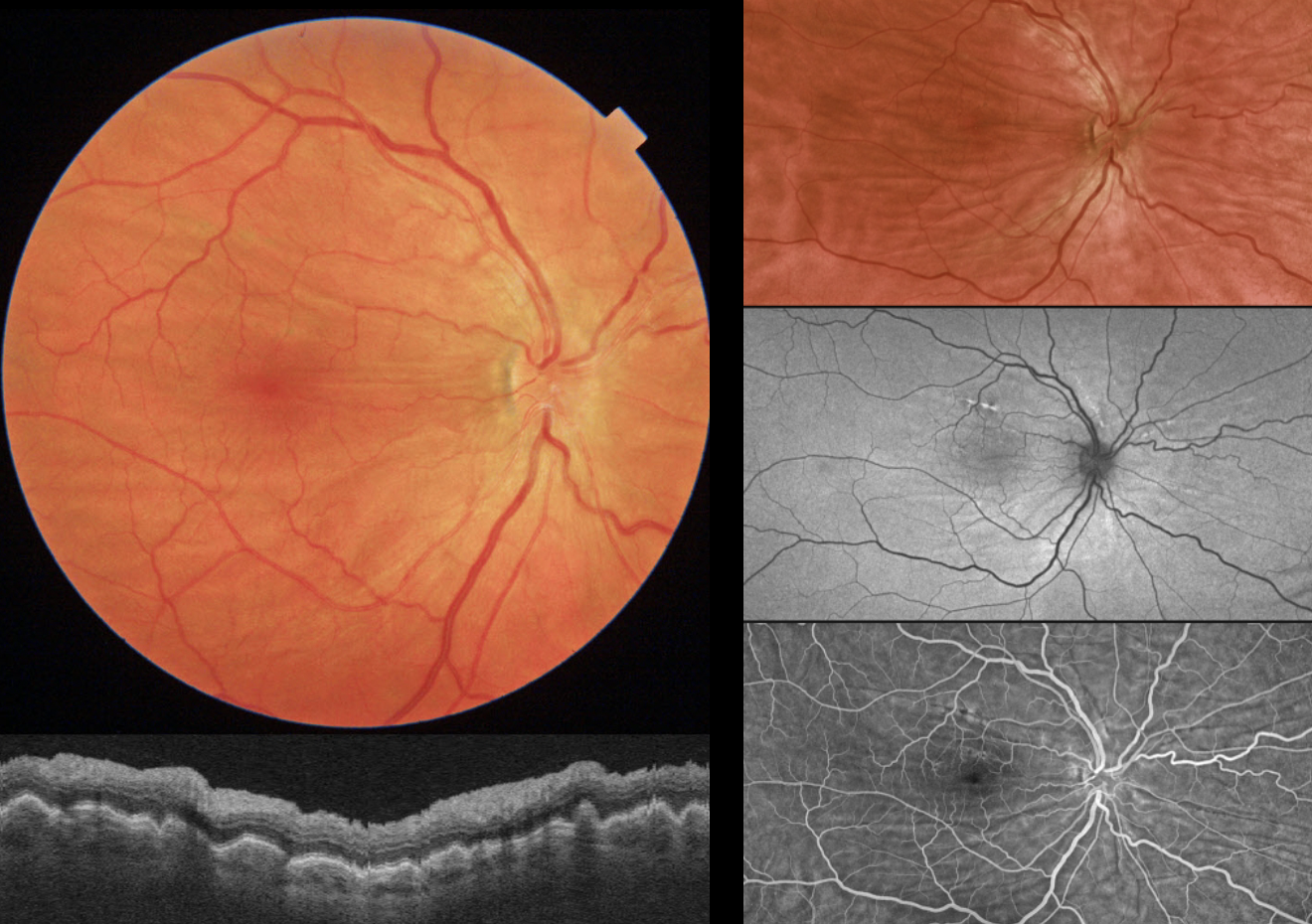
CHORIORETINAL FOLDS
Originally posted on @retina.rocks November 12, 2024
This 61YO male presented without a visual complaint. He was a +6 diopter hyperope OD and +5 diopter hyperope OS. Vision was 20/200 OD from refractive amblyopia and 20/40 OS.
Triton color imaging and swept-source OCT show bilateral chorioretinal folds. Optos fundus autofluorescence (FAF) shows variable hyper- and hypo-FAF changes from the folds, and fluorescein angiography shows alternating transmission and blocking defects.
Learning Points:
Chorioretinal folds appear clinically as often subtle horizontal and oblique subretinal folds. There are extensive intra- and extra-ocular causes for chorioretinal folds. The typical findings in idiopathic chorioretinal folds are a mild/moderate hyperopic prescription and bilateral, symmetric folds. The folds are thought to result from flattening of the macular sclera. When unilateral, an MRI is often indicated to rule out an orbital mass. Typical idiopathic chorioretinal folds, as in our patient, require no workup and usually cause no symptoms.
CHORIORETINAL FOLDS
Originally posted on @retina.rocks June 27, 2024
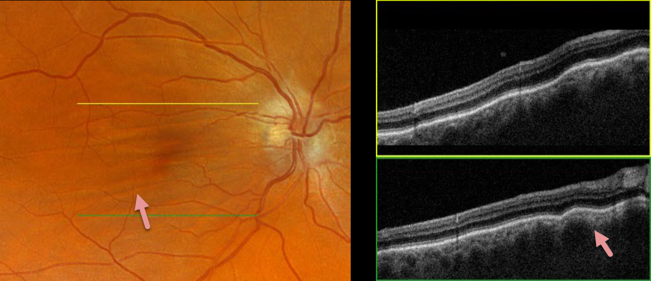
This 52YO male was referred for asymptomatic findings on retinal examination in his right eye. There was a history of refractive amblyopia OD from being a +3-diopter hyperope. He was emmetropic in his normal left eye.
Optos color RGB imaging shows horizontal chorioretinal folds throughout the right macula. These folds are prominently seen on Triton swept-source OCT. Orbital MRI was unremarkable.
Learning Points:
Chorioretinal folds appear clinically as often subtle horizontal and oblique subretinal folds. There are extensive intra- and extra-ocular causes for chorioretinal folds. The typical findings in idiopathic chorioretinal folds are a mild/moderate hyperopic prescription and bilateral, symmetric folds. The folds are thought to result from flattening of the macular sclera.
When unilateral, an MRI is often indicated to rule out an orbital mass. Typical idiopathic chorioretinal folds require no workup and usually cause no symptoms.
CHORIORETINAL FOLDS
Asma Samsudeen and Ashish Sharma
Originally posted on @retina.rocks January 4, 2024
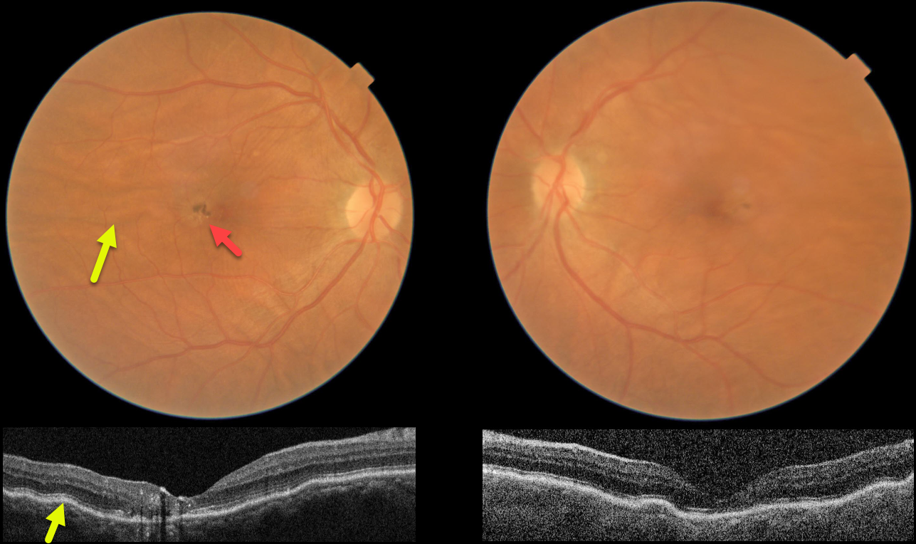
This 61YO female presented without a visual complaint. She was a +4 diopter hyperope bilaterally. Vision was 20/200 OD and 20/20 OS.
Color imaging and OCT show bilateral chorioretinal folds, which are particularly prominent in her right eye. Foveal pigmentary changes are noted in the central right macula, with OCT disorganization of the central macular layers and hyperreflective foci. Observation was recommended.
Learning Points:
Chorioretinal folds appear clinically as often subtle horizontal and oblique subretinal folds. There are extensive intra- and extra-ocular causes for chorioretinal folds. The typical findings in idiopathic chorioretinal folds are a mild/moderate hyperopic prescription and bilateral, symmetric folds. The folds are thought to result from flattening of the macular sclera.
When unilateral, an MRI is often indicated to rule out an orbital mass. Typical idiopathic chorioretinal folds require no workup and usually cause no symptoms.
In our practice, we have seen several patients with idiopathic chorioretinal folds and waxing/waning subretinal fluid (SRF), possibly as part of the pachychoroid spectrum.
Cohen et al. also reported a small series of patients with hyperopic chorioretinal folds, central serous retinopathy, and pachychoroid (Retinal Cases 2022;16:242-245). We therefore feel the pigmentary changes noted in our patient’s right eye most likely resulted from spontaneously resolved SRF.
CHORIORETINAL FOLDS
Originally posted on @retina.rocks August 29, 2022
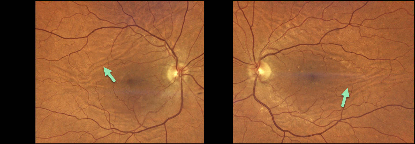
This 75YO female presented with 20/25 vision OU and asymptomatic chorioretinal folds. Optos color imaging shows extensive, irregularly oriented and oblique chorioretinal folds.
Learning Points:
Chorioretinal folds appear clinically as often subtle horizontal and oblique subretinal folds. There are extensive intra- and extra-ocular causes for these folds.
Typical findings include a mild to moderate hyperopic prescription with bilateral and symmetric folds. The folds are thought to result from flattening of the submacular sclera.
When unilateral, an MRI is often indicated to rule out an orbital mass. Typical idiopathic chorioretinal folds require no workup and usually cause no symptoms.
AIRBAG TRAUMA
Will Gibson
Originally posted on @retina.rocks August 25, 2022
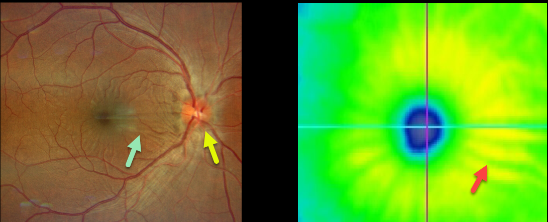
This 35YO male presented with a unilateral swollen nerve and retinal striae following blunt trauma from airbag deployment Vision was 20/25. The intraocular pressure was 15, and there were no signs of open-globe injury.
Optos imaging shows retinal folds radiating from the macular center. These striae are also evident on the OCT thickness map.
He was started on oral prednisone, and 10 days later, the ocular findings had improved (images not available).
CHORIORETINAL FOLDS
Originally posted on @retina.rocks July 6, 2022
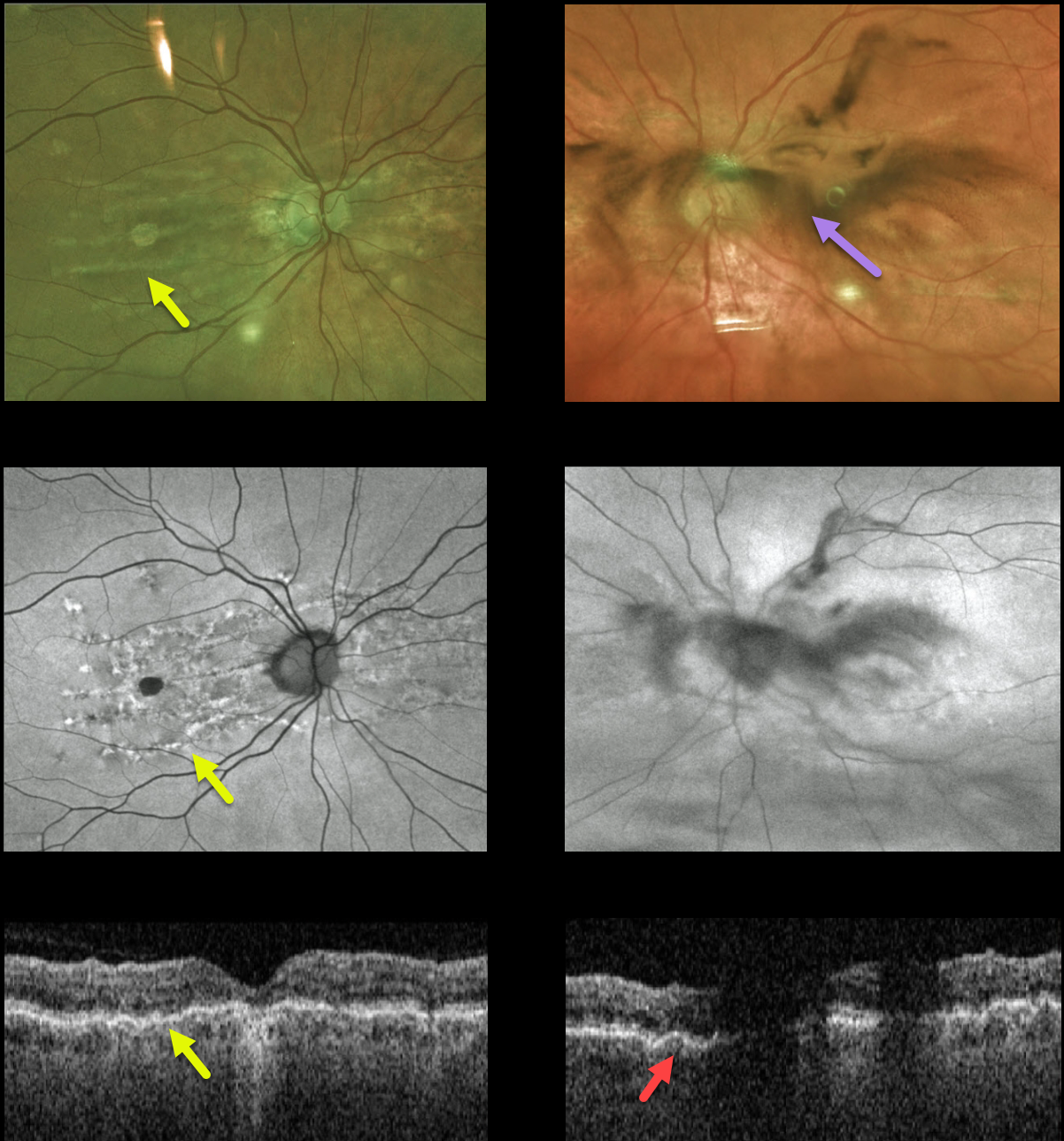
This 80YO female presented with a 2-day history of floaters and vision loss in her left eye. She had mild hyperopia prior to cataract surgery 5 years earlier. She had a history of diet-controlled type 2 diabetes. Vision was 20/100 OD and 20/30 OS.
Fundus Optos color imaging, fundus autofluorescence, and OCT of her right eye show changes associated with chronic chorioretinal folds. Similar imaging of her left eye is hazy due to a vitreous hemorrhage, but chorioretinal folds are also noted on the OCT B-scan.
There were no signs of diabetic retinopathy clinically or on fluorescein angiography (not shown), and no signs of retinal breaks or detachment peripherally. She was asked to sleep with her head elevated, and we are following her closely.
Learning Points:
Chorioretinal folds appear clinically as often subtle horizontal and oblique subretinal folds. There are extensive intra- and extra-ocular causes for chorioretinal folds. When unilateral, an MRI is often indicated to rule out an orbital mass. Typical idiopathic chorioretinal folds require no workup and usually cause no symptoms.
Our patient shows typical findings of idiopathic chorioretinal folds, including mild hyperopia and bilateral, symmetric folds. The folds are thought to result from flattening of the macular sclera.
HARADA DISEASE
Originally posted on @retina.rocks April 27, 2021
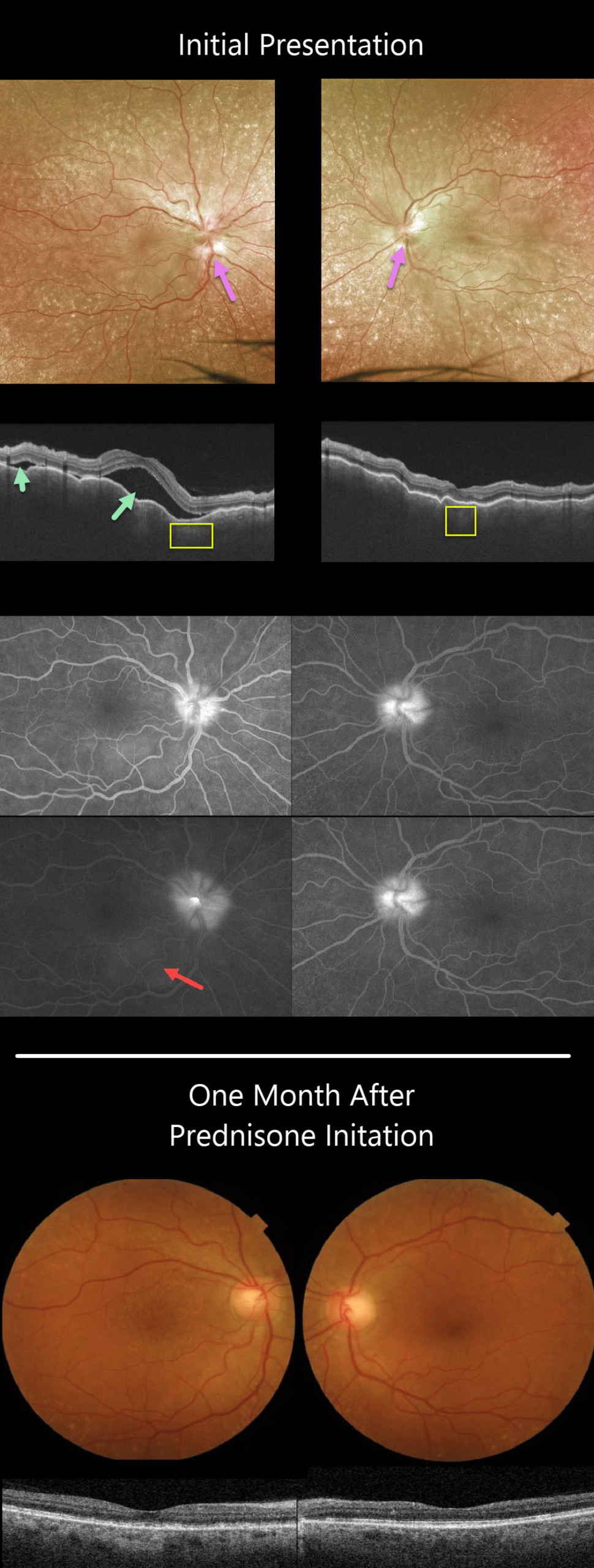
This 63YO female presented with a 3-week history of bilateral painful vision loss. Vision was 20/400 OD and 20/100 OS.
She had bilateral granulomatous anterior uveitis with a clear vitreous OU. Both nerves were swollen, and irregular chorioretinal folds were noted throughout each macula. Extensive incidental peripheral drusen were also present.
Triton swept-source OCT scanning shows a markedly irregularly thickened choroid OU with multifocal serous detachments in the right eye.
Optos fluorescein angiography (FA) shows bilateral optic nerve leakage and some faint subretinal leakage in the inferonasal right macula.
Oral prednisone was started, and there was near immediate improvement. One month later, vision improved to 20/40 OD and 20/60 OS, and all findings virtually normalized.
Learning Points:
This case has many classic findings for Harada’s disease, including bilateral panuveitis, optic nerve swelling, thickened choroid, chorioretinal folds, and multifocal serous retinal detachments.
We were surprised that the FA showed minimal subretinal leakage, since angiography typically shows extensive multifocal subretinal leaks with pooling in the overlying serous detachments and bacillary cavities.
Patients with just ocular findings have Harada’s disease, whereas those with additional systemic findings (including vitiligo, poliosis, headache, vertigo, and hearing loss) have Vogt-Koyanagi-Harada disease.
CHORIORETINAL FOLDS
Originally posted on @retina.rocks January 11, 2021
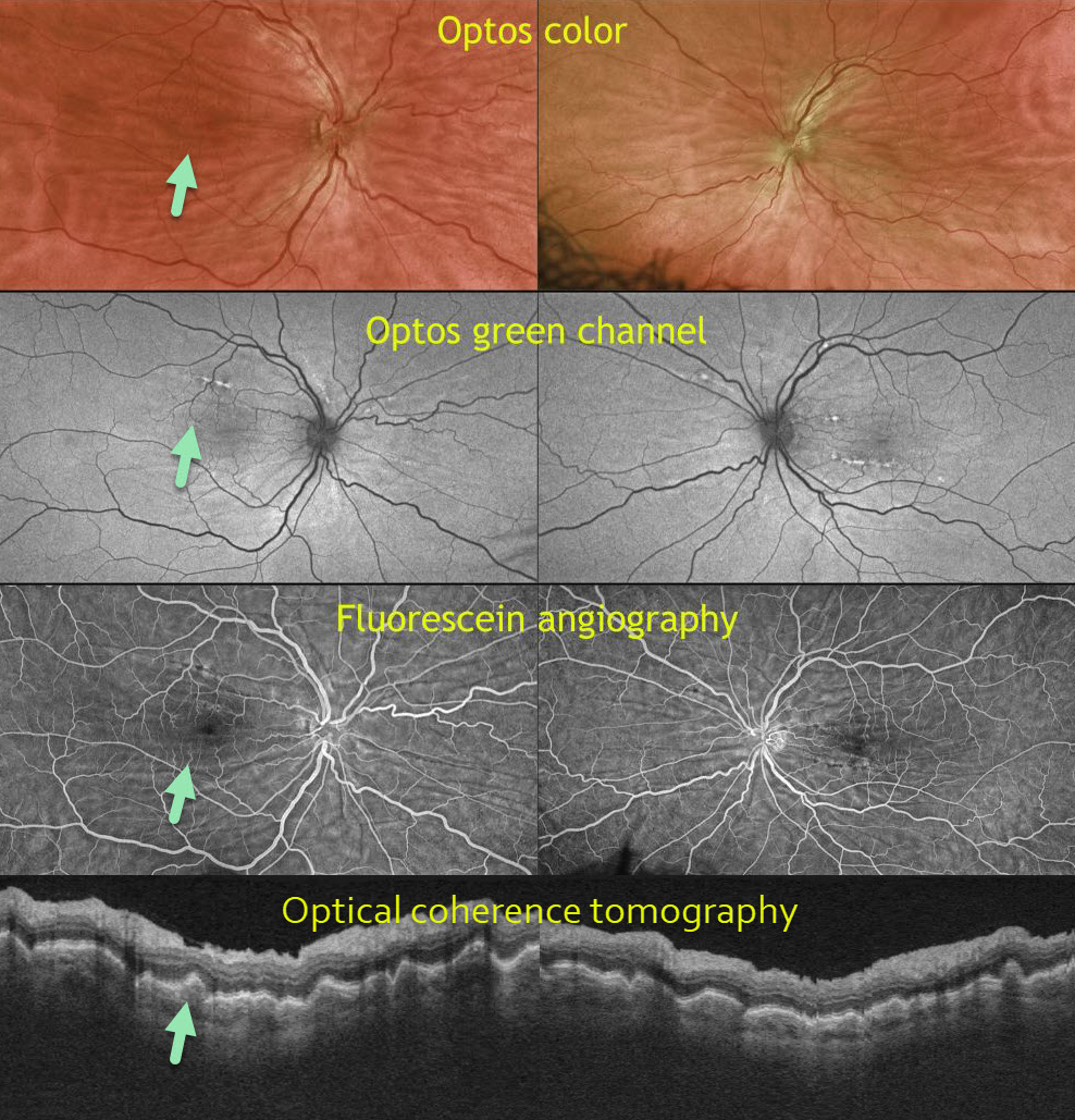
This 50-year-old male is a 6-diopter hyperope. Optos color, Optos green channel, fluorescein angiography (FA), and OCT reveal prominent chorioretinal folds.
Learning Points:
Chorioretinal folds appear clinically as often subtle horizontal and oblique subretinal folds. There are extensive intra- and extra-ocular causes for chorioretinal folds.
Our patient shows typical findings of idiopathic chorioretinal folds, including mild hyperopia and bilateral, symmetric folds. The folds are thought to result from flattening of the macular sclera.
When unilateral, an MRI is often indicated to rule out an orbital mass. Typical idiopathic chorioretinal folds require no workup and usually cause no symptoms.
PERIPAPILLARY PACHYCHOROID SYNDROME
Originally posted on @retina.rocks August 18, 2020
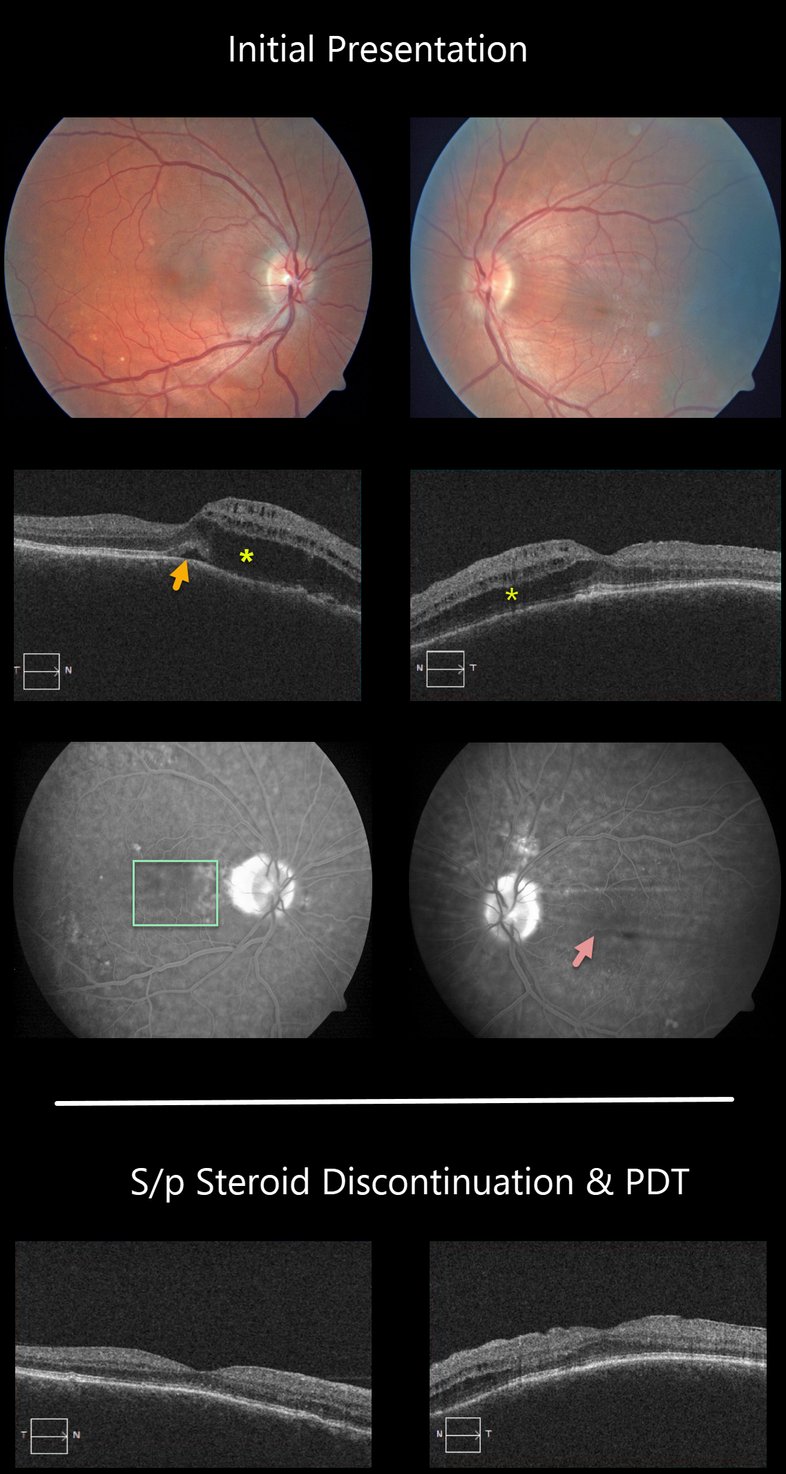
This patient has peripapillary pachychoroid syndrome (PPS), a recently described bilateral condition characterized by nasal macular intraretinal and subretinal fluid.
These eyes can look very similar to those of patients with optic pit maculopathy, but without the pit. Additional findings include a thicker nasal macular choroid than temporal macular choroid, choroidal folds (75% of cases), shorter axial lengths (<23mm), and hyperopia.
There is no angiographic evidence of leakage despite fluid noted on OCT.
The patient worsened when initially placed on steroids, but made a tremendous improvement after steroids were discontinued, followed by photodynamic therapy.
Learning Points:
PPS is part of the pachychoroid spectrum, where patients have a thick choroid. Probably the most common and well-known pachychoroid disease is idiopathic central serous chorioretinopathy. Just like ICSC, this condition worsens with steroid use.
See Phasukkijwatana et al Retina, 2018;38:1652-1657 for more on PPS.
CHORIORETINAL FOLDS
Originally posted on @retina.rocks March 13, 2020
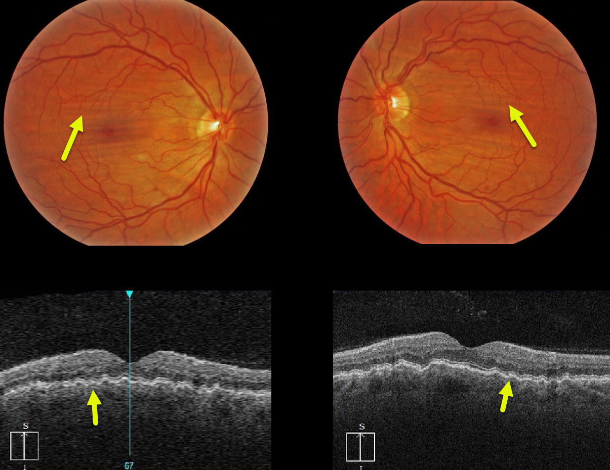
Our patient with mild hyperopia exhibits bilateral and symmetric idiopathic chorioretinal folds, shown on both the fundus photos and OCT images.
Learning Points:
Chorioretinal folds appear clinically as often subtle horizontal and oblique subretinal folds.
There are extensive intra- and extra-ocular causes for chorioretinal folds. Typical idiopathic chorioretinal folds require no workup and usually cause no symptoms.
When unilateral, an MRI is often indicated to rule out an orbital mass such as optic nerve meningioma.
CHORIORETINAL FOLDS
Originally posted on @retina.rocks March 5, 2020
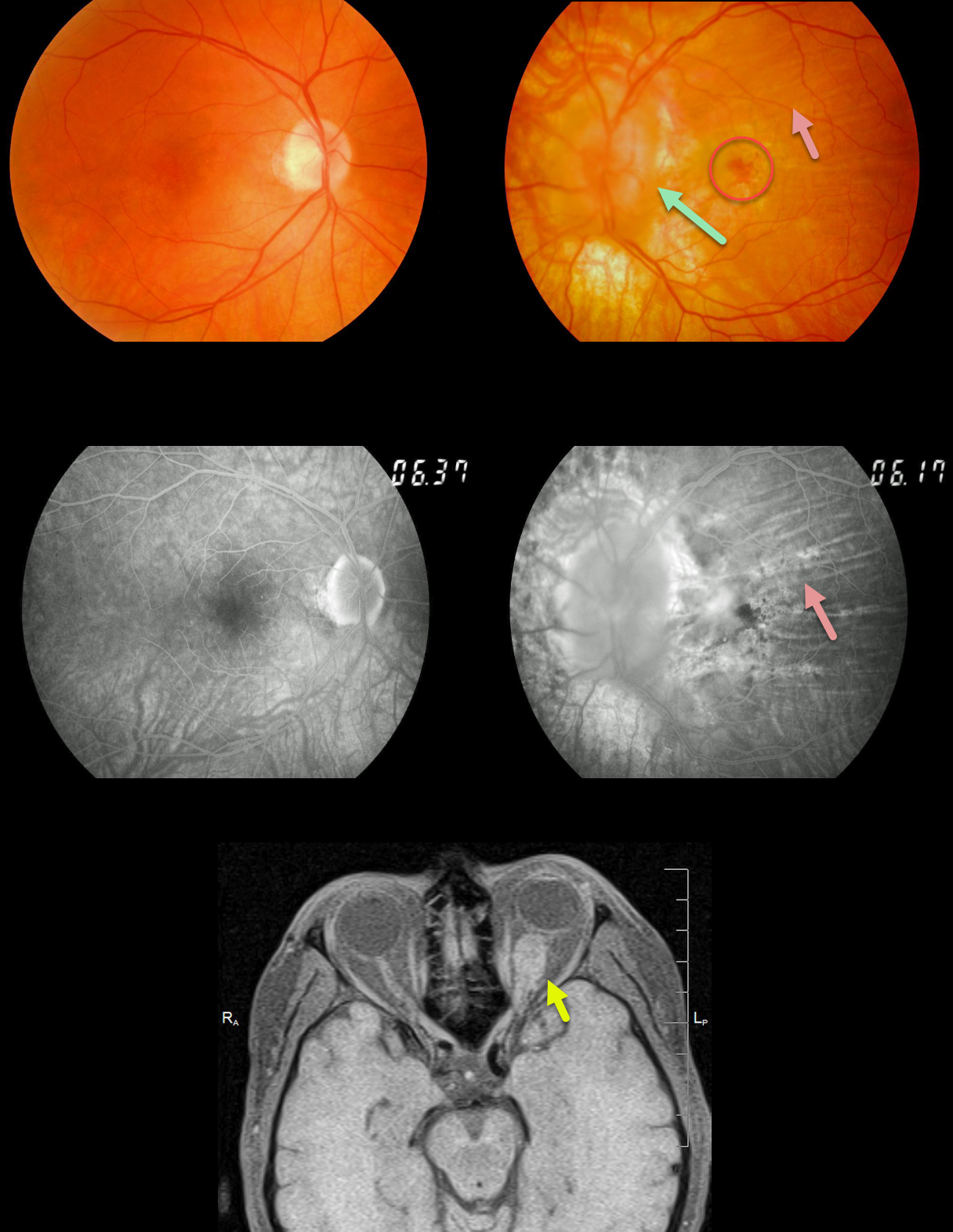
This patient presented with a history of a left optic nerve meningioma that had been followed for years. The left nerve is markedly swollen.
Extensive chorioretinal folds are present and best seen on angiography. A chronic subfoveal macular neovascularization (MNV) is noted, which we are observing since the lesion and vision remain stable at 20/400.
MRI shows the optic nerve meningioma; this is also being observed since surgical removal of this benign tumor would cause total blindness.
Learning Points:
Chorioretinal folds are usually idiopathic and bilateral, most commonly from mild hyperopia and flattening of the macular sclera.
When folds present unilaterally, as in our patient, an MRI of the orbit is recommended to rule out orbit pathology.


