

Idiopathic Macular Neovascularization (MNV)
<< Back to Cases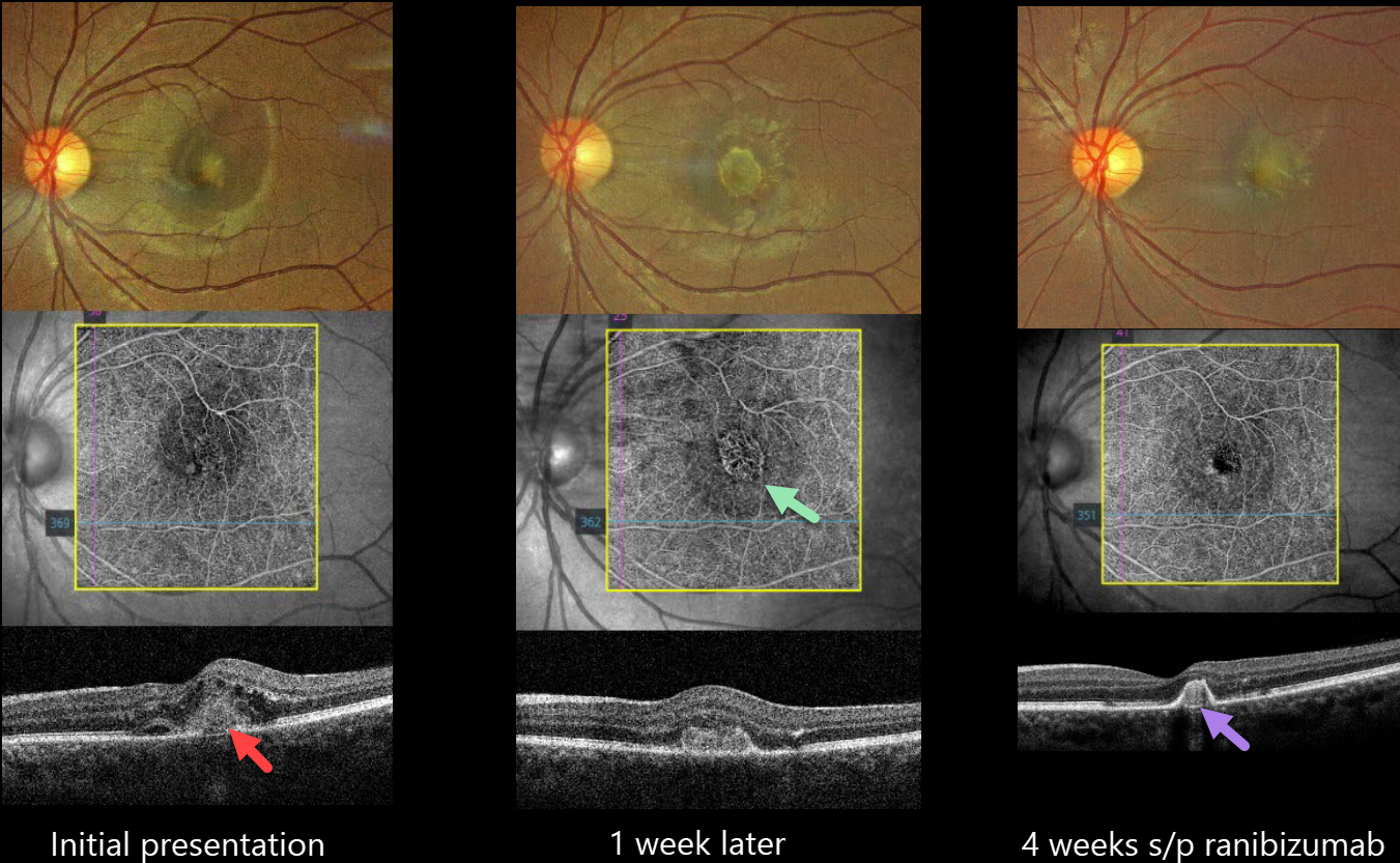
IDIOPATHIC MACULAR NEOVASCULARIZATION (MNV)
Nilesh Kumar
Originally posted on @retina.rocks August 7, 2024
This 20YO female presented with 2 weeks of decreased vision in her left eye. Vision was 20/25 in her normal right eye and 20/60 in her left eye.
Color imaging shows foveal fluid with an amelanotic subfoveal lesion. OCT angiography (OCTA) shows a small subfoveal macular neovascularisation (MNV). An OCT B-scan shows a type 2 MNV (above the RPE) with intraretinal fluid and numerous hyperreflective foci.
Although we recommended an intravitreal ranibizumab biosimilar that day, she refused treatment and returned one week later with 20/200 vision and marked enlargement of the MNV on OCTA.
The injection was given, and 4 weeks later, vision improved to 20/40, with a dramatic treatment response characterized by decreased size and exudation.
Learning Points:
Idiopathic MNV in younger patients was first described by Cleasby in 1976 (AJO 1976;81:590-596). They tend to be type 2 lesions and have a better natural history than typical neovascular AMD (Ophthalmology 1998;105:1816-1820). There is usually an excellent response to anti-VEGF therapy (Kodjikian et al, Retina 2022;42:290-297), as in our patient who initially had both a dramatic rapid enlargement of the untreated lesion, followed by an equally rapid treatment response.
IDIOPATHIC MACULAR NEOVASCULARIZATION (MNV)
Originally posted on @retina.rocks September 6, 2023
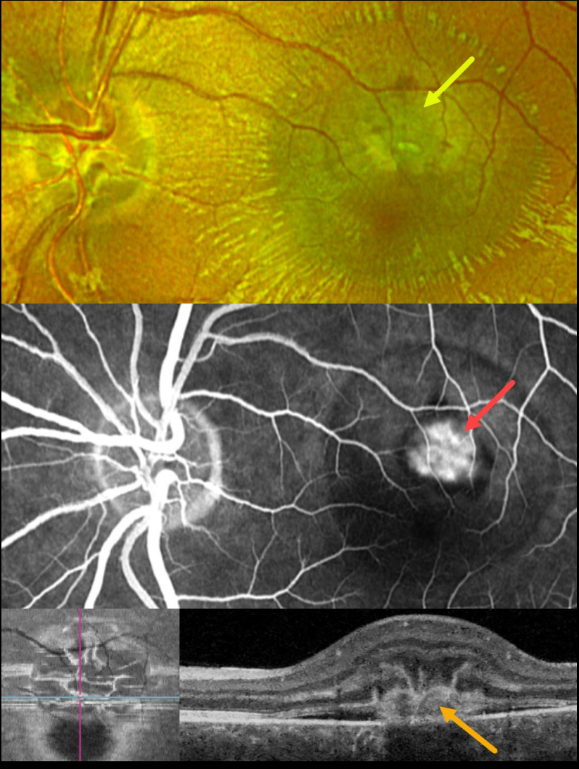
This 23YO male presented with a recent loss of vision in his left eye. Vision was 20/60.
Optos color RG imaging shows a depigmented subretinal lesion in the superior fovea with overlying dots of blood and radiating retinal striae.
Fluorescein angiography shows a well-defined superior extrafoveal macular neovascularization (MNV). Swept-source B-scan OCT through this lesion shows a type 2 (above the RPE) MNV associated with shallow subretinal fluid and overlying radiating hyperreflective lines (pitchfork sign). These radiating lesions are uniquely visualized with en face OCT.
Intravitreal Avastin was begun.
Learning Points:
The pitchfork sign was originally described by Hoang et al (Retina 2013;33:1049-1055) as a unique OCT finding in inflammatory MNV. Falavarajani et al more recently noted that this finding is also associated with non-inflammatory MNV (Ophthalmic Surg Lasers Imaging Retina 2019;50:719-725).
The pathogenesis of the avascular pitchfork spikes is unknown, but may be related to outer retinal traction or Müller cell activation.
IDIOPATHIC MACULAR NEOVASCULARIZATION (MNV)
Originally posted on @retina.rocks January 2, 2023
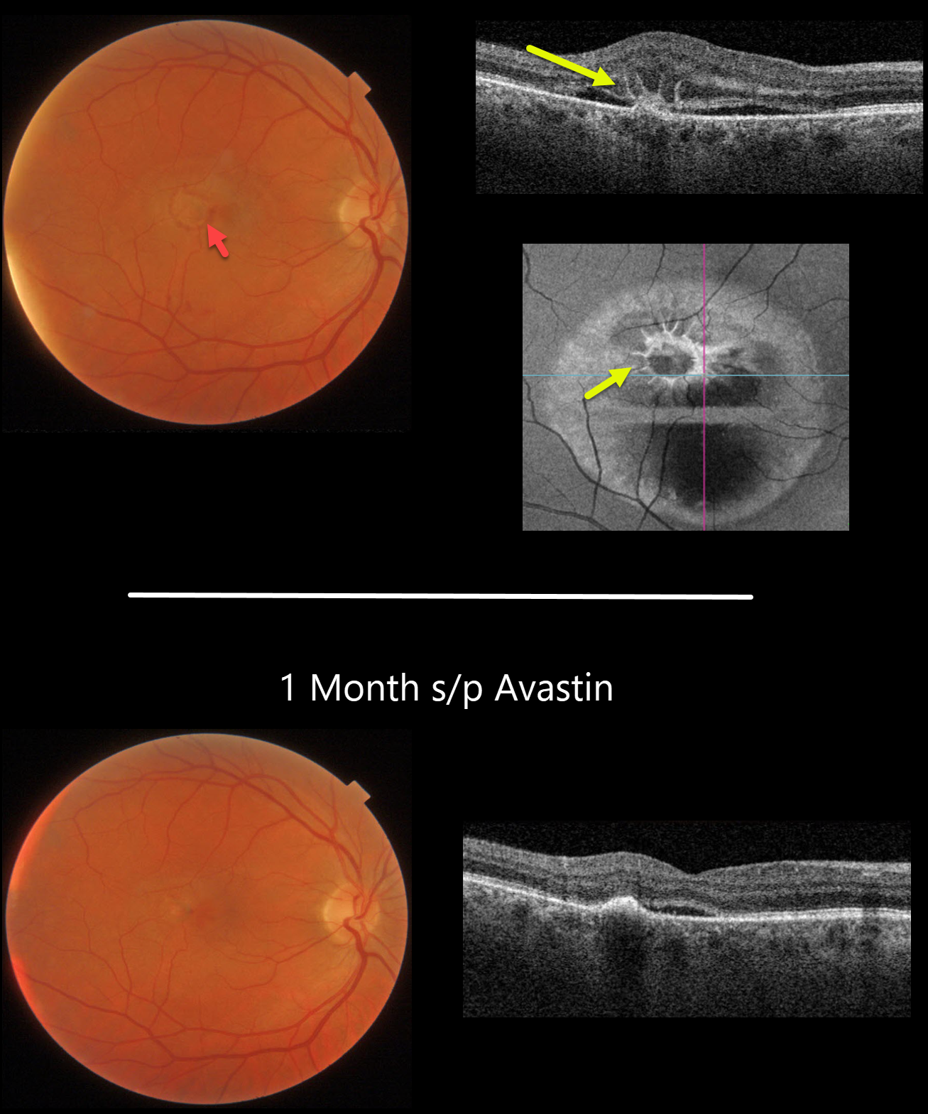
This 49YO male presented with acute vision loss of 20/400 in his right eye from an idiopathic macular neovascularization (MNV).
Color photography shows shallow foveal fluid with some areas of subretinal blood. Swept-source OCT shows outer macular edema, shallow subretinal fluid, and radiating hyperreflective lines (pitchfork sign). These radiating lesions are uniquely visualized with en face OCT.
The lesion was virtually dry, with resolution of the pitchfork lesion 1 month after a single intravitreal Avastin injection. Vision improved to 20/80.
Learning Points:
The pitchfork sign was originally described by Hoang et al (Retina 2013;33:1049-1055) as a unique OCT finding in inflammatory MNV.
Falavarajani et al more recently noted that this finding is also associated with non-inflammatory MNV (Ophthalmic Surg Lasers Imaging Retina 2019;50:719-725).
The pathogenesis of the avascular pitchfork spikes is unknown, but may be related to outer retinal traction or Müller cell activation.
IDIOPATHIC MACULAR NEOVASCULARIZATION (MNV)
Originally posted on @retina.rocks December 20, 2021
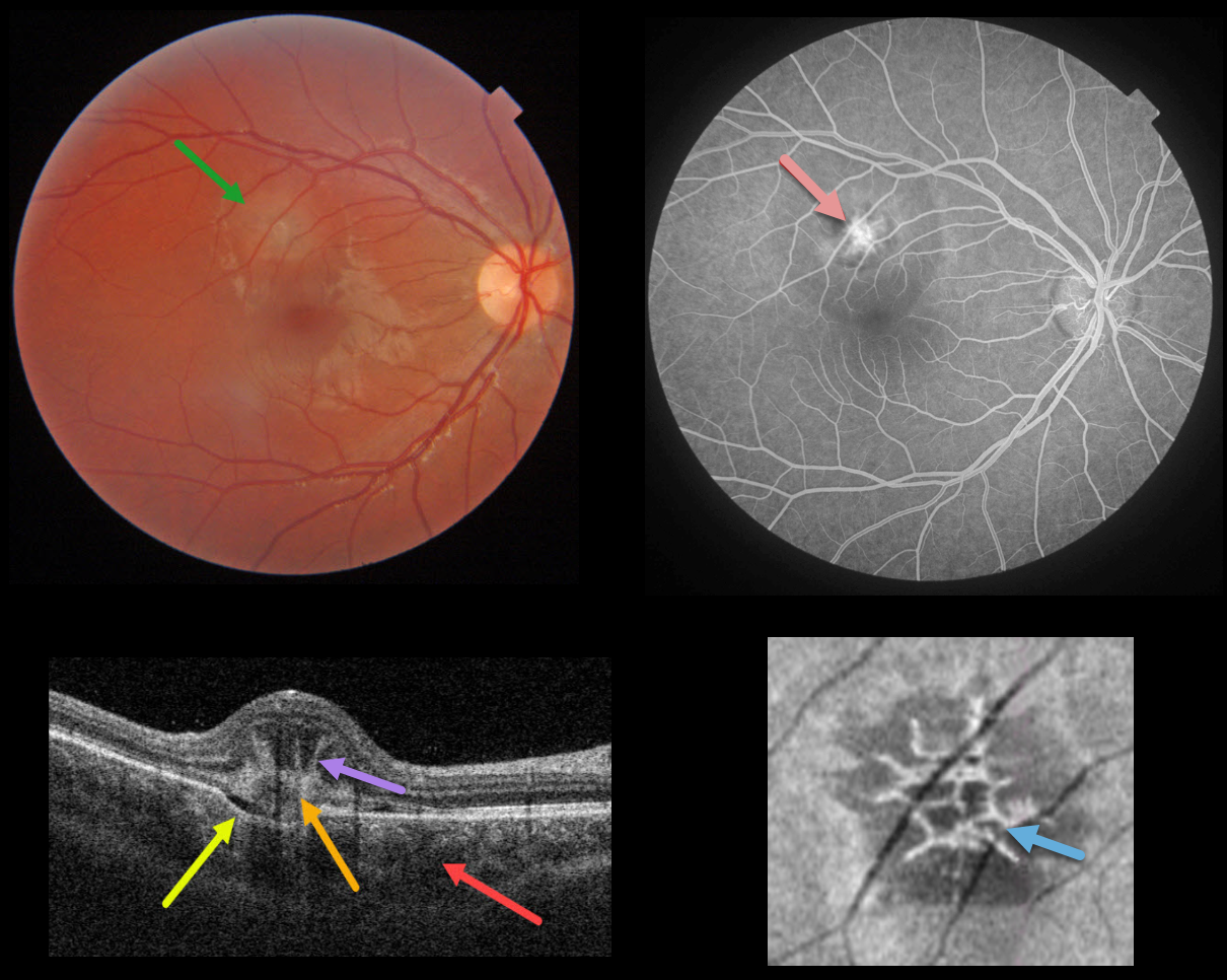
This healthy 14YO female presented with acute vision loss in her right eye. Vision was 20/40. Triton color photography shows a depigmented subretinal lesion in the superior macula.
Swept-source OCT shows a constellation of fascinating findings. A type 2 macular neovascularization is seen overlying a choroidal excavation. The choroid is thickened.
The most dramatic findings are the unusual linear, radiating, hyperreflective lesions extending anteriorly from the MNV, also known as the “pitchfork sign.” En face OCT through this lesion shows a spiky, wreath-like pattern of hyperreflectivity.
The MNV was confirmed on fluorescein angiography. Intravitreal anti-VEGF was begun.
Learning Points:
The pitchfork sign was originally described by Hoang et al (Retina 2013;33:1049-1055) as a unique OCT finding in inflammatory MNV. Falavarajani et al more recently noted that this finding is also associated with non-inflammatory MNV (Ophthalmic Surg Lasers Imaging Retina 2019;50:719-725). The pathogenesis of the avascular pitchfork spikes is unknown, but may be related to outer retinal traction or Müller cell activation.
Focal choroidal excavation, originally described by Jampol et al in 2006, is likely part of the pachychoroid spectrum and is associated with type 2 MNV. An excellent review is by Chung et al (Retina 2017;37;199-221).
IDIOPATHIC MACULAR NEOVASCULARIZATION (MNV)
Originally posted on @retina.rocks November 10, 2021
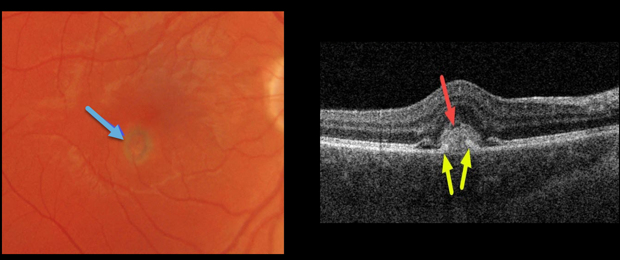
This 30YO male presented with acute unilateral vision loss of 20/50 in his right eye. Ocular examinations were normal bilaterally except for a small area of retinal edema with a subretinal pigment ring in the right inferotemporal fovea.
Swept-source OCT shows a hyperreflective macular neovascularization (MNV) mostly above the RPE. Elevated RPE partially extends into the midportion of the MNV.
In this case, due to our patient’s acute symptoms, we chose to begin anti-VEGF therapy.
Learning Points:
Type 2 MNV often have RPE cells that migrate along its anterior surface, and this appears clinically as a pigment ring. Although the RPE never completely envelops the MNV, its presence often helps to stabilize the lesion’s exudation. In this case, due to our patient’s acute symptoms, we chose to begin anti-VEGF therapy.
IDIOPATHIC MACULAR NEOVASCULARIZATION (MNV)
Originally posted on @retina.rocks July 24, 2020
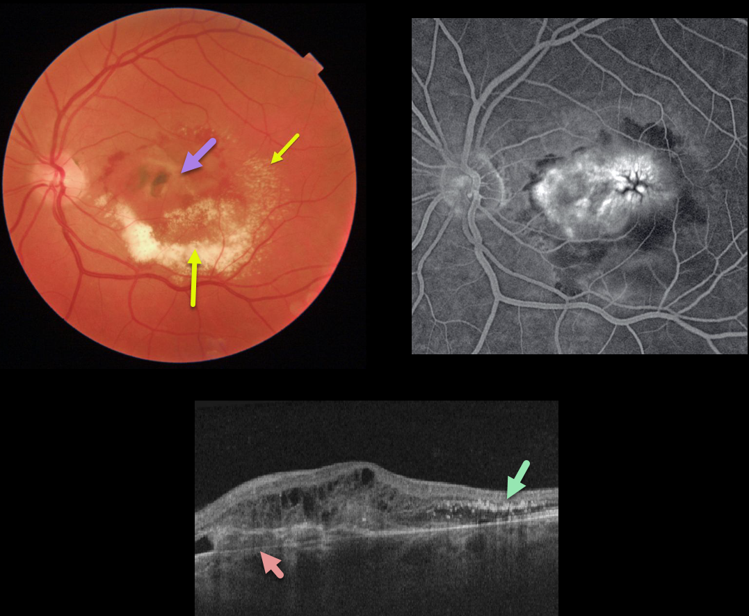
This patient was being followed for an inactive nasal macular chorioretinal scar with 20/25 vision. After being lost to follow-up for many months, he presented with counting-finger vision from an active type 1 macular neovascularization.
The macular OCT shows a complex lesion with the MNV located primarily beneath the RPE. The lipid precipitated out in the subretinal space and follows Henle’s layer temporally.
Learning Points:
The amount of lipid layered in the subretinal space is unusual in MNV. Extensive submacular lipid like this is more common with Coats disease or retinal capillary hemangiomas.
MACULAR NEOVASCULARIZATION (MNV)
Originally posted on @retina.rocks July 8, 2020
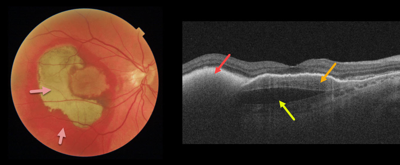
This patient’s vision remained 20/100 one month following vitrectomy with subretinal tissue plasminogen activator.
Surgery accomplished its goal of displacing most of the subfoveal blood, which is variably red and devitalized (yellow).
OCT shows the remaining subretinal hyperreflective blood, the causative sub-RPE type 1 macular neovascularization, and a subfoveal prechoroidal cleft.
Learning Points:
A prechoroidal cleft is a hyporeflective space between the RPE and Bruch’s membrane.
These are seen in up to about 20% of treated eyes with wet AMD, particularly with type 3 neovascularization and polypoidal choroidal vasculopathy.
They are usually associated with worse visual acuity due to potential complications, including RPE tear and subretinal hemorrhages.
For a more detailed discussion of prechoroidal clefts, see Kim et al Retina 2017;37;2047-2055.
MACULAR NEOVASCULARIZATION (MNV)
Originally posted on @retina.rocks May 15, 2020
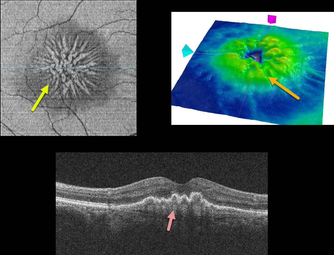
This patient is receiving ongoing intravitreal Avastin injections for a type 1 macular neovascularization (MNV) from wet AMD.
The injections caused the MNV to involute and contract, dragging the overlying RPE into radiating radial folds. This is best seen in the cool OCT en face view. The RPE folds are also seen in the OCT retinal map and OCT B-scan.
Learning Points:
Macular neovascularization is known by many other terms, including choroidal neovascular membrane, subretinal neovascular membrane, etc.
However, since these vessels are now known not to necessarily arise just from the choroid, the proper term is macular neovascularization (see Consensus on Neovascular Age-Related Macular Degeneration Nomenclature Study Group Ophthalmology 2020;127;616-636).
MNV is classified according the location of the new vessels in relation to its location relative to the RPE and neurosensory retina: type 1 (below the RPE), type 2 (between the RPE and neurosensory retina, and type 3 (within the neurosensory retina).
IDIOPATHIC MACULAR NEOVASCULARIZATION (MNV)
Originally posted on @retina.rocks April 29, 2020
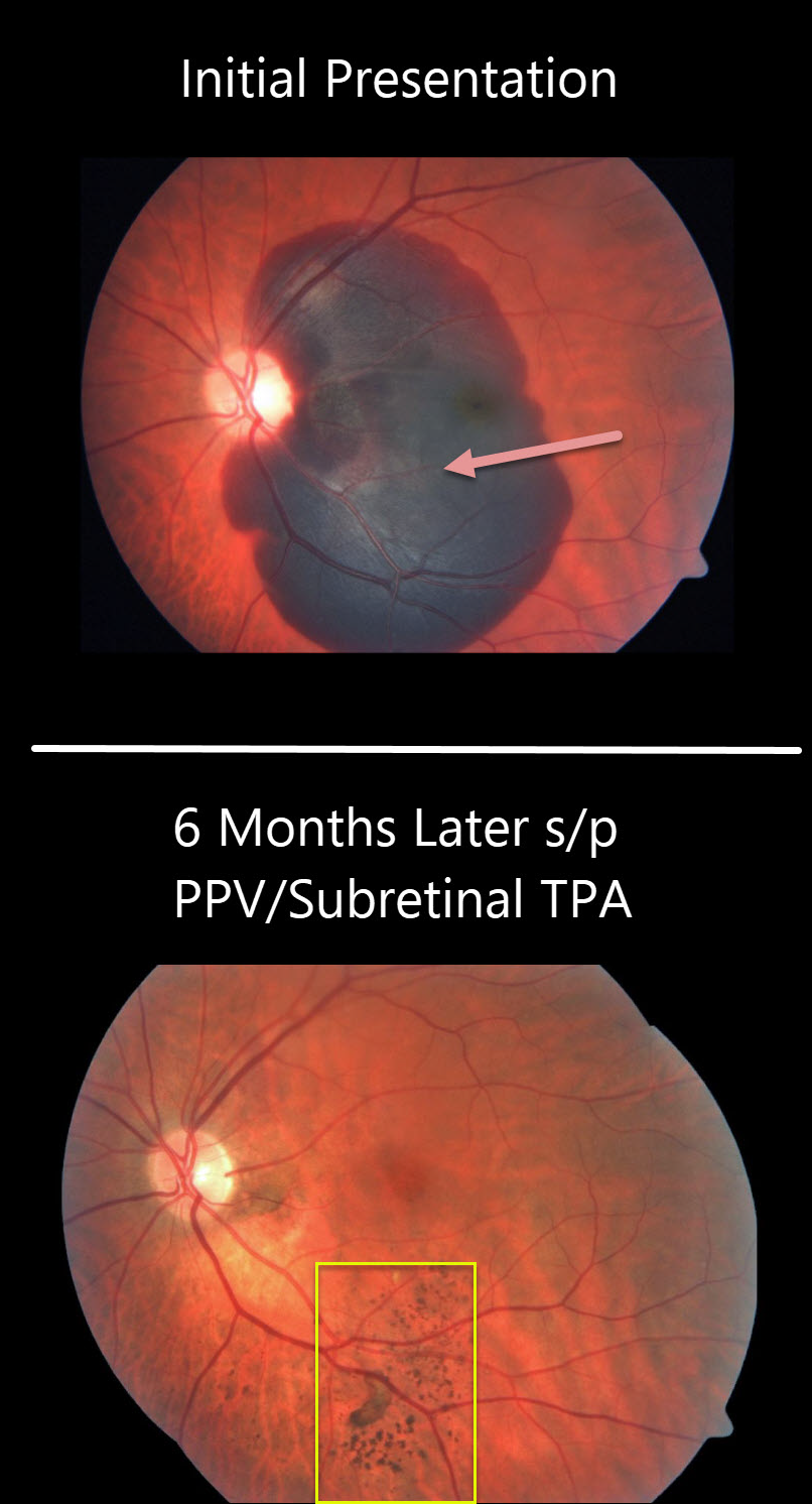
The large submacular hemorrhage is likely from a macular neovascularization from polypoidal choroidal vasculopathy.
The patient was treated with vitrectomy, subretinal tissue plasminogen activator (TPA), and fluid air exchange. The TPA helped liquefy the clot, which was then hydraulically displaced inferiorly outside the macula by the gas bubble.
At six months post-op, the central macula is remarkably normal with multifocal clumps of subretinal pigment more inferiorly.
Learning Points:
Multifocal subretinal pigment is a classic finding for regressed subretinal blood. This pigment is hypoautofluorescent without RPE atrophy (see Hussnain et al, Retina 2019;39;1925-1935).


