

Idiopathic Retinal Vasculitis Aneurysms and Neuroretinitis (IRVAN)
<< Back to Cases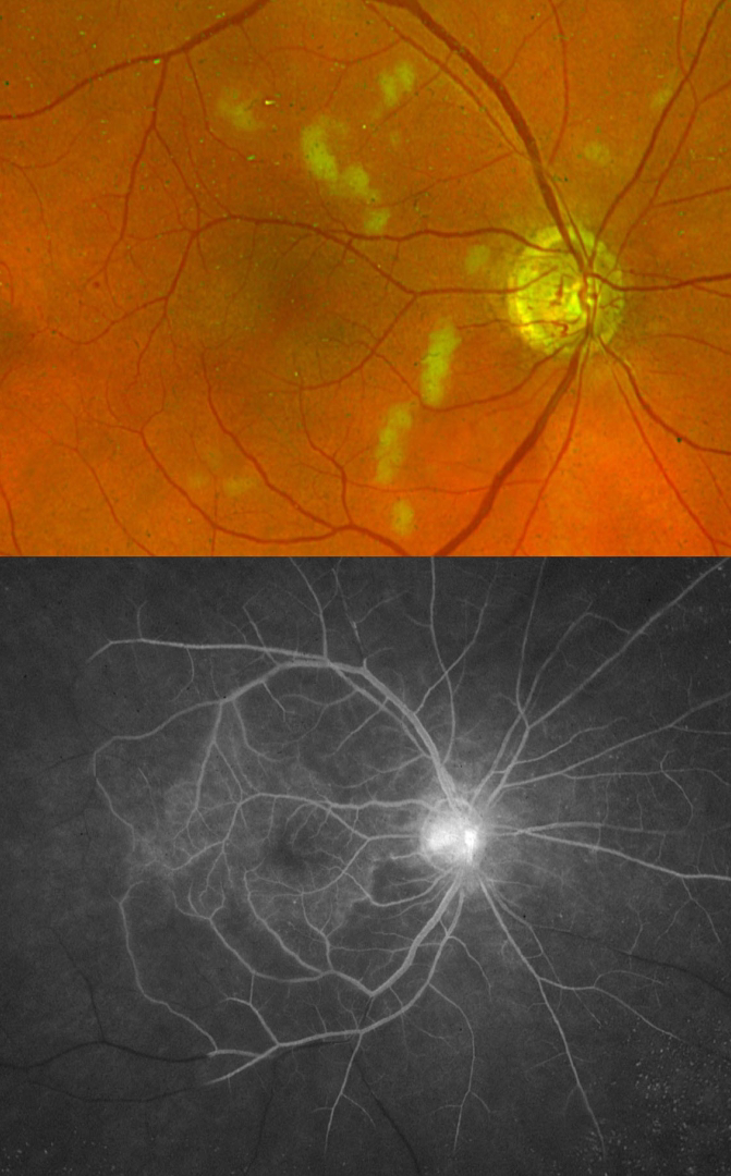
IDIOPATHIC RETINAL VASCULITIS ANEURYSMS AND NEURORETINITIS (IRVAN)
Mattie Adams
Originally posted on @retina.rocks November 5, 2025
This healthy 63YO male presented with an ill-defined history of blurriness in his right eye upon first waking in the morning. Vision was 20/100 OD and 20/20 in his normal OS. There was diffuse rubeosis OD on slit lamp examination, and intraocular pressure was 28 mmHg.
Optos color RG imaging shows scattered nerve fiber layer infarcts with a rare retinal hemorrhage. Fluorescein angiography shows complete capillary nonperfusion distal to the peripheral macula with late optic nerve leakage. Carotid Doppler and CT angiography were negative.
Intravitreal Avastin was given, followed by full panretinal photocoagulation. He subsequently developed florid rubeosis with neovascular glaucoma requiring an Ahmed tube.
Learning Points:
Idiopathic Retinitis, Vasculitis, Aneurysms, and Neuroretinitis (IRVAN) is a rare disease of unknown etiology characterized by retinal vasculitis, aneurysmal arterial dilations, and neuroretinitis. The diagnosis is clinical, as there are no systemic associations or diagnostic laboratory tests. Early panretinal photocoagulation (PRP) to areas of widespread retinal nonperfusion is recommended to prevent neovascular complications and vitreous hemorrhage (Samuel et al Ophthalmology 2007;114:1526-1529).
IDIOPATHIC RETINAL VASCULITIS ANEURYSMS AND NEURORETINITIS (IRVAN)
Originally posted on @retina.rocks July 1, 2021
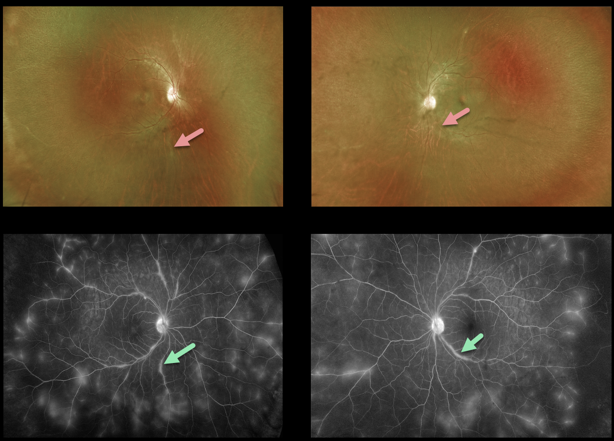
This 41YO female presented with vision of 20/70 OD and 20/25 OS from bilateral retinal vasculitis, highlighted well on fluorescein angiogram.
Extensive workup was negative, including sarcoidosis (chest X-ray, ACE, lysozyme), ANCA, ANA, HLA-B27, Lyme, and RPR. Following the negative labs, she was started on oral prednisone 20mg/day.
Learning Points:
Idiopathic Retinitis, Vasculitis, Aneurysms, and Neuroretinitis (IRVAN) is a rare disease of unknown etiology characterized by retinal vasculitis, aneurysmal arterial dilations, and neuroretinitis. The diagnosis is clinical, as there are no systemic associations or diagnostic laboratory tests.
Retinal vasculitis can be seen in a number of conditions, including pars planitis, frosted branch angiitis, lupus vasculitis, Behcet’s, and idiopathic retinal vasculitis aneurysms and neuroretinitis (IRVAN).
IDIOPATHIC RETINAL VASCULITIS ANEURYSMS AND NEURORETINITIS (IRVAN)
Originally posted on @retina.rocks December 1, 2020
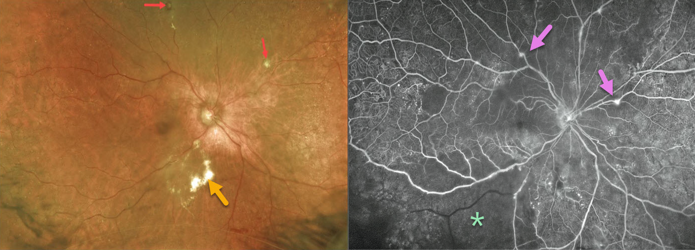
This 77YO female initially presented with a vitreous hemorrhage, which obscured her fundus. The blood spontaneously cleared, allowing us to see multiple fibrosed arterial macroaneurysms and subretinal lipid.
Fluorescein angiography (FA) shows extensive marked inferotemporal retinal ischemia, scattered capillary dropout, and focal areas of mostly venous staining superiorly and nasally. Panretinal photocoagulation (PRP) was applied to all ischemic areas.
Learning Points:
Idiopathic Retinitis, Vasculitis, Aneurysms, and Neuroretinitis (IRVAN) is a rare disease of unknown etiology characterized by retinal vasculitis, aneurysmal arterial dilations, and neuroretinitis. The diagnosis is clinical, as there are no systemic associations or diagnostic laboratory tests.
Early PRP to areas of widespread retinal nonperfusion is recommended to prevent neovascular complications and vitreous hemorrhage (Samuel et al Ophthalmology 2007;114;1526-1529).
IDIOPATHIC RETINAL VASCULITIS ANEURYSMS AND NEURORETINITIS (IRVAN)
Originally posted on @retina.rocks July 6, 2020
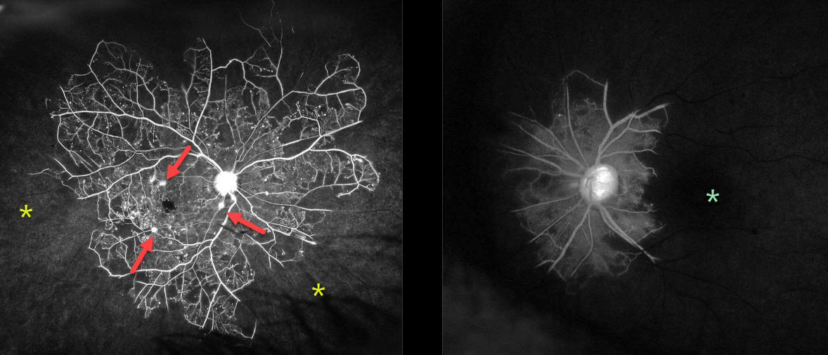
This 64YO female presented with 20/30 vision OD and no light perception OS. Her blood pressure was elevated at 147/101, and she had a history of type 2 diabetes. She had a prior macular laser for macular edema.
Although clinically the peripheral retina appeared ischemic, fluorescein angiography showed dramatic, extensive, and total peripheral ischemia OD, with even worse near total loss of retinal perfusion OS. Multiple aneurysmal dilations are seen in the right posterior pole.
We injected both eyes with Avastin that day and applied full panretinal photocoagulation bilaterally over the next 2 weeks. Carotid dopplers showed less than 50% bilateral internal carotid stenosis.
Learning Points:
Idiopathic Retinitis, Vasculitis, Aneurysms, and Neuroretinitis (IRVAN) is a rare disease of unknown etiology characterized by retinal vasculitis, aneurysmal arterial dilations and neuroretinitis. The diagnosis is clinical, as there are no systemic associations or diagnostic laboratory tests.
Early PRP to areas of widespread retinal nonperfusion is recommended to prevent neovascular complications and vitreous hemorrhage (Samuel et al Ophthalmology 2007;114;1526-1529).


