

03 Inherited Retinal Diseases
<< Back to Cases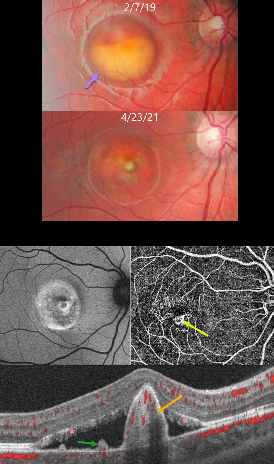
BEST DISEASE WITH MACULAR NEOVASCULARIZATION
João Pedro Marques
Originally posted on @retina.rocks August 26, 2026
This healthy 11YO boy presented to us on 2/7/19 with asymptomatic retinal findings. Vision was 20/25 OD and 20/20 OS.
Color photography of his right eye shows a partially scrambled vitelliform lesion consistent with Best vitelliform macular dystrophy (BVMD). A smaller, scarred, and atrophic lesion was noted in his left eye (not shown).
He returned 2 years later without visual complaints. Vision was 20/40 OD. The vitelliform lesion in his right macula is atrophic, with a central depigmented nodule. Fundus autofluorescence (FAF) shows variable hyper- and hypo-FAF. OCTA en face shows a central flow signal. OCTA B-scan shows a nodular subretinal hyperreflective pillar containing flow signals, subretinal fluid, and vitelliform material layered on the RPE and lining the outer retina. The vision and OCT findings did not change following 2 intravitreal Lucentis injections, so observation was recommended. We continue to follow him with stable findings 5 years later.
Learning Points:
BVMD is associated with a mutation in the BEST1 gene, which encodes the bestrophin-1 protein. Bestrophin-1, a calcium-activated chloride channel, is primarily found in the basolateral plasma membrane of the RPE. BEST1 mutations cause a variety of phenotypes, including autosomal recessive bestrophinopathy, best vitelliform macular dystrophy, and autosomal dominant vitreoretinochoroidopathy.
Secondary macular neovascularization (MNV) develops in up to half of eyes, depending on the imaging modality used, and can develop at any stage of the disease. MNV exists in two forms: the less common exudative form, which presents with acute vision loss, subretinal hemorrhage, and intraretinal/subretinal fluid, and the more common non-exudative form seen in more advanced (vitelliruptive and atrophic) stages (Parodi et al, Invest Ophthalmol Vis Sci 2020;61(6);61).
These subclinical MNVs are best detected with swept-source OCTA. Structural OCT findings that suggest non-exudative MNV include focal choroidal excavations and nodular subretinal pillars (as in our patient). Exudative MNV responds well to standard anti-VEGF injections, whereas non-exudative lesions tend to remain stable without treatment.
X-LINKED RETINOSCHISIS
Shraddha Raj Shrivastava, Akansha Sharma and Manish Nagpal
Originally posted on @retina.rocks August 19, 2026
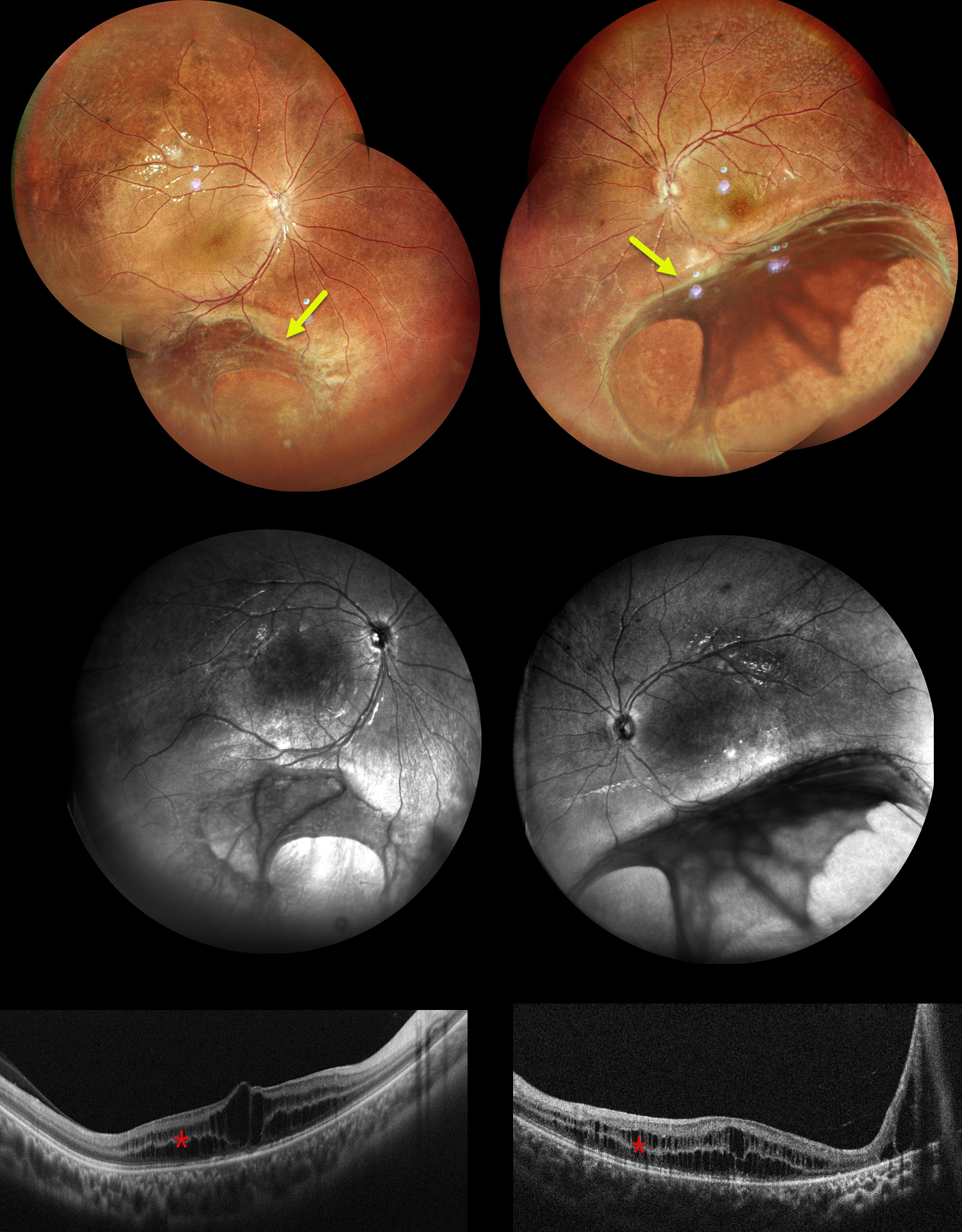
This 9YO boy presented with decreased vision in each eye since early childhood. Vision was 20/80 OU.
Pseudocolor SLO imaging shows blunted foveal reflexes due to inner and outer nuclear layer schisis on OCT. Widefield imaging shows extension of the schisis cavities into the inferotemporal periphery, involving the inner retina with vitreous veils. These changes are best seen in the infrared images. Our patient’s 13YO brother was examined the same day and had similar fundus findings.
Both brothers were prescribed topical carbonic anhydrase inhibitors (dorzolamide) and asked to follow up in 3 months. Genetic testing was recommended.
Learning Points:
Sex-linked retinoschisis is caused by a hemizygous mutation in the RS1 gene, which is located on the X chromosome and encodes for retinoschisin, a protein secreted by photoreceptors that is involved in intercellular adhesion and likely in retinal cellular organization (Heymann et al, Progress Retinal Eye Research 2023;95:101147). Although usually transmitted as an X-linked recessive disorder in males with an incidence of 1 in 15,000 to 30,000, it can also sometimes affect females.
The characteristic features include “spoke-wheel” foveoschisis and peripheral schisis with vitreous veils. Over time, the macular schisis flattens with secondary atrophy, and vision usually levels off at about 20/100. Patients can develop spontaneous vitreous hemorrhage and rhegmatogenous retinal detachment from peripheral schisis. Although there is no specific treatment, topical carbonic anhydrase inhibitors may decrease the foveal thickness and possibly help minimize foveal atrophy (see Andreuzzi et al Retina 2017;37:1555-1561).
PIGMENTED PARAVENOUS CHORIORETINAL ATROPHY
Brahim KhourI, Oriana Gomez, Sebastian Polo, and Axel Ramos Nuñez Cazares
Originally posted on @retina.rocks August 14, 2026
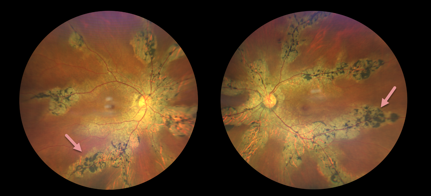
This healthy 55YO female presented with 2 years of mild bilateral vision loss. Family history was negative. Vision was 20/20 OD and 20/40 OS.
Color photography shows bilateral paravenous chorioretinal scarring with intraretinal pigment migration.
Learning Points:
Pigmented paravenous chorioretinal atrophy (PPCRA) is a rare condition characterized by paravenous pigment clumps with peripapillary and radial zones of retinal pigment epithelial (RPE) atrophy (Lee et al, AJO 2021;224:120-132). It is bilateral, often asymmetric, with various causes, including genetic and pseudo-PPCRA (inflammatory, infectious, or unknown). Patients may be asymptomatic or present with symptoms such as nyctalopia, while central visual acuity is often relatively preserved in the absence of macular involvement. The underlying pathophysiology remains incompletely understood, although primary choroidal and RPE abnormalities have been proposed to contribute to the characteristic retinal changes.
HYPOTRICHOSIS WITH JUVENILE MACULAR DEGENERATION
João Pedro Marques
Originally posted on @retina.rocks July 15, 2026
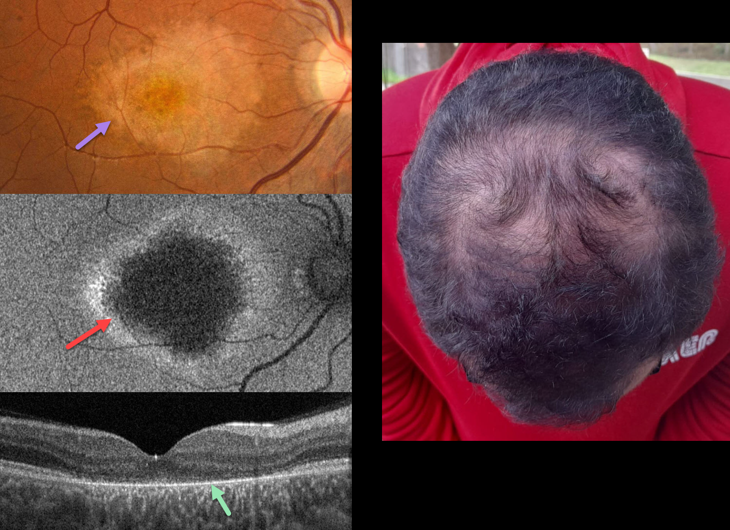
This 19YO male was referred for progressive vision loss and hemeralopia since early childhood. Ocular family history was negative, although his parents were consanguineous. Vision was 20/40 OU.
Color photography of his right eye shows an oval to tear-drop shaped area of central and nasal macular atrophy with clumps of more distal subretinal pigmentation. Fundus autofluorescence (FAF) shows central hypo-FAF with a ring of peripheral hyper-FAF. OCT scanning shows near-complete loss of the outer retinal bands external to the outer nuclear layer. Similar findings were noted in his left eye (not shown). The hair is thinned along the top of his head. Genetic testing revealed a likely pathogenic homozygous variant in the CDH3 gene (16q22.1).
Learning Points:
Hypotrichosis with juvenile macular dystrophy is a rare autosomal recessive disorder caused by biallelic mutations in the CDH3 gene, which encodes for P-cadherin, a calcium-dependent cell adhesion protein expressed in hair follicles and RPE. It is characterized by congenital sparse, short scalp hair (with normal facial and body hair). Progressive macular chorioretinal atrophy begins in childhood, often causing severe central visual impairment by the second to third decade of life.
This case was previously published in Ophthalmology Retina 2021;5:593.
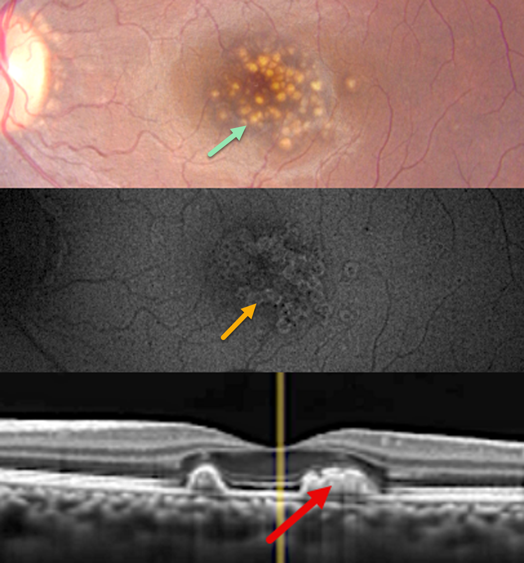
DOMINANTLY INHERITED RADIAL BASAL LAMINAR DRUSEN
Abhishek Karra and Ravindra Karra
Originally posted on @retina.rocks July 14, 2026
This healthy asymptomatic 27YO female presented for a routine eye examination. Ocular family history was negative. Vision was 20/20 OU.
Color photography of her left eye shows extensive, larger, well-defined drusen throughout the central macula. These drusen show a rim of hyper-fundus autofluorescence (FAF) with a central hypo-FAF core. The temporal foveal hyperreflective drusen are confluent, indenting the overlying retina into the outer nuclear layer. Similar findings were noted in her right eye (not shown).
Learning Points:
Doyne’s, also known as dominantly inherited radial basal laminar drusen or Malattia Leventinese, is a rare macular disorder caused by a mutation in the EFEMP1 gene on chromosome 2p16, although we have seen similar phenotypes with negative genetic testing. The EFEMP1 protein is a member of the fibulin family of extracellular matrix glycoproteins. The defective protein creates an abnormally thickened RPE basement membrane. Centrally large, nodular, and confluent drusen develop, along with a temporal radiating pattern of smaller cuticular drusen. Later, there may be variable amounts of RPE hyperplasia and fibrous metaplasia. Macular neovascularization may also occur.
Clues to the likely diagnosis of Doyne’s in our patient include extensive central, symmetrical drusen in a 27YO and pathognomonic drusen that indent the overlying retina on OCT. In addition, FAF usually shows hypo-FAF in age-related drusen. The lesions in Doyne’s, as in this patient, usually show hyper-FAF, likely from unmasking of the RPE by overlying outer retinal thinning.
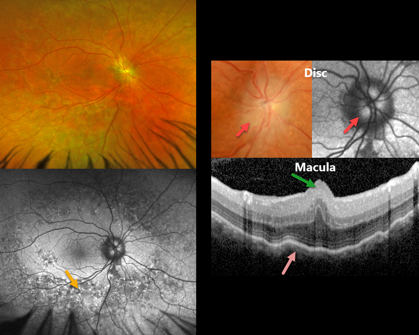
MFRP RETINITIS PIGMENTOSA
João Pedro Marques
Originally posted on @retina.rocks June 9, 2026
This 49YO male has a history of MFRP-related retinitis pigmentosa (RP). We have followed him for 13 years with stable findings of bilateral nanophthalmos (+18D hyperopia, axial lengths 14.82mm OD and 15.14 OS), optic disc drusen (ODD), and choroidal folds. He has compound heterozygosity for 2 pathogenic variants in the MFRP gene.
Optos color RG imaging in his OD shows pigmentary changes throughout the midperiphery along with an ill-defined disc margin. Fundus autofluorescence (FAF) shows variable hyper- and hypo-FAF in the midperiphery. Disc drusen are noted on a more magnified view, which hyper-FAF. Macular OCT shows a central fold, choroidal thickening, and chorioretinal folds. Identical findings were present in his OD (not shown).
Learning Points:
The MFRP (membrane frizzled-related protein) is expressed in the RPE and ciliary epithelium. MFRP functions as a molecular hub on the RPE apical membrane, coordinating protein trafficking and lipid homeostasis. Loss of MFRP leads to DHA accumulation in the RPE, downregulation of visual cycle genes and phototransduction genes, and progressive photoreceptor degeneration.
Biallelic MFRP mutations cause a well-characterized autosomal recessive retinitis pigmentosa (RP) syndrome with nanophthalmos and ODD (Li et al, BJO 2024;108;1679-1687). These eyes are at high risk of angle-closure glaucoma, usually requiring prophylactic laser peripheral iridotomy.
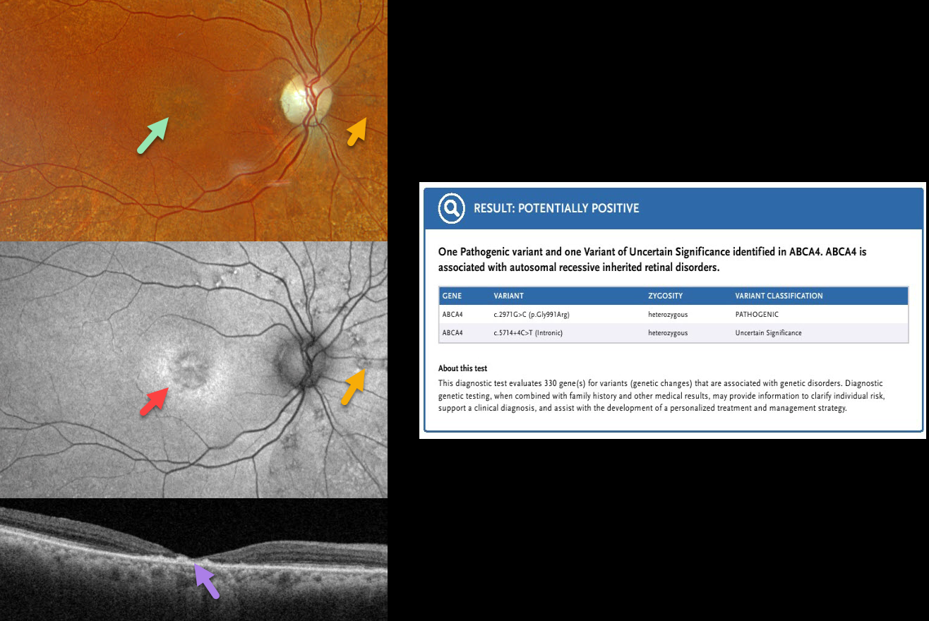
ABCA4 RETINOPATHY
Originally posted on @retina.rocks May 19, 2026
This 88YO female was diagnosed with age-related macular degeneration elsewhere. Vision was 20/400 OD and 20/50 OS.
Optos color RGB imaging OD shows a round area of subfoveal pigmentary changes with extensive drusen-like lesions extending outwards from the peripheral macula. Fundus autofluorescence (FAF) shows central hypo-FAF with a ring of hyper-FAF around this foveal lesion. Scattered lesions consisting of small dots of hyper-FAF with surrounding hypo-FAF are scattered around the nerve. Swept-source OCT shows severe central neurosensory atrophy with a macular pseudohole. Some clumps of hyperreflective material are noted within the area of atrophy, along with variable ellipsoid zone loss elsewhere. Similar findings were present in her left eye (not shown). Genetic testing revealed heterozygous ABCA4 mutations, including a pathogenic variant (c.2971G>C, p.Gly991Arg) and a variant of uncertain significance (c.5714.+4C-T, intronic).
Learning Points:
The ABCA4 protein is located in the photoreceptor outer segments and is involved in the recycling of 11-cis-retinal. The mutation of this gene causes the accumulation of lipofuscin in the RPE, which eventually leads to photoreceptor and RPE degeneration. ABCA4 disorders include Stargardt disease, fundus flavimaculatus, cone-rod dystrophy, retinitis pigmentosa, and age-related macular degeneration.
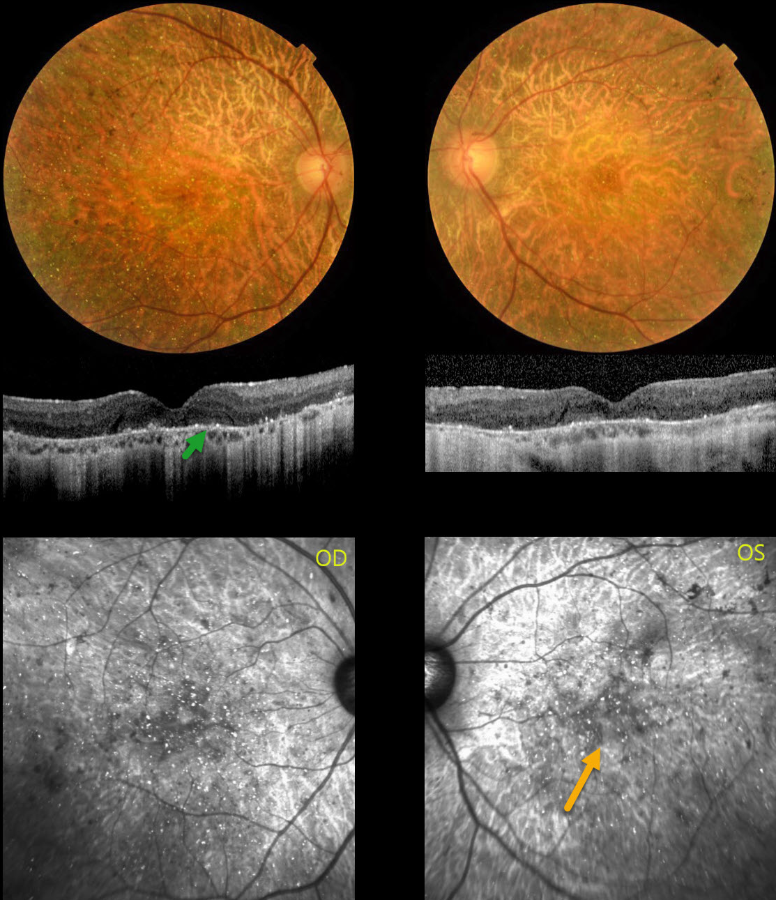
BIETTI CRYSTALLINE DYSTROPHY
João Pedro Marques
Originally posted on @retina.rocks April 29, 2026
This 42YO female has been followed by us for 7 years with a known history of Bietti crystalline retinopathy with homozygosity for a pathogenic CYP4V2 variant. Vision was stable at 20/400 OU.
Fundus imaging shows extensive pigmentary degeneration with subretinal crystalline deposits involving both posterior poles. The crystalline deposits are best seen on near-infrared imaging. OCT scanning shows hyperreflective dots overlying the RPE-Bruch’s membrane complex with variable outer retinal and RPE loss. The pigmentary changes extend into each retinal periphery with more typical retinitis pigmentosa-like changes, including intraretinal pigment migration (bone spicules).
Learning Points:
Bietti crystalline dystrophy is an autosomal recessive ocular disorder that affects both the anterior and posterior segments. Clinical features include crystalline deposits in the retina, cornea, and rarely the crystalline lens, as well as retinal pigment epithelial clumping and atrophy. Mutations in CYP4V2 causing dysfunctional lipid metabolism have been implicated in its pathogenesis. While there are no definite treatments, anti-VEGF therapy can be given for secondary macular neovascularization. See Saatci et al for a recent review (Clinical Ophthalmology 2023;17:953-967).
ACHROMATOPSIA
The European VitreoRetinal Society (EVRS), Vinod Kumar, Smile Singla and Bhupinder Singh
Originally posted on @retina.rocks April 24, 2026
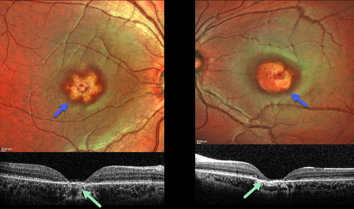
This healthy 5YO boy presented with decreased vision and repetitive eye movements. Family history was negative. Vision was 20/200 OU. Low-amplitude horizontal nystagmus was noted bilaterally.
MultiColor imaging shows star-shaped macular atrophy OD with a more rounded area of atrophy OS. OCT shows complete central RPE and outer retinal atrophy. A homozygous pathogenic missense mutation (c.1279C>T) in exon 8 of the CNGA3 gene led to the diagnosis of achromatopsia.
Learning Points:
CNGA3 mutations, which encode for components of the cone phototransduction cascade, cause achromatopsia, a rare autosomal recessive cone photoreceptor disorder. Clinical findings range from a normal-appearing fundus to progressive macular degeneration. OCT findings may be normal or show variable outer retinal changes, including foveal ellipsoid zone (EZ) absence or disruption, a hyporeflective zone, and outer retinal atrophy (Zobor et al, IOVS 2017;58:821-832). Most eyes have foveal hypoplasia. The retinal phenotype shows considerable heterogeneity even among patients with the same genotype.
RADIAL BASAL LAMINAR DRUSEN
Akansha Sharma, Manish Nagpal and Navneet Mehrotra
Originally posted on @retina.rocks March 30, 2026
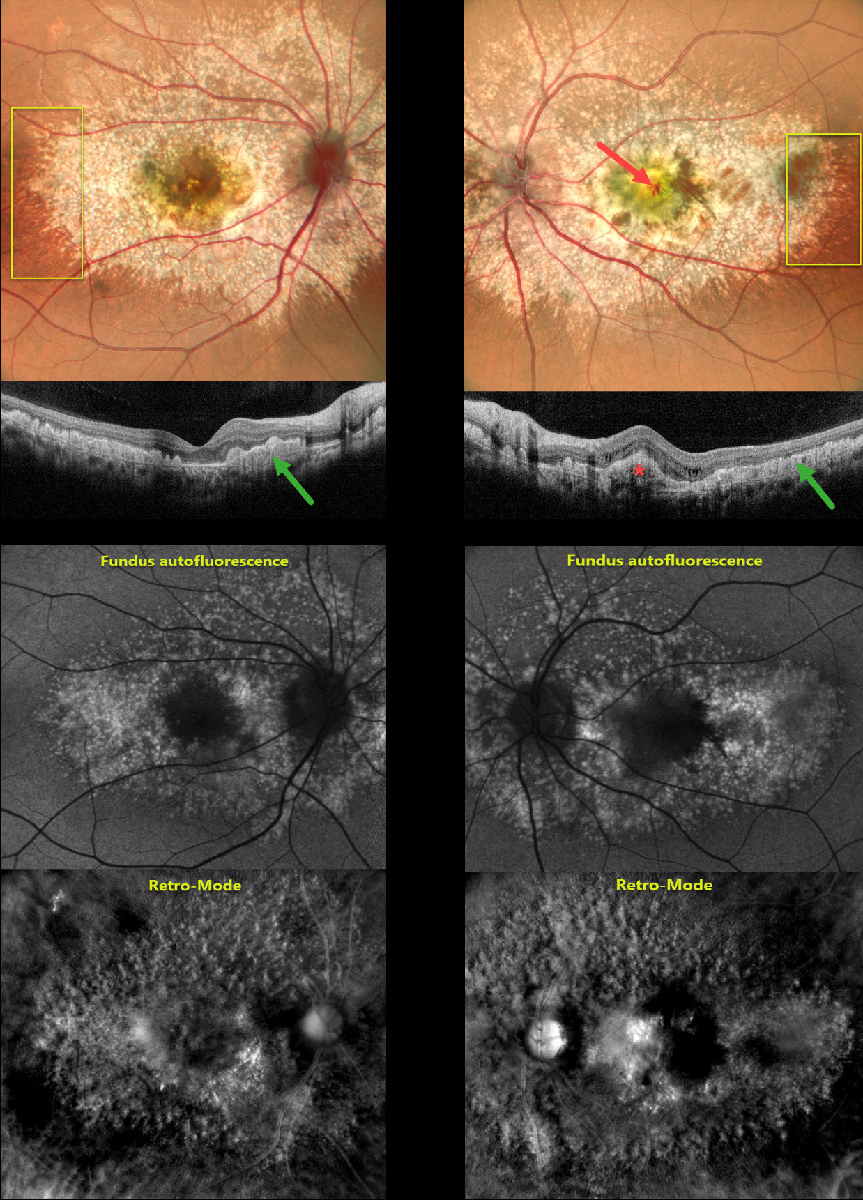
This 29YO male presented with a 1-month history of decreased vision in his left eye. Vision was 20/20 OD and counting fingers OS.
Pseudocolor SLO imaging shows extensive mixed and confluent drusen extending throughout the posterior pole in both eyes. A radiating linear pattern of fine drusen is present in each distal temporal macula. OCT scanning shows drusen indenting the overlying retina, extending into the outer nuclear layer and, in some places, up to the outer plexiform layer. Macular neovascularization (MNV) is present in the left fovea.
Fundus autofluorescence (FAF) shows macular hyper-FAF with surrounding relative hypo-FAF. Retro-Mode imaging reveals a dramatic, almost 3-dimensional appearance of depressions and mounds resembling the surface of the moon. Anti-VEGF therapy was started for the left eye.
Learning Points:
Doyne’s, also known as dominantly inherited radial basal laminar drusen or Malattia Leventinese, is a rare macular disorder caused by a mutation in the EFEMP1 gene on chromosome 2p16, although we have observed similar phenotypes in the absence of genetic testing. The EFEMP1 protein is a member of the fibulin family of extracellular matrix glycoproteins. The defective protein creates an abnormally thickened RPE basement membrane. Centrally large, nodular and confluent drusen develop, along with a temporal radiating pattern of smaller cuticular drusen. Later, there may be variable amounts of RPE hyperplasia and fibrous metaplasia. MNV may also occur.
Fundus autofluorescence (FAF) usually shows hypo-FAF in age-related drusen. The lesions in Doyne’s, as in this patient, usually show hyper-FAF, likely from unmasking of the RPE by overlying outer retinal thinning.
FAMILIAL EXUDATIVE VITREORETINOPATHY (FEVR)
João Pedro Marques
Originally posted on @retina.rocks March 23, 2026
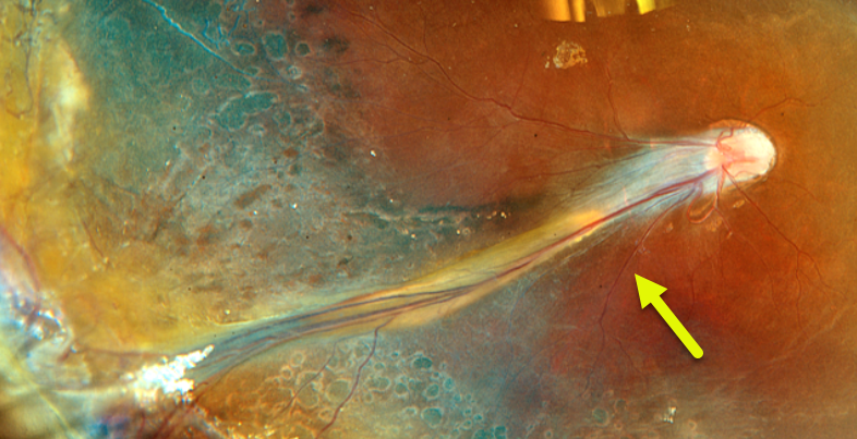
This healthy 6YO boy from Guinea-Bissau with long-standing low vision was referred due to a family history of familial exudative vitreoretinopathy (FEVR) in an older brother and maternal uncle. Vision was 20/400 OD and no light perception OS.
Fundus examination OD showed a falciform retinal fold, straightening of the temporal retinal vascular arcades, and temporal macular dragging. There was a dense white cataract OS, with a total funnel-shaped retinal detachment noted on ultrasonography. Scatter laser and peripheral cryotherapy were applied throughout the ischemic retina. Postoperative Optos color RGB imaging shows laser and cryotherapy scarring.
Familial variant testing confirmed the presence of the pathogenic c.362G>A p.(Arg121Gln) variant in hemizygosity in the NDP gene (NM_000266.4), establishing the diagnosis of X-linked FEVR.
Learning Points:
Originally described by Criswick and Schepens (AJO 1969;58:578-594), familial exudative vitreoretinopathy (FEVR) can be inherited as an autosomal-dominant, recessive, or X-linked trait with high penetrance and variable expressivity. There are numerous genes associated with FEVR, including LRP5, FZD4, NDP, TSPAN12, ZNF408, CTNNB1 and KIF11 [Tao et al, Invest Ophthalmol Vis Sci 2021;62(15);4].
FEVR is characterized by peripheral temporal retinal avascularity, lipid exudation, peripheral neovascularization, tractional retinal detachment, and temporal dragging of the macula and retinal vessels. These findings are somewhat similar to those seen in retinopathy of prematurity.
AUTOSOMAL RECESSIVE BESTROPHINOPATHY
The European VitreoRetina Society (EVRS) and Isha Acharya
Originally posted on @retina.rocks March 13, 2026
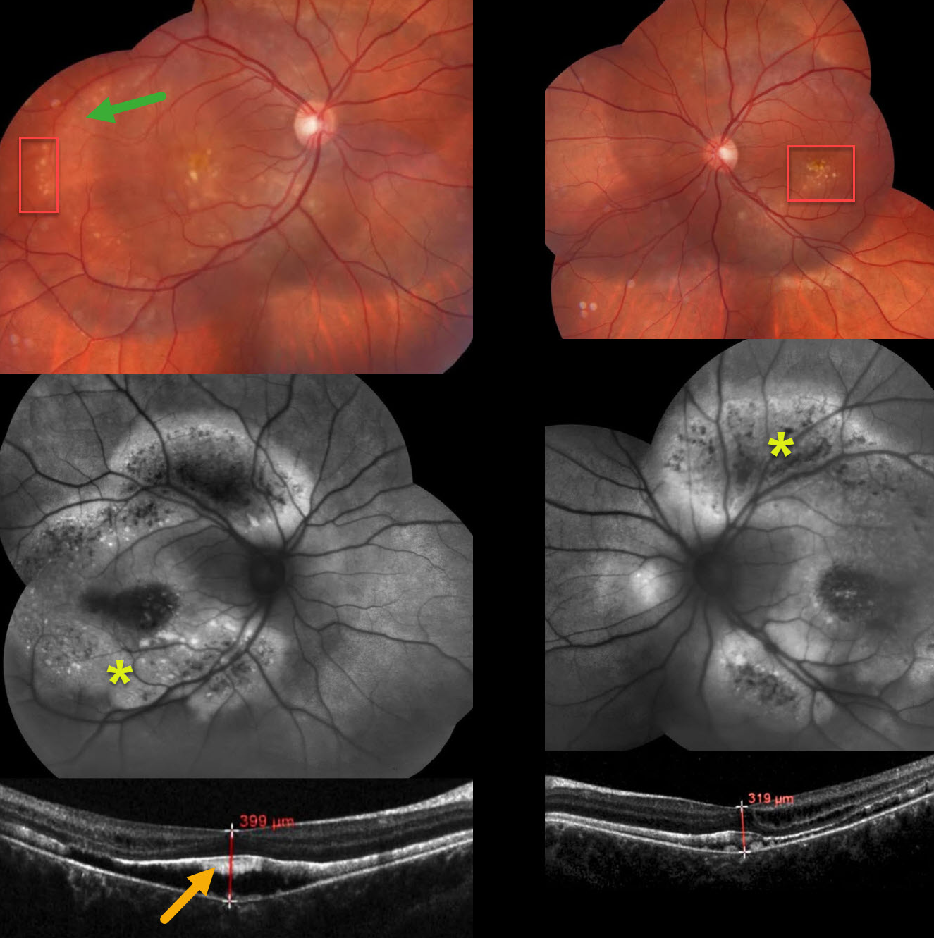
This healthy 17YO female presented with asymptomatic funduscopic findings. Family history was negative for eye disease. Vision was 20/40 OD and 20/50 OS.
Color imaging shows a rim of RPE pigment loss in the peripheral right temporal macula, along with bilateral yellow drusen-like changes. Fundus autofluorescence (FAF) shows much more pronounced findings with peripheral macular/midperipheral central hypo-FAF with surround hyper-FAF. Hyperreflective material is noted along the back surface of the detached foveal retina. Genetic testing was not performed due to cost concerns. Observation was recommended.
Learning Points:
Burgess et al first described autosomal recessive bestrophinopathy as a distinct BEST1-related disorder (American Journal of Human Genetics 2008;82:19-31). Unlike other BEST1 autosomal dominant disorders, patients have 2 abnormal alleles and thus no normally functioning bestrophin1 protein.
It is characterized by posterior pole vitelliform deposits and pigmentary changes that extend beyond the macula, subretinal fluid and vitelliform material, and intraretinal fluid (Casalino et al, Ophthalmology 2021;128:706-718). Vision gradually declines with age.
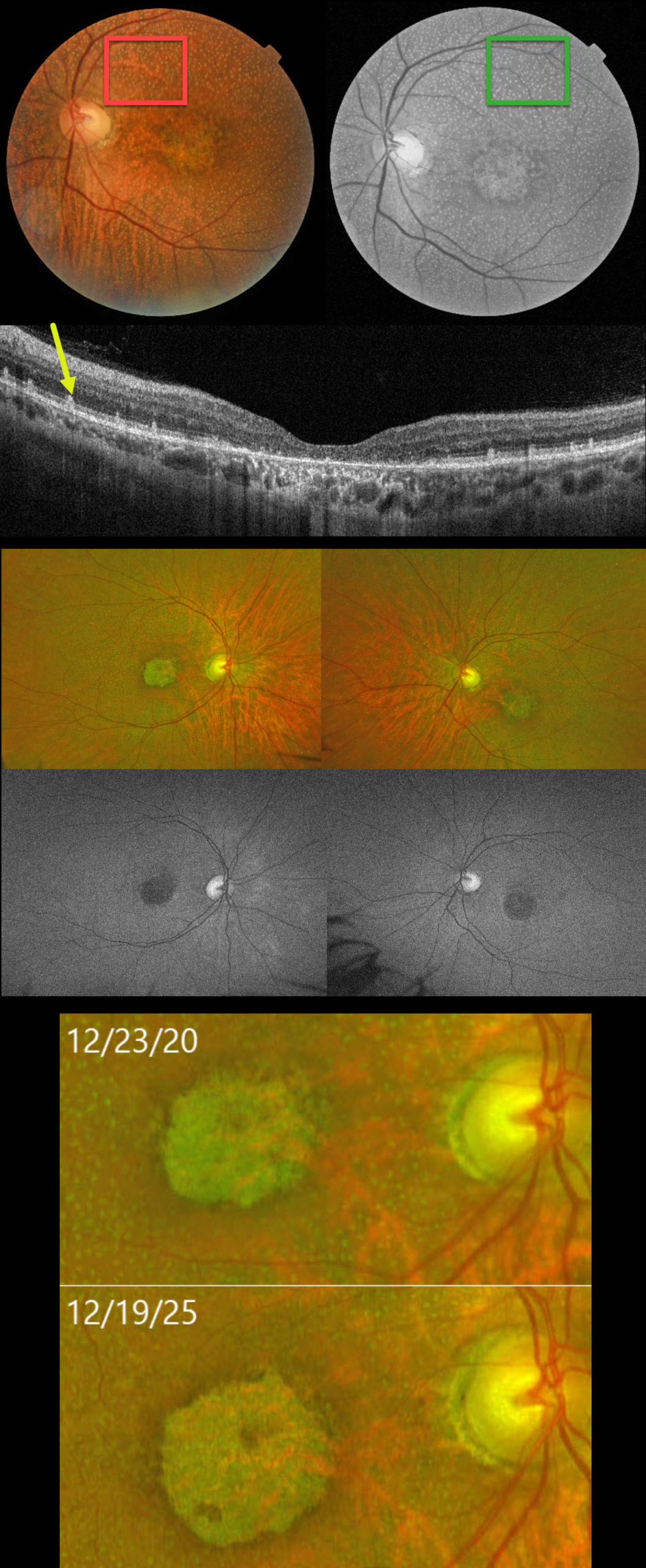
RDH5-ASSOCIATED DISEASE WITH MACULAR ATROPHY
João Pedro Marques
Originally posted on @retina.rocks February 17, 2026
This 58YO female was initially seen by us on 8/6/18 with a prior diagnosis of geographic atrophy and subretinal drusenoid deposits. She described a long-standing history of nyctalopia and poor vision. Vision was 20/640 OU.
Color photography shows innumerable subretinal yellow-white dots extending from the periphery of each macula into the midperiphery. These dots are most evident on red-free imaging. Bilateral foveal macular atrophy is noted. OCT scanning shows central outer retinal atrophy with more peripheral hyperreflective outer retinal deposits, corresponding to the funduscopic dots, which extend into the outer nuclear layer. Optos fundus autofluorescence (FAF) is hazy due to a decreased autofluorescent signal from each fundus. Genetic testing was homozygous for a class IV variant in the RDH5 gene.
When last examined on 12/31/25, vision was 20/800 OU. Her funduscopic findings were stable except for mild enlargement of the macular atrophy.
Learning Points:
Fundus albipunctatus (FA), an autosomal recessive form of congenital stationary night blindness, presents with scattered yellow-white dots that spare the central macula and extend into the midperipheries. Patients experience stable night blindness with normal central acuity. It is most commonly caused by homozygous mutations in the RDH5 gene, which encodes the retinol dehydrogenase protein. This protein is found in the RPE and helps recycle vitamin A in the visual cycle. The white dots are thought to contain 11-cis retinal precursors. This results in decreased retinoid-derived fluorophores, accounting for the diminished FAF in these eyes (Sergouniotis et al, Ophthalmology 2011;118:1161-1670).
A similar phenotype to FA, but with notable progression and a rod-cone dystrophy ERG, is retinitis punctata albescens (RPA). It can be associated with mutations in several genes, including RLBP1, RHO, RDS, RDH5, and RPE65. RDH5 can be associated with both FA and RPA. Some cases initially diagnosed as FA progress and develop macular atrophy (Bianco et al, AJO 2024;267:160-171), as in our patient. We therefore believe that RDH5-associated disease is a better term, as it encompasses the typical FA and RPA phenotypes associated with RDH5.
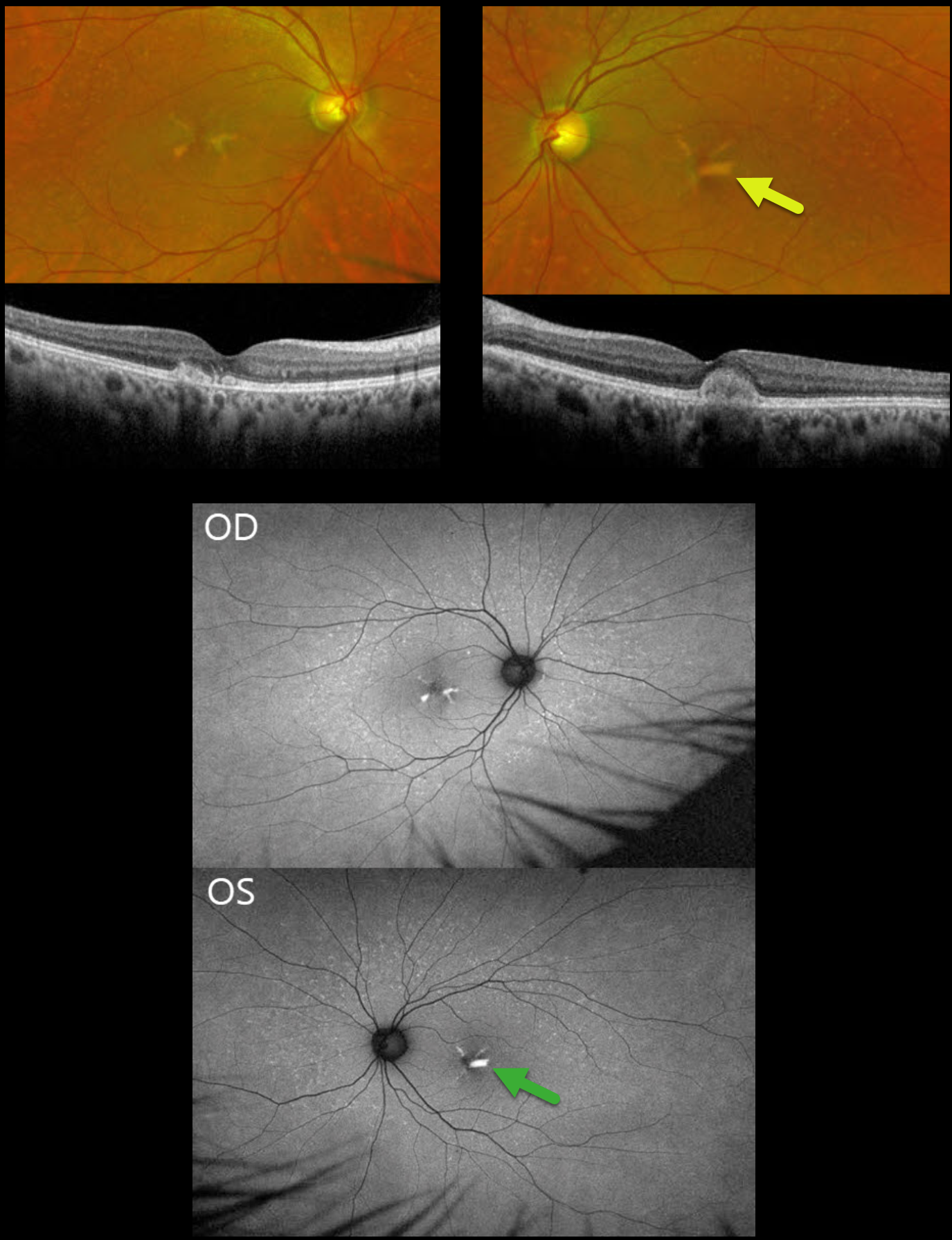
BUTTERFLY-SHAPED PATTERN MACULAR DYSTROPHY
João Pedro Marques
Originally posted on @retina.rocks January 27, 2026
This asymptomatic 53YO male was referred because of a positive family history of two brothers with genetically confirmed PRPH2-associated pattern dystrophy. His father was legally blind by age 65 from an unknown cause. Vision was 20/20 OU.
Optos color RG imaging shows multiple drusen-like deposits in each posterior pole, as well as larger yellow subfoveal deposits that mimic the wings of a butterfly. On OCT, the subfoveal lesions show central hyperreflective deposits in the outer retina. These obscure the outer band details OD. The lesion is more focal in his OS, indenting the overlying retina. On fundus autofluorescence (FAF), these deposits are hyper-FAF.
Learning Points:
Pattern macular dystrophies are a genetically heterogeneous group of disorders characterized by variably shaped yellow subretinal deposits, the most common of which, in our experience, is adult-onset foveomacular vitelliform dystrophy. Causative mutations, including BEST1, PRPH2, IMPG1, and IMPG2. PRPH2 mutations account for 25% of pattern dystrophies, which are inherited as an autosomal dominant trait (Alashwal et al, AJO 2024;271:7-30).
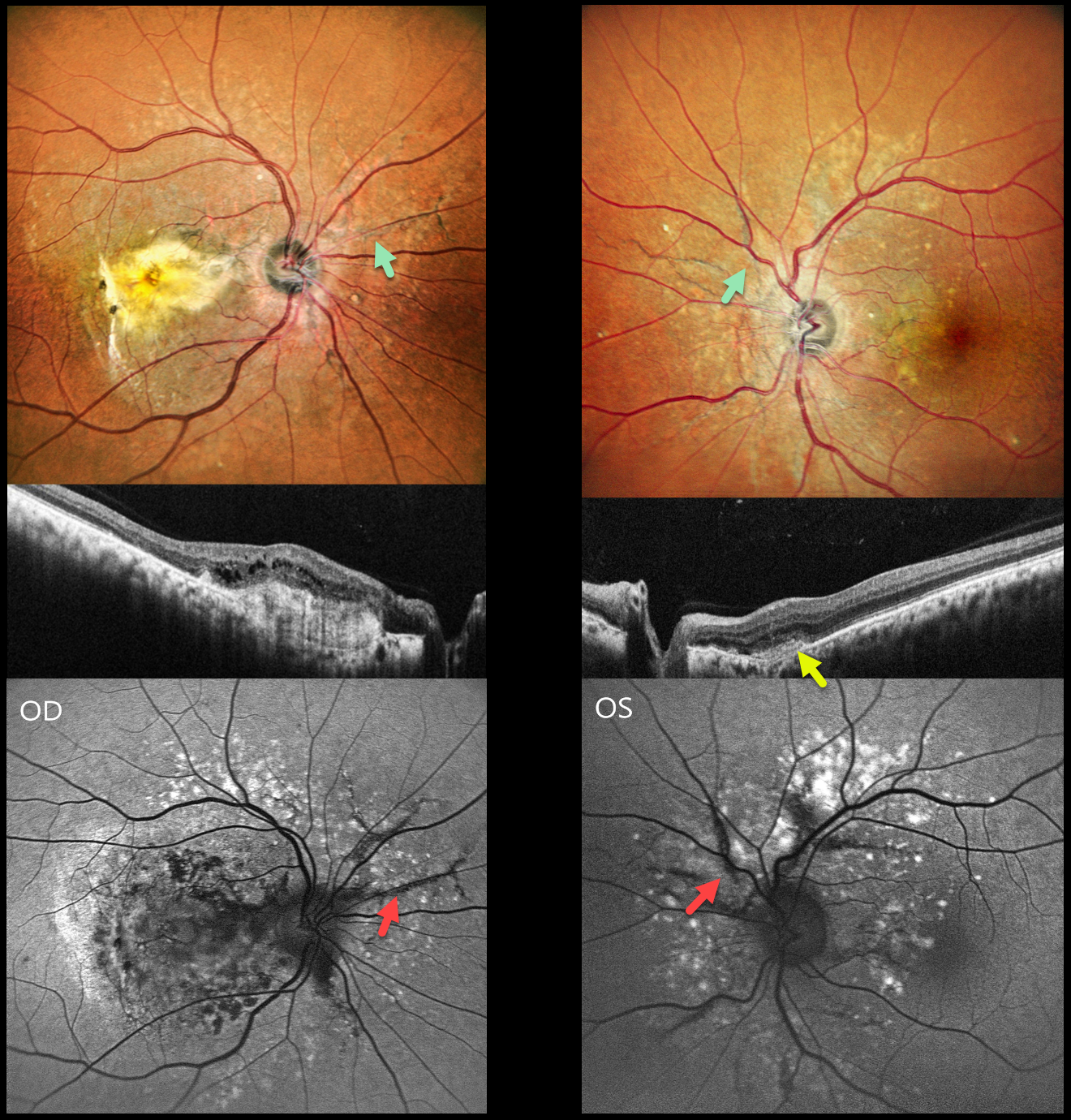
ANGIOID STREAKS WITH MNV AND PATTERN DYSTROPHY PHENOTYPE
Shraddha Raj Shrivastava and Manish Nagpal
Originally posted on @retina.rocks January 6, 2026
This healthy 42YO male presented with 10 months of vision loss in his right eye and 1 month of mild blurred vision in his left eye. Vision was counting fingers OD and 20/20 OS.
Pseudocolor SLO imaging shows angioid streaks extending around and radiating outwards from each optic nerve. There are drusen-like changes around each posterior pole, along with clumps of subretinal pigment temporally.
OCT scanning shows hyperreflective subretinal fibrosis with mild overlying cystic retinal edema OD. A much smaller area of subretinal hyperreflectivity (indicating a type 2 macular neovascularization, MNV) with overlying subretinal hyperreflective material is noted OS. The angioid streaks on fundus autofluorescence (FAF) are hypo-FAF due to RPE atrophy. The drusen-like changes are hyper-FAF.
Learning Points:
Retinal findings in pseudoxanthoma elasticum (PXE) most commonly include angioid streaks and a subretinal peaux d’orange appearance, most pronounced in each temporal macula. However, pattern dystrophy-like changes, as in our patient, can sometimes be the predominant phenotype (Murro et al, Graefe’s 2020;258:1881-1892). As with typical pattern dystrophies, the findings are dynamic and best captured with multimodal imaging.
PXE is caused by a mutation in the ABCA6 protein and is virtually always transmitted as an autosomal recessive disorder, although autosomal dominant transmission may rarely occur (Plomp et al, Am J Med Genet A 2004;126:403-412).
Our patient had no clinical signs of PXE and denied any family history of this disease. He was referred to his primary care doctor for further evaluation. Intravitreal anti-VEGF therapy was started OS.
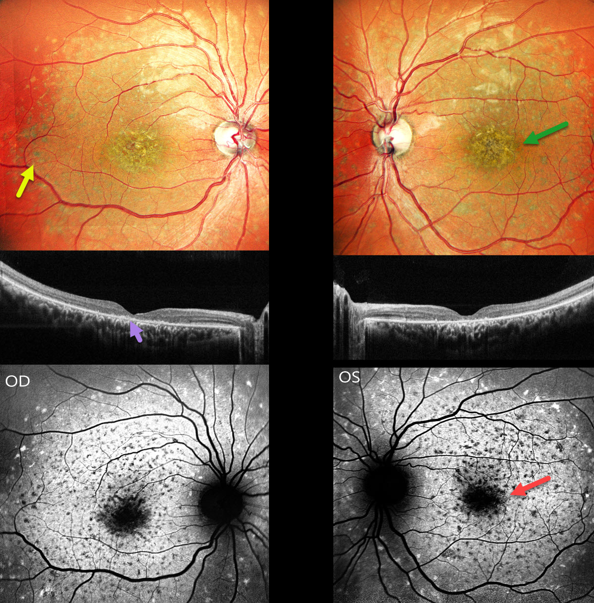
STARGARDT DISEASE
Shraddha Raj Shrivastava and Manish Nagpal
Originally posted on @retina.rocks December 31, 2025
This 26YO male presented with 3 years of bilateral decreased vision along with night vision difficulties since childhood. Family history was negative. Vision was counting fingers at 3 meters OU.
Pseudocolor SLO imaging shows a symmetrical ‘beaten-bronze’ sheen to each central macula with surrounding yellowish pigmented flecks. OCT shows central loss of the photoreceptor layers with hyperreflective deposits above the RPE. Green fundus autofluorescence (FAF) shows foveal hypo-FAF. The clinical flecks are hypo-FAF centrally and hyper-FAF more peripherally. There is relative peripapillary sparing.
Learning Points:
Stargardt disease is an autosomal recessive disorder caused by a mutation in the ABCA4 gene located on chromosome 1. The ABCA4 transmembrane protein localizes to the photoreceptor outer segments and is involved in the recycling of 11-cis-retinal. Mutations of this gene cause the accumulation of lipofuscin in the RPE, which eventually leads to photoreceptor and RPE degeneration. Other ABCA4 disorders include fundus flavimaculatus, cone-rod dystrophy, retinitis pigmentosa, and age-related macular degeneration.
Historically, Stargardt disease was used to describe patients with macular involvement only, and fundus flavimaculatus for those with flecks extending more peripherally. Both are now considered different retinal phenotypes within the ABCA4 spectrum.
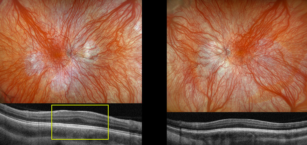
OCULOCUTANEOUS ALBINISM
The European VitreoRetinal Society (EVRS) and João Pedro Marques
Originally posted on @retina.rocks December 26, 2025
This 9YO girl was referred by her local ophthalmologist for esotropia, nystagmus, light hair, and skin, first noticed when she was 3 years old. She has a known history of Hermansky-Pudlak syndrome (HPS), diagnosed with genetic testing 3 years earlier. There is no history of parental consanguinity or of familial inherited retinal diseases. Vision was 20/63 OD and 20/80 OS. There were bilateral iris transillumination defects.
Optos color RGB imaging shows bilateral marked choroidal hypopigmentation with foveal hypoplasia. OCT scanning shows a flattened foveal contour bilaterally (fovea plana).
Learning Points:
Albinism is a group of genetic disorders characterized by abnormal melanin production due to faulty amino acid production. Patients have either ocular (eye) albinism or oculocutaneous (eye and skin) involvement. Inheritance patterns include autosomal dominant, autosomal recessive, and X-linked. Ocular findings include strabismus, nystagmus, iris transillumination defects, blonde fundus, and foveal hypoplasia (seen on OCT as fovea plana). There are often a higher number of crossed nerve fibers at the optic chiasm.
HPS is a rare autosomal recessive multisystem disorder characterized by oculocutaneous albinism, bleeding diathesis due to platelet storage pool deficiency, progressive pulmonary fibrosis, and granulomatous colitis. The underlying pathogenesis involves mutations in at least 10 genes encoding components of protein complexes (BLOC-1, BLOC-2, BLOC-3, and AP-3) essential for the biogenesis and trafficking of lysosome-related organelles in melanocytes, platelets, and other cell types. Our patient had a pathogenic c.223C>T p.(Gln75) homozygous variant in the HPS6 gene, which encodes for a key protein in the BLOC-2 pathway.
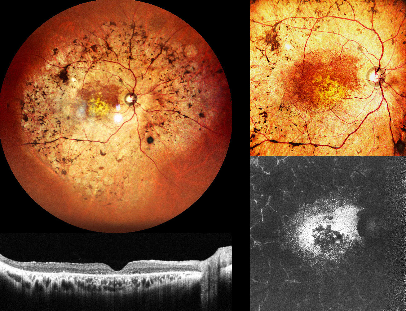
PERICENTRAL RETINAL DEGENERATION
Shraddha Raj Shrivastava, Akansha Sharma and Manish Nagpal
Originally posted on @retina.rocks December 23, 2025
This 47YO male presented with decreased vision in both eyes for 5 years. Ocular family history was negative. Vision was 20/300 OD and 20/660 OS.
Pseudocolor SLO imaging of his right eye shows intraretinal pigment migration (bone-spicules) extending from the macula into the midperiphery. OCT shows outer retinal thinning with disorganization of the outer retinal bands. Fundus autofluorescence (FAF) shows hyper-FAF from the relatively preserved paracentral retina, more central hypo-FAF, and a peculiar peripheral pattern of interlaced hyper-FAF lines around lobular atrophic RPE and retina. Identical findings were noted in his left eye (not shown).
Learning Points:
Pericentral retinitis pigmentosa (RP) is an atypical, mild form of RP characterized by retinal degeneration and intraretinal pigment migration confined to the retinal near midperiphery. Unlike classic RP, pericentral RP tends to progress more slowly, with preserved central visual acuity and less extensive visual field loss over time. Genetically, pericentral RP is heterogeneous (Matsui et al, IOVS 2025;56:6007-6018), with causative variants identified in multiple genes, many of which are also associated with typical RP and other retinal dystrophies, indicating that pericentral RP is a phenotypic variant rather than a distinct clinical entity. Long-term prognosis for central vision is generally favorable in the absence of macular involvement, which unfortunately
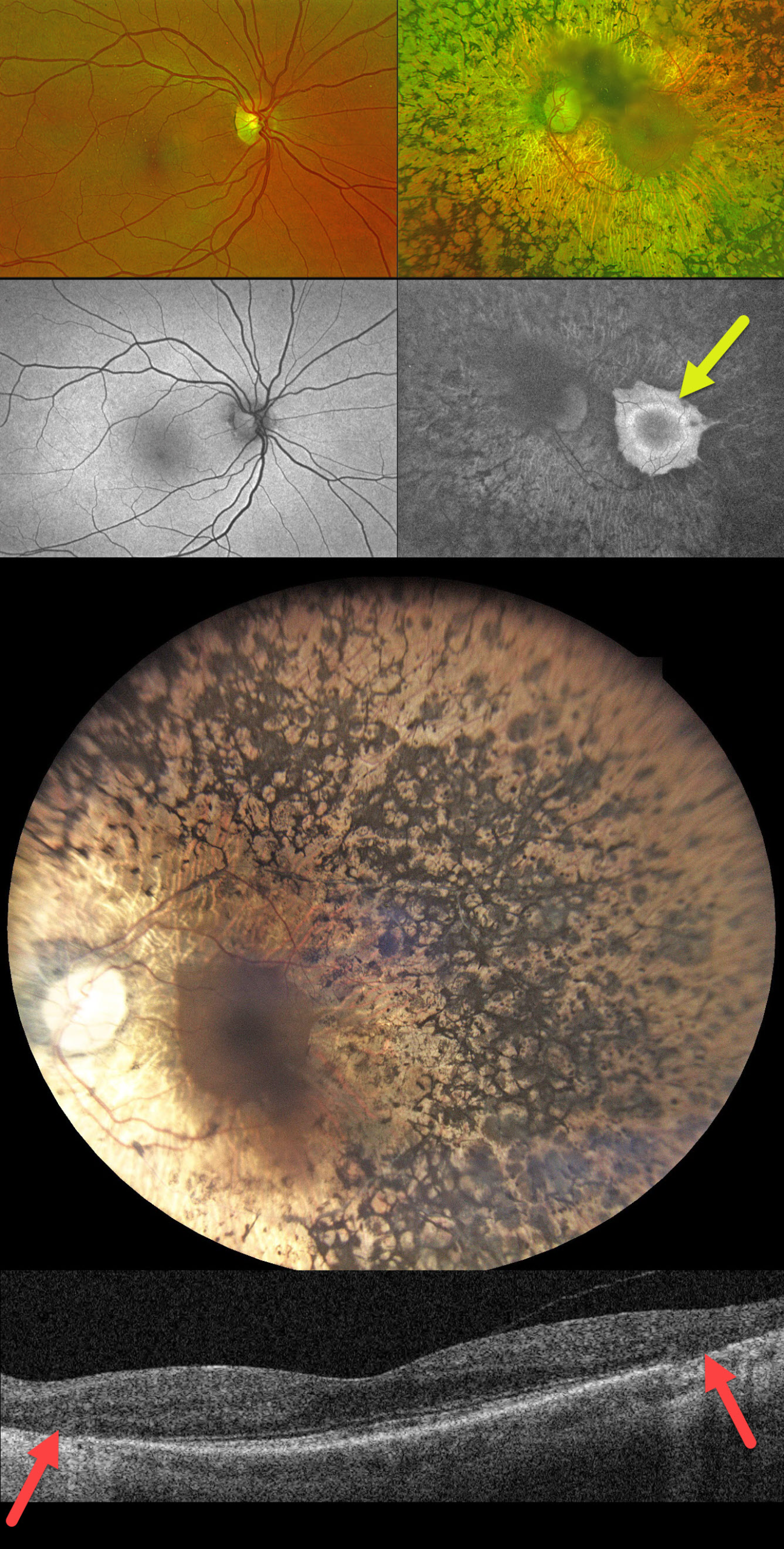
UNILATERAL PIGMENTARY RETINOPATHY
Mattie Adams
Originally posted on @retina.rocks November 19, 2025
This 55YO female has complained of some blurred vision in her left eye for about 6 years. Family history is completely negative for eye disease. Vision was 20/25 in her normal OD and 20/50 OS.
Optos color RG imaging and fundus autofluorescence (FAF) are completely normal for her right eye. The left eye shows severe apparent retinitis pigmentosa (RP) changes that extend from her peripheral macula into the periphery. FAF shows a somewhat scalloped appearance to the preserved central macula with a hyper-FAF ring around the fovea. OCT scanning shows peripheral macular outer retinal and RPE atrophy. Genetic testing was heterozygous for a pathogenic ABCC6 mutation (c.2787+1G>T, splice donor) and a benign ABCA4 variant (c.5603A>T, p.Asn1868lle).
Learning Points:
Unilateral pigmentary retinopathy, also referred to as unilateral RP, is an extremely rare disorder (Alina-Cristina et al, Romanian J Ophth 2016;60:47-52). Patients require long-term follow-up to rule out a delayed bilateral presentation of an inherited retinal disorder (IRD). Unilateral pseudo-RP, chorioretinal findings with intraretinal pigment migration, is much more common, including trauma, prior retinal detachment, and infectious or inflammatory disorders such as AZOOR. The funduscopic and FAF characteristics of our patient’s left eye strongly suggest a unilateral pigmentary retinopathy rather than one of these other imitators.
With our patient’s genetic testing showing two different heterozygous mutations for recessive disorders, her results were essentially negative. However, this does not definitively rule out an IRD. In a real-world study of the Invitae IRD panel, McClard et al yielded a positive diagnostic result in only 51% of patients (J Vitreoretinal Diseases 2022;6:351-357). Each genetic panel has its own limitations regarding which genes are tested, as well as false positives and false negatives (Pulido et al, Retina 2022;42:1-3).
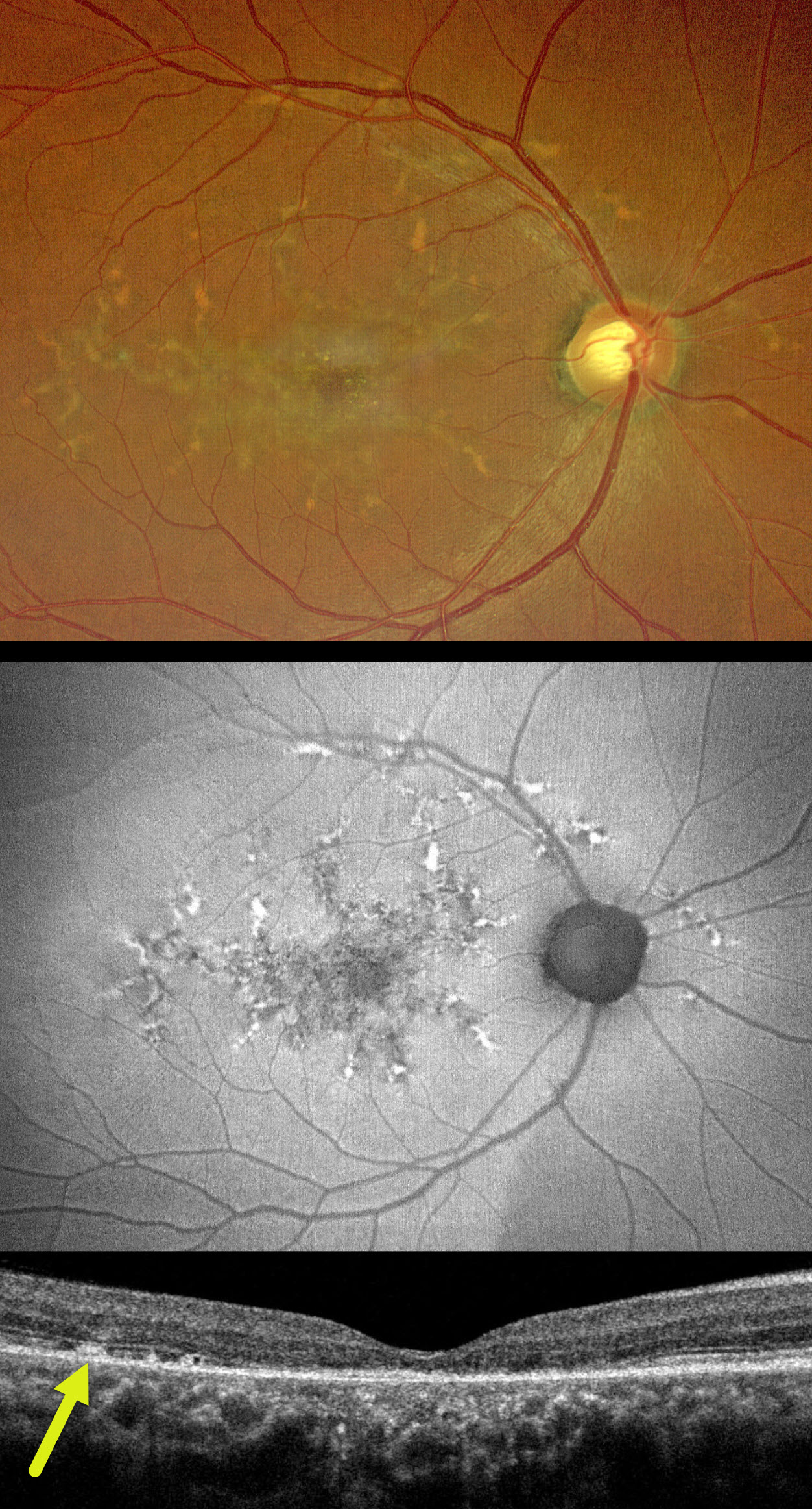
PATTERN MACULAR DYSTROPHY
Nilesh Kumar
Originally posted on @retina.rocks November 17, 2025
This healthy 50YO male presented with 10 months of bilateral vision loss. Family history was negative. Vision was 20/80 OU.
Color photography of his right eye shows submacular pigment and orange-yellow flecks radiating outwards from the fovea towards the peripheral macula and around the nerve. These lesions on fundus autofluorescence (FAF) are variably hyper- and hypo-FAF. OCT shows variable outer retinal loss. The subretinal flecks appear as hyperreflective deposits above the RPE. Identical findings are present in his left eye (not shown).
Learning Points:
Autosomal dominant pattern macular dystrophies present in mid-life with macular subretinal orange-yellow or pigmented deposits that accumulate in various patterns. Usually caused by mutations in the PRPH2 gene, the condition is classified by the pattern created by these deposits. However, they likely represent different phenotypes of the same disorder. The most common clinical presentation is adult-onset foveomacular vitelliform dystrophy, which presents as symmetrical, small, subfoveal, yellow-pigmented lesions. Our patient’s appearance is characteristic of butterfly-shaped pigment dystrophy. The visual prognosis is generally good, although some patients may develop vision loss due to secondary atrophy or macular neovascularization.
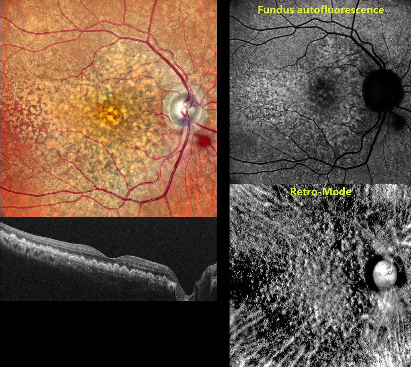
DOMINANTLY INHERITED RADIAL BASAL LAMINAR DRUSEN
Rohan Jain and Manish Nagpal
Originally posted on @retina.rocks October 28, 2025
This 58 YO female presented to us with 2 to 3 years of bilateral vision loss. There was no family history of eye disease. Vision was 20/20 OU.
Pseudocolor SLO imaging shows extensive mixed and confluent drusen extending throughout the right posterior pole. OCT scanning shows drusen indenting the overlying retina, extending into the outer nuclear layer and, in some areas, up to the outer plexiform layer. Fundus autofluorescence (FAF) shows macular hyper-FAF with surrounding relative hypo-FAF. Retro-Mode imaging reveals a dramatic, almost 3-dimensional appearance of depressions and mounds resembling the surface of the moon. Identical findings were noted in her left eye (not shown).
Learning Points:
Doyne’s, also known as dominantly inherited radial basal laminar drusen or Malattia Leventinese, is a rare macular disorder caused by a mutation in the EFEMP1 gene on chromosome 2p16, although we have seen similar phenotypes with negative genetic testing. The EFEMP1 protein is a member of the fibulin family of extracellular matrix glycoproteins. The defective protein creates an abnormally thickened RPE basement membrane. Centrally large, nodular, and confluent drusen develop, along with a temporal radiating pattern of smaller cuticular drusen. Later, there may be variable amounts of RPE hyperplasia and fibrous metaplasia. Macular neovascularization may also occur.
Fundus autofluorescence (FAF) usually shows hypo-FAF in age-related drusen. The lesions in Doyne’s, as in this patient, usually show hyper-FAF, likely from unmasking of the RPE by overlying outer retinal thinning.
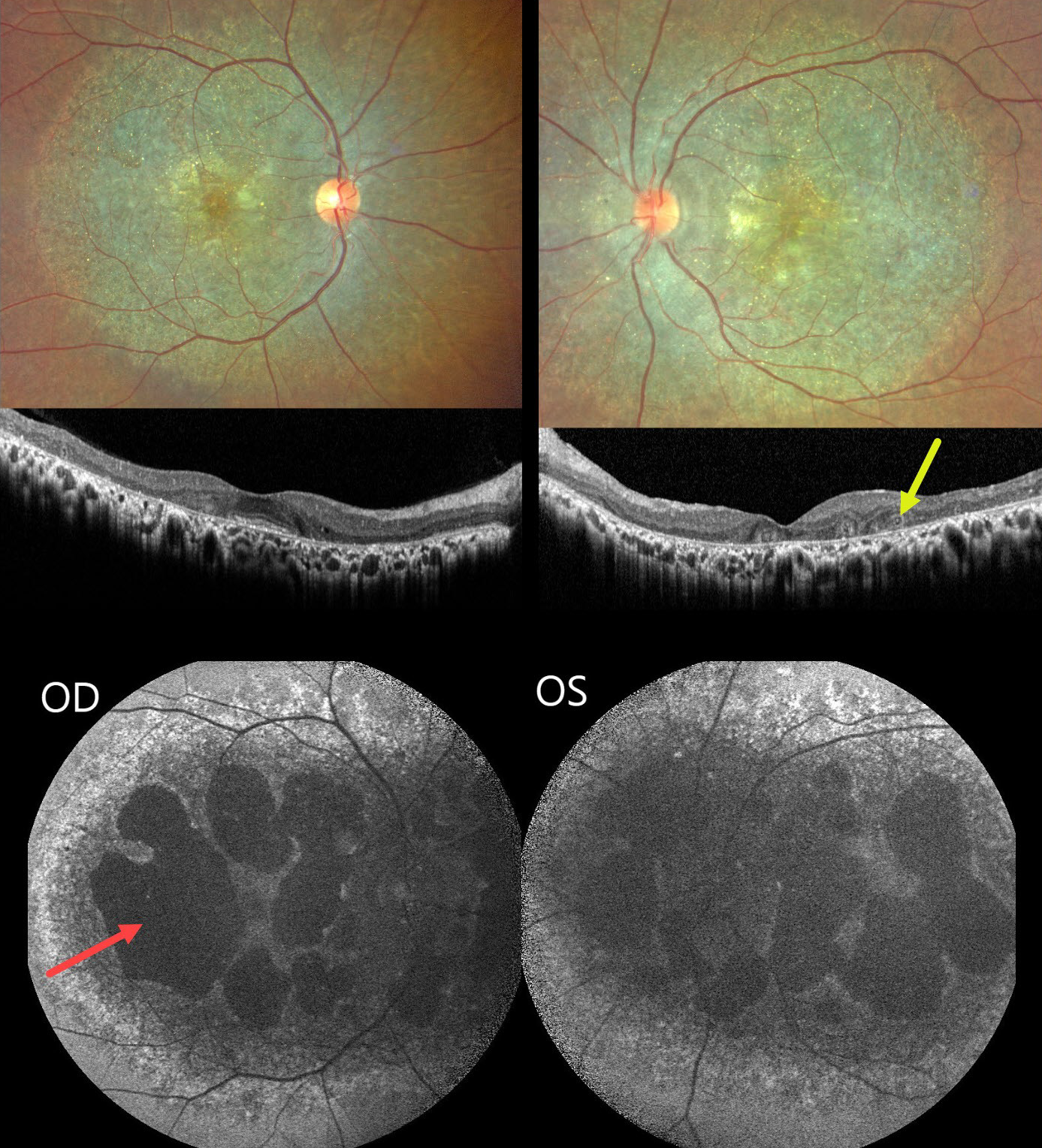
BIETTI CRYSTALLINE DYSTROPHY
The European VitreoRetina Society (EVRS) and Shishir Verghese
Originally posted on @retina.rocks October 24, 2025
This healthy 36YO female presented with a long history of bilateral vision loss with night blindness. Family history was negative. Vision was 20/80 bilaterally. Anterior segments were normal.
Fundus imaging shows extensive pigmentary degeneration with subretinal crystalline deposits involving both posterior poles. OCT scanning shows hyperreflective dots overlying the RPE-Bruch’s membrane complex with variable outer retinal and RPE loss. A few areas of outer retinal tubulation are noted temporally OS. Fundus autofluorescence (FAF) shows diffuse hypo-FAF throughout each posterior pole, dense hypo-FAF within areas of discrete atrophy, and dots of more peripheral hyper-FAF.
Learning Points:
Bietti crystalline dystrophy is an autosomal recessive ocular disorder that affects both the anterior and posterior segments. Clinical features include crystalline deposits in the retina, cornea, and rarely the crystalline lens, as well as retinal pigment epithelial clumping and atrophy. Mutations in CYP4V2 that cause dysregulated lipid metabolism have been implicated in its pathogenesis. While there is no definitive treatment, anti-VEGF therapy can be administered for secondary macular neovascularization. See Saatci et al for a recent review (Clinical Ophthalmology 2023;17:953-967).
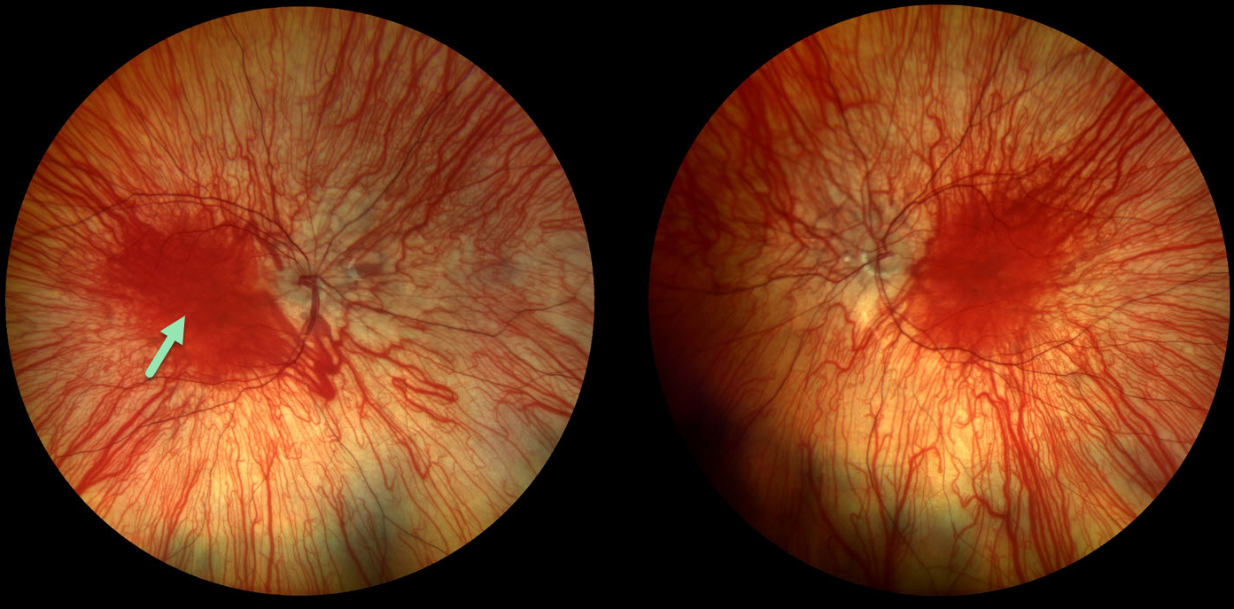
OCULOCUTANEOUS ALBINISM
Nilesh Kumar
Originally posted on @retina.rocks September 23, 2025
This 14YO female has a history of nystagmus and vision loss from oculocutaneous albinism. There was no significant family history. Vision was 20/120 OU.
Color photography shows bilateral marked choroidal hypopigmentation with foveal hypoplasia. No genetic testing or pedigree analysis was performed.
Learning Points:
Albinism is a group of genetic disorders characterized by abnormal melanin production due to faulty amino acid production. Patients have either only eye involvement (ocular albinism) or eye and skin (oculocutaneous albinism). Inheritance patterns include autosomal dominant, autosomal recessive, and X-linked. Ocular findings include strabismus, nystagmus, iris transillumination defects, blonde fundus, and foveal hypoplasia. There are often a higher number of crossed nerve fibers at the optic chiasm.
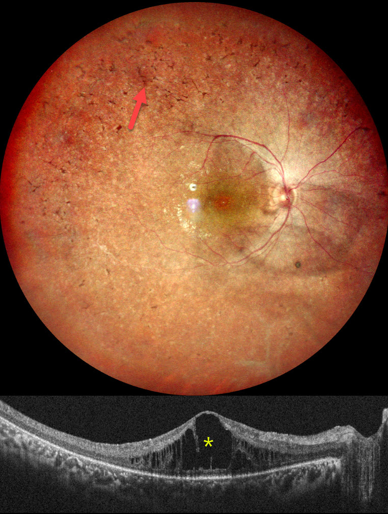
RETINITIS PIGMENTOSA WITH CME
Rohan Jain and Manish Nagpal
Originally posted on @retina.rocks September 15, 2025
This healthy 18YO female complained of night blindness for a few years. Her mother and sister have a history of retinitis pigmentosa (RP). Vision was 20/90 OU.
Pseudocolor SLO imaging of her right eye shows typical RP scarring with intraretinal pigment migration extending from the macula into the retinal periphery. There is loss of the foveal reflex and cystoid macular edema (CME). Identical findings were noted in the OS (not shown).
Learning Points:
RP is a clinically and genetically heterogeneous group of inherited retinal disorders, which can present sporadically or with any inheritance pattern (autosomal dominant, autosomal recessive, sex-linked, or mitochondrial). It is characterized by diffuse, progressive dysfunction of predominantly rod photoreceptors, followed by RPE degeneration with intraretinal pigment migration. Visual impairment usually presents as night blindness and progressive visual field loss.
CME is observed in up to 20% of RP patients, often without angiographic leakage. Pathological mechanisms include vitreous traction/epiretinal membranes or a breakdown of the blood-retinal barrier due to inflammation or RPE/Muller cell dysfunction (Gaudric et al, Progress in Retinal and Eye Research 2022;91:101092). First-line treatment is topical or oral carbonic anhydrase inhibitors (CAIs). Other options for refractory CME include steroids (topical, periocular, or intravitreal), anti-VEGF injections, or vitrectomy for a tractional component. Our patient was given topical CAIs but was immediately lost to follow-up.
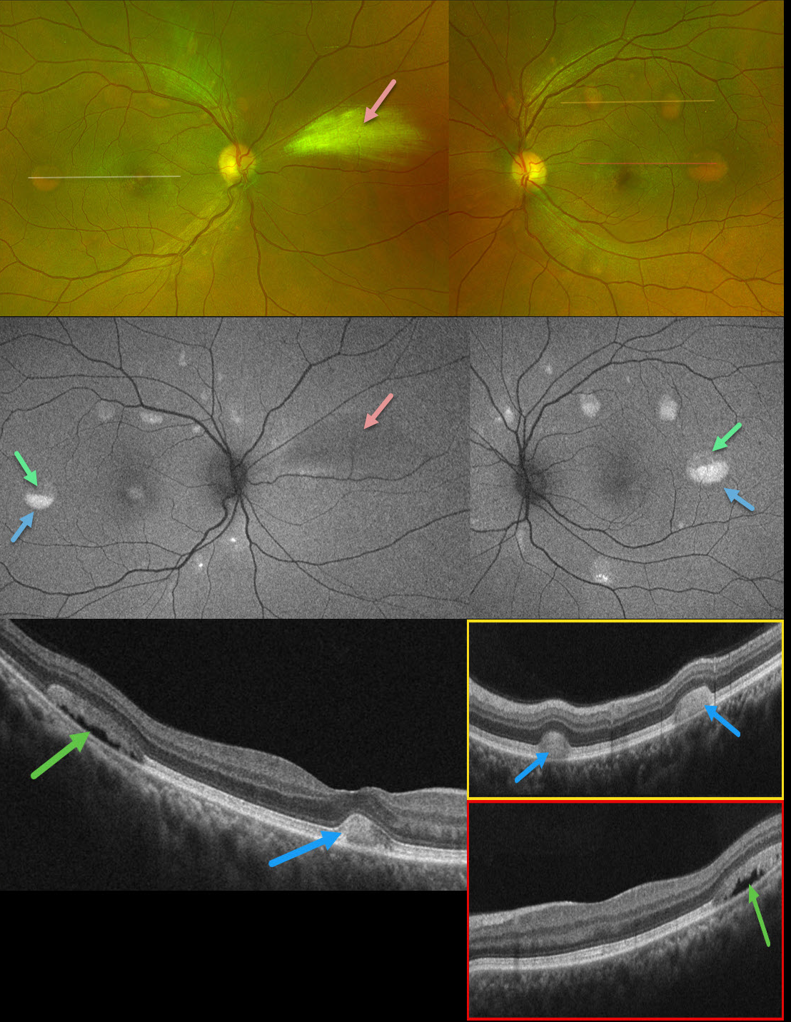
BEST DISEASE WITH MYELINATED NERVE FIBER LAYER
The European Society (EVRS) and Vaibhav Sethi
Originally posted on @retina.rocks July 25, 2025
This 39YO male presented with asymptomatic retinal lesions. Family history was negative. Vision was 20/20 OU.
Optos color RG imaging shows bilateral partially scrambled vitelliform lesions in each posterior pole. On OCT, the superior, scrambled portion of these lesions is hyporeflective, and the inferior, yellow portion is hyperreflective. On fundus autofluorescence (FAF), the superior portions are hypo-FAF, and the inferior vitelliform material is hyper-FAF. The incidental area of the nasal myelinated nerve fiber layer OD is hypo-FAF from blocking the underlying RPE.
Learning Points:
Vitelliform lesions are characterized by the accumulation of yellow subretinal material thought to be caused by impaired metabolism of photoreceptors or retinal pigment epithelium. They can be associated with a wide variety of retinal disorders, including Best vitelliform macular dystrophy (BVD, our patient), adult-onset foveomacular dystrophy, acute exudative polymorphous vitelliform maculopathy, as well as paraneoplastic, toxic, tractional, and degenerative etiologies (Iovino et al, Surv Ophthalmology 2023;68:361-379).
BVD is usually transmitted as an autosomal-dominant disorder due to a mutation in the BEST1 gene, which encodes bestrophin-1. Bestrophin-1, a calcium-activated chloride channel, is primarily found in the basolateral plasma membrane of the RPE. Unfortunately, our patient denied genetic testing due to cost.
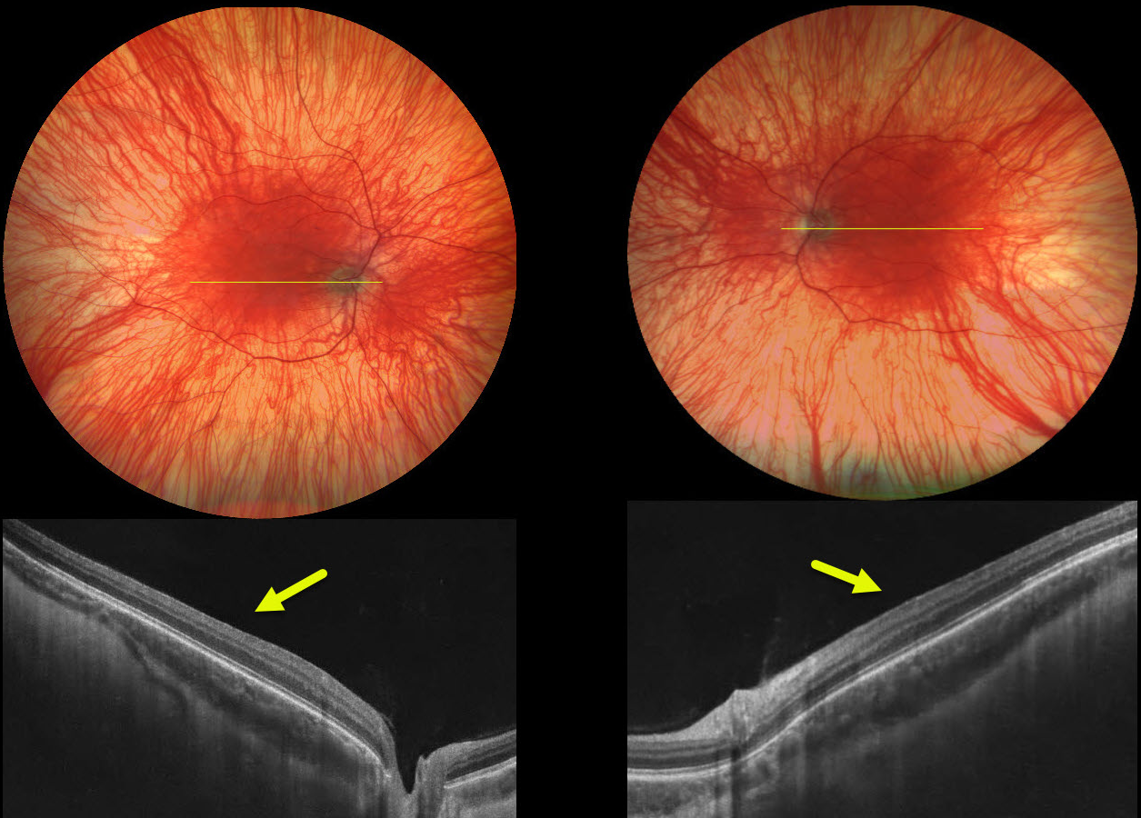
OCULOCUTANEOUS ALBINISM
Ayushi Gupta and Vishal Agrawal
Originally posted on @retina.rocks July 21, 2025
This 25YO male has a history of nystagmus and vision loss from oculocutaneous albinism. Her grandfather also had albinism. Vision was 20/70 bilaterally.
Color photography shows bilateral marked choroidal hypopigmentation with foveal hypoplasia. OCT scanning shows the absence of the normal foveal depression.
Learning Points:
Albinism is a group of genetic disorders characterized by abnormal melanin production due to faulty amino acid production. Patients have either eye-only involvement (ocular albinism) or eye and skin involvement (oculocutaneous albinism). Inheritance patterns include autosomal dominant, autosomal recessive, and X-linked. Ocular findings include strabismus, nystagmus, iris transillumination defects, blonde fundus, and foveal hypoplasia. There are often a higher number of crossed nerve fibers at the optic chiasm.
Fovea plana is characterized by the absence of the foveal pit, which is formed embryologically by the inner retinal tissues and vasculature being displaced centrifugally. It is most commonly associated with albinism, prematurity, or as an isolated finding. Despite the abnormal foveal architecture, vision is usually normal, unless due to albinism.
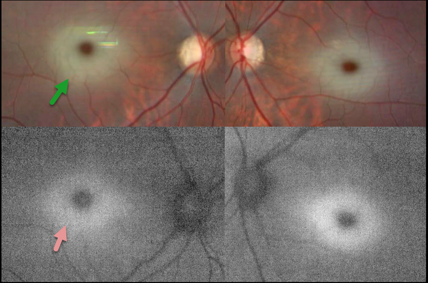
TAY-SACHS DISEASE
The European VitreoRetina Society (EVRS) and David Aggarwal
Originally posted on @retina.rocks July 18, 2025
This 15-month-old girl was brought to a multi-specialty hospital by her parents with complaints of fever, seizures, and progressive weakness in her limbs for two days. She was admitted to the pediatric intensive care unit, and symptomatic treatment was started. She was the firstborn of a third-degree consanguineous marriage.
Fundus imaging is blurred due to constant eye movement, but shows a symmetric central macular whitish appearance surrounding an orange- brown foveal center. On fundus autofluorescence (FAF), this white material is hyper-FAF. Whole genome sequencing revealed GM2-gangliosidosis, AB variant (Tay-Sachs disease).
Learning Points:
A cherry-red spot refers to a reddish-brown appearance of the normal macular center, which is surrounded by opaque white parafoveal retina. This is usually caused by an acute central retinal artery occlusion (CRAO), but it is also a common feature of sphingolipidoses, a group of lysosomal metabolic disorders that cause abnormal lipid accumulation in retinal ganglion cells. As the foveal pit lacks the ganglion cell layer, it retains its normal transparency, giving a cherry-red appearance.
In Tay-Sachs disease, GM2 trihexosylceramide accumulates due to a deficiency of the A component of hexosaminidase. This material is hyper-FAF (Abalem et al, JAMA Ophthalmology 2014;132:876), unlike a CRAO, which appears hypo-FAF due to blockage of the underlying RPE.
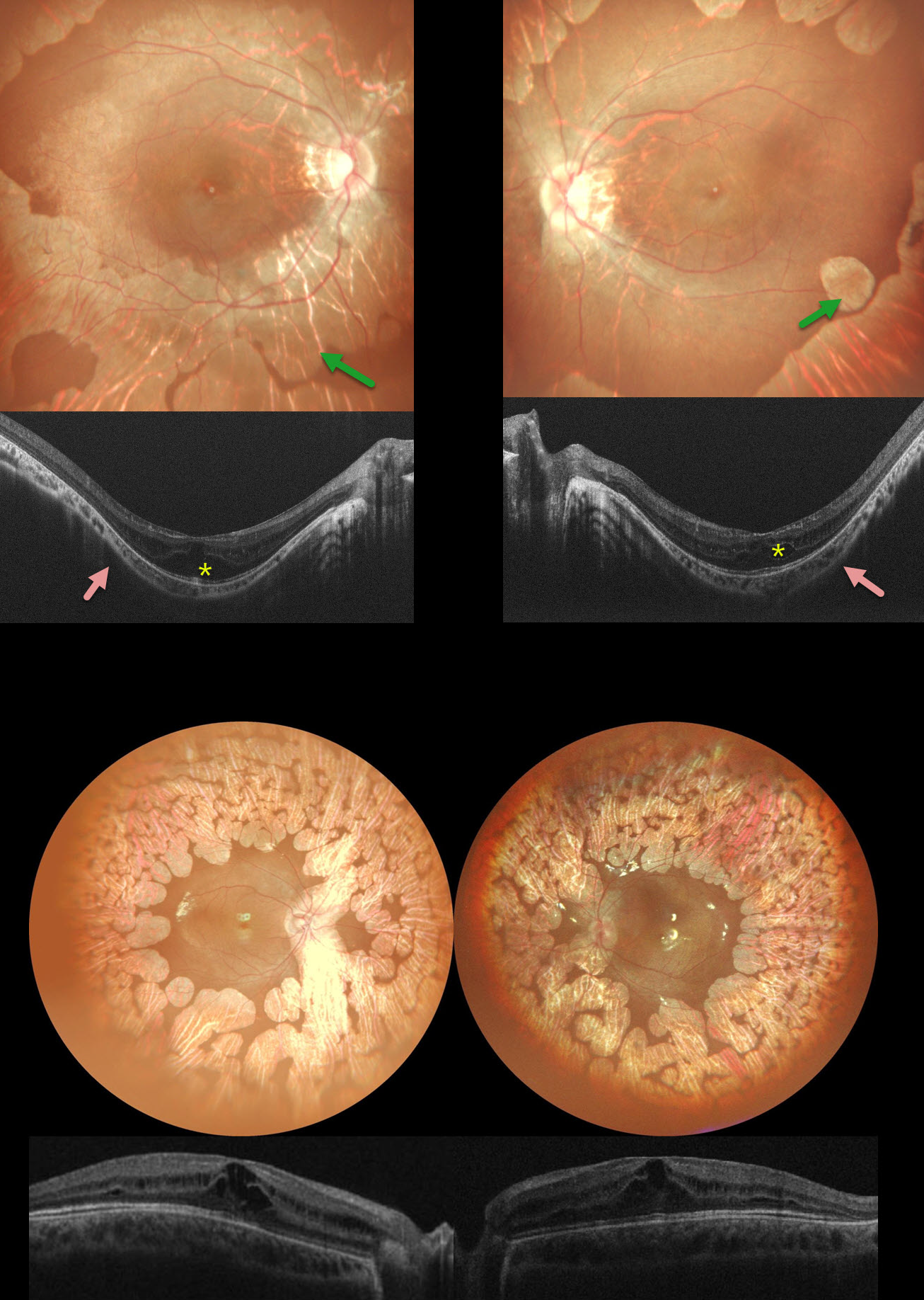
GYRATE ATROPHY
Anju Bansal
Originally posted on @retina.rocks June 30, 2025
This 6YO girl has a history of night blindness, and her twin brother has a history of milestones delay and seizures. Family history was suggestive of consanguinity. The girl’s vision was 20/120 OD and 20/80 OS, and 20/120 OU in the boy.
The girl’s fundus photography shows variably coalescent, scalloped areas of bilateral chorioretinal atrophy extending from the peripheral maculas into each periphery. OCT shows bilateral cystic retinal edema primarily in the inner and outer nuclear layers. There are shallow poster staphylomas. Her brother shows similar findings, but without the staphylomas.
Blood tests of both siblings revealed a deficiency of the ornithine aminotransferase (OAT) enzyme. Topical non-steroidal anti- inflammatory eye drops were started along with an arginine-free diet and B6 supplementation.
Learning Points:
Gyrate atrophy is a rare autosomal recessive retinal dystrophy, characterized by extensive areas of lobular peripheral chorioretinal degeneration that gradually spread posteriorly. Macular involvement on OCT is common, with foveal thickening in younger patients followed by atrophy with outer retinal tubulation later in life (Sergouniotis et al, Ophthalmology 2012;119:596-605).
End-stage disease can be indistinguishable from choroideremia. Other findings include myopia and night blindness. The degeneration is caused by a lack of a vitamin B6-dependent ornithine aminotransferase mitochondrial enzyme, which leads to elevated ornithine levels. Vitamin B6 supplementation and an arginine-restricted diet
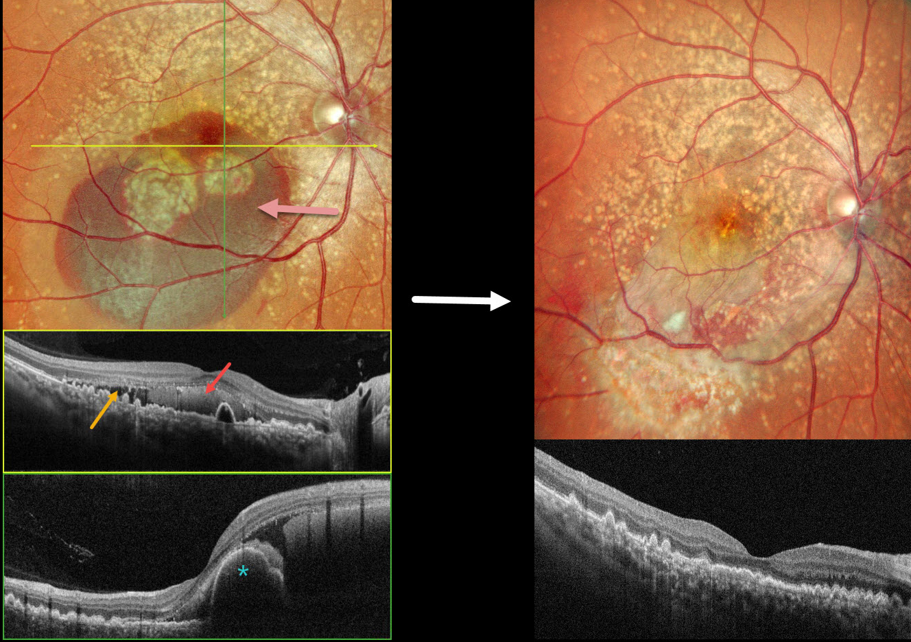
DOMINANTLY INHERITED RADIAL BASAL LAMINAR DRUSEN WITH POSSIBLE POLYPOIDAL CHOROIDAL VASCULOPATHY
Malvika Singh and Manish Nagpal
Originally posted on @retina.rocks June 25, 2025
This 49YO female presented with 3 days of decreased vision in her OD. Vision was 20/70 OD and 20/15 OS. There was no family history of eye disease.
Pseudocolor SLO imaging shows extensive mixed and confluent drusen extending through each macula into the midperipheries. Inferior submacular blood extends into the right foveal center. OCT scanning shows extensive conically shaped drusen. The subretinal blood in her right eye is hyperreflective, with additional temporal subretinal fluid. A bilobed nodular RPE detachment is present within the nasal blood.
She underwent pars plana vitrectomy, fluid-air exchange, and intravitreal Avastin. One month postoperatively, vision improved to 20/15 OD with resolved fluid and trace residual blood.
Learning Points:
Doyne’s honeycomb macular dystrophy, also known as dominantly inherited radial basal laminar drusen or Malattia Leventinese, is a rare macular disorder caused by a mutation in the EFEMP1 gene on chromosome 2p16. The EFEMP1 protein is a member of the fibulin family of extracellular matrix glycoproteins. The defective protein creates an abnormally thickened RPE basement membrane.
Centrally large, nodular, and confluent drusen are noted, along with a temporal radiating pattern of smaller cuticular drusen. Later, there may be variable amounts of RPE hyperplasia and fibrous metaplasia. Macular neovascularization, as in this case, may also develop. Our patient’s pre-op right OCT was suggestive of polypoidal choroidal vasculopathy, although this is not a known association with Doyne’s.
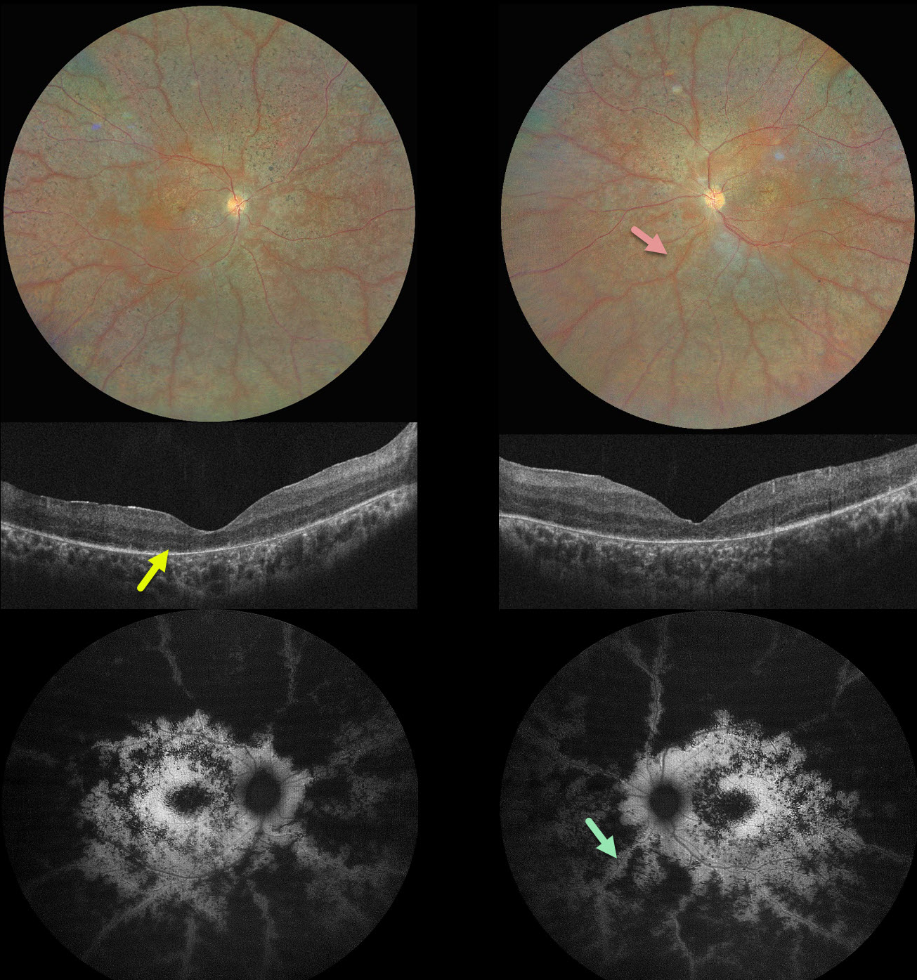
PRESERVED PARA-ARTERIOLE RPE IN RP
Ayushi Gupta and Vishal Agrawal
Originally posted on @retina.rocks June 16, 2025
This 16YO male complained of night blindness for a few years. There was no family history of eye disease. Vision was 20/200 OU.
Color photography shows bilateral findings consistent with retinitis pigmentosa (RP), including diffuse chorioretinal scarring, intraretinal pigment migration, and narrow retinal vessels. The retinal tissue beneath and immediately surrounding the arterioles is relatively spared, and this is most dramatically seen on fundus autofluorescence. OCT scanning shows variable loss of the outer retinal bands and RPE. Our patient could not afford genetic testing and was immediately lost to follow-up.
Learning Points:
Preserved para-arteriole retinal pigment epithelium (PPRPE) in retinitis pigmentosa, originally described by Heckenlively (Br J Ophthalmol 1982;66:26-30), has the unique phenotype, as seen in our patient, of diffuse RP degeneration with relative sparing of the RPE beneath the retinal arterioles. It was later found to be caused by a mutation in the crumbs cell polarity complex component 1 (CRB1) gene (den Hollander et al, Nature Genetics 1999;23:217-221). CRB1 is part of a group of proteins that help determine the structure and orientation of photoreceptors. It may also be involved in forming connections between different types of retinal cells.
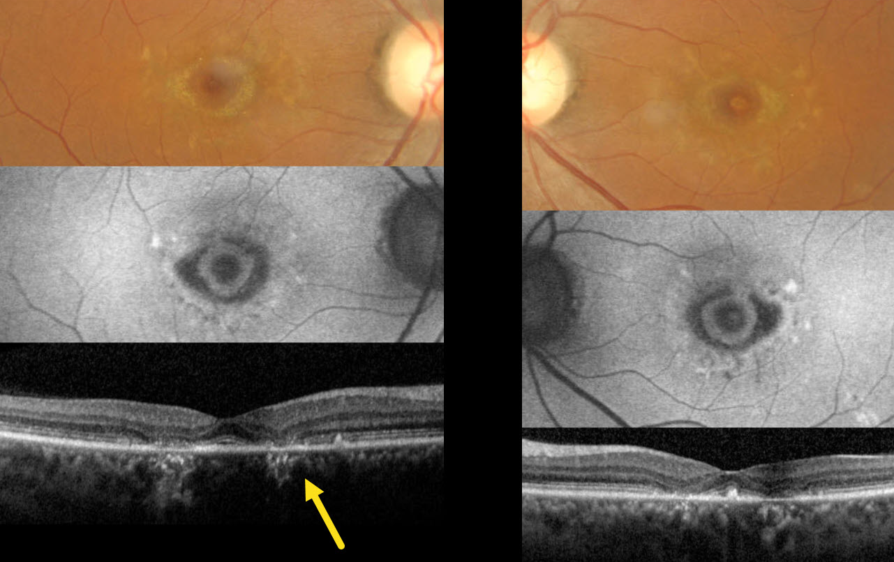
BULLSEYE MACULOPATHY PROBABLY FROM ABCA4 MUTATION
Moazzam Parvez and Krishnendu Nandi
Originally posted on @retina.rocks June 11, 2025
This 40YO female presented with 5 years of stable, mild, distorted vision bilaterally. There was no family history of eye disease. Vision was stable at 20/20 OU.
Color imaging shows bilaterally symmetric bullseye lesions with surrounding subretinal yellow macular flecks. On fundus autofluorescence (FAF), these lesions are hypo-FAF with an inner hyper-FAF ring. Pericentral outer retinal atrophy is noted on OCT.
The funduscopic appearance is most consistent with ABCA4 disease, including Stargardt disease and fundus flavimaculatus. The patient declined genetic testing due to cost concerns, and continued observation was recommended.
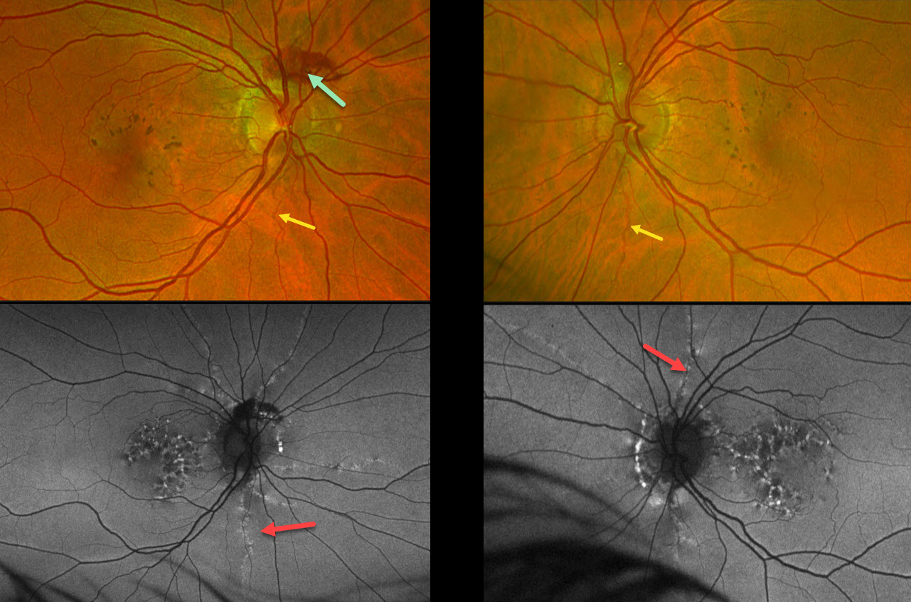
PSEUDOXANTHUM ELASTICUM WITH ANGIOID STREAKS AND PATTERN DYSTROPHY PHENOTYPE
Mattie Adams
Originally posted on @retina.rocks June 4, 2025
This 53YO female was referred for asymptomatic macular findings. Her mother has a history of pseudoxanthoma elasticum (PXE). Vision was 20/25 OD and 20/40 OS.
Optos color RG imaging shows coarse subretinal pigment clumps bilaterally. Subretinal blood is noted along the superior aspect of the right nerve from a peripapillary choroidal neovascularization. A few barely visible angioid streaks radiate from each nerve.
Fundus autofluorescence (FAF) shows variable hyper- and hypo-FAF flecks. The angioid streaks appear as hyper-FAF lesions radiating outwards from each nerve.
Learning Points:
Retinal findings in PXE most commonly include angioid streaks and a subretinal peaux d’orange appearance, most pronounced in each temporal macula. However, pattern dystrophy-like changes, as in our patient, can sometimes be the predominant phenotype (Murro et al, Graefe’s 2020;258:1881-1892). As with typical pattern dystrophies, the findings are dynamic and best captured with multimodal imaging.
PXE is caused by mutations in the ABCA6 gene, and our patient tested positive for homozygous pathogenic ABCA6 mutations. PXE is virtually always transmitted as an autosomal recessive disorder, although autosomal dominant transmission may rarely occur (Plomp et al, Am J Med Genet A 2004;126A:403-412).
POSSIBLE FAMILIAL EXUDATIVE VITREORETINOPATHY (FEVR)
Mattie Adams
Originally posted on @retina.rocks May 29, 2025
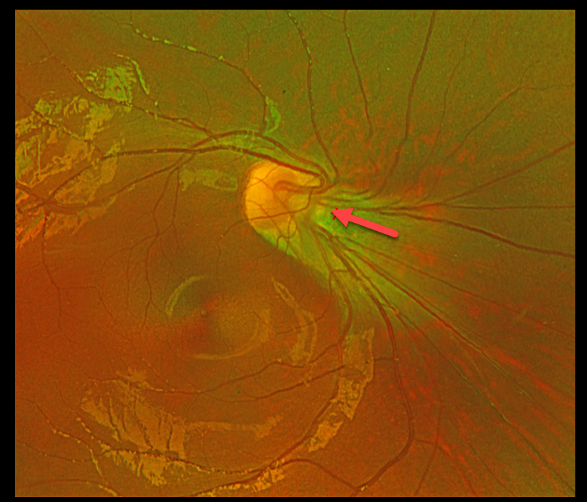
This healthy 9YO boy was referred for an asymptomatic abnormal right optic nerve. There was no ocular family history. Vision was 20/150 OD and 20/30 in his normal OS.
Optos color RG imaging shows an anomalous right optic nerve, which is tilted and displaced superonasally from its apparent normal location. The fovea is markedly ectopic, located about 60 degrees inferotemporal to the nerve. Genetic testing revealed a heterozygous FZD4 variant of uncertain significance.
Learning Points:
Originally described by Criswick and Schepens (AJO 1969;58:578-594), familial exudative vitreoretinopathy (FEVR) can be inherited as an autosomal-dominant, recessive, or X-linked trait with high penetrance and variable expressivity. There are numerous genes associated with FEVR, including LRP5, FZD4, NDP, TSPAN12, ZNF408, CTNNB1 and KIF11 [Tao et al, Invest Ophthalmol Vis Sci 2021;62(15);4].
FEVR is characterized by peripheral temporal retinal avascularity, lipid exudation, neovascularization, tractional retinal detachment, and temporal dragging of the macula and retinal vessels. These findings are somewhat like those found with retinopathy of prematurity.
STELLATE NON-HEREDITARY IDIOPATHIC FOVEOMACULAR RETINOSCHISIS
Originally posted on @retina.rocks May 7, 2025
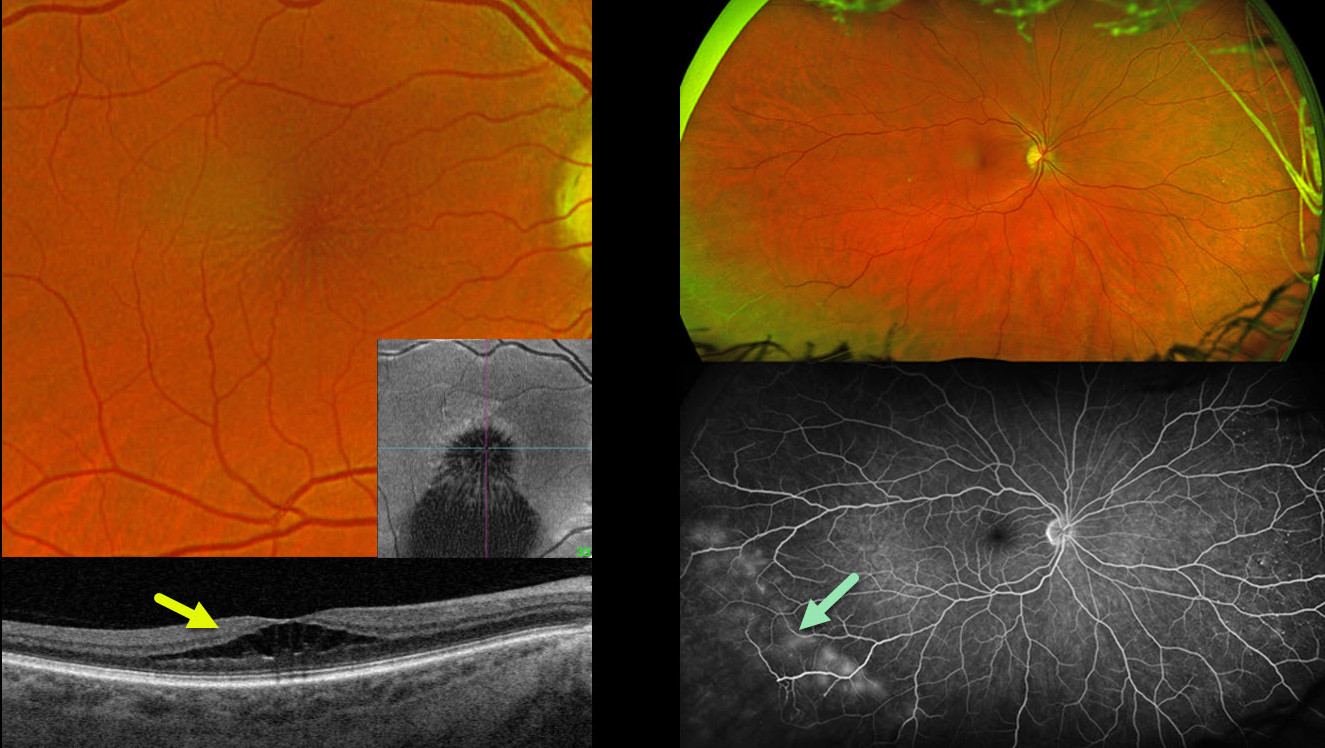
This 58YO female presented with 6 months of blurred vision in her right eye. Vision was 20/40 OD and 20/25 OS.
Optos color RG imaging of her right eye shows a stellate pattern of foveal schisis, which is confirmed on Triton swept-source B-scan and en face OCT. The macular appearance and OCT in her left eye were normal (not shown). Inferotemporal retinoschisis is noted OU peripherally. The peripheral schisis shows late leakage angiographically. Genetic testing was heterozygous for a pathogenic USH2A mutation.
Learning Points:
A new classification of stellate nonhereditary idiopathic foveomacular retinoschisis (SNIFR) was initially described by Ober et al (Ophthalmology 2014;121:1406-1413). Unlike X-linked schisis, which, in our experience, mostly affects the inner and outer nuclear layers bilaterally, SNIFR causes splitting of the outer plexiform layer, usually in just one eye. Many eyes will have peripheral retinoschisis, even when the macula is normal (Bloch et al, Retina 2021;41:2361-
MATERNALLY INHERITED DIABETES AND DEAFNESS (MIDD)
Originally posted on @retina.rocks April 17, 2025
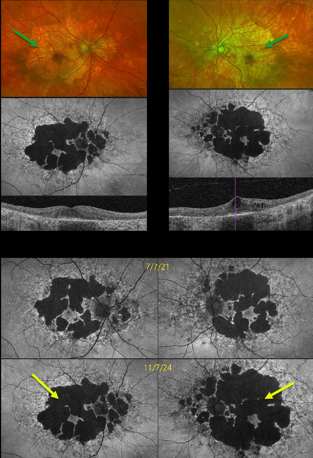
This 58YO male presented with several years of progressive vision loss. He has a history of well-controlled type 2 diabetes. Vision was 20/30 OD and 20/60 OS.
Optos color RG imaging shows fairly symmetric areas of variable, confluent, non-central macular atrophy. Fundus autofluorescence (FAF) shows hypo-FAF from the areas of atrophy, along with areas of hyper-FAF around each nerve and macula. Swept-source OCT shows outer retinal and RPE atrophy bilaterally with central cystic edema OS. FAF shows significant progression of atrophy compared with 3 years earlier. The subclinical edema OS was felt to be diabetic and has been observed since it was spontaneously fluctuating with unchanged vision and symptoms.
Learning Points:
Maternally inherited diabetes and deafness (MIDD) accounts for up to 3% of all cases of diabetes and results from the A3243 G mutation in mitochondrial DNA. MIDD often masquerades as a pattern macular dystrophy. In our experience, fundus autofluorescence (FAF) is the best way to visualize these changes. The FAF appearance somewhat resembles that seen with Elmiron toxicity. Peripapillary hypoautofluorescence, more densely packed macular autofluorescent changes, and earlier central macular involvement suggest Elmiron toxicity over other causes (Barnes et al Ophthalmology Retina 2020;4:1196-1201).
Despite our patient’s multimodal findings being classical for MIDD, mitochondrial genetic testing revealed variants of uncertain significance (homozygous AMPD1 and heterozygous ACAD5). Invitae Inherited Retinal Disorders Panel revealed homozygous variants of u
ALPORT SYNDROME
Fraser McKay
Originally posted on @retina.rocks April 9, 2025
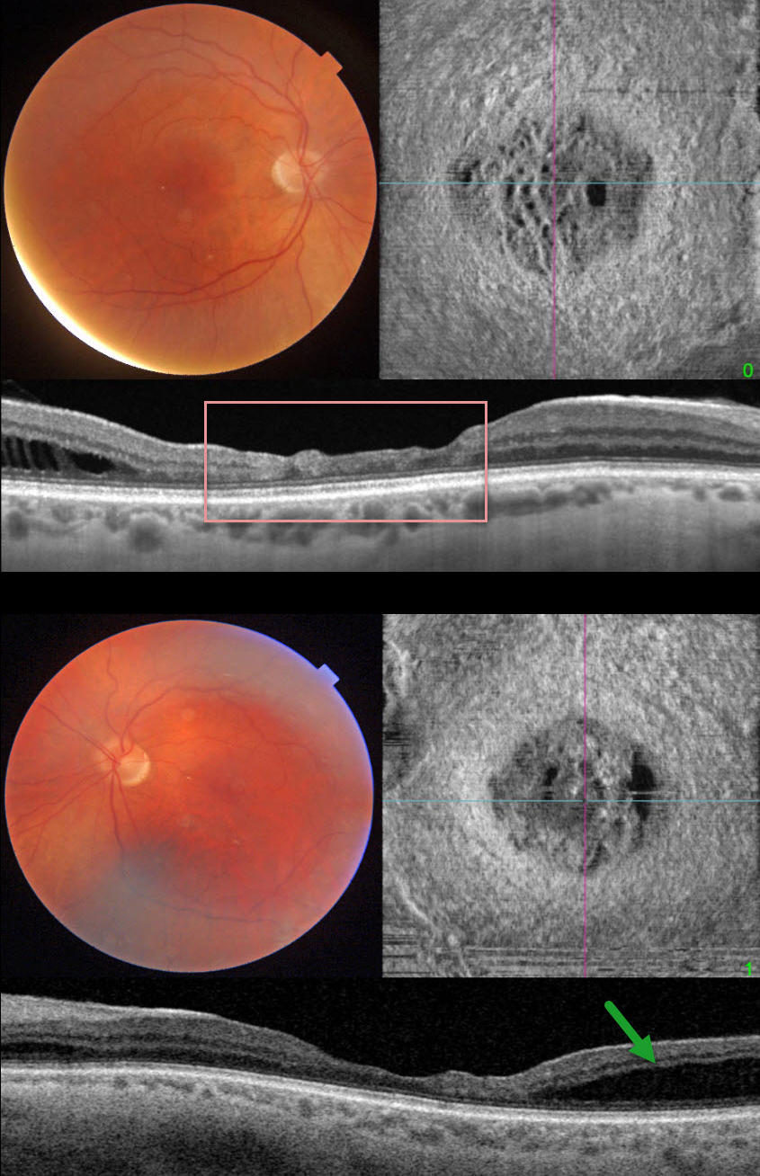
This 46YO male presented with increasing difficulties with watching TV and working on the computer. He had a known history of Alport syndrome, including renal failure with prior renal transplantation and hearing loss. Vision was 20/40 OD and 20/50 OS. Slit lamp examinations showed bilateral cataracts without anterior lenticonus. Cataract surgery was performed, and vision remarkably improved to 20/20 OU.
Triton color imaging shows a blunted foveal reflex. Swept-source OCT shows central inner macular thinning and disorganization. Outer nuclear layer schisis is noted temporally. En face OCT reveals another view of these central macular findings. Similar changes are noted in his left eye.
Learning Points:
Alport syndrome is a rare genetic disorder, most commonly inherited in an X-linked recessive pattern with mutations in the COL4A5 gene, which encodes for collagen type IV alpha. Basement membrane thinning and lamellation cause hemorrhagic nephritis and renal failure, sensorineural hearing loss, and ocular findings, including corneal abnormalities and anterior lenticonus. Retinal findings include macular and peripheral retinoschisis and macular yellow-white perimacular dots and flecks in the inner retina.
In more severe forms, a dull macular reflex with inner macular thinning (lozenge sign), best seen with red-free imaging, can occur (Colville et al, Br J Ophthalmol 2009;93:383-386). Multimodal imaging, including wide-field OCT, shows a spectrum of internal limiting membrane (ILM) findings, including ILM granularity, progressive ILM lamellation, retinal nerve fiber layer (RNFL) dehiscence, vascular lacunae, and course arrangement of RNFL toward the disc (Cicinelli et al, Retina 2022;42:274-282).
ADULT-ONSET FOVEOMACULAR VITELLIFORM DYSTROPHY
Tejaswita Verma and Manish Nagpal
Originally posted on @retina.rocks February 4, 2025
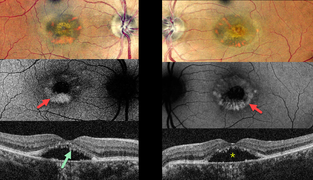
This 62YO female complained of stable bilateral blurred vision for seven years, having been diagnosed with central serous chorioretinopathy elsewhere. Vision was 20/30 OD and 20/40 OS.
Pseudocolor SLO imaging shows symmetrical, partially scrambled vitelliform lesions. The lesions are hyperautofluorescent along their outer edges and hypoautofluorescent centrally. OCT shows shaggy hyperreflective material lining the outer retina overlying foveal subretinal fluid.
Learning Points:
Adult-onset foveomacular vitelliform dystrophy is characterized by bilateral, symmetrical, grayish-yellow, one-third to one-half disc diameter subfoveal lesions with a central pigmented spot. In our experience, it is the most common pattern of dystrophy and closely resembles Best disease, although the lesions are generally smaller and diagnosed in adulthood. Causative mutations include BEST1, PRPH2, IMPG1 and IMPG2.
For a great review article, see Chowers et al, Progress in Retinal and Eye Research 2015;47:64-85.
X-LINKED RETINOSCHISIS
Anand Temkar and Manish Nagpal
Originally posted on @retina.rocks January 28, 2025
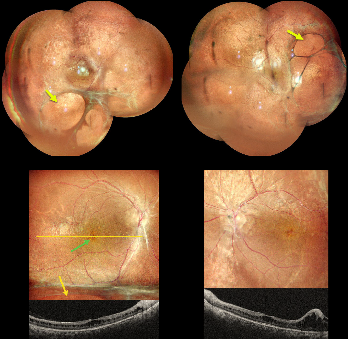
This 15YO boy presented with a lifelong history of decreased vision. His father and paternal grandfather also had poor vision but were unavailable for examination. Vision was 20/80 OD and 20/120 OS.
Pseudocolor SLO imaging shows a central, radially oriented foveal schisis with an inferior schisis hole OD. OCT scanning confirms foveal schisis, which primarily involves the inner nuclear layer. Larger areas of inner schisis extend inferiorly OD and temporally OS.
Learning Points:
X-linked retinoschisis is caused by a mutation of the RS1 gene, which is located on the X chromosome and encodes for retinoschisin, a protein secreted by photoreceptors that is involved in intercellular adhesion and likely retinal cellular organization (Heymann et al, Progress Retinal Eye Research 2023;95:101147). Although usually transmitted as an X-linked recessive disorder in males with an incidence of 1 in 15,000 to 30,000, it can also sometimes affect females.
The characteristic features include foveoschisis and peripheral schisis with vitreous veils. Over time, the macular schisis flattens with secondary atrophy. Vision usually levels off at about 20/100. Patients can develop spontaneous vitreous hemorrhage and rhegmatogenous retinal detachment from peripheral schisis. Although there is no specific treatment, topical carbonic anhydrase inhibitors may decrease the foveal thickness and possibly help minimize foveal atrophy (see Andreuzzi et al Retina 2017;37:1555-1561).
CHOROIDAL COLOBOMA
Anand Temkar and Manish Nagpal
Originally posted on @retina.rocks January 16, 2025
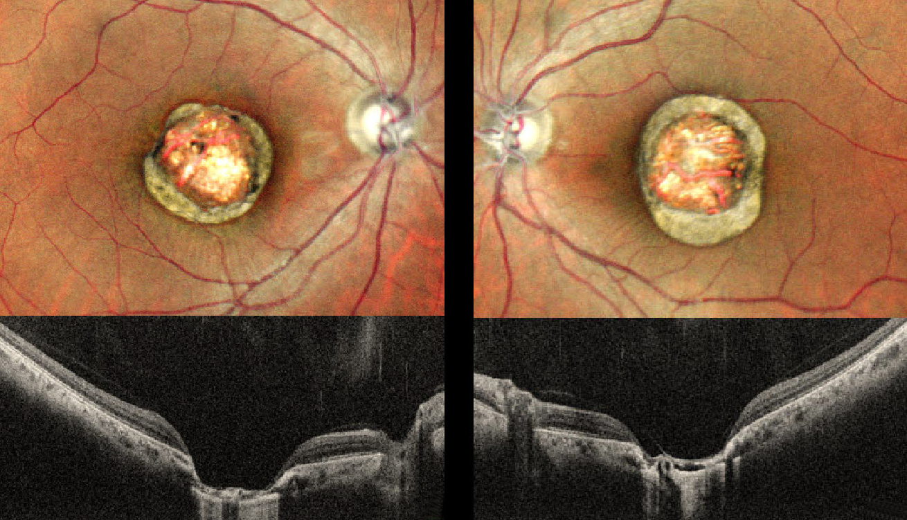
This healthy 24YO male presented with a lifelong history of stable bilateral vision loss. Vision was 20/80 OU.
Pseudocolor SLO imaging shows symmetrical macular colobomas. OCT scanning shows near-total choroidal loss within the coloboma, with a thinned, atrophic retina draped against bare sclera. Prior serologic testing elsewhere was negative for both toxoplasmosis and cytomegalovirus.
Learning Points:
Choroidal colobomas are most commonly due to failure of the optic vesicle and choroidal fissure to close during embryonic development and are usually found inferonasally. They can present as solitary or multiple lesions.
The differential diagnosis for congenital macular coloboma also includes post-inflammatory lesions (toxoplasmosis, cytomegalovirus, and Zika) and retinal dystrophies, including North Carolina macular dystrophy (NCMD). The retina within the coloboma is variably thinned, increasing the risk of rhegmatogenous retinal detachment (RRD).
The symmetry of the lesions argues for NCMD, but unfortunately, genetic testing was not performed due to cost.
RETINITIS PIGMENTOSA WITH MACULAR COLOBOMA
Ankit Jain and Manish Nagpal
Originally posted on @retina.rocks January 7, 2025
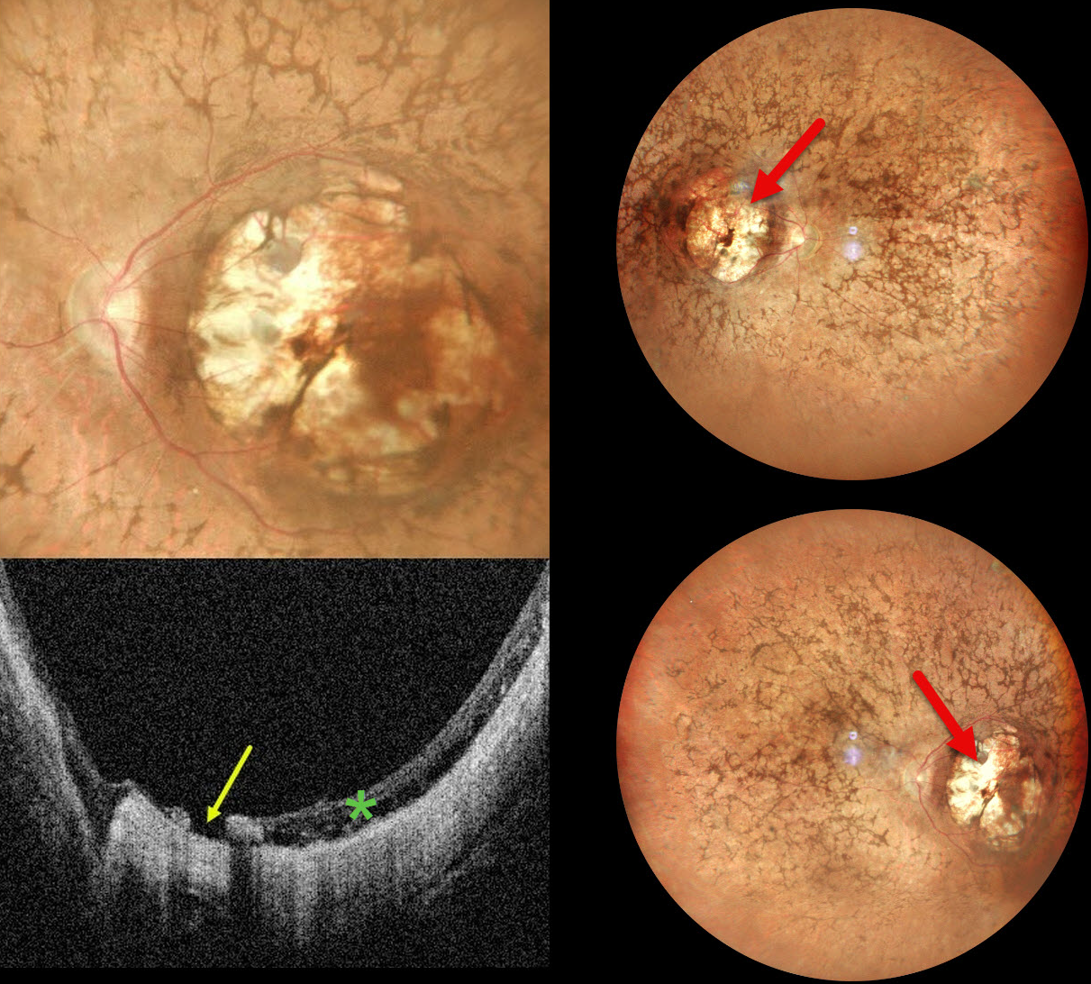
This 32YO female was born with severe vision loss and nystagmus. There was no family history of eye disease. Vision was counting fingers bilaterally.
Pseudocolor SLO imaging shows bilateral symmetric macular colobomas with diffuse retinitis pigmentosa (RP) findings, including vascular narrowing, diffuse pigmentary changes, and intraretinal pigment migration (bone spicules). OCT shows thinned and variably disorganized retinal bands within the colobomas, and a small macular hole is noted nasally OS.
Learning Points:
Macular colobomas are rarely associated with RP (Parmeggiani et al, Eye 2004;18:421-428). North Carolina macular dystrophy, caused by a mutation in the PRDM13 gene (Small et al, Ophthalmology 2016;123:9-18), is characterized by macular coloboma-like lesions. However, these eyes do not have peripheral RP findings. Genetic testing in our patient was not performed, and observation was recommended.
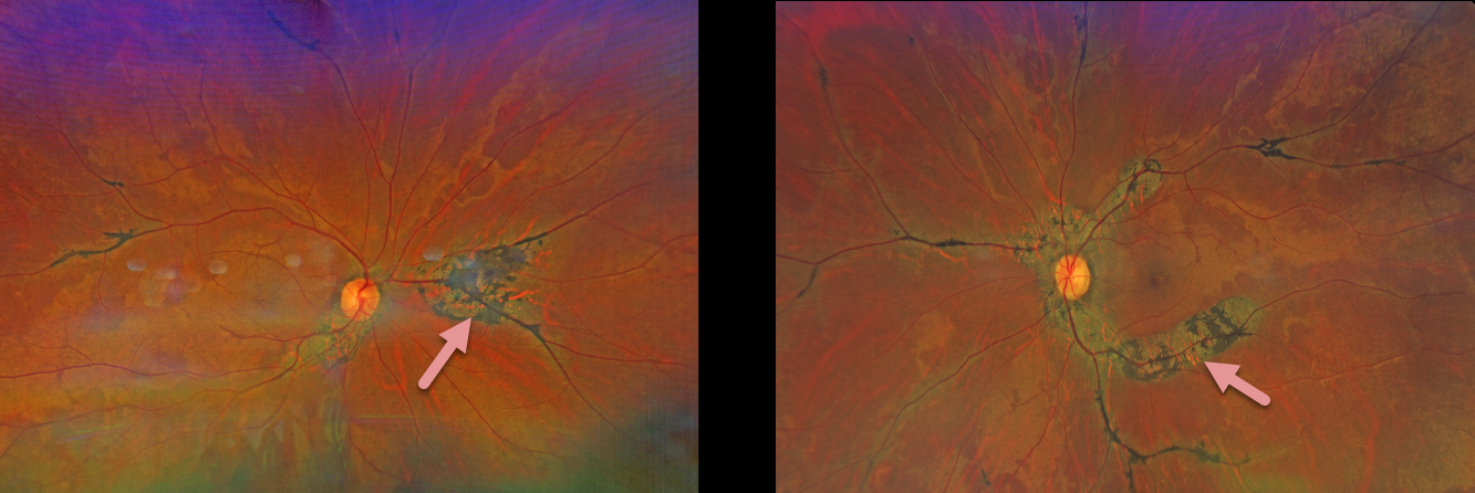
PIGMENTED PARAVENOUS CHORIORETINAL ATROPHY
Will Gibson
Originally posted on @retina.rocks December 26, 2024
This 67YO male presented with asymptomatic retinal findings. There was no family history of ocular disease. Vision was 20/20 OU.
Color photography shows bilateral paravenous chorioretinal scarring with intraretinal pigment migration.
Learning Points:
Pigmented paravenous chorioretinal atrophy (PPCRA) is a rare condition characterized by paravenous pigment clumps with peripapillary and radial zones of RPE atrophy. It is bilateral, often asymmetric, with various causes, including genetic and pseudo-PPCRA (inflammatory, infectious, or unknown). Patients often have symptoms, usually nyctalopia, but most retain good vision, especially those without macular involvement. The underlying pathophysiology is likely a primary choroidopathy, which then causes overlying RPE and retinal changes. The chorioretinal changes are best visualized with fundus autofluorescence, which was unfortunately unavailable for our patient.
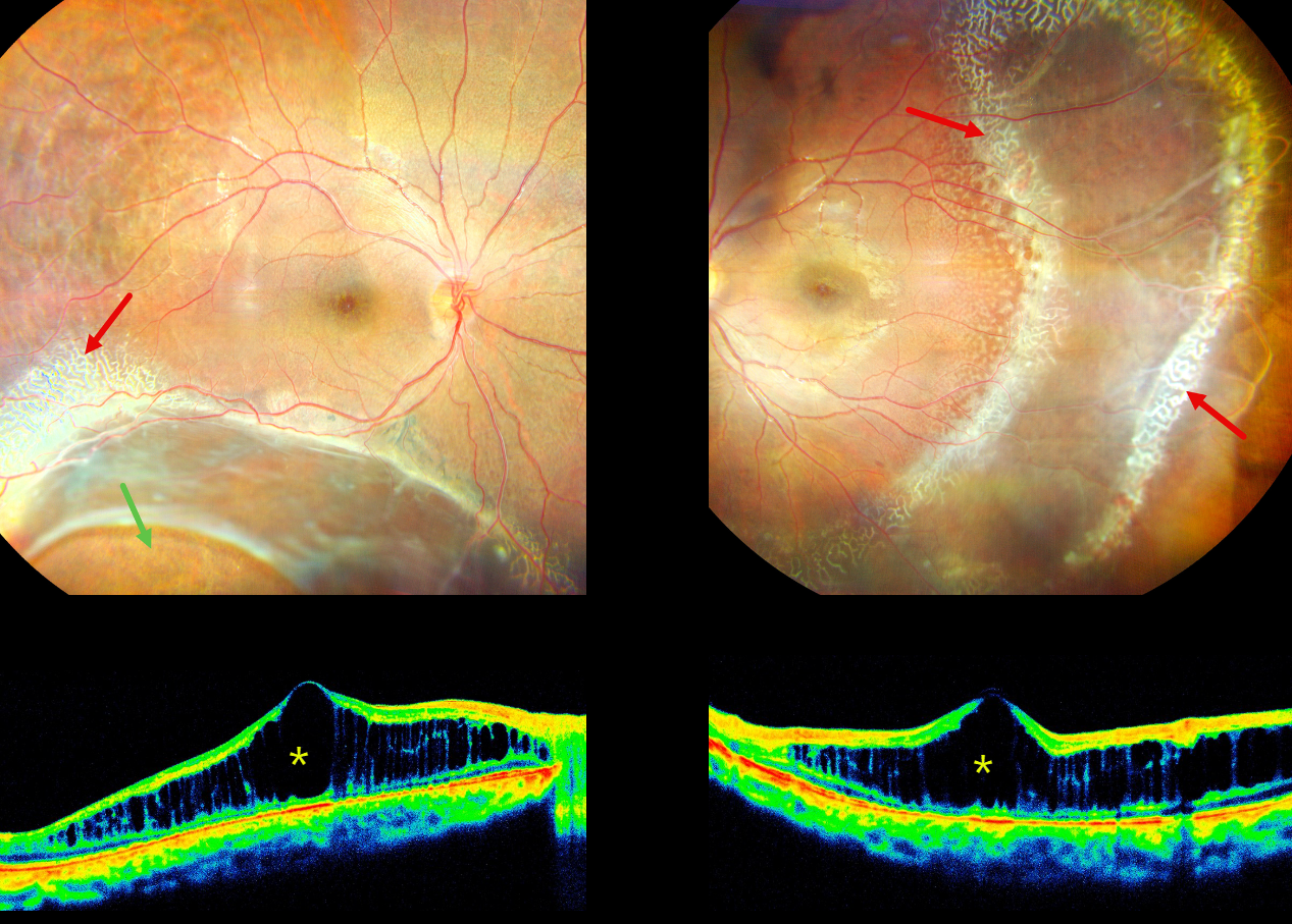
X-LINKED RETINOSCHISIS
Kanwaljeet Harjot Maden
Originally posted on @retina.rocks December 17, 2024
This 16YO male presented with stable bilateral vision loss since childhood, with new floaters in his right eye for 2 weeks. There was no ocular family history. Vision was 20/200 OD and 20/60 OS.
His right eye shows a large area of inferotemporal combined retinoschisis-rhegmatogenous retinal detachment (RD) extending up to the inferotemporal arcade. Subretinal pigmentation is seen along the posterior edge of the schisis inferiorly, and a large oval inner retinal defect is present inferotemporally. A combined schisis-RD is noted in his left eye which extends temporally and inferiorly. Radiating lines of foveal schisis are present bilaterally.
A dendriform pattern of occluded vessels is noted along the superotemporal edge of the schisis OD and surrounding the schisis OS. There are also numerous tiny round inner retinal breaks posterior to these dendriform lesions in the distal temporal left macula. The retinal vessels are variably fibrosed overlying the schisis cavities. OCT scanning shows severe macular schisis, primarily affecting the inner nuclear layer. Bilateral laser demaration was performed along the posterior edges of the schisis-retinal detachments.
Learning Points:
X-linked retinoschisis is caused by a mutation of the RS1 gene which is located on the X chromosome and encodes for retinoschisin, a protein secreted by photoreceptors which is involved in intercellular adhesion and likely retinal cellular organization (Heymann et al, Progress Retinal Eye Research 2023;95:101147). Although usually transmitted as an X-linked recessive disorder in males with an incidence of 1 in 15,000 to 30,000, it can also sometimes affect females.
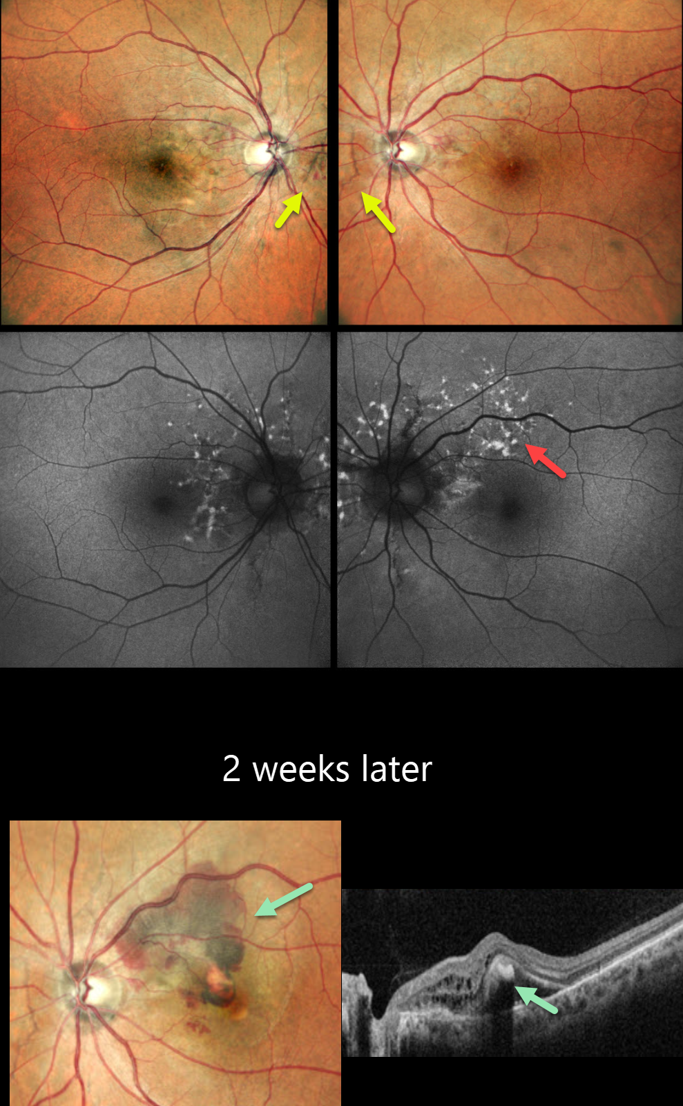
ANGIOID STREAKS
Anand Temkar and Manish Nagpal
Originally posted on @retina.rocks November 21, 2024
This 45YO male presented for a second opinion for bilateral wavy vision for 2 months. He was diagnosed elsewhere with macular neovascularization (MNV) and received two prior anti-VEGF injections (last 10 days earlier). Vision was 20/20 OU.
Pseudocolor SLO imaging shows subtle bilateral peripapillary angioid streaks associated with a few retinal hemorrhages. Fundus autofluorescence (FAF) shows numerous mostly peripapillary hyper-FAF flecks. Initial observation was recommended.
He returned two weeks later with new symptoms OS, and vision decreased to 20/120. New submacular fluid and blood are noted clinically and on OCT, and monthly anti-VEGF therapy was restarted. Unfortunately, he was lost to follow-up after 3 injections.
Learning Points:
Angioid streaks are orange-yellow, irregular, crack-like dehiscences in Bruch’s membrane associated with atrophic degeneration of the overlying RPE. These patients, especially those with pseudoxanthoma elasticum (PXE), are at significant risk for MNV as occurred in each of our patient’s eyes. MNV responds best to anti-VEGF therapy, and treatment is usually ongoing as with the typical neovascular AMD protocols. However, long-term vision is often poor due to RPE and choroidal atrophy (Mori et al, Graefe’s 2020;258:1863-1869).
Our patient had no family history of PXE or suggestive skin lesions, so skin biopsy or genetic testing was not performed. However, the funduscopic changes and macular dystrophy-like FAF findings are fairly pathognomonic for PXE.
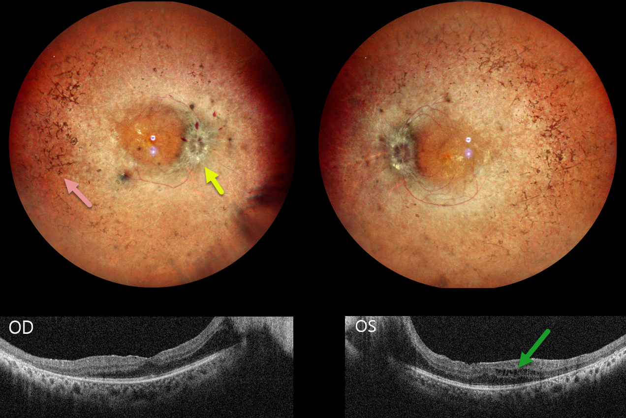
RETINITIS PIGMENTOSA WITH PAPILLEDEMA
Tejaswita Verma and Manish Nagpal
Originally posted on @retina.rocks November 20, 2024
This 30YO male presented with 3 weeks of decreased vision. His father has a history of retinitis pigmentosa (RP). Vision was 20/30 OD and 20/80 OS.
Pseudocolor SLO imaging shows typical bilateral RP scarring with intraretinal pigment migration extending from the peripheral maculas into the retinal peripheries. Bilateral macular puckers and disc swelling with right peripapillary inner retinal fluid are also seen. OCT scanning confirms the epimacular membranes. Bilateral pericentral outer macular atrophy is noted, along with some cystic edema in the left inner and outer nuclear layers.
Learning Points:
RP is a clinically and genetically heterogeneous group of inherited retinal disorders. The disease can present sporadically or with any inheritance pattern (autosomal dominant, autosomal recessive, sex-linked, or mitochondrial). It is characterized by diffuse, progressive dysfunction of predominantly rod photoreceptors, with subsequent RPE degeneration and intraretinal pigment migration. Visual impairment usually presents as night blindness and progressive visual field loss.
Bilateral optic disc edema is not typical for RP. Unfortunately, we were unable to work up our patient for a secondary cause because he was lost to follow-up immediately.
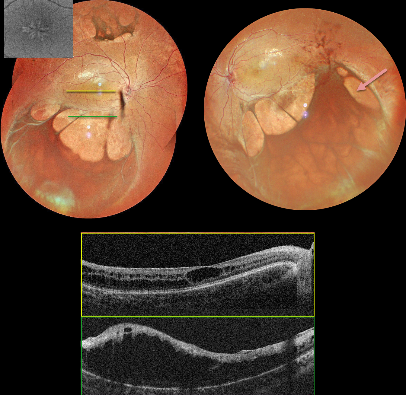
X-LINKED RETINOSCHISIS
Tejaswita Verma and Manish Nagpal
Originally posted on @retina.rocks October 10, 2024
This 17YO boy presented with a few days of mild bilateral blurred vision. Vision was 20/40 OU.
MultiColor imaging shows central radiating foveal schisis, and larger areas of schisis extend beyond the arcades. OCT scanning confirms foveal schisis and more peripheral macular schisis. Identical findings were noted in the left macula (not shown).
Learning Points:
Sex-linked retinoschisis is caused by a mutation of the RS1 gene which is located on the X chromosome and encodes for retinoschisin, a protein secreted by photoreceptors that is involved in intercellular adhesion and likely retinal cellular organization (Heymann et al, Progress Retinal Eye Research 2023;95:101147). Although usually transmitted as an X-linked recessive disorder in males with an incidence of 1 in 15,000 to 30,000, it can also sometimes affect females.
The characteristic features include foveoschisis and peripheral schisis with vitreous veils. Over time, the macular schisis flattens with secondary atrophy, and vision usually levels off at about 20/100. Patients can develop spontaneous vitreous hemorrhage and rhegmatogenous retinal detachment from peripheral schisis. Although there is no specific treatment, topical carbonic anhydrase inhibitors may decrease the foveal thickness and possibly help minimize foveal atrophy (see Andreuzzi et al Retina 2017;37:1555-1561).
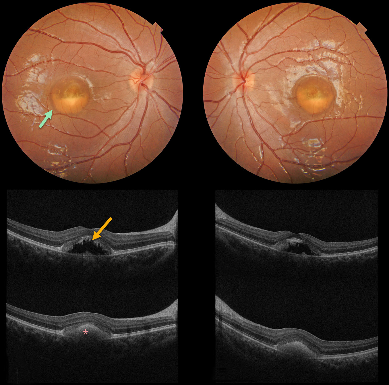
BEST DISEASE
Evgenii Chernov
Originally posted on @retina.rocks September 17, 2024
Color photography of our patient’s eyes shows partially scrambled Best lesions. OCT scanning through the superior lesion shows a mostly hyporeflective elevation of the neurosensory retina, with shaggy hyperreflective material along the posterior edge of the outer segments. OCT scanning through the inferior lesion shows hyperreflective layering of the vitelliform material.
Learning Points:
Best disease is associated with a mutation in the BEST1 gene, which encodes bestrophin-1. Bestrophin-1, a calcium-activated chloride channel, is primarily found in the basolateral plasma membrane of the RPE. BEST1 mutations cause a variety of phenotypes, including autosomal recessive bestrophinopathy, best vitelliform macular dystrophy, and autosomal dominant vitreoretinochoroidopathy.
Our patient has progressed to the ‘scrambled egg’ appearance. The remaining vitelliform material gravitates inferiorly, often leaving an optically empty, hyporeflective space between the RPE and outer retina. Eventually, the retina flattens, with secondary outer retinal atrophy.
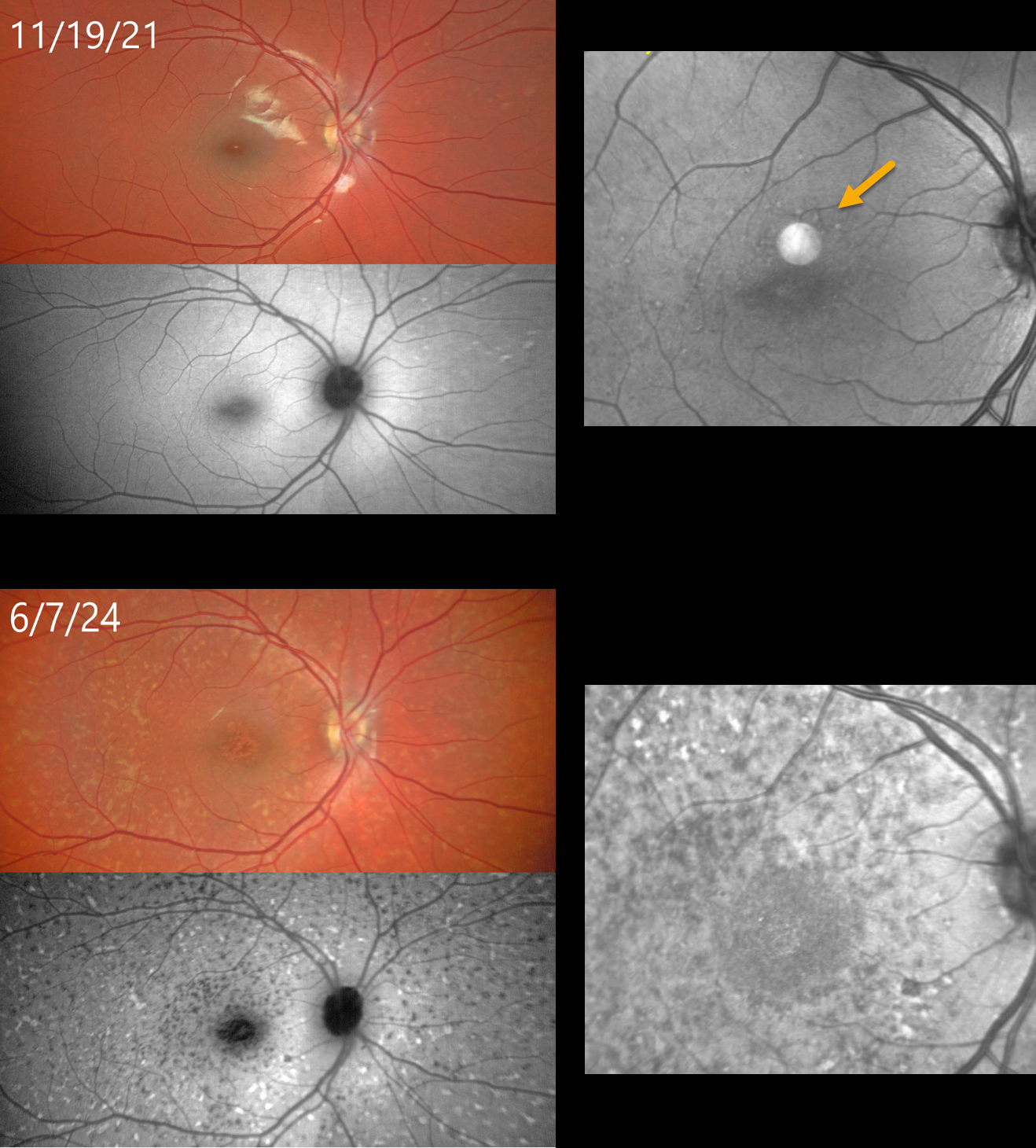
STARGARDT DISEASE
Yuenpang Cheung and Stephen Tsang
Originally posted on @retina.rocks September 4, 2024
This 9YO female had a known history of Stargardt disease at the time of examination on 11/19/21. Prior genetic testing revealed two pathogenic ABCA4 mutations.
On 11/19/21, color photography, fundus autofluorescence, and OCT en face of the outer retina are fairly normal except for some faint hyper-FAF flecks outside the macula and faint hyperreflective macular lesions.
When last examined on 6/7/24, vision was 20/100 OD and 20/80 OS. Despite the relatively stable vision, there was a dramatic progression of her disease on all multimodal images.
Similar findings were noted in her left eye (not shown).
Learning Points:
Stargardt disease is an autosomal recessive disorder caused by a mutation in the ABCA4 gene. The ABCA4 protein is located in the photoreceptor outer segments and is involved in the recycling of 11-cis-retinal. The mutation in this gene causes lipofuscin accumulation in the RPE, which eventually leads to photoreceptor and RPE degeneration. Other ABCA4 disorders include fundus flavimaculatus, cone-rod dystrophy, retinitis pigmentosa, and age-related macular degeneration.
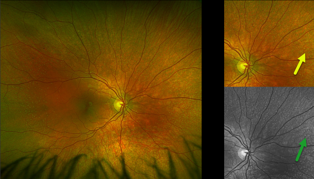
FUNDUS ALBIPUNCTATUS
Originally posted on @retina.rocks August 26, 2024
This healthy 10YO girl was referred for asymptomatic findings on retinal examination. There was no ocular family history. Vision was 20/40 OU.
Optos color RG imaging of the right eye shows innumerable subretinal yellow-white dots extending from outside each macula into the midperiphery. These dots are most evident in green-channel imaging. Identical findings were noted in the left eye (not shown). Genetic testing revealed double heterozygous RDH5 mutations.
Learning Points:
Fundus albipunctatus, an autosomal recessive form of congenital stationary night blindness, presents with scattered yellow-white dots that extend into the midperipheries, sparing the macula. Patients experience stable night blindness with normal central acuity. It is caused by a mutation in the RDH gene, which encodes the retinol dehydrogenase protein. This protein, found in the RPE helps recycle vitamin A in the visual cycle, and the white dots are thought to contain 11-cis retinal precursors. This results in decreased retinoid-derived fluorophores, accounting for the diminished FAF in these eyes (Sergouniotis et al, Ophthalmology 2011;118:1161-1670).
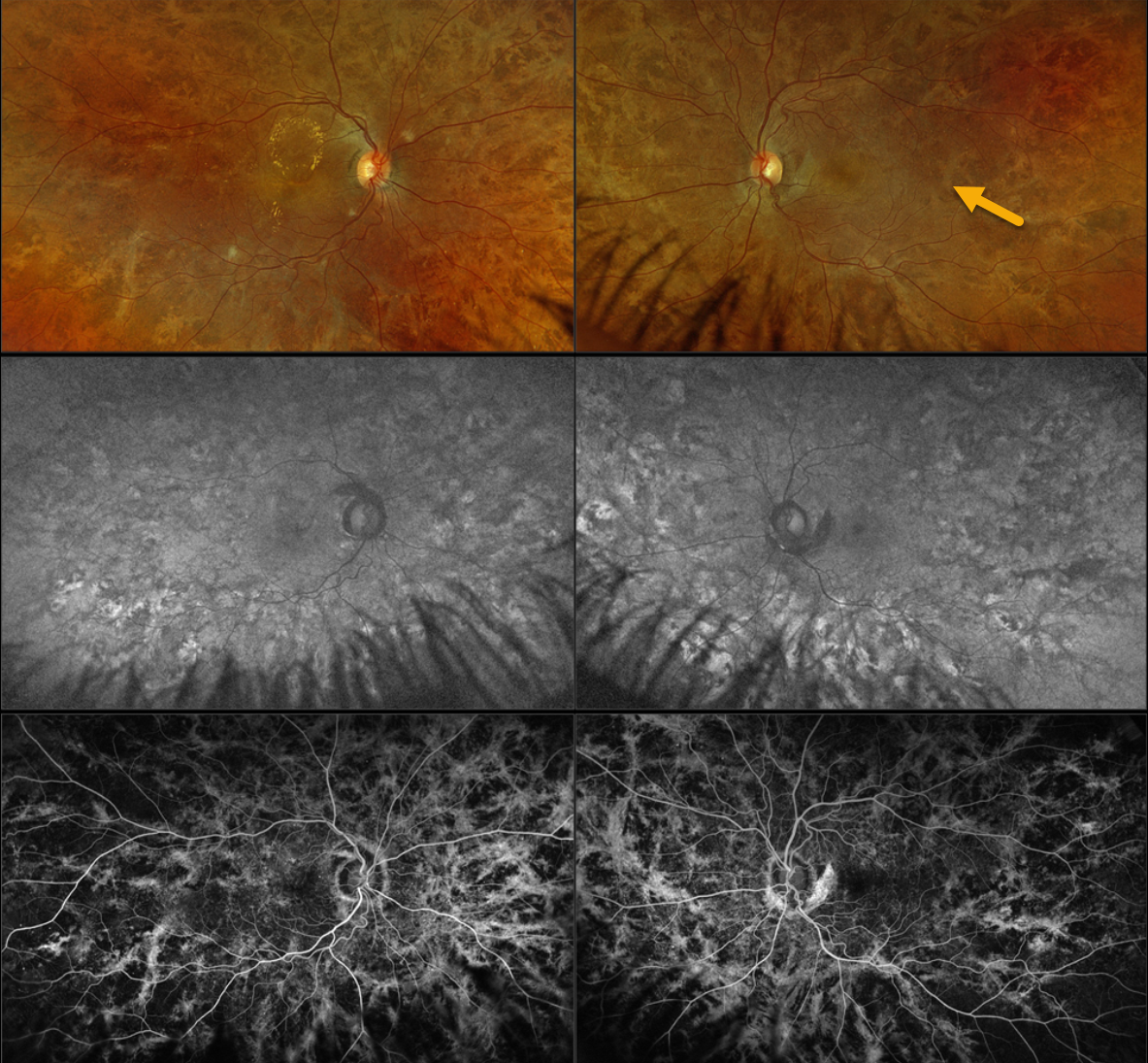
CHOROIDEREMIA CARRIER
Originally posted on @retina.rocks August 20, 2024
This 42YO female with type 2 diabetes was referred for asymptomatic diabetic macular edema (DME). There was no family history of ocular disease. Vision was 20/40 OD and 20/20 OS.
Optos color RGB imaging shows diffuse coarse subretinal hyperpigmentations throughout each posterior pole. Fundus autofluorescence (FAF) shows variable hyper- and hypo-FAF from these pigmentary changes, with fluorescein angiographic window defects. Genetic testing revealed a heterozygous pathogenic CHM mutation.
Learning Points:
Choroideremia is an X-linked recessive chorioretinal dystrophy caused by a mutation in the CHM gene located on the long arm of the X chromosome. Sons of female carriers have a 50% chance of developing choroideremia, and daughters have a 50% chance of becoming carriers. Some female carriers can develop asymptomatic pigmentary changes, as in our patient, or frank choroideremia due to irregular inactivation of the X chromosome (see Jauregui et al, AJO 2019;207:77-86).
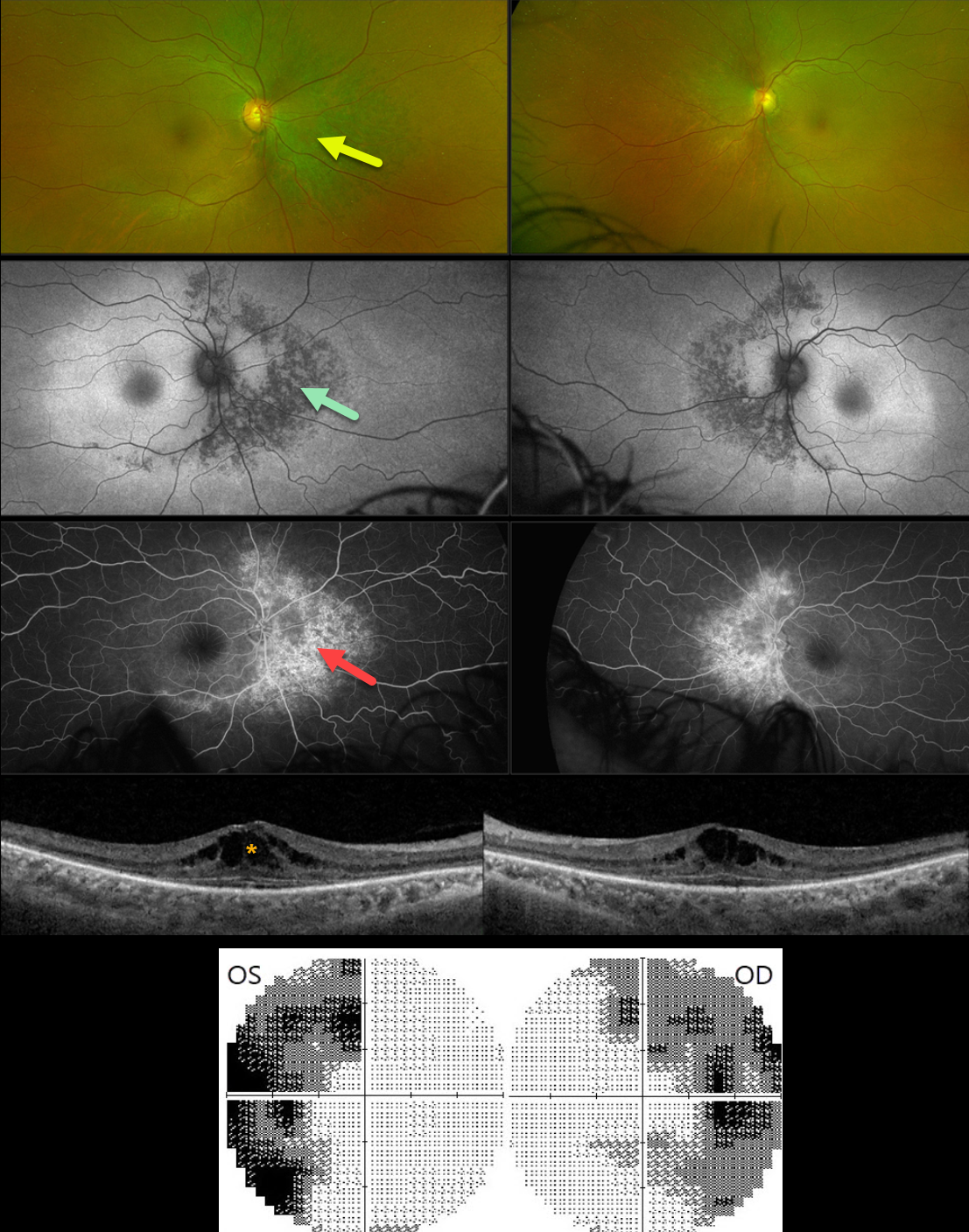
BITEMPORAL HEMIANOPIA WITH RETINITIS PIGMENTOSA
Mattie Adams
Originally posted on @retina.rocks August 12, 2024
This 54YO female was referred for asymptomatic retinal changes. Vision was 20/30 OD and 20/20 OS.
Visual fields showed bitemporal hemianopia. What is your diagnosis?
Optos color RG imaging shows pigmentary changes with intraretinal pigment migration extending superiorly, nasally, and inferiorly from the optic nerves. These changes are hypoautofluorescent and appear as angiographic window defects. Bilateral cystoid edema is seen on Triton swept-source OCT.
Learning Points:
Although bitemporal hemianopia usually signifies retrobulbar pathology at the optic chiasm, retinal disease can sometimes also cause similar field loss. Our patient’s retinitis pigmentosa (RP) changes were centered around each optic nerve, causing the hemianopia. Genetic testing revealed double heterozygous pathogenic ARSG mutations. This genotype has been associated with atypical Usher syndrome (Fowler et al, Ophthalmic Genetics 2021;42:338-343).
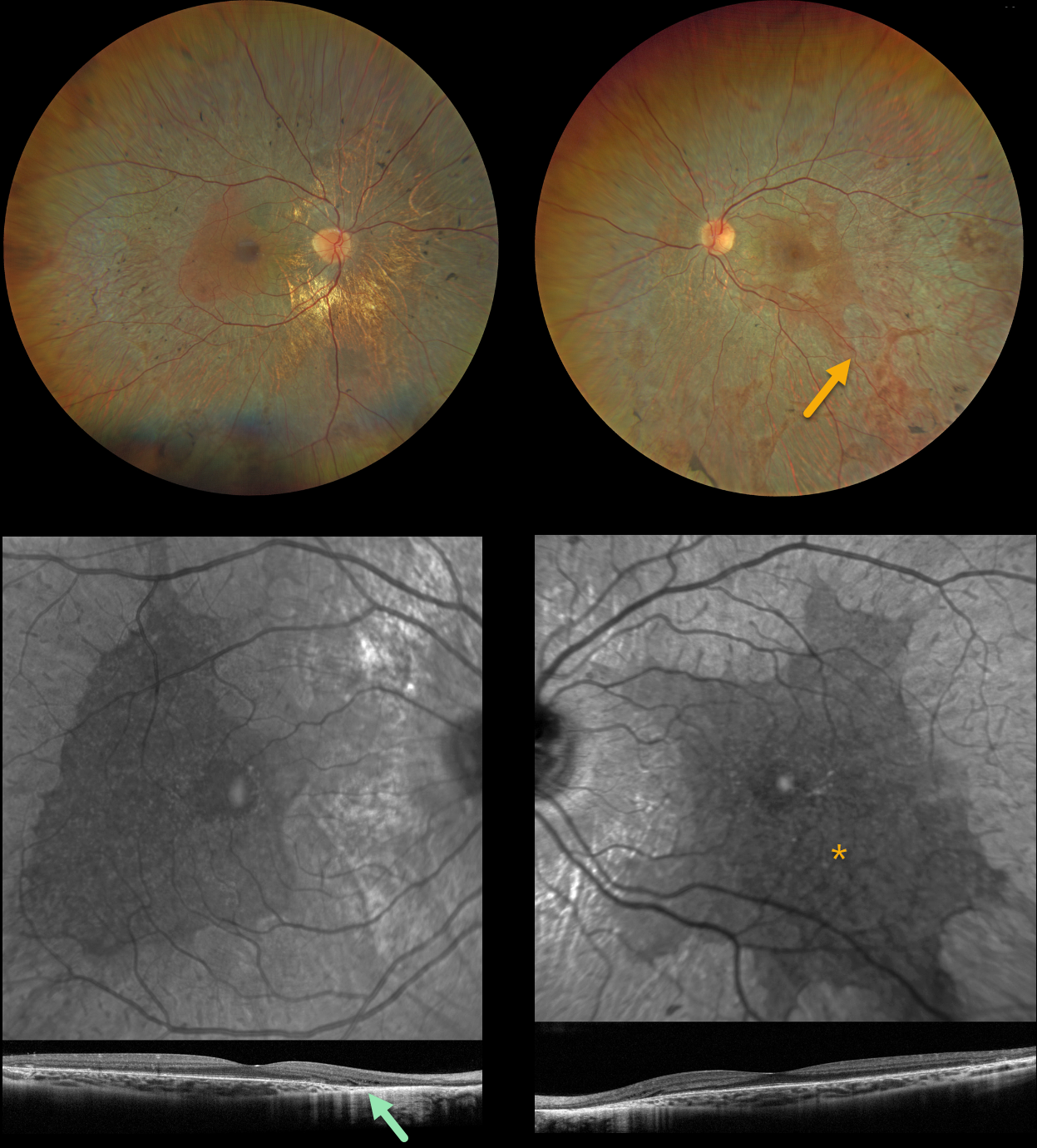
CHOROIDEREMIA IN A FEMALE CARRIER
Yuenpang Cheung and Stephen Tsang
Originally posted on @retina.rocks August 6, 2024
This 27YO female has a known history of choroideremia and is heterozygous for a pathogenic CHM mutation. Vision was 20/150 OD and 20/30 OS.
Color imaging shows large, symmetrically bilateral, scalloped areas of chorioretinal atrophy that spare the central maculae. Some peripheral areas of intraretinal pigment migration (bone spicules) are noted as well. OCT scanning shows variable outer retinal loss on B-scan with a scalloped preserved central macula on en face.
Learning Points:
Choroideremia is an X-linked recessive chorioretinal dystrophy caused by a mutation in the CHM gene located on the long arm of the X chromosome. Sons of female carriers have a 50% chance of developing choroideremia, and daughters have a 50% chance of becoming carriers. Some female carriers, as in our patient, can still develop choroideremia due to irregular inactivation of the X chromosome (see Jauregui et al, AJO 2019;207:77-86).
The onset of night blindness usually begins between the ages of 10 and 30, followed by peripheral visual field loss. Zones of patchy RPE and chorioretinal atrophy initially appear in the mid-periphery, gradually spreading anteriorly and posteriorly. Eventually, the patient is left with only a small island of vision, narrowed retinal vessels, and optic atrophy.
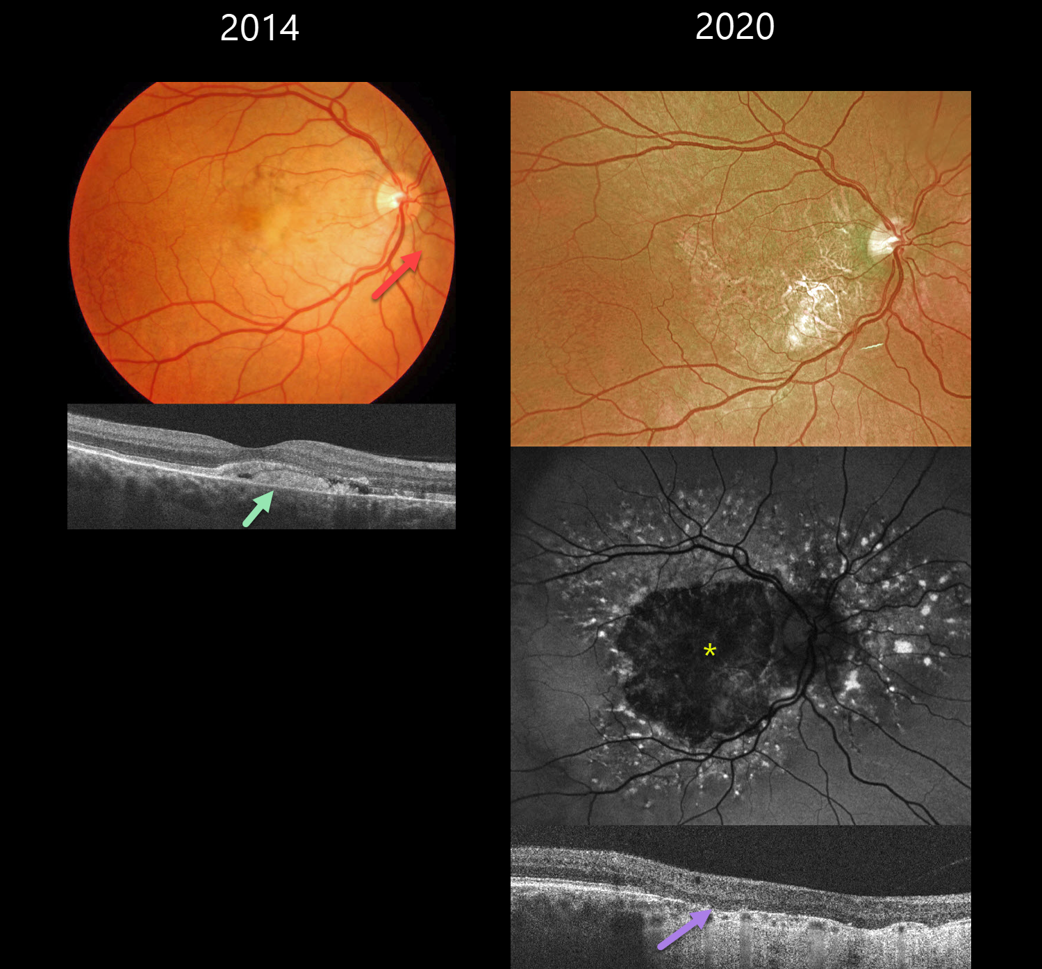
PSEUDOXANTHUM ELASTICUM WITH ANGIOID STREAKS AND PATTERN DYSTROPHY PHENOTYPE
Originally posted on @retina.rocks July 11, 2024
This 39YO male with a known history of pseudoxanthoma elasticum presented on 2/19/14. Findings were bilateral at this, and subsequent visits, so only his right eye images will be shown and discussed.
Color photography shows nasal yellowish subfoveal pigmentary changes with a peaux d’orange appearance temporally. Some peripapillary angioid streaks are noted. OCT shows hyperreflective vitelliform material and fluid below the retina, with some of this material also infiltrating the outer retina.
He has been followed without treatment, and when examined on 7/17/20, vision was 20/200 OD and 20/80 OS. Optos color RG imaging shows a variably atrophic macula. The atrophy on fundus autofluorescence is hypo-FAF. There is a fairly dramatic pattern of hyper-FAF spots radiating out from this atrophy. OCT shows outer retinal atrophy and no fluid. When last examined on 7/6/23, vision was 20/200 OU.
Learning Points:
Retinal findings in PXE most commonly include angioid streaks and a subretinal orange skin (peaux d’orange) appearance, most pronounced in each temporal macula. However, pattern dystrophy-like changes, as in our patient, can sometimes be the predominant phenotype (Murro et al, Graefe’s 2020;258:1881-1892). As with typical pattern dystrophies, the findings are dynamic and best captured with multimodal imaging.
BIETTI CRYSTALLINE DYSTROPHY
Yuenpang Cheung and Stephen Tsang
Originally posted on @retina.rocks July 10, 2024
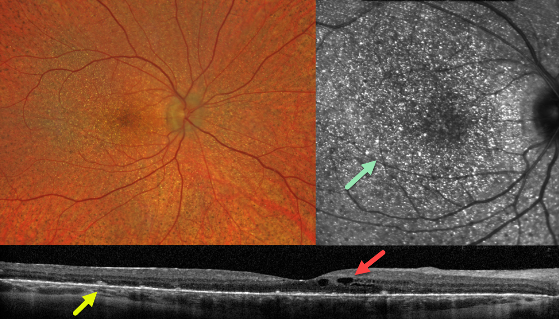
This 28YO female has a known history of Bietti crystalline dystrophy and was without new visual complaints. Vision was 20/50 OD and 20/40 OS.
Fundus imaging of her right eye shows extensive subretinal crystalline deposits primarily in the posterior poles, with tiny subretinal pigment clumps more peripherally.
OCT scanning shows hyperreflective dots overlying the RPE-Bruch’s membrane complex, with variable outer retinal and RPE loss. Cystic changes are noted in the inner nuclear layer.
En face OCT through the outer retina reveals innumerable hyperreflective crystalline deposits. Identical findings were noted in her left eye.
Learning Points:
Bietti crystalline dystrophy is an autosomal recessive ocular disorder that affects both the anterior and posterior segments. Clinical features include crystalline deposits in the retina and cornea, and, rarely, in the crystalline lens, as well as retinal pigment epithelial clumping and atrophy.
Mutations in CYP4V2, which cause dysfunctional lipid metabolism, have been implicated in its pathogenesis. While there are no definite treatments, anti-VEGF therapy can be given for secondary macular neovascularization.
See Saatci et al for a recent review (Clinical Ophthalmology 2023;17:953-967).
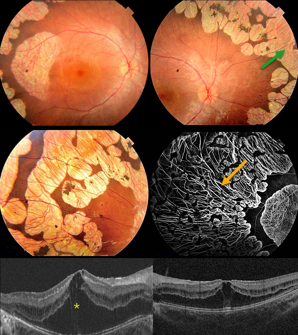
GYRATE ATROPHY
Originally posted on @retina.rocks June 12, 2024
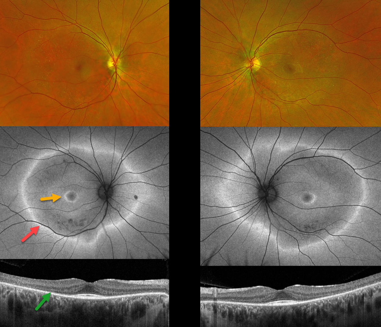
INHERITED RETINAL DISEASE
Yuenpang Cheung and Stephen Tsang
Originally posted on @retina.rocks June 3, 2024
This 32YO female has a known history of an unspecified inherited retinal disease (IRD) diagnosed elsewhere 3-4 years earlier. Genetic testing was negative. She has difficulty seeing in the dark and avoids night driving. More recently, she’s aware of a midperipheral ring of bilateral vision loss. Vision was 20/20 OU.
Optos color RG imaging is fairly normal bilaterally except for drusen-like deposits in the peripheral maculas. Fundus autofluorescence shows a large hyper-FAF ring surrounding each posterior pole, along with a smaller hyper-FAF macular ring. OCT scanning shows outer retinal atrophy sparing the macular center.
Learning Points:
Fundus autofluorescence (FAF) visualizes endogenous fluorophores, compounds that spontaneously fluoresce without an external dye. The main fundus fluorophore is lipofuscin, which resides within the RPE lysosomes. The photoreceptor outer segments absorb some of the autofluorescent excitatory light, and thus normally diminish the FAF signal.
In IRDs, loss of outer segments unmasks this signal, leading to increased FAF. Many patients with an IRD will show a hyper-FAF ring between clinically normal and abnormal retina, which likely represents retina ‘at risk’, showing the junction of a functional and dysfunctional retina (Oishi et al, Int J Retina Vitreous 2019;5(S1);23).
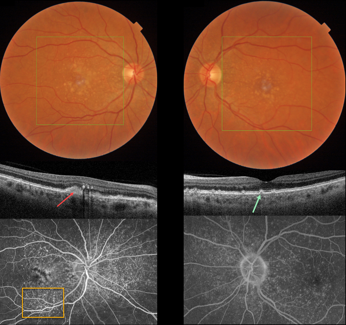
CUTICULAR DRUSEN WITH VITELLIFORM LESION
Originally posted on @retina.rocks May 28, 2024
This 57YO female was referred for asymptomatic macular changes. Vision was 20/20 bilaterally.
Triton color imaging shows bilateral confluent central soft drusen and smaller, more peripheral macular drusen. Swept-source OCT shows bilateral, small arrowhead-shaped cuticular drusen. A hyperreflective vitelliform lesion is noted OD, along with some hyperreflective deposits in the outer plexiform layer. Optos fluorescein angiography shows extensive bilateral hyperfluorescent cuticular drusen along with a staining vitelliform lesion OD.
Learning Points:
Originally described by Gass as basal laminar drusen (AJO 1985;99:445-459), cuticular drusen were initially thought to represent nodular thickening of the RPE basement membrane. They appear clinically as numerous, round, yellow-white ‘hard drusen’, and on OCT as small, arrowhead-shaped drusen located above Bruch’s membrane and below the RPE (Spaide and Curcio, Retina 2010;30:1441-1454).
Our patient’s angiographic appearance echoes Dr. Gass’ original description of “a beautiful stars-in-the-sky or Milky-Way pattern” of hyperfluorescence. Cuticular drusen are classically associated with avascular vitelliform lesions (Finger et al, Retina 2010;30:1455-1464), as in this case, but can also develop macular neovascularization.
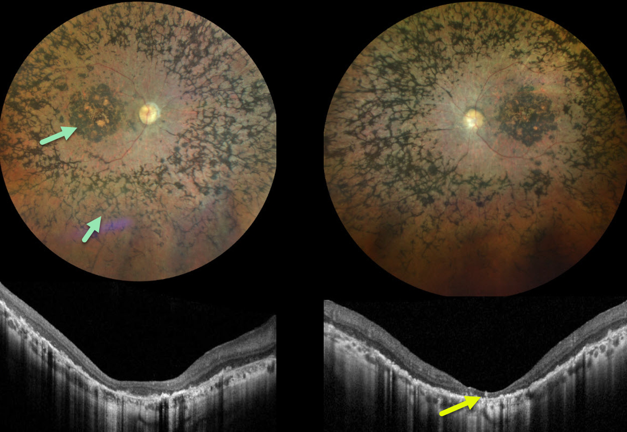
LEBER CONGENITAL AMAUROSIS
Yuenpang Cheung and Stephen Tsang
Originally posted on @retina.rocks May 9, 2024
This 32YO female was diagnosed at age 3 with Leber congenital amaurosis from a pathogenic RDH12 mutation. Progressive central and peripheral field loss with night blindness began in her late teens. Her brother also has this disorder. Vision was 20/200 OD and hand motion OS.
Color photography shows extensive pigmentary changes with intraretinal pigment migration throughout the macula and extending into the retinal peripheries. Variable outer retinal atrophy, especially in the central macula, is noted on OCT scanning.
Learning Points:
Initially described by Theodor Karl Gustav von Leber in 1869, Leber congenital amaurosis is a genetically heterogeneous group of inherited retinal dystrophies causing severe early vision loss.
The most common causative genes include CEP290, GUCY2D, CRB1, and RPE65. Mutations in the RDH12 gene, which encodes retinol dehydrogenase 12, account for about 4% of cases of Leber congenital amaurosis (Perrault et al, Am J Hum Genet 2004;76:639-646).
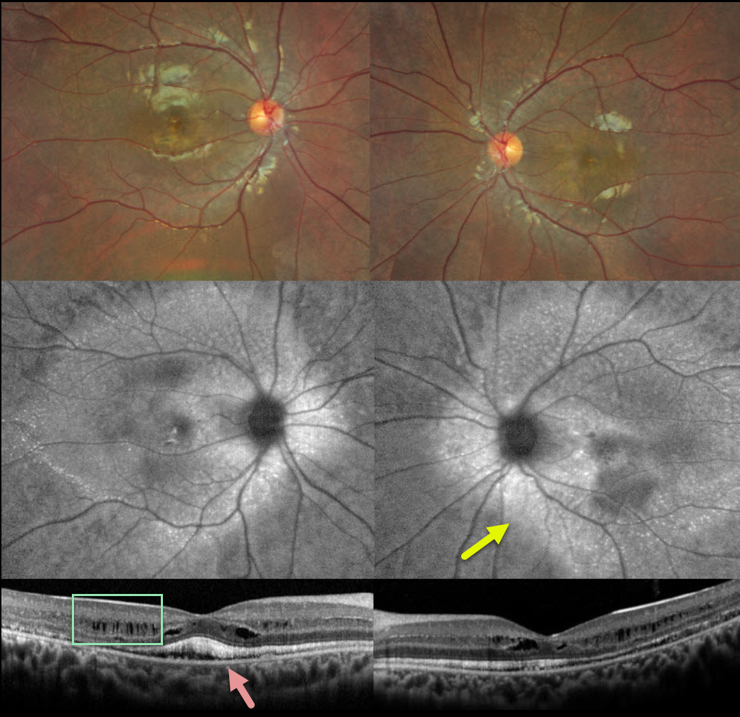
AUTOSOMAL RECESSIVE BESTROPHINOPATHY
Yuenpang Cheung and Stephen Tsang
Originally posted on @retina.rocks May 7, 2024
This 12YO boy has a known history of Best disease, which is being treated with topical dorzolamide for macular edema. He’s noticed some bilateral vision loss over the past 3 years. Vision is 20/25 OU.
Color imaging shows yellowing macular pigmentary changes. Fundus autofluorescence (FAF) shows much more pronounced changes with hyper-FAF extending beyond the arcades. OCT shows predominantly inner plexiform fluid versus schisis and hyperreflective subretinal vitelliform material.
Genetic testing revealed a heterozygous pathogenic BEST1 c.763C>T mutation, a heterozygous BEST1 c.113T>G mutation, and a single heterozygous PDE6B mutation. Oral acetazolamide 500mg daily was started.
Learning Points:
Burgess et al first described autosomal recessive bestrophinopathy as a distinct BEST1-related disorder (American Journal of Human Genetics 2008;82:19-31). Unlike other BEST1 autosomal dominant disorders, patients have 2 abnormal alleles and thus no normally functioning bestrophin1 protein.
It is characterized by posterior pole vitelliform deposits and pigmentary changes that extend beyond the macula, subretinal fluid and vitelliform material, and intraretinal fluid (Casalino et al, Ophthalmology 2021;128:706-718). Vision gradually declines with age.
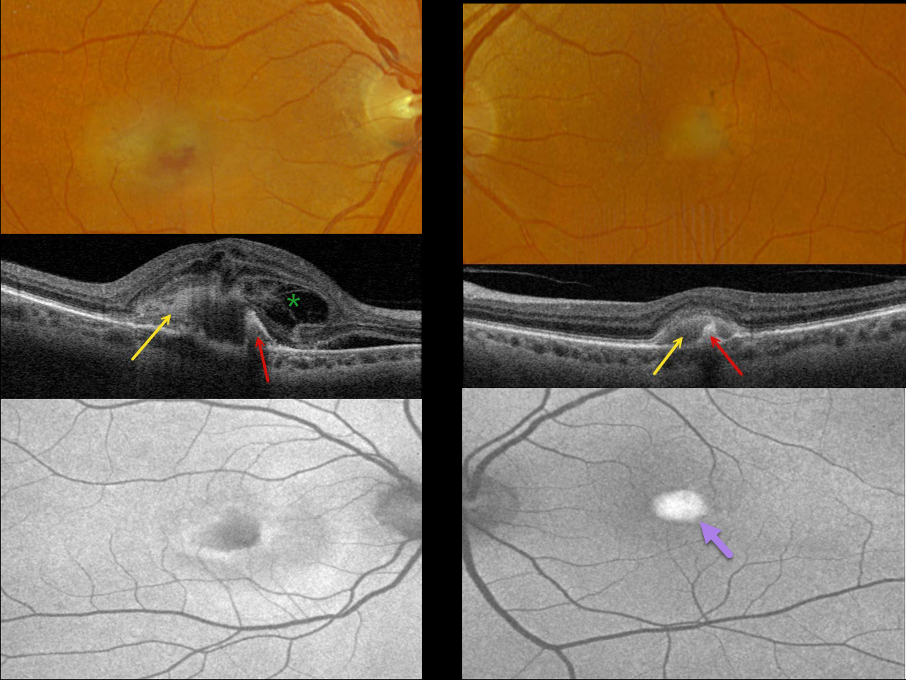
ADULT-ONSET FOVEOMACULAR VITELLIFORM DYSTROPHY
Originally posted on @retina.rocks April 29, 2024
This 71YO female was referred for neovascular AMD. She noticed recent vision loss in her right eye and was fairly asymptomatic in her left eye. Vision was counting fingers OD and 20/80 OS.
Optos color RGB imaging shows somewhat turbid, yellowish foveal fluid OD and a vitelliform lesion OS. Triton swept-source OCT shows hyperreflective subretinal vitelliform material and hyperreflectivity from a subretinal pigment clump. Subretinal fluid and a complex pattern of loculated pockets of intraretinal fluid are noted OD.
On Optos fundus autofluorescence (FAF), the lesion shows central hypo-FAF and peripheral hyper-FAF OD, and marked hyper-FAF OS.
Anti-VEGF therapy was started for macular neovascularization (MNV) in her right eye.
Learning Points:
Best disease is associated with a mutation in the BEST1 gene, which encodes bestrophin-1. Bestrophin-1, a calcium-activated chloride channel, is primarily found in the basolateral plasma membrane of the RPE.
BEST1 mutations cause a variety of phenotypes, including adult-onset foveomacular vitelliform dystrophy (as in our patient), best vitelliform macular dystrophy, autosomal recessive bestrophinopathy, and autosomal dominant vitreoretinochoroidopathy.
Although our patient had a Best phenotype, genetic testing revealed uncertain significant heterozygous variants SAG, SLC7A14, and VCAN.
In our experience, MNV is rarely associated with Best lesions, although Miyagi et al identified MNV in 18% of eyes in their retrospective Asian population (Graefe’s 2022;260:1125-1137).
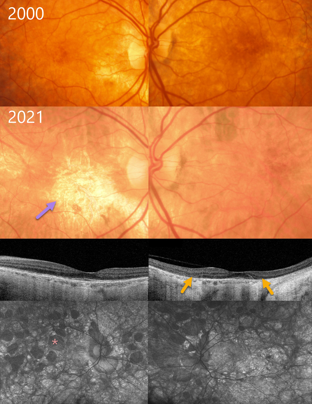
CHOROIDEREMIA IN A FEMALE CARRIER
Originally posted on @retina.rocks April 24, 2024
This 37YO female, whose son was diagnosed with choroideremia, was examined in 2000. Color photography shows some mild bilateral pigmentary changes.
Over the subsequent 20+ years, her ocular findings have progressed into more typical choroideremia. When last examined in 2021, vision was 20/30 OU.
The macula is now atrophic OD with minimal progressive changes OS. Swept-source OCT, however, shows fairly symmetrical outer retinal atrophy that spares each macular center. Optos autofluorescence reveals multifocal lobular areas of atrophy extending into each periphery. Genetic testing was heterozygous for a pathogenic CHM variant.
Learning Points:
Choroideremia is an X-linked recessive chorioretinal dystrophy caused by a mutation in the CHM gene located on the long arm of the X chromosome. Sons of female carriers have a 50% chance of developing choroideremia, and daughters have a 50% chance of becoming carriers. Some female carriers, as in our patient, can still develop choroideremia due to irregular inactivation of the X chromosome (see Jauregui et al, AJO 2019;207:77-86).
The onset of night blindness usually begins between the ages of 10 and 30, followed by peripheral visual field loss. Zones of patchy RPE and chorioretinal atrophy initially appear in the mid-periphery, gradually spreading anteriorly and posteriorly. Eventually, the patient is left with only a small island of vision, narrowed retinal vessels, and optic atrophy.
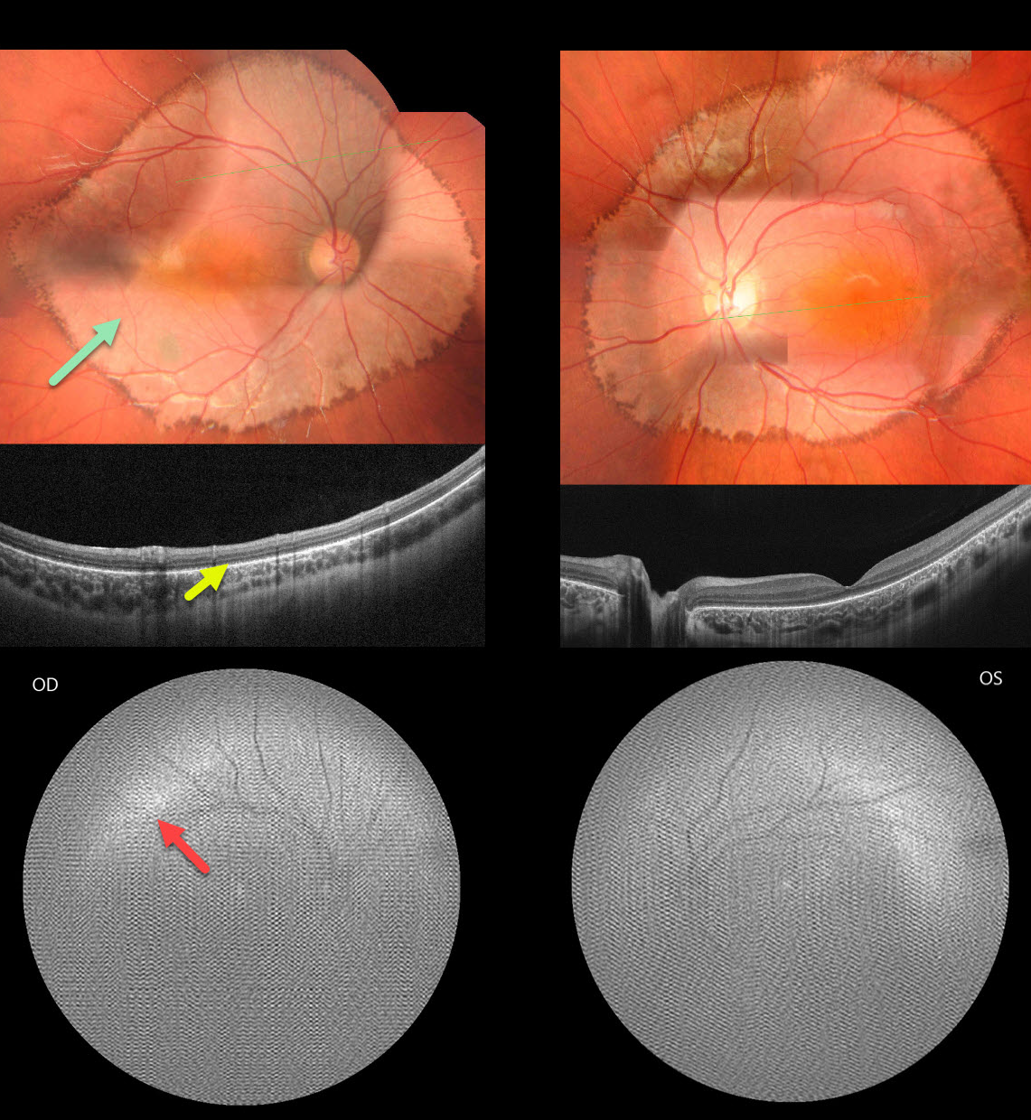
INHERITED RETINAL DISEASE
Seif Anwar
Originally posted on @retina.rocks April 4, 2024
This healthy 6YO boy presented with diminished vision noticed by his parents and teacher. There was no family history of eye disease. Vision was 20/200 OU.
Fundus photography shows fairly symmetrical, creamy, pale subretinal lesions encircling each posterior pole. The margins of these lesions have a somewhat irregular hyperpigmented border.
OCT shows outer retinal thinning and loss of the outer retinal band detail, along with a thinned or absent RPE band.
Fundus autofluorescence (FAF) was of extremely poor quality, and possibly shows superior and temporal hyper-FAF just outside each macula. It was quite difficult to obtain imaging in our patient, and these studies required multiple sessions.
Learning Points:
Unfortunately, we do not have a specific diagnosis for our patient’s presumed sporadic inherited retinal disease (IRD). The location and bilaterality of the multimodal findings suggest a cone-mediated disorder. Our patient’s funduscopic appearance somewhat resembles that found in autosomal recessive CDH3 disease, which includes sparse scalp hair and occasional limb abnormalities (Hull et al, JAMA Ophthalmol 2916;134:992-1000). Genetic testing could not be performed due to cost constraints.
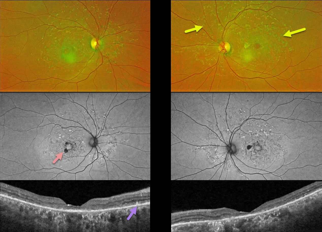
FUNDUS FLAVIMACULATUS
Originally posted on @retina.rocks March 26, 2024
This 58YO female recently noticed difficulties with depth perception. There was no family history of eye disease. Vision was 20/50 OD and 20/80 OS. Genetic testing revealed two heterozygous pathogenic ABCA4 mutations.
Optos color RG imaging shows multiple yellow fleck-like lesions throughout each posterior pole. Fundus autofluorescence (FAF) of these flecks shows variable hyper-FAF, with patches of paracentral hypo-FAF.
Triton swept-source OCT shows variable outer retinal atrophy and disorganization. Some hyperreflective spots are noted above the RPE.
Learning Points:
Fundus flavimaculatus is a genetic disorder, usually transmitted as an autosomal recessive trait, that is caused by mutations involving the ABCA4 gene. This gene encodes one of a family of ATP-binding cassette (ABC) transmembrane proteins that mediate the active transport of various substrates across cellular membranes. The ABCA4 protein is present in the photoreceptor outer segment disc membranes, where it is involved in recycling 11-cis-retinal.
Stargardt disease is believed to be fundus flavimaculatus without peripheral findings. Vision in fundus flavimaculatus is often better than that in Stargardt due to later disease onset and less macular involvement. Both likely represent an RPE lipofuscin storage disease. The RPE cells become engorged with lipofuscin, causing a dark choroid on fluorescein angiography.
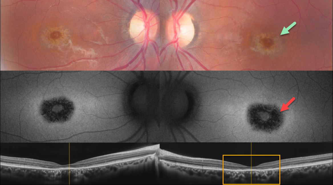
CONE DYSTROPHY
Gökşen Gökçen
Originally posted on @retina.rocks February 21, 2024
This 22YO male medical student gave a history of relatively rapid bilateral central vision loss 6 years earlier. His symptoms have been stable since, and he continues to function well visually, personally, and academically.
Color imaging shows a bullseye of pigmentary loss around each fovea. The area of pigment loss is hypo-autofluorescent. OCT scanning shows central outer retinal atrophy, particularly in the perifovea. The changes are markedly symmetrical between the eyes.
Genetic testing revealed two pathogenic ABCA4 mutations.
Learning Points:
Cone-rod dystrophy is a group of inherited retinal disorders characterized by loss of cone photoreceptors followed by rod photoreceptors. Symptoms include decreased visual acuity, central vision loss, color vision abnormalities, and photophobia.
Several genes have been implicated, including ABCA4, CRX, GUCY2D, and RPGR. ABCA4 mutations are found in autosomal recessive cone-rod dystrophy and Stargardt disease.
For an excellent review of the complex clinical and genetic spectrum of ABCA4 disorders, see Cremers et al, Progress Retinal Eye Research 2020;79;100861.
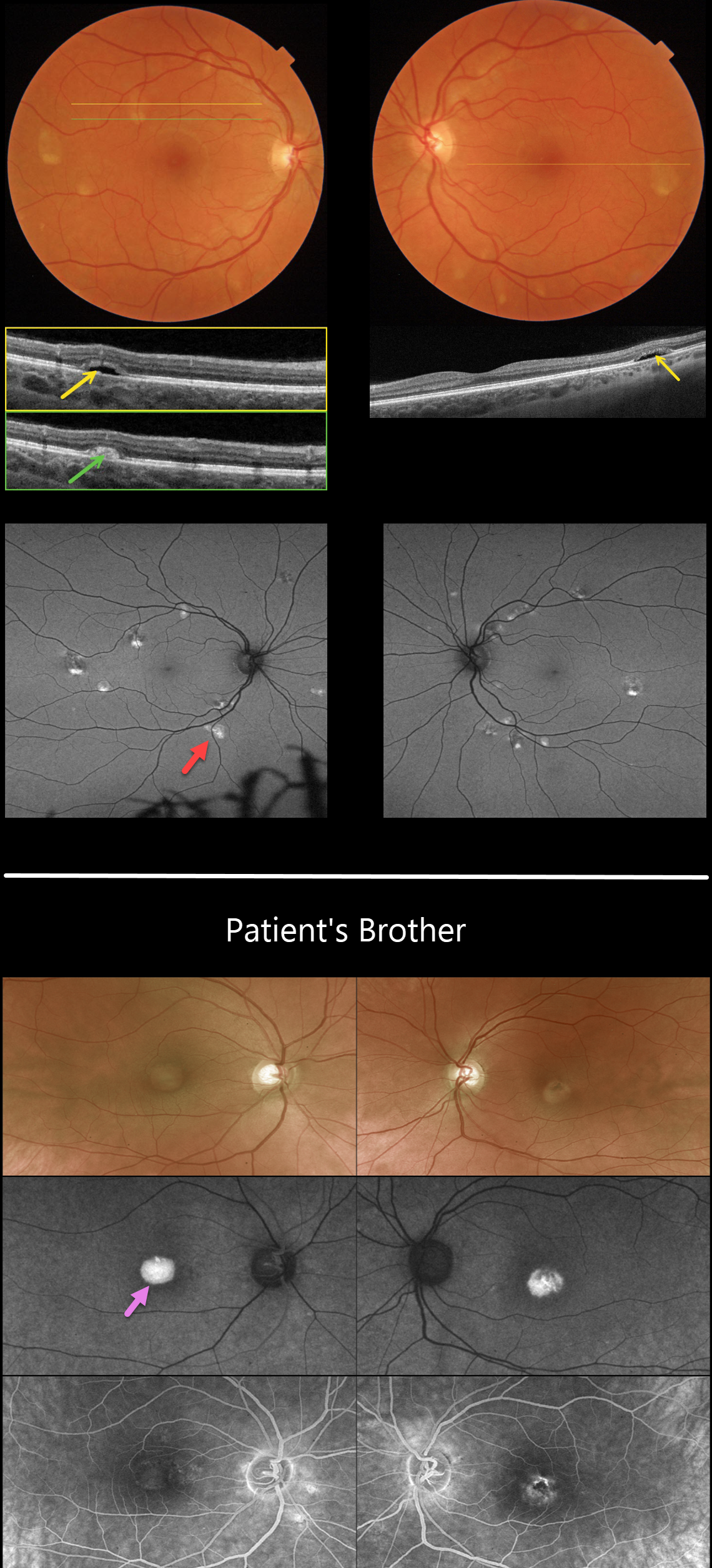
IMPG1 BEST DISEASE WITH DIFFERENT PHENOTYPES IN SIBLINGS
Jessica Johnson and Mattie Adams
Originally posted on @retina.rocks February 5, 2024
This 41YO female complained of increasing difficulties with dark adaptation and night vision. Vision was 20/25 OU.
Color imaging shows bilateral, multifocal, partially scrambled vitelliform lesions. The superior scrambled aspect of each lesion on OCT shows subretinal hyporeflectivity, and the inferiorly layered vitelliform material is hyperreflective. The inferior vitelliform material is markedly hyper-autofluorescent with Optos imaging.
Genetic testing was positive for a heterozygous pathogenic IMPG1 deletion in exon 7.
Interestingly, her brother was recently diagnosed with Best disease, having more typical single, subfoveal yellow vitelliform lesions. The brother’s genetic testing showed a similar IMPG1 mutation.
Learning Points:
Best disease is most often associated with a mutation in the BEST1 gene, which encodes bestrophin-1. Bestrophin-1, a calcium-activated chloride channel, is primarily found in the basolateral plasma membrane of the RPE.
BEST1 mutations cause a variety of phenotypes, including autosomal recessive bestrophinopathy, best vitelliform macular dystrophy, and autosomal dominant vitreoretinochoroidopathy.
Multifocal lesions are a rare phenotype and are not usually associated with subfoveal lesions. Multifocal Best needs to be differentiated from other causes of multifocal serous detachments, including multifocal central serous, autosomal recessive bestrophinopathy (see Boon et al, Ophthalmology 2013;120:809-820), and idiopathic or paraneoplastic acute exudative polymorphous vitelliform maculopathy (see Barbazetto et al, Ophthalmology 2018;125:75-88).
Vitelliform macular dystrophies (VMD) are most commonly caused by BEST1, PRPH2, or VMD2 mutations. IMPG1 and IMPG2, which encode proteins that are components of the interphotoreceptor matrix, have also been implicated in both autosomal dominant and recessive VMD.
Manes et al. reported a wide variety of IMPG1 VMD phenotypes, including multifocal lesions and more classic single subfoveal lesions, as seen in our two patients (Am J Hum Genet 2013;93:571-578).
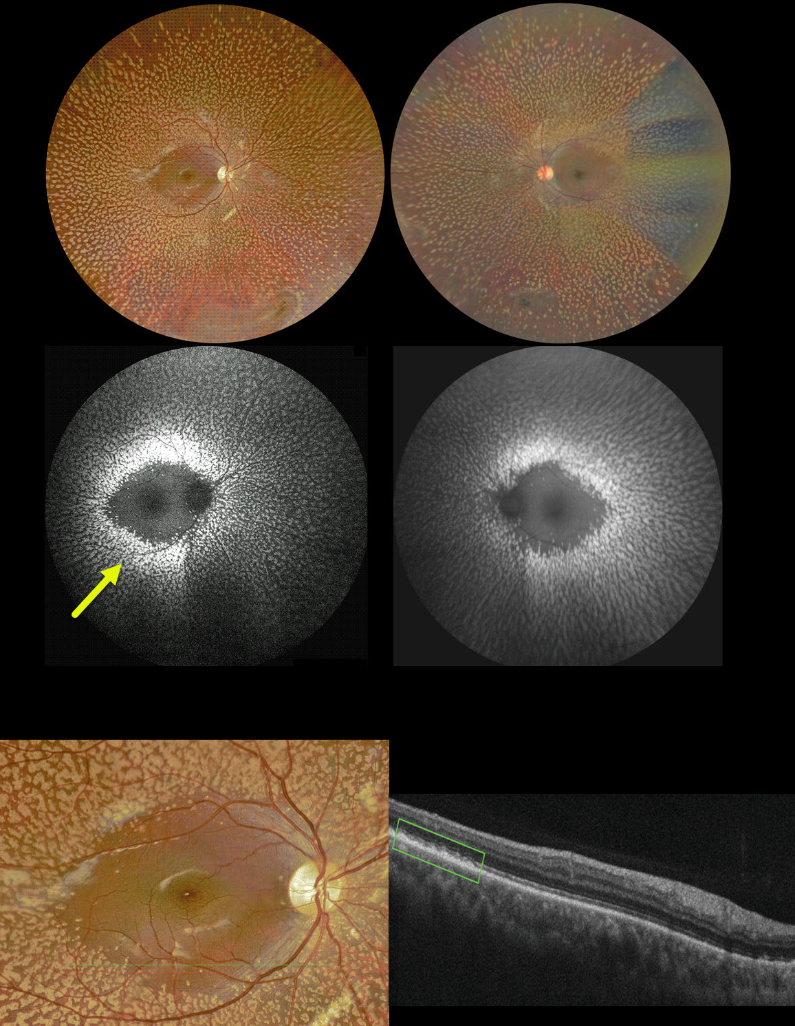
BENIGN FAMILIAL FLECK RETINA
Ayushi Gupta and Vishal Agrawa
Originally posted on @retina.rocks February 1, 2024
This 10YO boy complained of needing frequent changes of his glasses. Vision was 20/20 OD and 20/30 OS. He was a -8D myope OD and -6D myope OS. Corneal topography was consistent with keratoconus.
Color imaging shows symmetric, multiple, discrete yellow-white fleck lesions involving the entire fundus except the maculae. A patch of inferior lattice is also noted OD.
On fundus autofluorescence, these flecks show marked hyper-FAF. On OCT, the flecks appear as hyperreflective outer retinal deposits that extend into and distort the overlying ellipsoid zone.
Learning Points:
Initially reported by Aish and Dajani (BJO 1980;64:652-659), benign familial fleck retina (BFFR) is an extremely rare autosomal recessive disorder that belongs to a heterogeneous group of flecked retina syndromes.
The BFFR gene encodes for a group V phospholipase A2 (PLA2G5). Despite the dramatic fundus appearance, affected individuals have normal acuity, visual fields, and retinal function, including electrophysiology.
Other flecked retinal disorders include fundus albipunctatus, fundus flavimaculatus, familial drusen, retinitis punctata albescens, and fleck retina of Kandori. These disorders can be associated with night blindness, central visual problems, abnormal ERG, and abnormal perimetry.
The ERG helps distinguish BFFR from other flecked retina syndromes, such as retinitis punctata albescens and fundus albipunctatus, as scotopic ERG responses are decreased in the latter conditions but normal in BFFR.
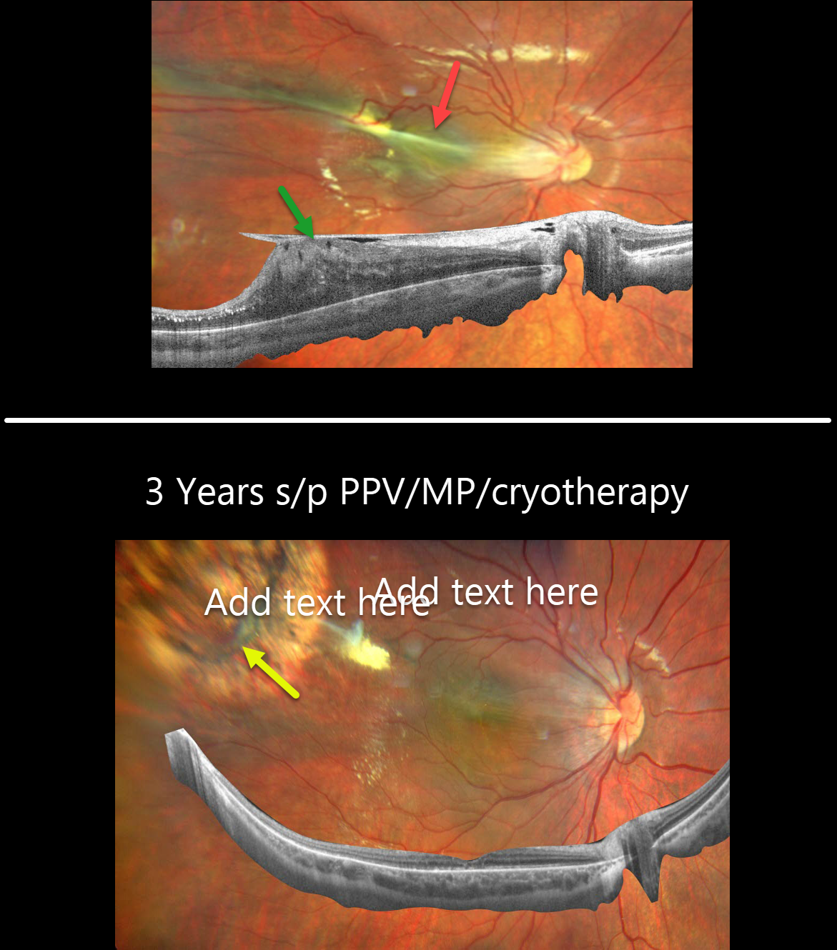
FAMILIAL EXUDATIVE VITREORETINOPATHY (FEVR)
Barbara Parolini, Veronika Matello, Giulia Freschi, and Roberta Penzani
Originally posted on @retina.rocks January 23, 2024
This otherwise healthy 8YO male presented with a chronic history of vision loss in his right eye. Vision was counting fingers OD and 20/25 OS.
Fundus photography shows dragging of the optic nerve and macula temporally towards an area of dilated, anomalous retinal vessels. Widefield OCT of the right shows an adherent epiretinal membrane elevating and dragging the nerve and macula temporally.
Pars plana vitrectomy with membrane peeling and cryotherapy of the area with anomalous vessels was performed. Three years later, vision improved to 20/25 OD. The macula is clinically and on OCT completely flat, with a dry cryotherapy scar noted in the superotemporal midperiphery.
This Eyecare Clinic (in Brescia, Italy) case was submitted by Barbara Parolini, Veronika Matello, Giulia Freschi, and Roberta Penzani.
Learning Points:
Originally described by Criswick and Schepens (AJO 1969;58:578-594), familial exudative vitreoretinopathy (FEVR) can be inherited as an autosomal-dominant, recessive, or X-linked trait with high penetrance and variable expressivity. There are numerous genes associated with FEVR, including LRP5, FZD4, NDP, TSPAN12, ZNF408, CTNNB1 and KIF11 [Tao et al, Invest Ophthalmol Vis Sci 2021;62(15);4].
FEVR is characterized by peripheral temporal retinal avascularity, lipid exudation, neovascularization, tractional retinal detachment, and temporal dragging of the macula and retinal vessels. These findings are somewhat similar to those found with retinopathy of prematurity (ROP).
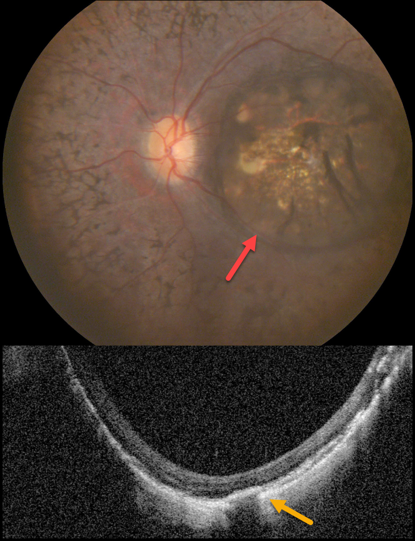
POSTERIOR STAPHYLOMA
Muhammad Arif Ozir
Originally posted on @retina.rocks December 26, 2023
This 20YO male presented with a four-year history of bilateral night blindness and progressive vision loss. Vision was no light perception OU.
Color photography of his left eye shows a variably pigmented colobomatous lesion occupying virtually the entire macula. Macular OCT scanning shows loss of the outer retinal bands, a thinned to non-visualized choroid, and posterior bowing of the sclera. Identical findings were noted in his right eye (not shown).
Learning Points:
Posterior staphylomas are most commonly noted with pathologic myopia, although they have been reported in non-highly myopic eyes with retinitis pigmentosa (Xu et al, Retina 2019;39:1299-1304).
Macular staphylomatous/colobomatous lesions have also been described in other inherited retinal diseases, including North Carolina macular dystrophy, which is caused by an MCDR1 mutation (Small et al, JAMA Ophthalmology 2016;134:355-356).
Unfortunately, our patient was subsequently lost to follow-up, so genetic testing was not possible.
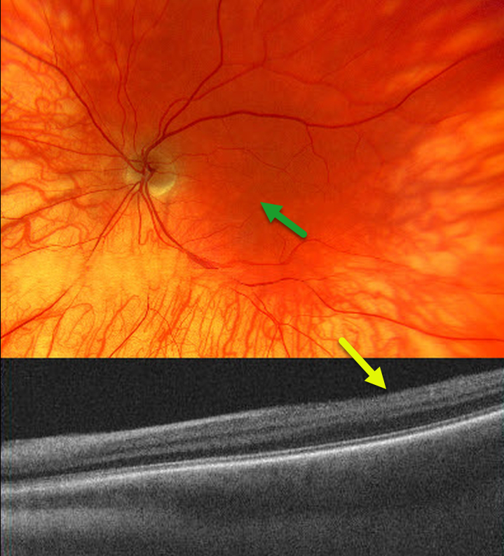
OCULAR ALBINISM
Borivoje Ivezić, Emma Oreškovič, and Nataša Drača
Originally posted on @retina.rocks December 21, 2023
The 53YO male has a known history of ocular albinism, reporting poor visual acuity and horizontal nystagmus since childhood. Vision was 20/200 bilaterally.
Color imaging of the left eye shows a diffusely hypo-pigmented fundus with foveal hypoplasia.
OCT imaging shows a flat foveal contour (fovea plana). Similar findings were noted in the right eye (not shown).
Learning Points:
Ocular albinism is an X-linked recessive disorder that affects only the eyes and is caused by a mutation in the OA1 gene.
Findings seen in ocular albinism include strabismus, nystagmus, iris transillumination defects, blonde fundus, and foveal hypoplasia. There are often a higher number of crossed nerve fibers at the optic chiasm.
Fovea plana is characterized by the absence of the foveal pit, which is formed embryologically by the inner retinal tissues and vasculature being displaced centrifugally. It is most commonly associated with albinism, prematurity, or as an isolated finding. Despite the abnormal foveal architecture, vision is usually normal, unless due to albinism.
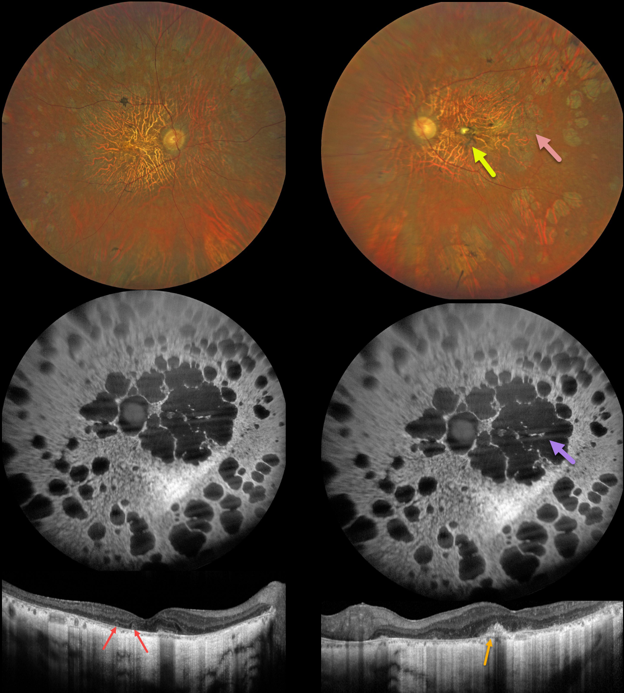
INHERITED RETINAL DISEASE
Yuenpang Cheung, Stephanie Choi and Stephen Tsang
Originally posted on @retina.rocks November 14, 2023
This 56YO male has a history of nyctalopia since childhood with a known mutation in the RDS/peripherin gene. Vision was 20/60 in the right eye and counting fingers in the left eye. The patient has been taking vitamin A supplementation. He has an older brother with a more advanced inherited retinal disorder (IRD).
Fundus imaging shows bilateral diffuse areas of retinal pigment epithelial atrophy. In addition, there is a yellow elevated dome-like lesion with surrounding pigment in the left fovea.
Fundus autofluorescence (FAF) shows hypo-FAF in the areas of atrophy. OCT shows diffuse outer retinal/RPE atrophy with choroidal thinning. In addition, there are a few outer retinal tubulations OD and a subretinal hyperreflective elevated lesion OS. Electrophysiology revealed extinguished rod and diminished cone responses.
Learning Points:
Mutations in the RDS/peripherin gene have been implicated in a number of inherited conditions, including retinitis pigmentosa, pattern dystrophy, retinitis punctata albescens, cone-rod dystrophy, and adult-onset vitelliform macular dystrophy.
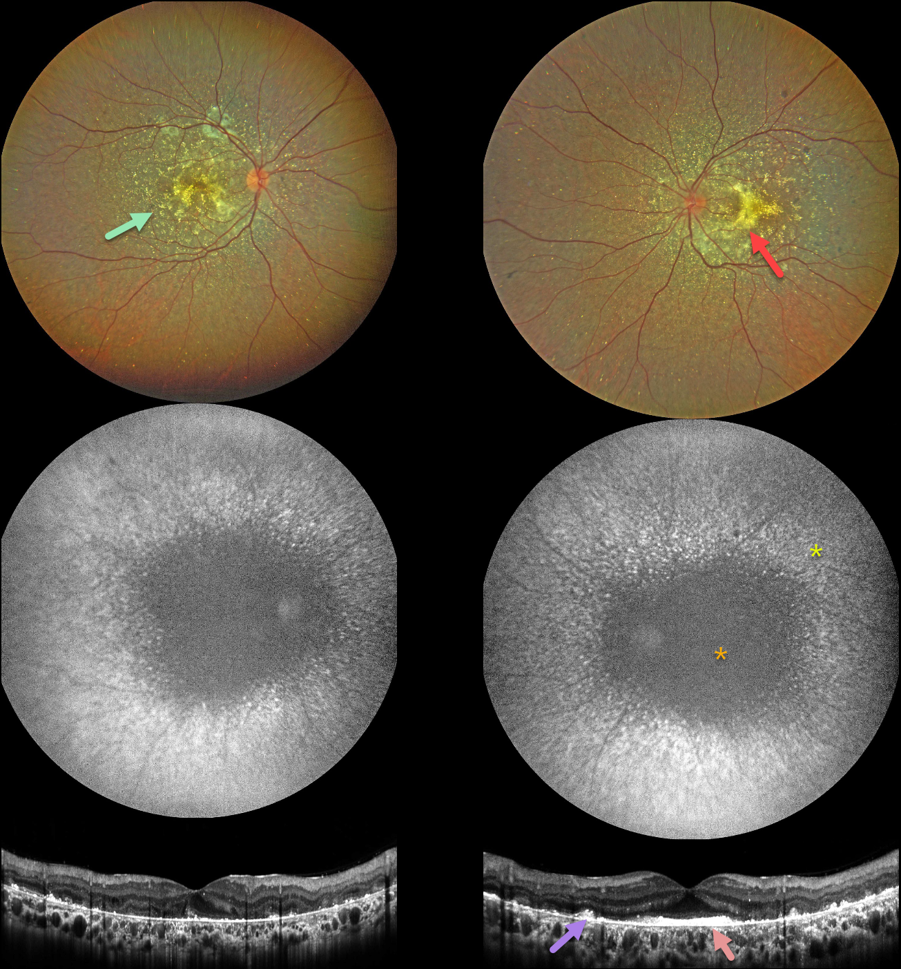
BIETTI CRYSTALLINE DYSTROPHY
Yuenpang Cheung, Stephanie Choi, and Stephen Tsang
Originally posted on @retina.rocks November 7, 2023
This 28YO Egyptian male reported blurry vision, nyctalopia, and photosensitivity. His great-grandmother was blind from an unknown etiology. Vision was 20/40 OD and 20/60 OS.
Anterior segments were notable for bilateral limbal crystalline deposits (not shown). Fundus imaging shows extensive subretinal crystalline deposits primarily in the posterior poles bilaterally, with areas of subretinal fibrosis.
Fundus autofluorescence (FAF) shows central hypo-FAF with radiating dots of more peripheral hyper-FAF.
OCT scanning shows hyperreflective dots overlying the RPE-Bruch’s membrane complex, with variable outer retinal and RPE loss. A placoid area of subfoveal hyperreflectivity is also noted OS.
Genetic testing revealed pathogenic heterozygous CYP4V2 mutations, consistent with Bietti crystalline dystrophy.
Learning Points:
Bietti crystalline dystrophy is an autosomal recessive ocular disorder that affects both the anterior and posterior segments. Clinical features include crystalline deposits in the retina, cornea, and rarely the crystalline lens, as well as retinal pigment epithelial clumping and atrophy.
Mutations in CYP4V2, which cause dysfunctional lipid metabolism, have been implicated in its pathogenesis. While there is no definitive treatment, anti-VEGF therapy can be administered for secondary macular neovascularization. See Saatci et al for a recent review (Clinical Ophthalmology 2023;17:953-967).
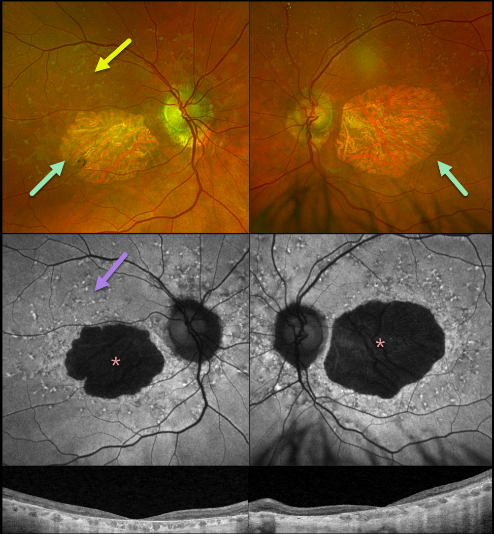
STARGARDT DISEASE
Originally posted on @retina.rocks October 10, 2023
This 83YO female presented for a retinal examination without a new complaint. There was a known history of Stargardt disease from a pathogenic ABCA4 mutation. Vision was stable at 20/200 OD and 20/400 OS.
Optos color RG imaging shows bilateral macular atrophy with yellow subretinal flecks more peripherally. Fundus autofluorescence (FAF) shows hypo-FAF from the macular atrophy with variable hyper-FAF from the subretinal flecks. Diffuse outer retinal atrophy is seen on Triton swept-source OCT.
Learning Points:
Stargardt disease is an autosomal recessive disorder caused by a mutation in the ABCA4 gene. The ABCA4 protein is located in the photoreceptor outer segments and is involved in the recycling of 11-cis-retinal. The mutation in this gene causes lipofuscin accumulation in the RPE, which eventually leads to photoreceptor and RPE degeneration. Other ABCA4 disorders include fundus flavimaculatus, cone-rod dystrophy, retinitis pigmentosa, and age-related macular degeneration.
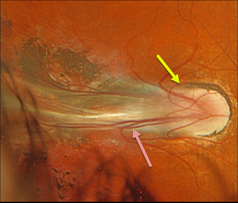
FAMILIAL EXUDATIVE VITREORETINOPATHY (FEVR)
Originally posted on @retina.rocks October 3, 2023
This 22YO female presented with a lifelong history of stable severe bilateral vision loss, congenital nystagmus, and a prior inoperable retinal detachment in her left eye. There was no past medical or family history of eye disease.
Optos color RGB imaging of her right eye shows temporal dragging of the disc and vessels from a falciform retinal fold extending towards the periphery. Secondary surrounding pigmented chorioretinal scarring is noted.
The left eye had a dense white cataract with no view of the posterior pole. Genetic testing was positive for a pathogenic KIF11 mutation.
Learning Points:
Originally described by Criswick and Schepens (AJO 1969;58;578-594), familial exudative vitreoretinopathy (FEVR) can be inherited as an autosomal-dominant, recessive, or X-linked trait with high penetrance and variable expressivity.
FEVR is characterized by peripheral temporal retinal avascularity, lipid exudation, neovascularization, tractional retinal detachment, and temporal dragging of the macula and retinal vessels. These findings are somewhat similar to those found with retinopathy of prematurity (ROP).
There are numerous genes associated with FEVR, including LRP5, FZD4, NDP, TSPAN12, ZNF408, CTNNB1 and KIF11 [Tao et al, Invest Ophthalmol Vis Sci 2021;62(15);4].
The exact mechanism by which KIF11 mutations cause FEVR is not fully understood. However, it is thought that mutations in the KIF11 gene disrupt the transport of vesicles and organelles essential for retinal blood vessel development.
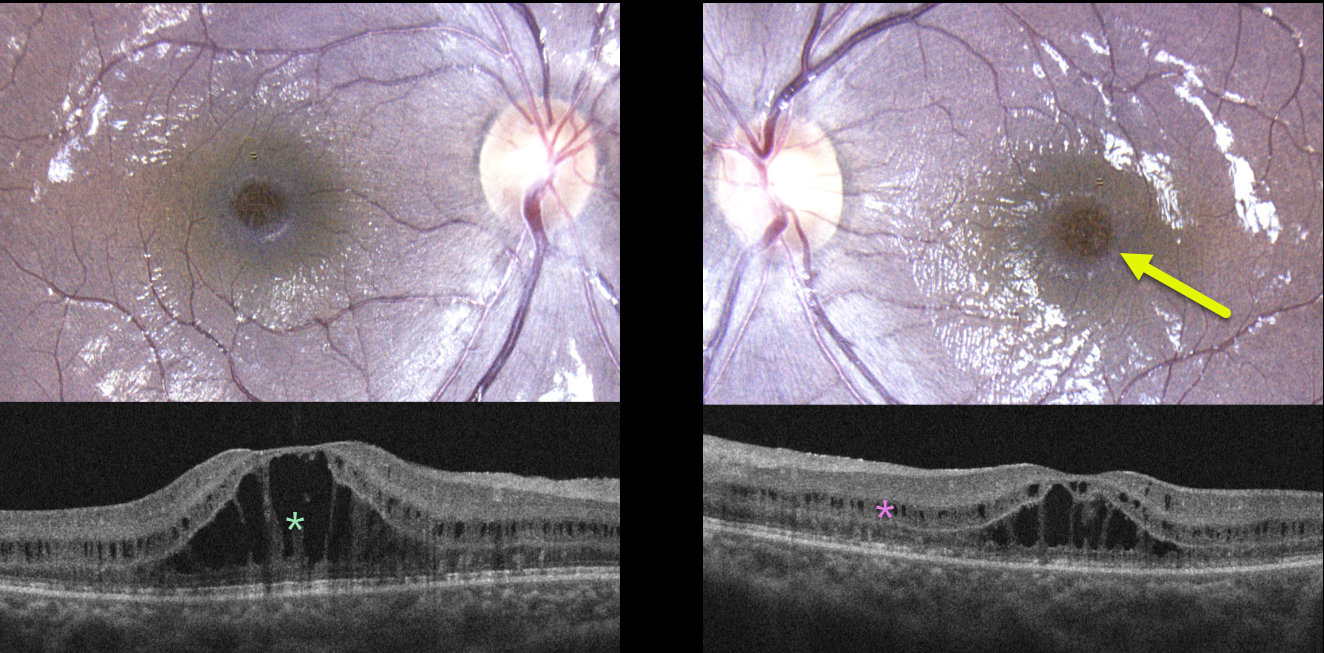
CRB1-RELATED FOVEOSCHISIS
Mattie Adams
Originally posted on @retina.rocks September 19, 2023
This healthy 12YO girl was referred for bilateral macular changes. She was having some mild subjective central vision loss. Vision was 20/40 OD and 20/30 OS. There was no ocular family history.
Color photography shows symmetrical radiating foveoschisis. OCT scanning shows central outer nuclear layer schisis with milder, more peripheral inner nuclear layer schisis.
Learning Points:
X-linked retinoschisis is caused by a mutation of the RS1 gene, which is located on the X chromosome and encodes for retinoschisin, a protein likely involved in retinal cellular adhesion (Heymann et al, Progress Retinal Eye Research 2023;95;101147).
Although usually transmitted as an X-linked recessive disorder found in males, non-sex-linked foveoschisis is present in other heritable disorders, such as enhanced S-cone syndrome and autosomal recessive bestrophinopathy, or non-heritable disorders, such as stellate nonhereditary idiopathic foveomacular retinoschisis.
Genetic testing of our patient revealed two pathogenic mutations of CRB1. Although few cases have been described (Vincent et al, Invest Ophthalmol Vis Sci 2016;57;2637-2646), CRB1-related foveoschisis appears to have a high female preponderance.
Similar to X-linked retinoschisis, these patients tend to maintain good vision into early adulthood, but can progress to secondary macular atrophy later in life with collapse of the schisis.
Thankfully, the foveoschisis phenotype represents the mildest of CRB1-related ocular diseases, which include Leber’s congenital amaurosis, retinitis pigmentosa, early-onset retinal dystrophy, and cone-rod dystrophy (Varela et al., AJO 2023;246:107-121).
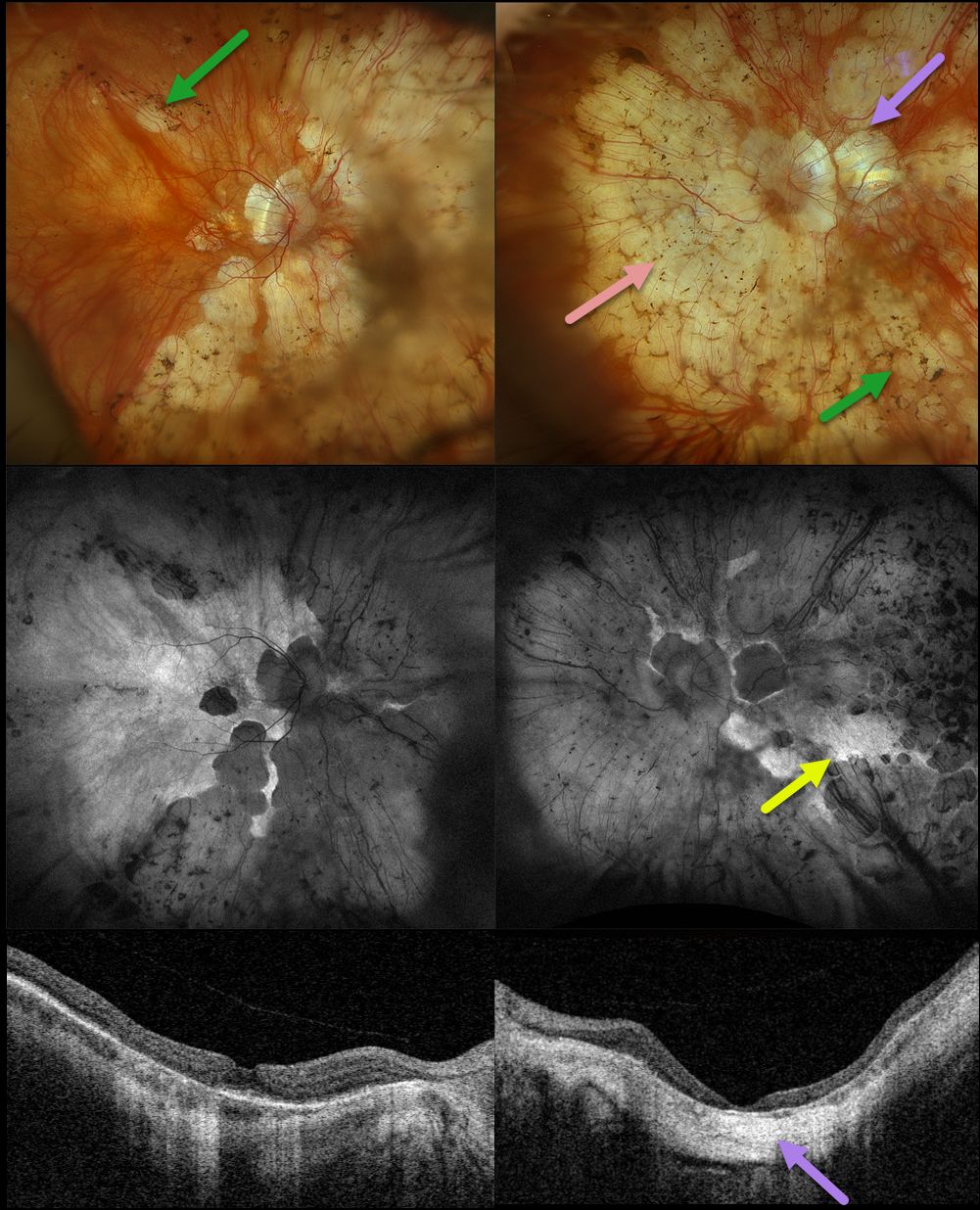
RETINITIS PIGMENTOSA + MYOPIC DEGENERATION
Originally posted on @retina.rocks August 30, 2023
This 78YO female has been followed by us for several years for retinitis pigmentosa (RP) and degenerative myopia. Vision is stable at 20/30 OD and hand motion OS. Multimodal imaging shows a fascinating fusion of findings for these two disorders.
Optos color RGB imaging shows typical RP findings of bone spicules (intraretinal pigment migration) extending from outside the arcades into the peripheries, superimposed on severe degenerative myopic changes.
Fundus autofluorescence (FAF) shows hypo-FAF within the areas of myopic atrophy, with hyper-FAF more peripherally from the RP-induced outer retinal atrophy.
A macular staphyloma is also seen in the left eye and confirmed on swept-source OCT. The degenerative myopic changes just spare the right macular center, which has no staphyloma.
Learning Points:
Genetic testing showed pathogenic heterozygous mutations for OCA2 and RPGR. Fortunately, the RP spares each macula. Her myopic degeneration has been stable, and we are hopeful that her central vision will be preserved in her right eye.
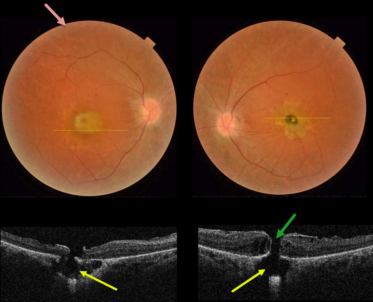
RETINITIS PIGMENTOSA WITH CHOROIDAL CAVITATIONS
Omar Mulki and Faisal Fayyad
Originally posted on @retina.rocks August 16, 2023
This 32YO female presented with a known history of severe vision loss from retinitis pigmentosa (RP). Her parents are first-degree cousins, and there was no family history of RP. Vision was hand motion OD and counting fingers OS.
Fundus photography shows bilateral irregular yellow central macular pigmentary changes, with a central hyperpigmented spot OS.
OCT scanning through each macula shows a full-thickness macular hole with an underlying defect in the RPE. Each macular hole is suspended over a choroidal cavitation.
Surgical repair was offered with appropriate counseling regarding the poor visual prognosis, and the patient elected to be observed.
Learning Points:
Initially described by Freund et al (Arch Ophthalmol 2003;121:197-204), intrachoroidal cavitations are most commonly found in high myopes as a peripapillary yellow-orange subretinal lesion.
Ornek and Ornek reported a patient with pathologic myopia and a unilateral macular hole communicating with a choroidal cavitation (Retinal Cases 2020;14:328-330).
Although macular holes can rarely be associated with RP (Jin et al, Retina 2008;28:610-614), to the best of our knowledge, ours is the first occurrence of RP with bilateral macular holes and underlying choroidal cavitations.
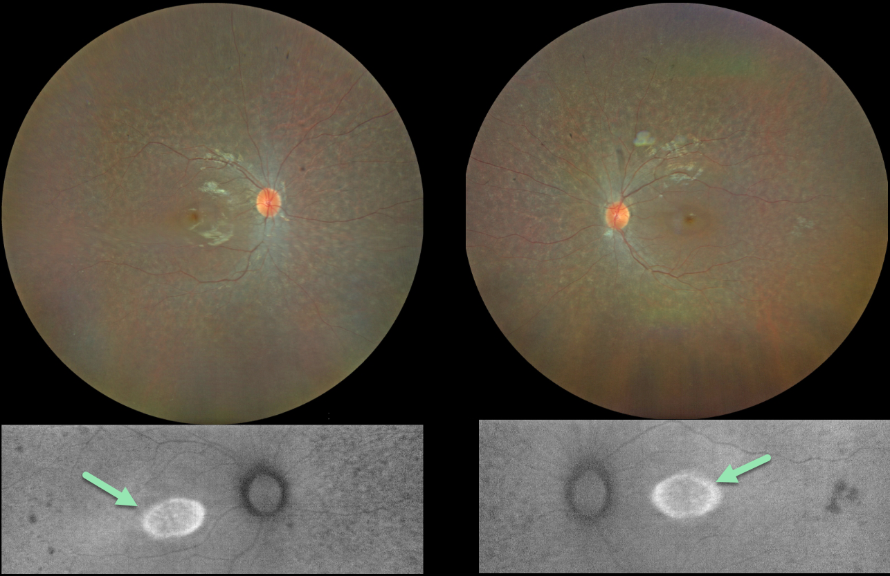
RETINITIS PIGMENTOSA
Yuenpang Cheung
Originally posted on @retina.rocks August 9, 2023
This 13YO girl has a history of congenital deafness, autism, and ADHD. Vision was 20/30 OU.
Color imaging shows bilateral and symmetrical moth-eaten pigmentary changes with an oval area of foveal hyper-autofluorescence.
Fundus autofluorescence (FAF) visualizes endogenous fluorophores, compounds that spontaneously fluoresce without an external dye.
The main fundus fluorophore is lipofuscin, which resides within the RPE lysosomes. The photoreceptor outer segments absorb some of the autofluorescent excitatory light, and thus normally diminish the FAF signal.
In inherited retinal diseases (IRDs), loss of outer segments unmasks this signal, leading to increased FAF. Many patients with an IRD will show a hyper-FAF ring between clinically normal and abnormal retina, which likely represents retina ‘at risk’, showing the junction of a functional and dysfunctional retina (Oishi et al, Int J Retina Vitreous 2019;5(S1);23).
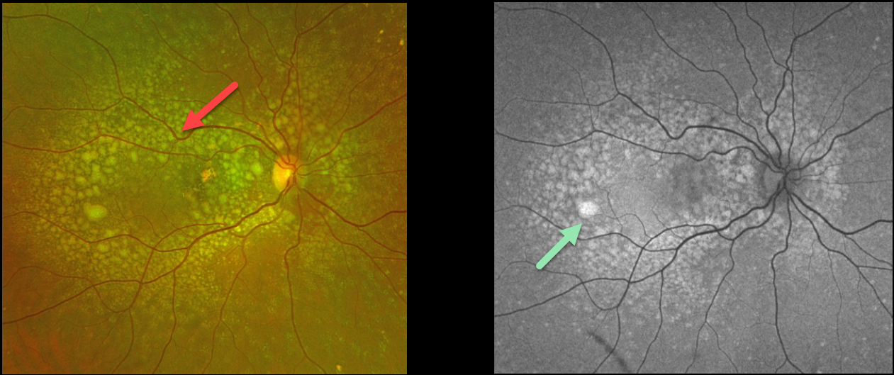
DOMINANTLY INHERITED RADIAL BASAL LAMINAR DRUSEN
Originally posted on @retina.rocks August 3, 2023
This 53YO female presented with these asymptomatic macular changes. She denied any family history of eye disease. Vision was 20/30 OD and 20/25 OS.
Optos color RG imaging shows extensive mixed drusen throughout the right posterior pole. The drusen hyper-autofluoresce.
OCT B-scan shows the sub-RPE drusen indenting and extending into the outer nuclear layer and up to the outer plexiform layer in some places. Similar findings were noted in her left eye (images not shown).
Learning Points:
Doynes, also known as dominantly inherited radial basal laminar drusen or Malattia Leventinese, is a rare macular disorder caused by a mutation in the EFEMP1 gene on chromosome 2p16. The EFEMP1 protein is a member of the fibulin family of extracellular matrix glycoproteins. The defective protein creates an abnormally thickened RPE basement membrane.
Centrally large, nodular, and confluent drusen develop, along with a temporal radiating pattern of smaller cuticular drusen. Later, variable amounts of RPE hyperplasia and fibrous metaplasia may be present. Macular neovascularization may also occur.
Fundus autofluorescence (FAF) usually shows hypo-FAF in age-related drusen. The lesions in Doyne’s, as in this patient, usually show hyper-FAF, likely from unmasking of the RPE by overlying outer retinal thinning.
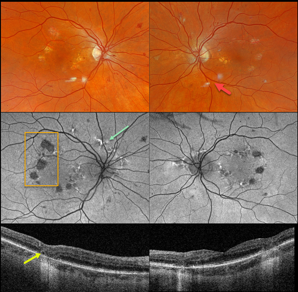
MATERNALLY INHERITED DIABETES AND DEAFNESS (MIDD)
Originally posted on @retina.rocks July 20, 2023
This 66YO female was seen for a diabetic retinopathy screening. She had a prior history of age-related macular degeneration (AMD). There were no visual symptoms. Vision was 20/40 OD and 20/30 OS.
Optos color RGB imaging shows moderate non-proliferative diabetic retinopathy (NPDR) with some scattered retinal hemorrhages and nerve fiber layer infarcts. Some focal areas of non-central macular atrophy are also seen.
Fundus autofluorescence (FAF) shows hypo-FAF from the areas of atrophy, along with areas of hyper-FAF around each nerve and macula.
Swept-source OCT shows outer retinal and RPE atrophy. A small area of outer retinal tubulation (ORT) is noted along the edge of an area of atrophy OD.
On further questioning, the patient’s mother had type 2 diabetes. Hearing was normal in our patient and her mother. Genetic testing of our patient showed a pathologic mitochondrial A3243G mutation.
Learning Points:
Maternally inherited diabetes and deafness (MIDD) accounts for up to 3% of all cases of diabetes and results from a mutation in mitochondrial DNA at position A3243G. MIDD often masquerades as a pattern macular dystrophy.
In our experience, fundus autofluorescence (FAF) is the best way to visualize these changes. The FAF appearance somewhat resembles that seen with Elmiron toxicity. Peripapillary hypoautofluorescence, more densely packed macular autofluorescent changes, and earlier central macular involvement suggest Elmiron toxicity over other causes (see Barnes et al Ophthalmology Retina 2020;4:1196-1201).
ORT is often noted overlying inactive MNV with ongoing anti-VEGF therapy and should not be confused with exudative fluid or cysts, which lack a hyperreflective border. The outer hyperreflective band likely represents inner segment mitochondria undergoing fission and translocation toward the nucleus (Litts et al, Retina 2018;38:445-461).
ORT, initially described by Zweifel et al (Arch Ophthalmol 2009;127:1596-1602), is a neurodegenerative condition of the photoreceptors and Muller cells associated with atrophy affecting the outer retina and retinal pigment epithelium, including advanced AMD and inherited retinal diseases.
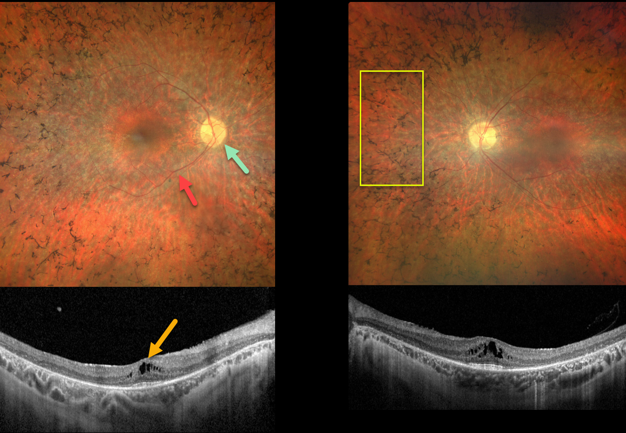
USHER SYNDROME
Emma Oreskovic and Natasa Draca
Originally posted on @retina.rocks July 19, 2023
This 33YO female has a known history of retinitis pigmentosa (RP) and congenital neurosensory hearing loss. Her nyctalopia and peripheral vision loss are stable, and vision was 8/20 OD and 15/20 OS.
Color photography of both eyes shows symmetrical pigmentary changes and intraretinal pigment migration (bone spicules) extending from the peripheral maculas into the retinal peripheries. The central maculas look fairly normal, but show schisis in the central inner nuclear layer. The nerves are slightly pale, and the retinal vessels are attenuated.
Genetic testing was heterozygous for a PCHD15 pathogenic variant.
Learning Points:
Usher syndrome (USH) affects over 400 000 persons worldwide and is a major cause of deafness with blindness. Ten USH genes have been identified to date, and three clinical subtypes (USH1-3) have been described. Mutations in the PCDH15 gene that encode for the protocadherin-15 protein cause autosomal recessive Usher syndrome type 1.
Although our patient clinically has USH, genetic testing failed to reveal a causative gene (she only had a single pathogenic gene for a recessive disorder). The negative genetic test result does not mean this patient doesn’t have USH; rather, it reminds us that this test must be interpreted with caution and always with the clinical findings in mind (see Mustafi et al, Ophthalmology Retina 2022;6:702-710).
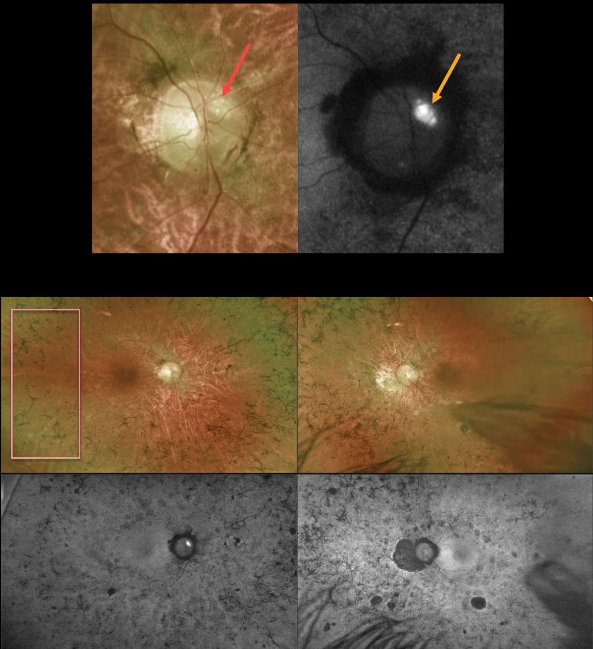
OPTIC NERVE DRUSEN + RETINITIS PIGMENTOSA
Originally posted on @retina.rocks April 17, 2023
This 43YO male with a known history of retinitis pigmentosa (RP) was referred for further evaluation. He noted a gradual worsening of vision bilaterally. Vision was 20/80 OD and 20/200 OS.
Optos color imaging of the right optic nerve shows several large optic disc drusen (ODD) that markedly hyperautofluoresce. Ultrawide-field imaging shows diffuse, classic bilateral RP scarring.
Learning Points:
ODD are refractile calcific concretions that form anterior to the lamina cribrosa. They are thought to be extruded, extracellular, calcified mitochondria and may be inherited as an autosomal dominant trait. They are present in about 3% of RP patients (Russell et al, Eye 2022;36:2213).
Most patients are asymptomatic, but some can develop progressive visual field defects or central vision loss. There is no treatment for the drusen themselves, although symptomatic macular neovascularization can be treated with thermal laser, photodynamic therapy, or anti-VEGF injections.
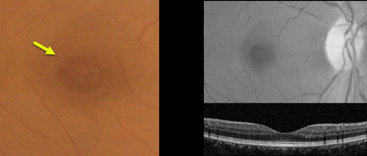
BENIGN YELLOW DOT MACULOPATHY
Asma Samsudeen and Ashish Sharma
Originally posted on @retina.rocks March 21, 2023
This 49YO female presented with asymptomatic macular changes. Vision was 20/30 OD and 20/40 OS. There was no past medical history and no family history of eye disease.
Color imaging shows tiny yellow perifoveal dots, which are somewhat better seen with red-free imaging. OCT scanning is unremarkable.
Learning Points:
Benign yellow dot maculopathy was reported as a new nonprogressive macular phenotype by Borman et al (Ophthalmology 2017;124:1004-1013).
Patients present with normal vision and asymptomatic tiny yellow foveal dots. These dots are hyperautofluorescent, but OCT scanning is generally normal. There is no known causative gene.
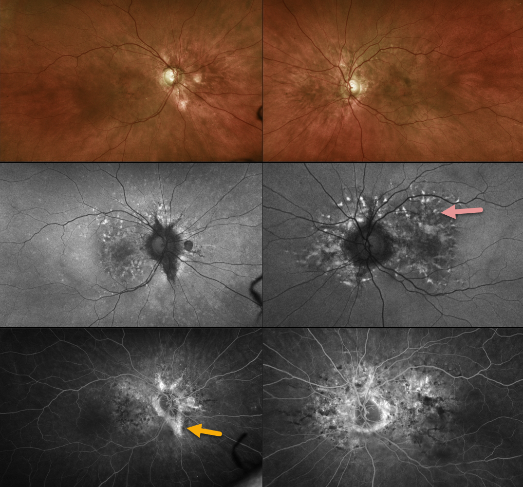
PSEUDOXANTHUM ELASTICUM WITH ANGIOID STREAKS AND PATTERN DYSTROPHY PHENOTYPE
Originally posted on @retina.rocks March 20, 2023
This 55YO female has been followed for years with macular pattern dystrophy-type changes due to pseudoxanthoma elasticum (PXD). Vision is 20/25 OU.
Color imaging shows variable mild pigmentary changes. Optos fundus autofluorescence imaging shows variable hyperautofluorescent flecks with variable angiographic staining. Some staining angioid streaks are noted in her right eye.
Learning Points:
Retinal findings in PXE most commonly include angioid streaks and a subretinal orange skin (peaux d’orange) appearance, most pronounced in each temporal macula.
However, pattern dystrophy-like changes, as in our patient, can sometimes be the predominant phenotype in some PXE patients (Murro et al, Graefe’s 2020;258:1881-1892). As with typical pattern dystrophies, the findings are dynamic and best captured with multimodal imaging.
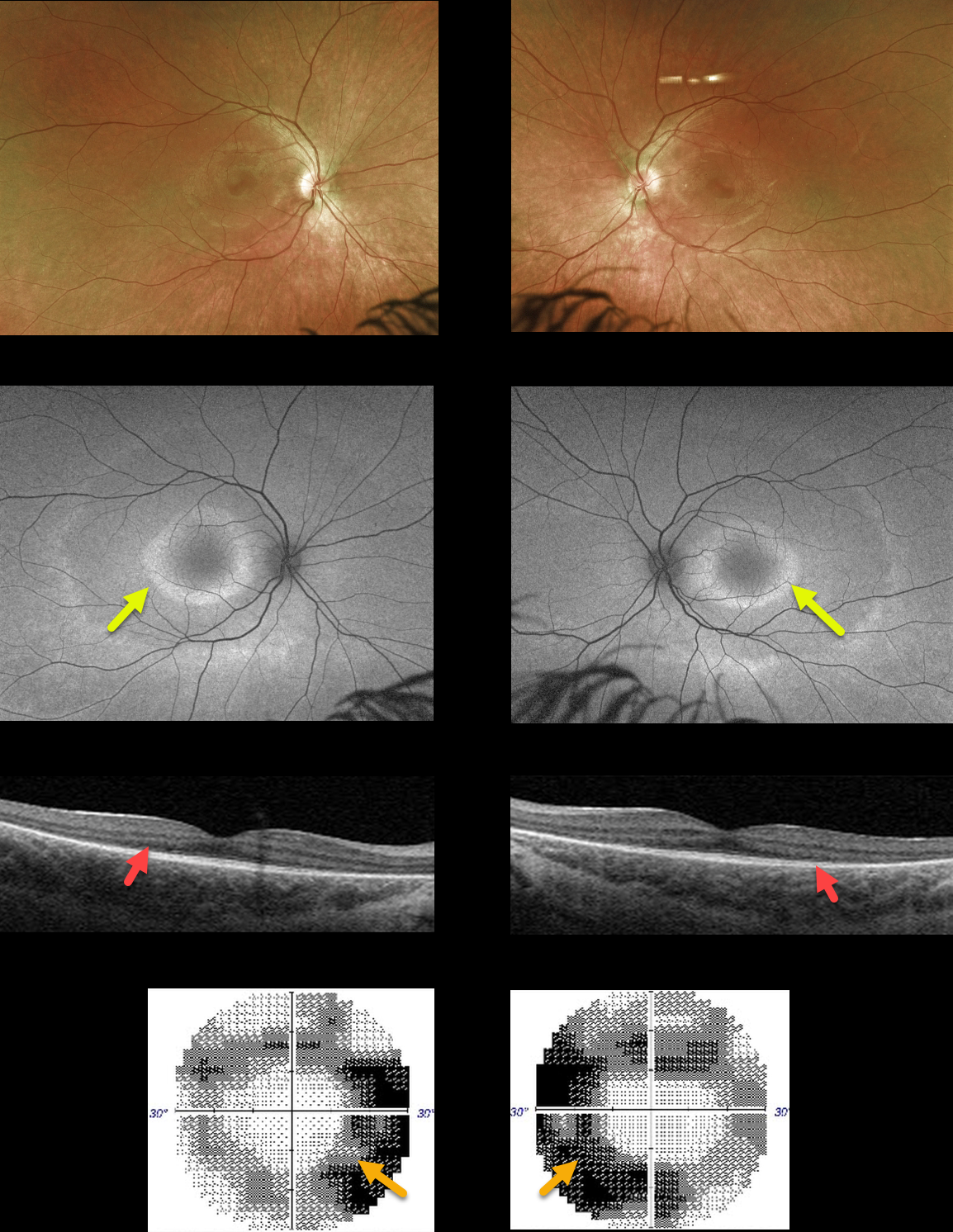
RETINITIS PIGMENTOSA
Originally posted on @retina.rocks March 14, 2023
This 22YO healthy female presented with a year or two of mildly symptomatic problems with distance vision. Vision was 20/25 OD and 20/30 OS. There was no family history of eye disease, and her hearing was normal.
Optos color imaging shows bilaterally symmetrical doughnut-shaped rings of subtle pigmentary changes around each macular center. Fundus autofluorescence (FAF) shows these rings to be hyper-FAF. Swept-source OCT shows outer retinal atrophy. 30-2 visual fields are constricted.
Learning Points:
FAF visualizes fluorophores, endogenous compounds that spontaneously fluoresce without the need for an external dye. The main fundus fluorophore is lipofuscin, which resides within the RPE lysosomes. The photoreceptor outer segments absorb some of the autofluorescent excitatory light, and thus normally diminish the FAF signal.
In RP, loss of the outer segments unmasks this signal, leading to increased FAF. Many patients with RP will show a hyper-FAF ring between clinically normal and abnormal retina, which likely represents retina ‘at risk’, showing the junction of a functional and dysfunctional retina.
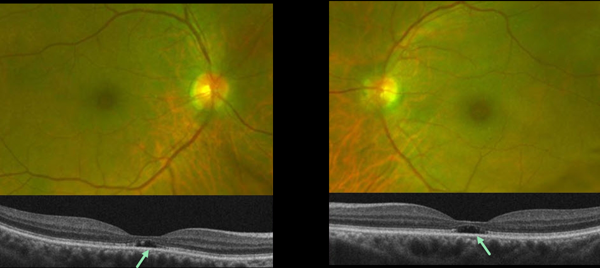
OUTER FOVEAL MICRODEFECTS FROM POSSIBLE IRD
Emily Shepherd
Originally posted on @retina.rocks March 10, 2023
This 65YO presented complaining of difficulty seeing at a distance for some time. She gave a history of radial keratotomy in both eyes about 20 years prior. There was no significant past medical history or family history of eye disease. Vision was 20/60 OD and 20/70 OS.
Optos color imaging shows mild symmetrical pericentral pigmentary change. Fundus autofluorescence and fluorescein angiography were unremarkable (not shown). However, OCT B-scans show bilateral subfoveal loss of the outer retinal tissue external to the external limiting membrane.
An ERG showed rod receptor dysfunction and abnormal dark adaptation with delayed adaptation, while a multifocal ERG was consistent with normal cone function.
Our patient underwent genetic testing and was found to have a heterozygous mutation for FLVCR1. This gene is associated with autosomal recessive posterior column ataxia with retinitis pigmentosa.
A recent report by Kuehlewein et al (Graefe’s Arch Clin Exp Ophthalmol 2019;257:629-638) showed a variety of phenotypes, none of which matched our patient. It is therefore difficult to say whether our patient’s findings are isolated (given the lack of funduscopic, autofluorescence, and angiographic findings beyond the OCT outer defects) or represent an atypical manifestation (given the abnormal electrophysiology) of FLVCR1 disease.
She is scheduled to return for follow-up imaging and visual field testing.
Learning Points:
Outer foveal microdefects (Cohen et al, Ophthalmology Retina 2021;5:553-561) have been described in numerous conditions, including macular telangiectasia, tamoxifen use, ABCA4 disorders (cone-rod dystrophies, Stargardt disease, and fundus flavimaculatus), phototoxicity, trauma, and vitreomacular traction disorders.
In our experience, in patients with inherited retinal diseases (IRD), these findings are most common with a pathogenic ABCA4 mutation (Leng et al, Retina 2012;32:1411-1419 and Wang et al Invest Ophthalmol Vis Sci 2022;53(5):28). These defects, caused by focal loss of the outer retinal layers, leave an optically empty space which can be confused with subretinal fluid but are more of an optical gap within the outer retina.
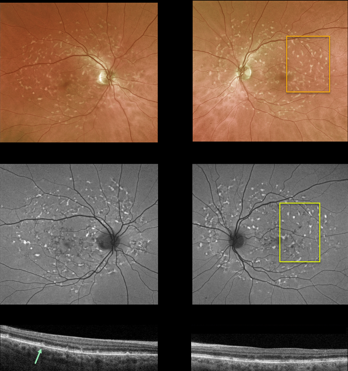
FUNDUS FLAVIMACULATUS
Originally posted on @retina.rocks February 21, 2023
This 55YO female was referred for a possible white dot syndrome. She had no ocular symptoms, and vision was 20/30/ OU. There was no family history of ocular disease.
Optos color imaging shows multiple yellow fleck-like lesions throughout each posterior pole. Fundus autofluorescence of these lesions shows variable hyper-FAF.
OCT scanning shows scattered defects in the EZ with hyperreflective dots extending from the RPE into the EZ and outer nuclear layer.
Learning Points:
Fundus flavimaculatus is a genetic disorder, usually transmitted as an autosomal recessive trait, that is caused by mutations involving the ABCA4 gene. This gene encodes one of a family of ATP-binding cassette (ABC) transmembrane proteins, which mediate the active transport of various substrates across cellular membranes. The ABCA4 protein is present in the photoreceptor outer segment disc membranes, where it participates in the recycling of 11-cis-retinal.
Stargardt disease is believed to be fundus flavimaculatus without peripheral findings. Vision in fundus flavimaculatus is often better than that in Stargardt due to later disease onset and less macular involvement. Both likely represent an RPE lipofuscin storage disease. The RPE cells become engorged with lipofuscin, causing a dark choroid on fluorescein angiography.
See Cremers et al (Progress Retinal Eye Research 2020;79;100861) for a wonderful review of the complex spectrum of ABCA4 disorders.
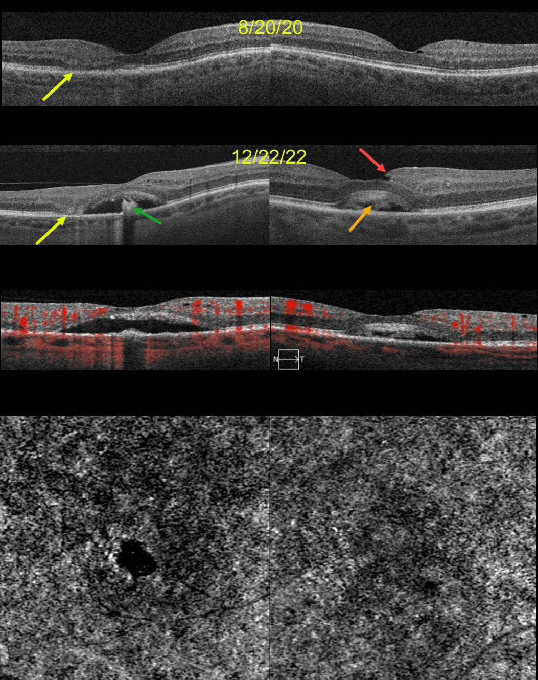
ACQUIRED VITELLIFORM LESION
Originally posted on @retina.rocks January 30, 2023
This 68YO female was seen on 8/20/20. Vision was 20/70 OD and 20/40 OS. Although there were no visual complaints, OCT scanning showed bilateral defects in the EZ bands, along with some temporal outer retinal atrophy OD.
She returned two years later, complaining of gradual vision loss OU, although vision was stable at 20/70 OD and 20/30 OS. OCT scanning shows bilateral acquired vitelliform lesions (AVL). The lesion OD is mostly scrambled with a hyporeflective space between the RPE and outer retina, along with a central hyperreflective clump on top of the RPE. There is now complete outer retinal atrophy temporally.
The AVL in her left macula shows variable hyperreflective material. An unrelated small partial lamellar macular hole is noted temporally. OCT angiography (OCTA) shows no flow signals from either AVL.
Learning Points:
Vision loss from AVLs most commonly results from lesions collapsing due to secondary atrophy or macular neovascularization, which can develop in about 8% of eyes (Balaratnasingam et al., AJO 2016;172:28-38).
The absence of OCTA flow signals in either eye confirmed that these lesions were avascular, and we continue to follow her.
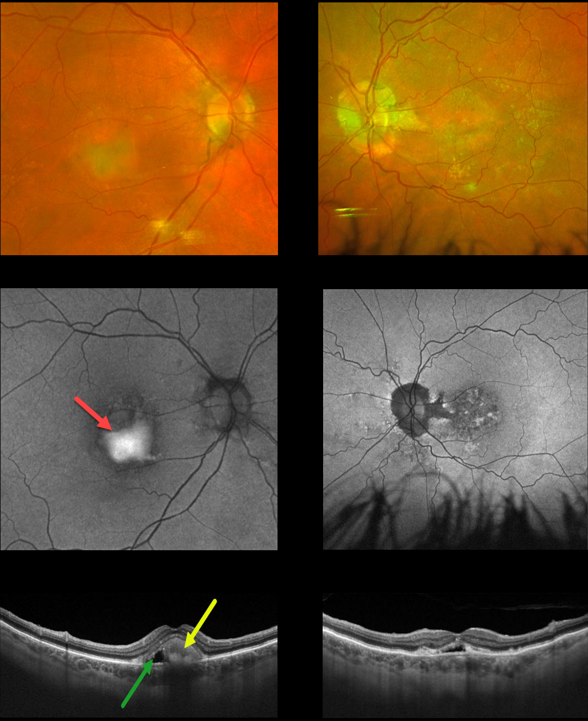
BEST DISEASE
Originally posted on @retina.rocks January 19, 2023
This 75YO male presented with chronic stable central blurriness. Vision was 20/150 OU.
Color photography of his right eye shows superior foveal pigment loss with inferior yellowish, vitelliform material, with more atrophic foveal changes in his left eye.
Fundus autofluorescence (FAF) shows the vitelliform material to be dramatically hyper-autofluorescent OD, with a more speckled hypo-FAF appearance OS.
Swept-source OCT scanning of his right eye shows hyperreflective inferior layering of the vitelliform material with a hyporeflective space more superiorly. Similar OCT findings are also seen in his left eye.
Our patient has progressed to a more atrophic, ‘scrambled egg’ appearance, most evident in his right eye. The remaining vitelliform material gravitates inferiorly, often leaving an optically empty, hyporeflective space between the RPE and outer retina. Eventually, the retina flattens over time with secondary atrophy, which is more pronounced in his left eye.
Learning Points:
Best disease is associated with a mutation in the BEST1 gene, which encodes the bestrophin-1 protein. Bestrophin-1, a calcium-activated chloride channel, is primarily found in the basolateral plasma membrane of the RPE.
BEST1 mutations cause a variety of phenotypes, including autosomal recessive bestrophinopathy, best vitelliform macular dystrophy, and autosomal dominant vitreoretinochoroidopathy.
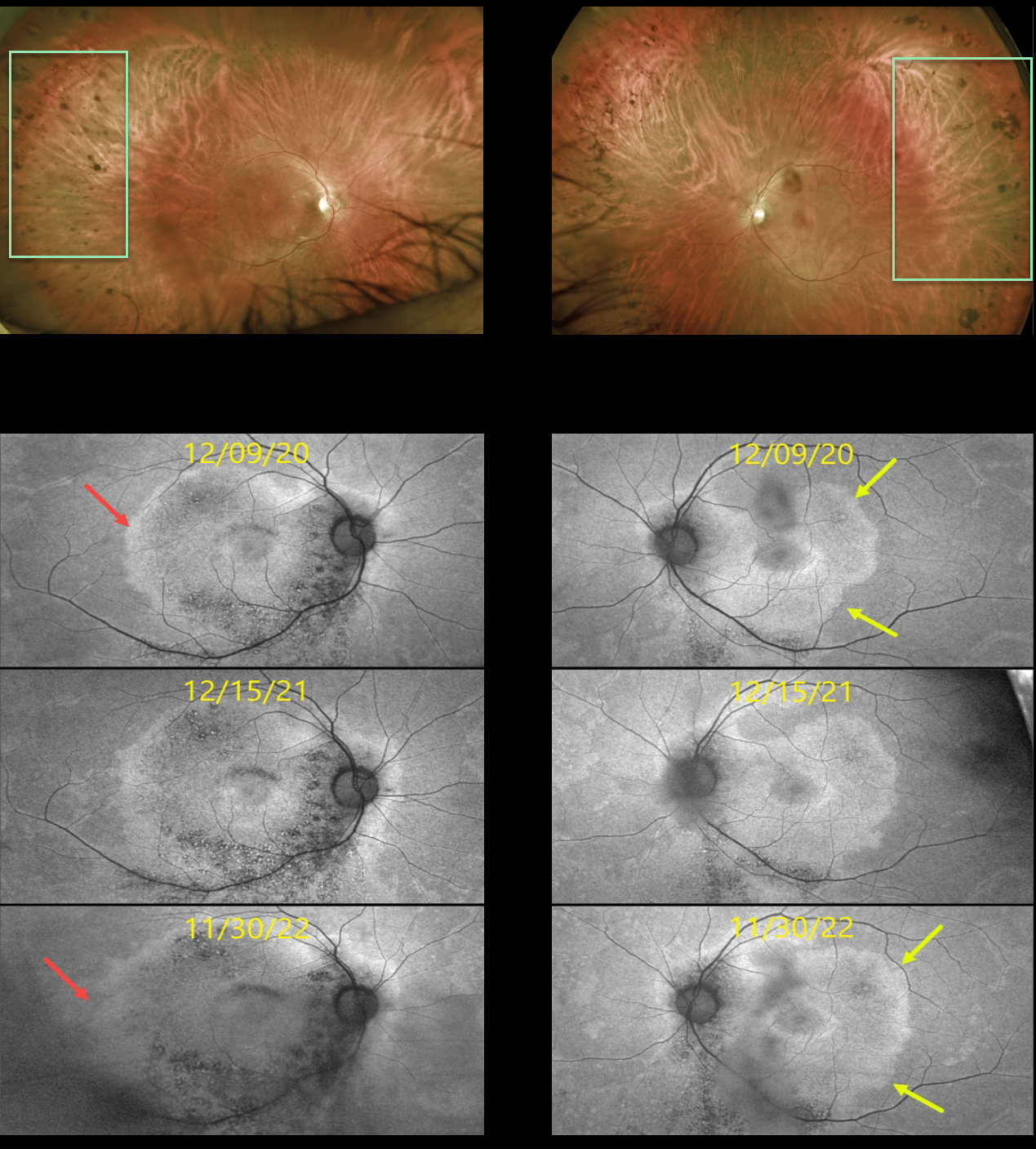
RETINITIS PIGMENTOSA
Originally posted on @retina.rocks January 4, 2023
This 53YO male presented on 12/9/20 with a history of known retinitis pigmentosa (RP). He noticed bilateral blurred vision that had been worsening for about 6 months. Vision was 20/50 OD and 20/30 OS.
Optos color imaging shows fairly normal appearing maculas with a fairly-well circumscribed border of pigmentary loss and migration extending anteriorly from the mid-peripheries.
However, fundus autofluorescence (FAF) reveals subclinical findings of increased macular hyper-FAF sparing each fovea, with hypo-FAF more inferiorly. Subsequent genetic testing revealed a pathogenic RP1 mutation.
FAF shows gradual progressive changes over the 2 years we have been following him. The temporal margins of the hyper-FAF in his right and left eyes have significantly enlarged. This is due to the unmasking of the underlying RPE with progressive photoreceptor atrophy.
Learning Points:
FAF visualizes fluorophores, endogenous compounds that spontaneously fluoresce without the need for an external dye. The main fundus fluorophore is lipofuscin, which resides within the RPE lysosomes.
The photoreceptor outer segments absorb some of the autofluorescent excitatory light, and thus normally diminish the FAF signal.
In RP, loss of the outer segments unmasks this signal, leading to increased FAF. Many patients with RP will show a hyper-FAF ring between clinically normal and abnormal retina, which likely represents retina ‘at risk’, showing the junction of functional and dysfunctional retina.
See Schmitz-Valckenberg et al (Progress Retinal Eye Research 2021;81:100893) for an excellent general review of FAF.
For further reading on monitoring RP with FAF, see Robson et al (Retina 2011;31:1670-1679) and Lee et al (Ophthalmology Retina 2018;2;1062-1070).
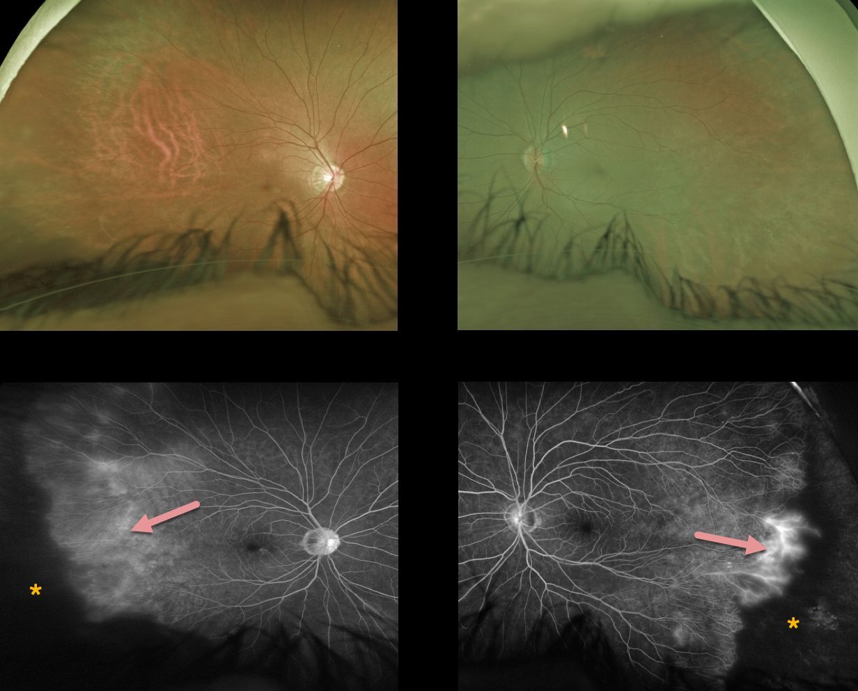
FAMILIAL EXUDATIVE VITREORETINOPATHY (FEVR)
Originally posted on @retina.rocks December 20, 2022
This 53YO male came in for an initial examination since both of his sons and one of his nephews have been followed by us for years with familial exudative vitreoretinopathy (FEVR).
Both sons developed peripheral retinal neovascularization that responded well to scatter laser. Their father was visually asymptomatic.
Optos color imaging shows normal posterior poles with featureless, ischemic temporal retinas. Fluorescein angiography shows marked bilateral peripheral temporal ischemia with leaking vessels at the border of the perfused and ischemic retina.
Prophylactic scatter laser to the ischemic retina was recommended to help prevent retinal neovascularization.
Learning Points:
Originally described by Criswick and Schepens (AJO 1969;58:578-594), familial exudative vitreoretinopathy (FEVR) can be inherited as an autosomal-dominant, recessive, or X-linked trait with high penetrance and variable expressivity.
FEVR is characterized by peripheral temporal retinal avascularity, lipid exudation, neovascularization, tractional retinal detachment, and temporal dragging of the macula and retinal vessels. These findings are somewhat similar to those found with retinopathy of prematurity (ROP).
Kashani et al found that peripheral retinal vascular findings are highly prevalent in asymptomatic FEVR relatives (Ophthalmology 2014;121:262-268), as was the case with our patient. They therefore recommended widefield angiographic screening in immediate relatives.
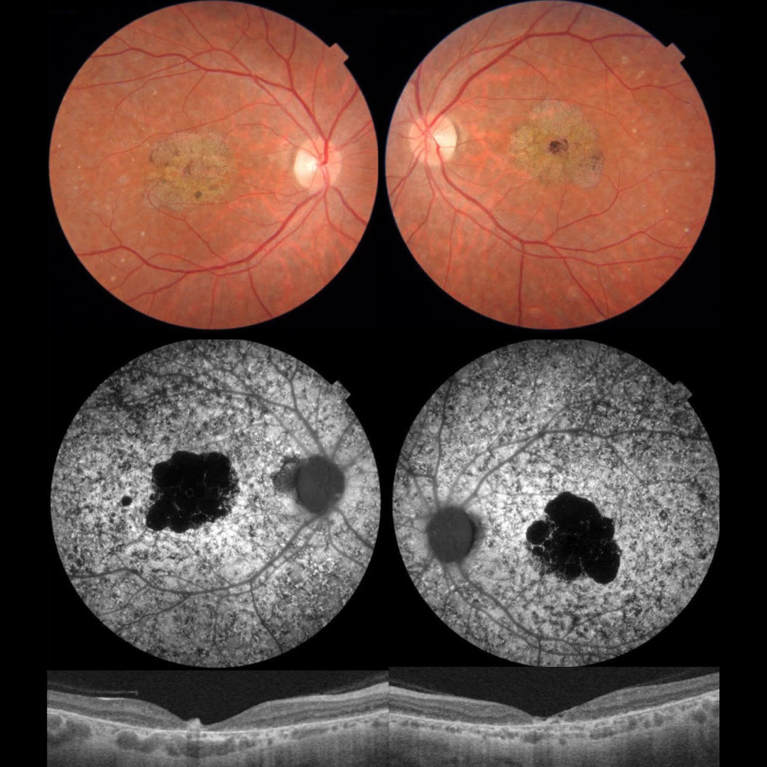
STARGARDT DISEASE
Omar Mulki
Originally posted on @retina.rocks November 25, 2022
This 33YO male presented with counting fingers bilaterally from Stargardt disease. There was a positive family history for Stargardt, and genetic testing in the past was positive for a pathogenic ABCA4 mutation.
Color photographs show a bilateral beaten-bronze appearance at the macular centers, with more subtle pigmentary changes throughout the fundi.
Fundus autofluorescence (FAF) shows hypo-FAF in the macular lesions and, more dramatically, diffuse pigmentary changes elsewhere.
OCT scanning shows marked central foveal retinal thinning, with variable more peripheral outer retinal atrophy.
Learning Points:
Stargardt disease is an autosomal recessive disorder caused by a mutation in the ABCA4 gene. The ABCA4 protein is located in the photoreceptor outer segments and is involved in the recycling of 11-cis-retinal.
The mutation of this gene causes the accumulation of lipofuscin in the RPE, which eventually leads to photoreceptor and RPE degeneration. Other ABCA4 disorders include fundus flavimaculatus and cone-rod dystrophy.
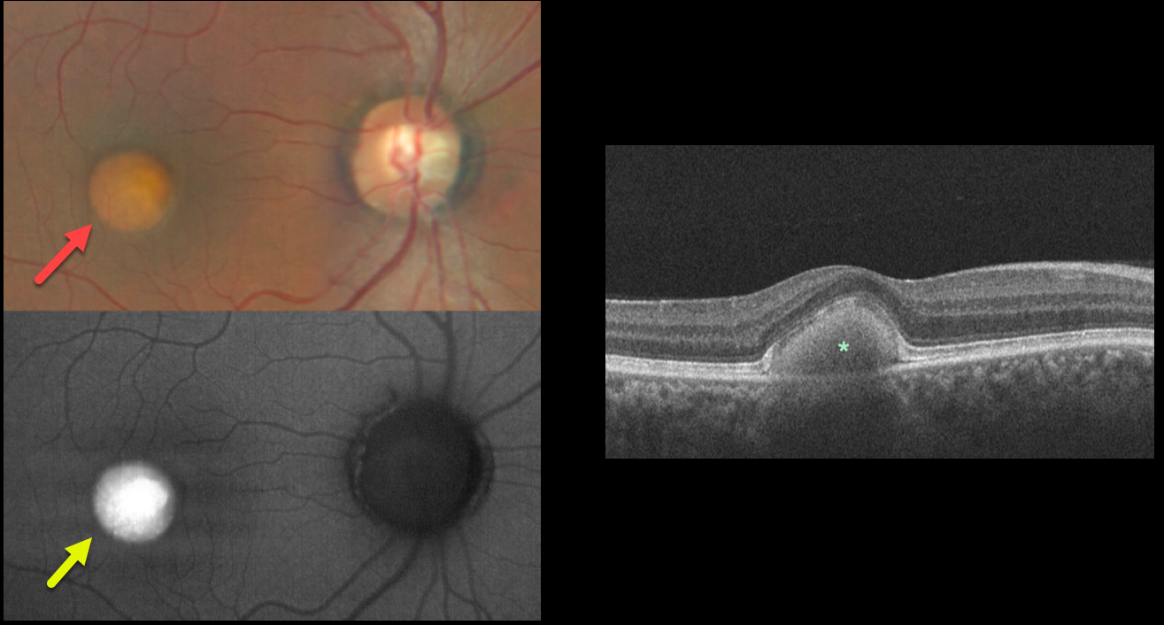
BEST DISEASE
Will Gibson
Originally posted on @retina.rocks November 9, 2022
This 39YO male presented with these asymptomatic subretinal yellow posterior pole lesions. Vision was 20/30 OU.
The sharply demarcated yellowish lesions are markedly hyper-autofluorescent. This vitelliform material is hyperreflective on OCT. The subretinal material extends into and elevates the central retina.
Learning Points:
Best disease is associated with a mutation in the BEST1 gene, which encodes the bestrophin-1 protein.
Bestrophin-1, a calcium-activated chloride channel, is primarily found in the basolateral plasma membrane of the RPE. BEST1 mutations cause a variety of phenotypes, also including autosomal recessive bestrophinopathy, best vitelliform macular dystrophy, and autosomal dominant vitreoretinochoroidopathy.
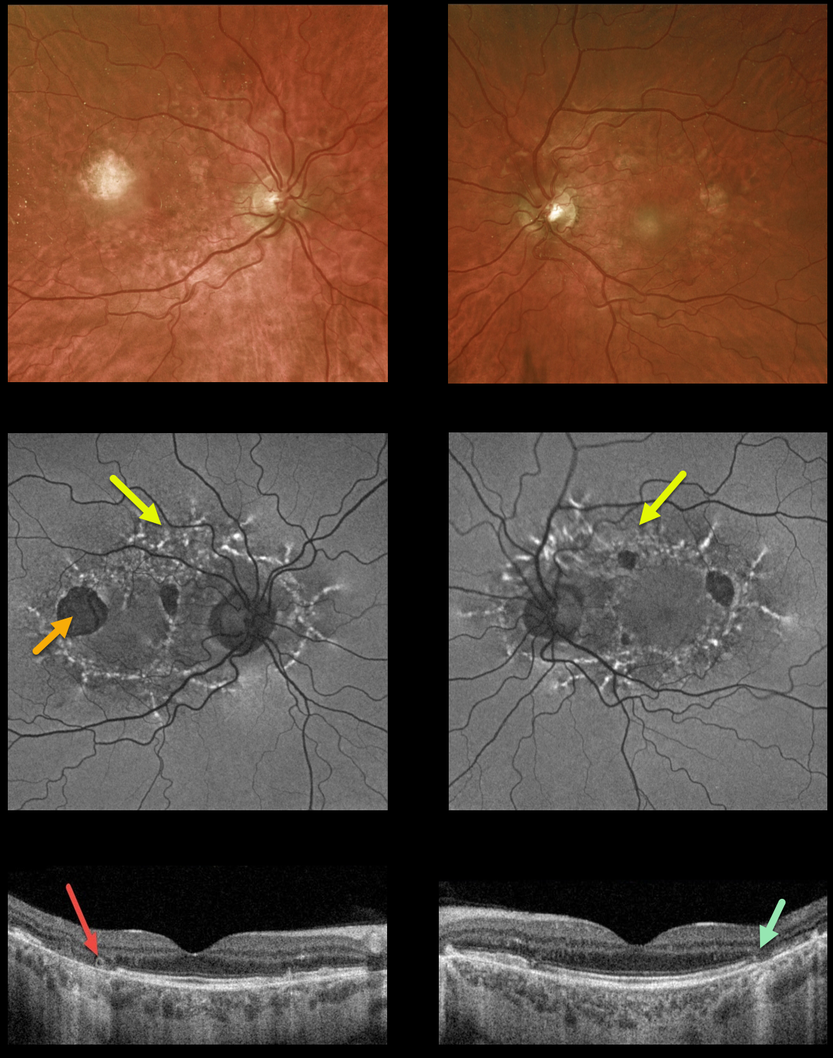
MATERNALLY INHERITED DIABETES AND DEAFNESS (MIDD) + OUTER RETINAL TUBULATION (ORT)
Originally posted on @retina.rocks October 3, 2022
This 59YO male presented with vision of 20/400 OD and 20/30 OS. He has severe lifelong hearing loss, and both he and his mother have type 2 diabetes.
Optos ultrawidefield imaging shows temporal macular atrophy. Fundus autofluorescence shows the true extent of the pathology, with linear interconnected subretinal streaks of hyper-FAF associated with areas of hypo-FAF macular atrophy.
Swept-source OCT shows variable, mostly temporal outer retinal and RPE atrophy, along with a small area of temporal outer retinal tubulation (ORT) OD.
Learning Points: Maternally inherited diabetes and deafness (MIDD) is responsible for up to 3% of all cases of diabetes, and results from a mutation of mitochondrial DNA A3243G. MIDD often masquerades as a pattern macular dystrophy.
Fundus autofluorescence (FAF) in our experience is the best way to visualize these changes. The FAF appearance somewhat resembles that seen with Elmiron toxicity.
Peripapillary hypoautofluorescence, more densely-packed macular autofluorescent changes, and earlier central macular involvement suggest Elmiron toxicity over other causes (see Barnes et al Ophthalmology Retina 2020;4:1196-1201). Note that our case spares the central macula and peripapillary retina, which is more consistent with MIDD.
ORT is often noted overlying inactive MNV with ongoing anti-VEGF therapy and should not be confused with exudative fluid or cysts which lack a hyperreflective border. The outer hyperreflective band likely represents inner segment mitochondria undergoing fission and translocation toward the nucleus (Litts et al, Retina 2018;38:445-461).
ORT, initially described by Zweifel et al (Arch Ophthalmol 2009;127:1596-1602), is a neurodegenerative condition of the photoreceptors and Muller cells associated with atrophy affecting the outer retina and retinal pigment epithelium, including advanced AMD and inherited retinal diseases.
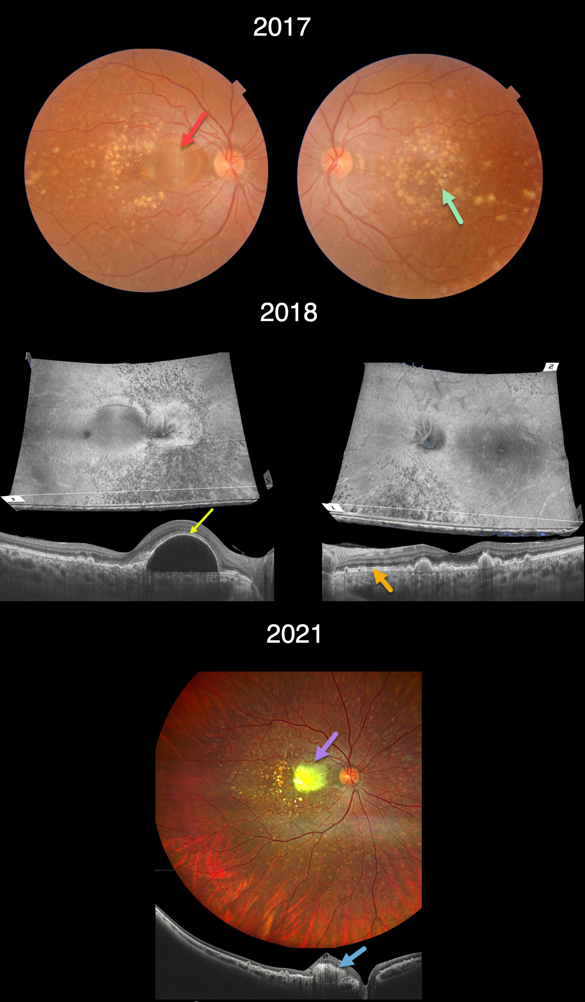
DOMINANTLY INHERITED RADIAL BASAL LAMINAR DRUSEN
Barbara Parolini and Veronika Matello
Originally posted on @retina.rocks September 16, 2022
This 33YOF presented in 2017 with a recent history of vision loss in her right eye. She had no past ocular or medical history, nor family history of eye disease. Vision of 20/70 OD and 20/20 OS.
Triton color imaging shows extensive, mostly large drusen scattered throughout each macula, extending into the midperipheries. A large serous retinal pigment epithelial detachment (PED) is noted in the nasal right macula.
Triton swept-source OCT analysis in 2018 shows numerous unique perspectives. 3D reconstruction reveals extensive lumpy drusen extending throughout the posterior poles, especially outside each macula. B-scans show extensive variably elevated drusen. The large PED is evident in the nasal right macula. The retina external to the ellipsoid zone is thickened and hyperreflective.
Three years later vision decreased to 20/200 OD and was stable at 20/20 OS. The PED became clinically yellow and markedly hyperreflective on OCT (Canon Xephilio OCT-S1). The lesion failed to respond to anti-VEGF therapy.
Learning Points:
Doynes, also known as dominantly inherited radial basal laminar drusen or Malattia Leventinese, is a rare macular disorder caused by a mutation in the EFEMP1 gene on chromosome 2p16.
The EFEMP1 protein is a member of the fibulin family of extracellular matrix glycoproteins. The defective protein creates an abnormally thickened RPE basement membrane.
Centrally large, nodular,and confluent drusen are noted, along with a temporal radiating pattern of smaller cuticular drusen.
Later, there may be variable amounts of RPE hyperplasia and fibrous metaplasia, which is likely the cause of the vision loss in our patient’s right eye. Macular neovascularization may also develop.
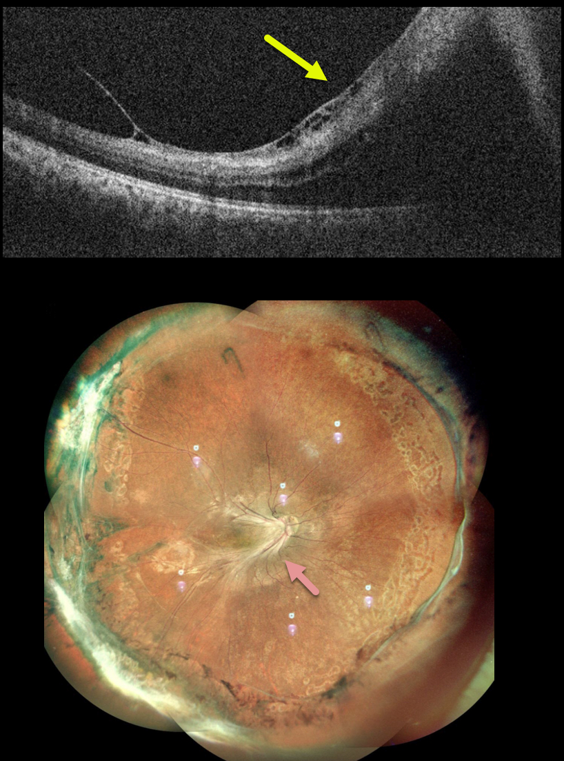
FAMILIAL EXUDATIVE VITREORETINOPATHY (FEVR)
Akansha Sharma and Manish Nagpal
Originally posted on @retina.rocks September 15, 2022
This 20YO female presented with a 2-month history of vision loss in her right eye. Vision was counting fingers.
The macular vessels were dragged temporally with a taut membrane elevating the fovea, as demonstrated on OCT. Fluorescein angiography showed bilateral encircling peripheral ischemia with retinal neovascularization (not shown).
Encircling peripheral scatter laser was performed for her left eye. Pars plana vitrectomy with membrane peeling and intravitreal gas was performed for her right eye.
Two months following surgery, all traction remained relieved, and the retina was completely attached.
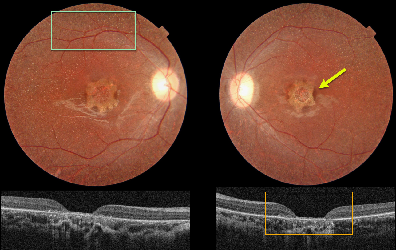
UNKNOWN INHERITED RETINAL DISEASE
Nilesh Kumar and Ashish Sharma
Originally posted on @retina.rocks September 8, 2022
This 19YO female has stable 20/200 vision bilaterally with some form of inherited retinal disease (IRD).
Color photography shows a central one-disc-diameter area of scalloped atrophy with tiny white dots extending from the peripheral macula to beyond the arcades.
OCT shows central neurosensory retinal atrophy with a thin remaining rim of featureless tissue and underlying RPE loss.
Learning Points:
These findings do not fit neatly into any specific diagnosis. Stargardt/ABCA4 disease usually shows more pericentral than central atrophy, and usually involves just the outer retina. The subretinal flecks are much larger and yellow in appearance than the tiny whitish dots seen in our patient.
Fundus albipunctatus is a form of congenital stationary night blindness with somewhat similar white dots, but foveal atrophy and severe central vision loss are usually absent (though it may be associated with an RDH5 mutation).
Finally, retinitis punctata albescens presents with white dots similar to those in fundus albipunctatus, but it is a progressive disorder that can later develop more typical retinitis pigmentosa findings. Foveal atrophy is not a feature of this disease.
Fundus autofluorescence (FAF) is often helpful in differentiating these disorders. The lesions in ABCA4 disease are hyper-FAF. The global FAF signal is markedly diminished in fundus albipunctatus.
Unfortunately, our patient did not get FAF imaging. And of course, genetic testing would hopefully be diagnostic, but this was also not available.
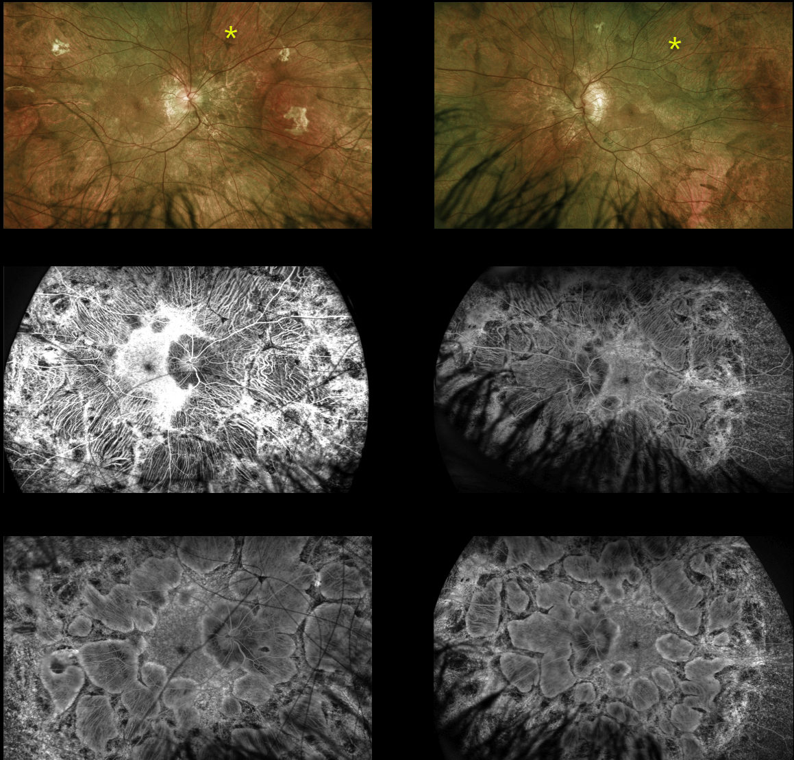
CHOROIDEREMIA
Originally posted on @retina.rocks August 31, 2022
This 14YO male is surprisingly asymptomatic with visual acuity of 20/30 OD and 20/40 OS. His retinal findings were found on a routine exam.
Optos imaging shows large symmetrically bilateral scalloped areas of chorioretinal atrophy, which spare the central maculas. On fundus fluorescein angiography, the early phase (middle) shows exquisite detail of the choroidal vortex system and late phase (bottom) shows staining of the scalloped areas of atrophy.
The patient is adopted and is unaware of any family history of inherited retinal disease. Subsequent genetic testing revealed a pathogenic hemizygous CHM variant on the X chromosome.
Learning Points:
Choroideremia is an X-linked recessive chorioretinal dystrophy caused by a mutation in the CHM gene located on the long arm of the X chromosome. Sons of female carriers have a 50% chance of developing choroideremia, and daughters have a 50% chance of becoming carriers.
Some female carriers can still develop clinical disease due to irregular inactivation of the X-chromosome (see Jauregui et al, AJO 2019;207:77-86).
The onset of night blindness usually begins between the ages of 10 and 30, followed by peripheral visual field loss. Zones of patchy RPE and chorioretinal atrophy initially appear in the mid-periphery, gradually spreading anteriorly and posteriorly.
The choroidal vortex system is relatively resistant to most disorders, including choroideremia. This system is beautifully imaged angiographically in our patient since the RPE, which normally blocks the underlying choroid, is atrophic and no longer blocks these vessels.
Eventually, the patient is left with a small central island of vision, narrowed retinal vessels and optic atrophy.
For a great choroideremia review article, see Pennesi et al, Retina 2019;39:2019-2069.
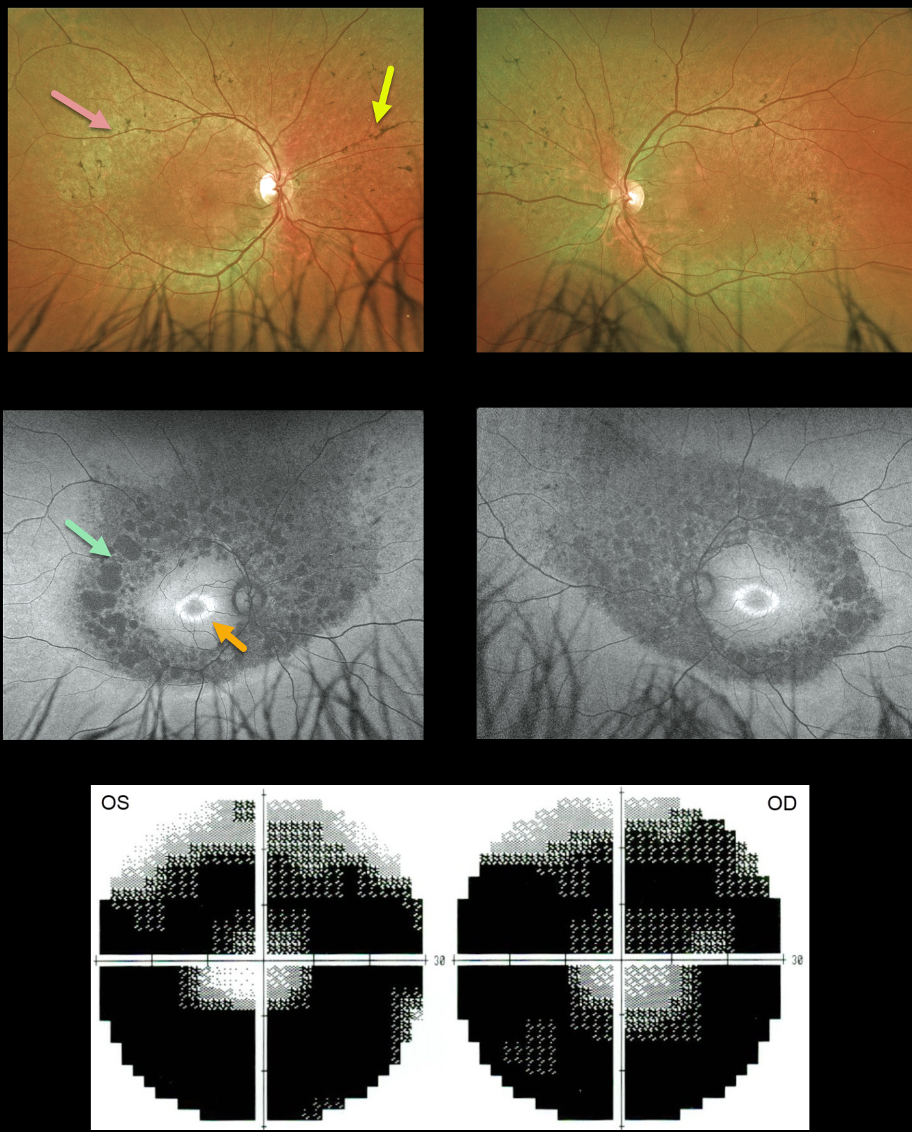
RETINITIS PIGMENTOSA
Originally posted on @retina.rocks July 4, 2022
This 34YO male was referred for relatively asymptomatic retinal findings. He had complained of some blurred distance vision for several years. Vision was 20/25 OU.
Optos color imaging shows fairly normal maculas with midperipheral encircling pigment loss and intraretinal pigment migration. Fundus autofluorescence (FAF) shows a bull’s-eye-shaped area of macular hyper-FAF, and areas of midperipheral and nasal hypo-FAF corresponding to the pigmentary changes clinically. Visual field testing shows marked bilateral constriction. Invitae genetic testing was negative.
Learning Points:
Patients with retinitis pigmentosa (RP) classically have pale optic nerves, narrowed retinal blood vessels, variable outer retinal and RPE loss, and intraretinal pigment migration (bone spicules) due to loss of the inhibitory effect of the degenerated outer segments, allowing the RPE to migrate along the retinal capillaries.
A negative genetic test does not mean this patient doesn’t have RP; rather, it reminds us that such testing must be interpreted with caution and always with the clinical findings in mind.
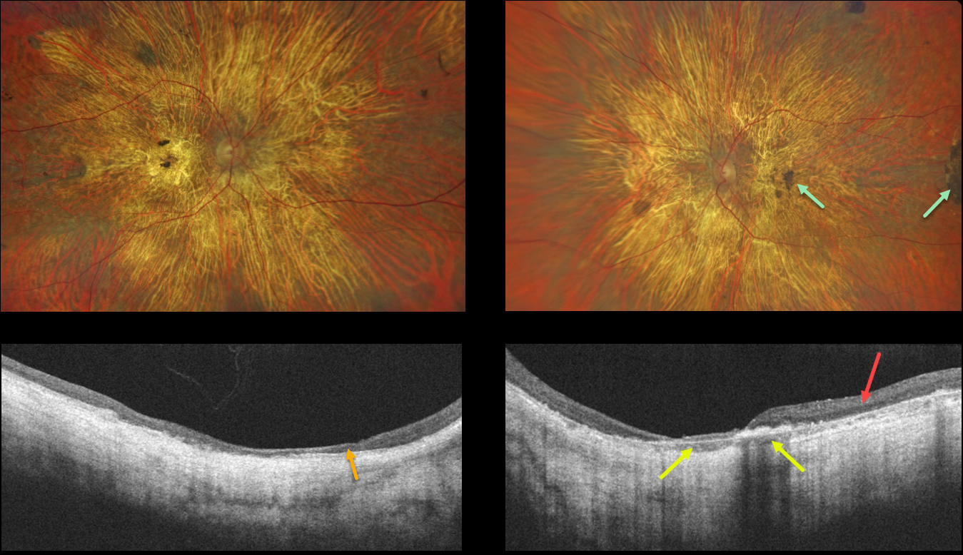
CHOROIDEREMIA
Will Gibson
Originally posted on @retina.rocks June 27, 2022
This 56YO male presented with a long history of vision loss and night blindness. Vision was counting fingers bilaterally.
Optos imaging shows severe, bilateral diffuse chorioretinal atrophy with some scattered large clumps of pigmentation. OCT scanning of the right eye shows diffuse thinned, disorganized retina and no choroidal structures. OCT scan of the left eye shows relatively preserved temporal retina and some preserved choroid.
Learning Points:
Choroideremia is an X-linked recessive chorioretinal dystrophy caused by a mutation in the CHM gene located in the long arm of the X chromosome. Sons of female carriers have a 50% chance of developing choroideremia, and daughters have a 50% chance of becoming carriers. Some female carriers can still develop choroideremia due to irregular inactivation of the X chromosome (see Jauregui et al, AJO 2019;207:77-86).
Night blindness usually begins between the ages of 10 and 30, followed by peripheral visual field loss. Zones of patchy RPE and chorioretinal atrophy initially appear in the mid-periphery, gradually spreading anteriorly and posteriorly. Eventually, the patient is left with only a small island of vision, narrowed retinal vessels, and optic atrophy.
For a great choroideremia review article, see Pennesi et al., Retina 2019;39:2019-2069.
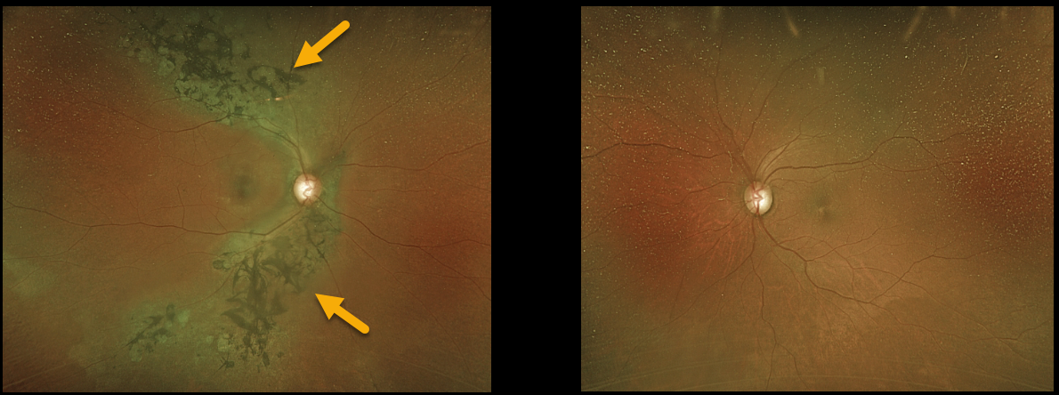
TRAUMATIC PSEUDO-RP
Originally posted on @retina.rocks May 31, 2022
A 67YO female was referred for asymptomatic retinal findings in her right eye. She gave a history of unspecified head trauma as a child.
Optos imaging shows chorioretinal scarring with intraretinal pigment migration, extending superotemporally and inferotemporally from the optic nerve. The left fundus was completely normal.
Learning Points:
Photoreceptor loss from numerous causes, including blunt trauma, retinal detachment, macular telangiectasia, and retinitis pigmentosa (RP), can allow for intraretinal RPE migration with this shared phenotype.
Unilateral RP is exceedingly rare and requires normal electrophysiologic findings and long-term observation to ensure delayed findings in the fellow eye.
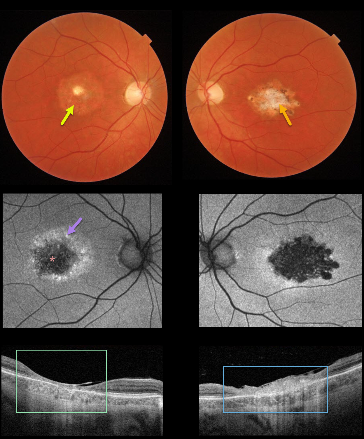
CENTRAL AREOLAR CHOROIDAL DYSTROPHY
Originally posted on @retina.rocks May 11, 2022
This 40YO female gave a 4-5 year history of gradual bilateral vision loss. Vision was 20/70 OD and 20/200 OS. There was no family history of retinal disease.
Color imaging shows a round area of central pigment loss OD and a more irregular area of atrophic pigmentary changes OS.
Optos fundus autofluorescence (FAF) showed central hypo-FAF with surrounding hyper-FAF.
Swept-source OCT showed a markedly thinned and disorganized central macula OD, with a more thickened, hyperreflective, disorganized central macula OS.
Genetic testing revealed a heterozygous pathogenic mutation in IQCB1, a defect commonly associated with autosomal recessive Senior-Løken Syndrome. We felt the clinical picture was most consistent with central areolar choroidal dystrophy (CACD).
Learning Points:
CACD is usually autosomal dominant and genetically heterogeneous, caused by mutations in the peripherin/RDS (PRPH2) gene. The current genetic testing panel results, as this case illustrates, must still be approached with caution (see Rodriguez-Munoz et al, Retina 2021;41:1966-1975).
See Smailhodzic et al Invest Ophthalmol Vis Sci 2011;52;8908-8918 for how to differentiate CACD from age-related macular degeneration best clinically.
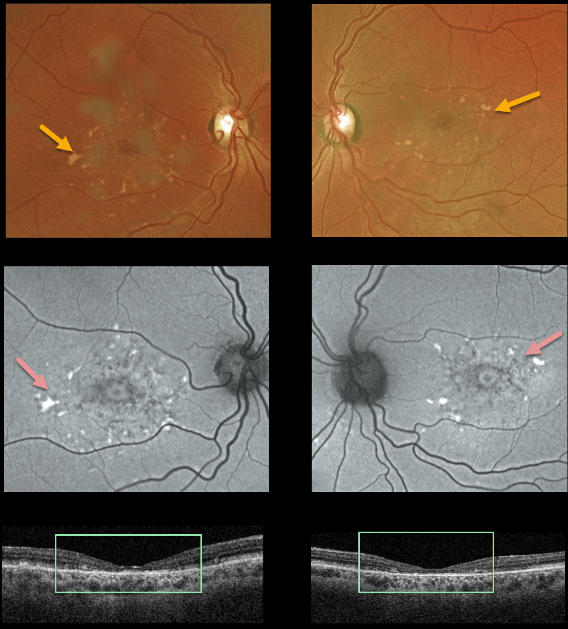
STARGARDT DISEASE
Originally posted on @retina.rocks April 26, 2022
This 37YO female presented with vision of 20/80 OU and classic findings for Stargardt disease, captured with Optos multimodal imaging and Triton swept-source OCT, including bilateral perifoveal yellow subretinal flecks that are variably hyperautofluorescent and variable outer retinal and RPE atrophy.
Learning Points:
Stargardt disease is an autosomal recessive disorder caused by a mutation in the ABCA4 gene. The ABCA4 protein is located in the photoreceptor outer segments and is involved in the recycling of 11-cis-retinal.
Mutation of this gene causes the accumulation of lipofuscin in the RPE, which eventually leads to photoreceptor and RPE degeneration. Other ABCA4 disorders include fundus flavimaculatus and cone-rod dystrophy.
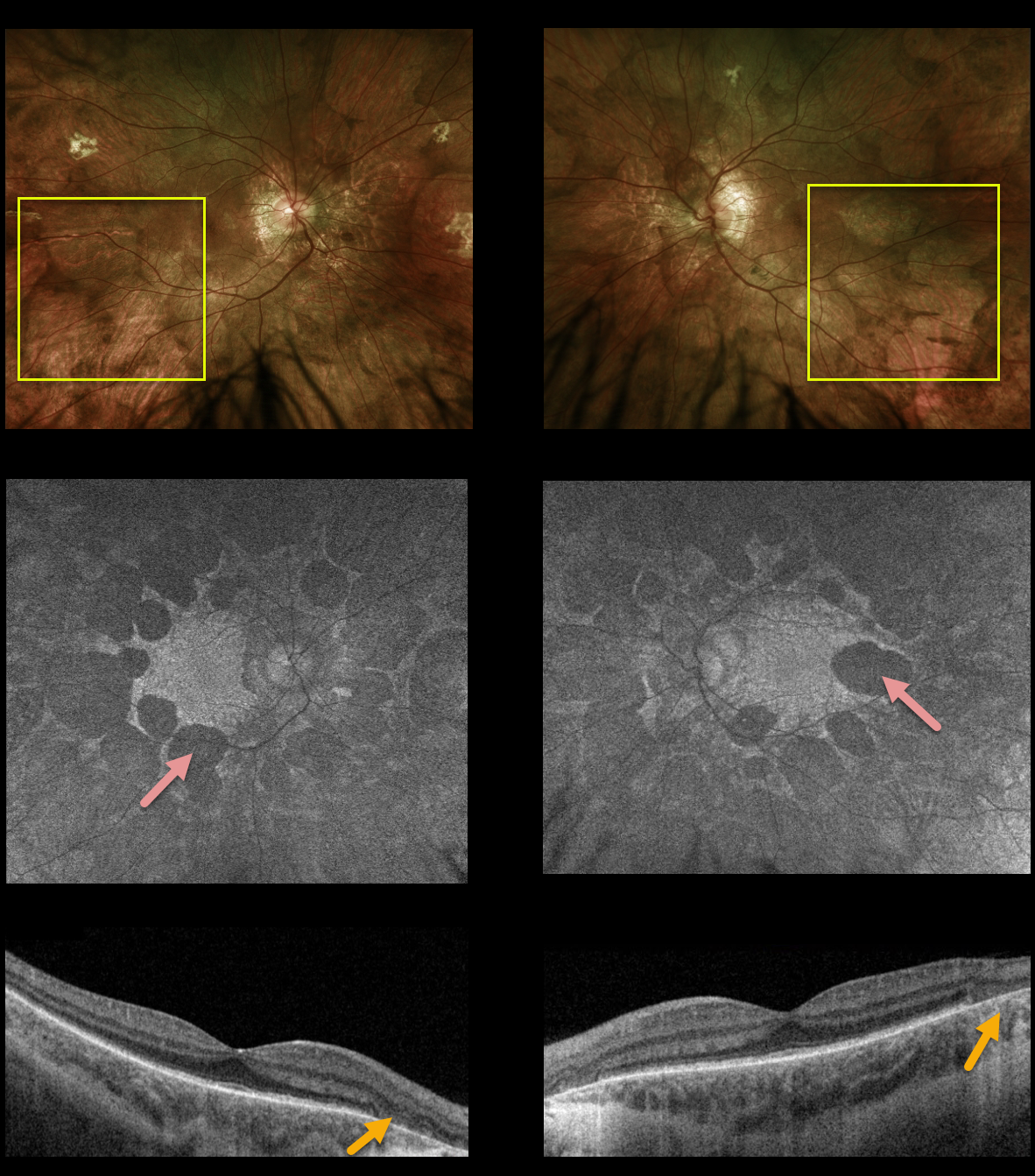
CHOROIDEREMIA
Originally posted on @retina.rocks April 21, 2022
This 14YO male is surprisingly asymptomatic with visual acuity of 20/30 OD and 20/40 OS. His retinal findings were found on a routine exam.
Optos imaging shows large, symmetrically bilateral, scalloped areas of chorioretinal atrophy that spare the central maculae.
On fundus autofluorescence (FAF), the scalloped areas of atrophy show hypo-FAF.
Swept-source OCT shows outer retinal and RPE atrophy within the clinical areas of atrophy.
The patient is adopted and is unaware of any family history of inherited retinal disease. Subsequent genetic testing revealed a pathogenic hemizygous CHM variant on the X chromosome, consistent with choroideremia.
Learning Points:
Choroideremia is an X-linked recessive chorioretinal dystrophy caused by a mutation in the CHM gene located on the long arm of the X chromosome.
Sons of female carriers have a 50% chance of developing choroideremia, and daughters have a 50% chance of becoming carriers. Some female carriers can still develop clinical disease due to irregular inactivation of the X chromosome (see Jauregui et al, AJO 2019;207:77-86).
Night blindness usually begins between the ages of 10 and 30, followed by peripheral visual field loss.
Zones of patchy RPE and chorioretinal atrophy initially appear in the mid-periphery, gradually spreading anteriorly and posteriorly. Eventually, the patient is left with only a small central island of vision, narrowed retinal vessels, and optic atrophy.
For a great choroid plexus review article, see Pennesi et al., Retina 2019;39:2019-2069.
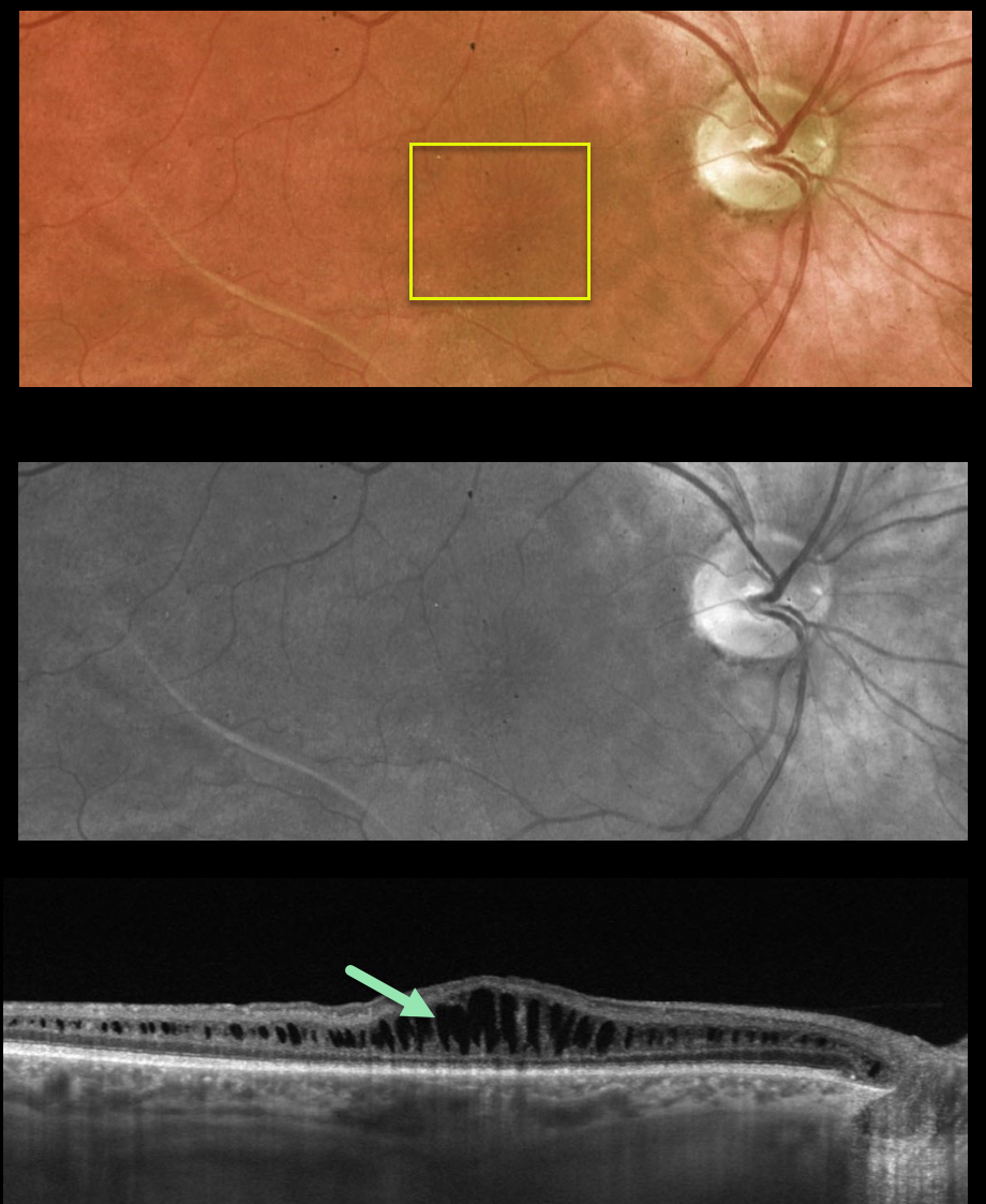
X-LINKED RETINOSCHISIS
Originally posted on @retina.rocks April 11, 2022
This 39YO male presented to our clinic with a known history of sex-linked retinoschisis. Vision was 20/100 OU.
Optos color and green channel images show faint radiating foveal cystic changes bilaterally (only right eye shown).
Swept-source OCT scanning shows prominent splitting of the inner nuclear layer.
Learning Points:
Although historically taught as involving the nerve fiber layer, sex-linked retinoschisis in the posterior pole causes schisis throughout the macula including the inner nuclear, outer nuclear and outer plexiform layers (see Yu et al, AJO 2010;149:973-978).
With time, the schisis often collapses with secondary atrophy (see Menke et al, Retina 2011;31:1186-1192).
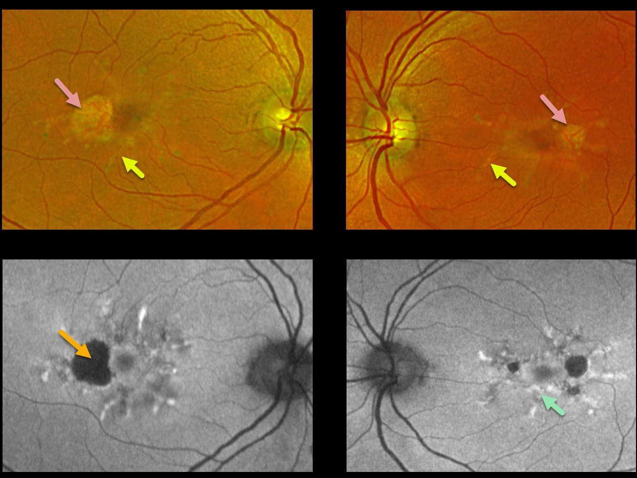
MACULAR DYSTROPHY
Nicole Wood
Originally posted on @retina.rocks February 22, 2022
This 62YO female presented with 20/20 vision OU and no visual symptoms. Optos imaging shows bilateral radiating subretinal yellowish macular lesions with temporal atrophy.
Fundus autofluorescence (FAF) shows variable hyper-FAF from these lesions, along with hypo-FAF within the areas of atrophy.
Learning Points:
Pattern macular dystrophies are associated with numerous mutations, including ABCA4, PRPH2, BEST1, PROM1, CERKL, CNGB3, and CRB1, among others (see Del Pozo-Valero et al, Invest Ophthalmol Vis Sci 2022;63(2):11).
These patients tend to maintain fairly good vision, although outer retinal and RPE atrophy can cause central vision loss, as can the rare development of macular neovascularization. Patients should therefore regularly monitor their vision with an Amsler grid.
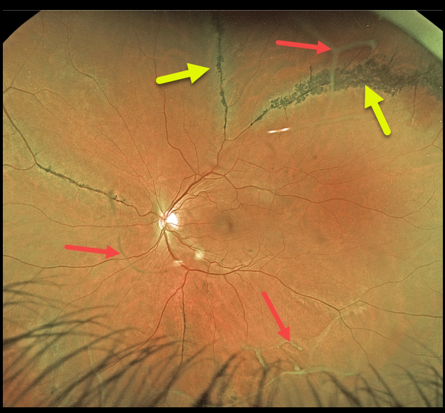
STICKLER SYNDROME
Originally posted on @retina.rocks February 18, 2022
This 32YO male with Stickler syndrome is blind in his right eye following failed retinal detachment surgery elsewhere. Vision is 20/25 in his asymptomatic left eye.
There are prominent areas of pigmented paravenous lattice superiorly, with scattered veils of vitreous strands and condensations.
Learning Points:
Stickler syndrome is a usually autosomal-dominant genetic disorder characterized by defective vitreous collagen (types II, IX, and XI). The vitreous may be optically empty, or associated with vitreous strands and membranes, as in our patient.
Other ocular findings include high myopia and paravenous lattice degeneration. Patients have premature arthropathy with classic orofacial and auditory features.
Stickler syndrome is the most common cause for inherited rhegmatogenous retinal detachment, and prophylactic encircling retinopexy with laser or cryotherapy may be considered (see Fincham et al Ophthalmology 2014;121:1588-1597).
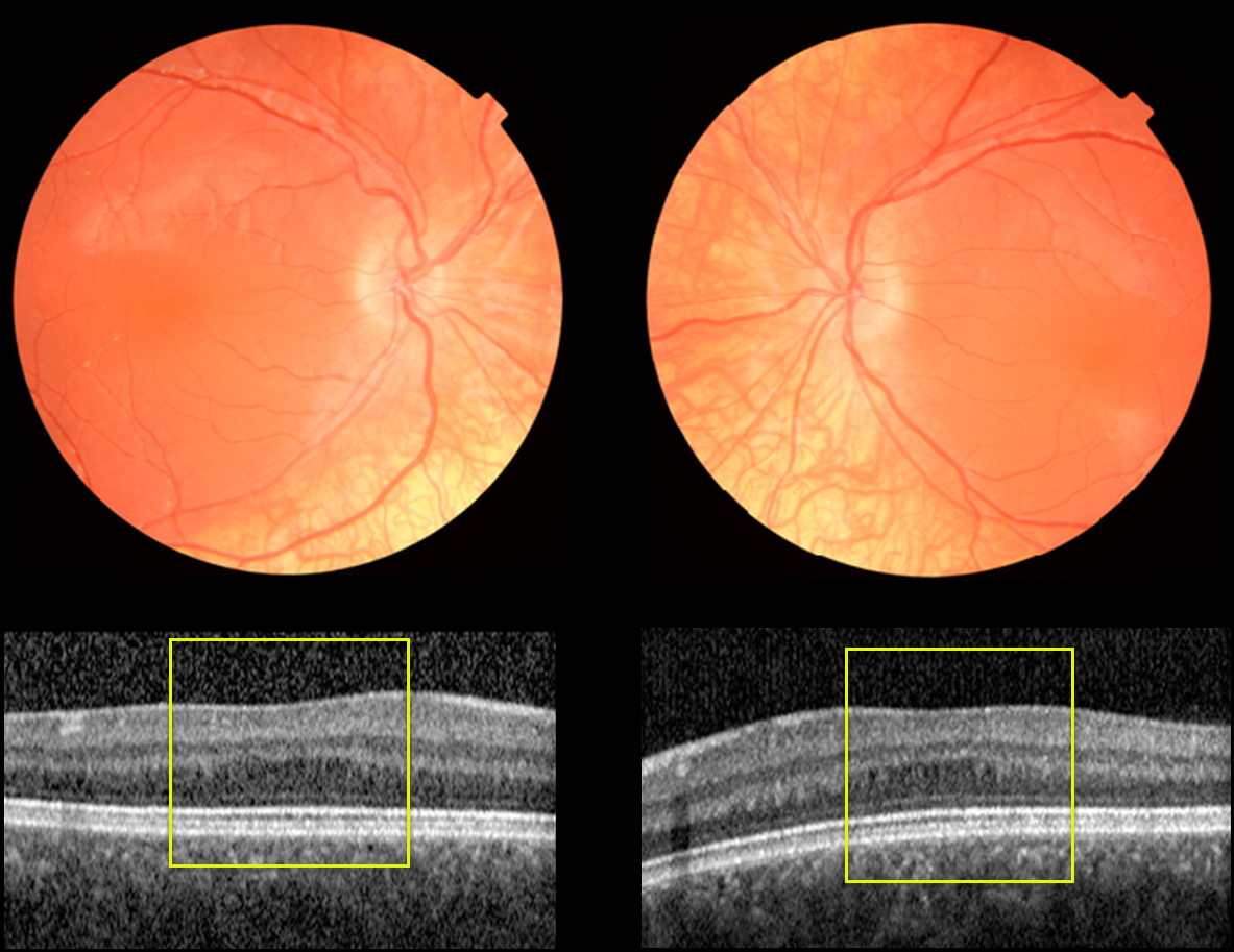
OCULAR ALBINISM
Erdem Dinc
Originally posted on @retina.rocks February 3, 2022
This 11YO female presented with 20/50 vision OU. She was healthy with normal skin and hair pigmentation and was the product of a consanguineous marriage. Motility was normal without nystagmus. Bilateral iris transillumination defects were noted.
Fundus photography shows marked decreased choroidal pigmentation and absent foveal reflex. OCT scanning shows bilateral absence of the normal foveal depression (fovea plana).
Learning Points:
Ocular albinism is an X-linked recessive disorder that affects only the eyes and is caused by a mutation in the OA1 gene. Findings seen in ocular albinism include strabismus, nystagmus, iris transillumination defects, blonde fundus, and absent or blunted foveal reflex (fovea plana). There are often a higher number of crossed nerve fibers at the optic chiasm.
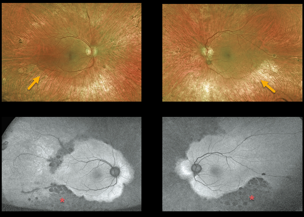
ALAGILLE SYNDROME
Originally posted on @retina.rocks January 26, 2022
This 24YO man with Alagille syndrome presented with vision of 20/200 OD and 20/50 OS. There was a long history of bilateral poor vision. He was adopted and is a ward of the state.
Optos color imaging shows fairly normal maculas with midperipheral encircling pigment loss with some pigment migration.
Fundus autofluorescence (FAF) shows extensive areas of midperipheral and nasal hypo-FAF.
Learning Points:
Alagille syndrome, also known as arteriohepatic dysplasia, is an autosomal dominant, genetically and phenotypically heterogeneous disorder characterized by distinct facial features, including a prominent forehead, deep orbital sulcus, and pointed chin.
It is caused by loss of function of the JAG1 gene, which is located on chromosome 20p12. The JAG1 gene encodes transmembrane proteins responsible for cell signaling during embryonic development, notably in the Notch signaling pathway.
Patients develop intrahepatic cholestatic liver disease, heart abnormalities, hypogonadism, and intellectual disability. Virtually all patients have peripheral retinal pigmentary changes, and three-quarters have anterior chamber angle abnormalities, including Axenfeld and Rieger anomalies (our patient’s slit lamp examinations were normal).
For a recent, comprehensive review of the spectrum of ocular findings in Alagille syndrome, see da Palma et al., Invest Ophthalmol Vis Sci 2021; 62 (7):27.
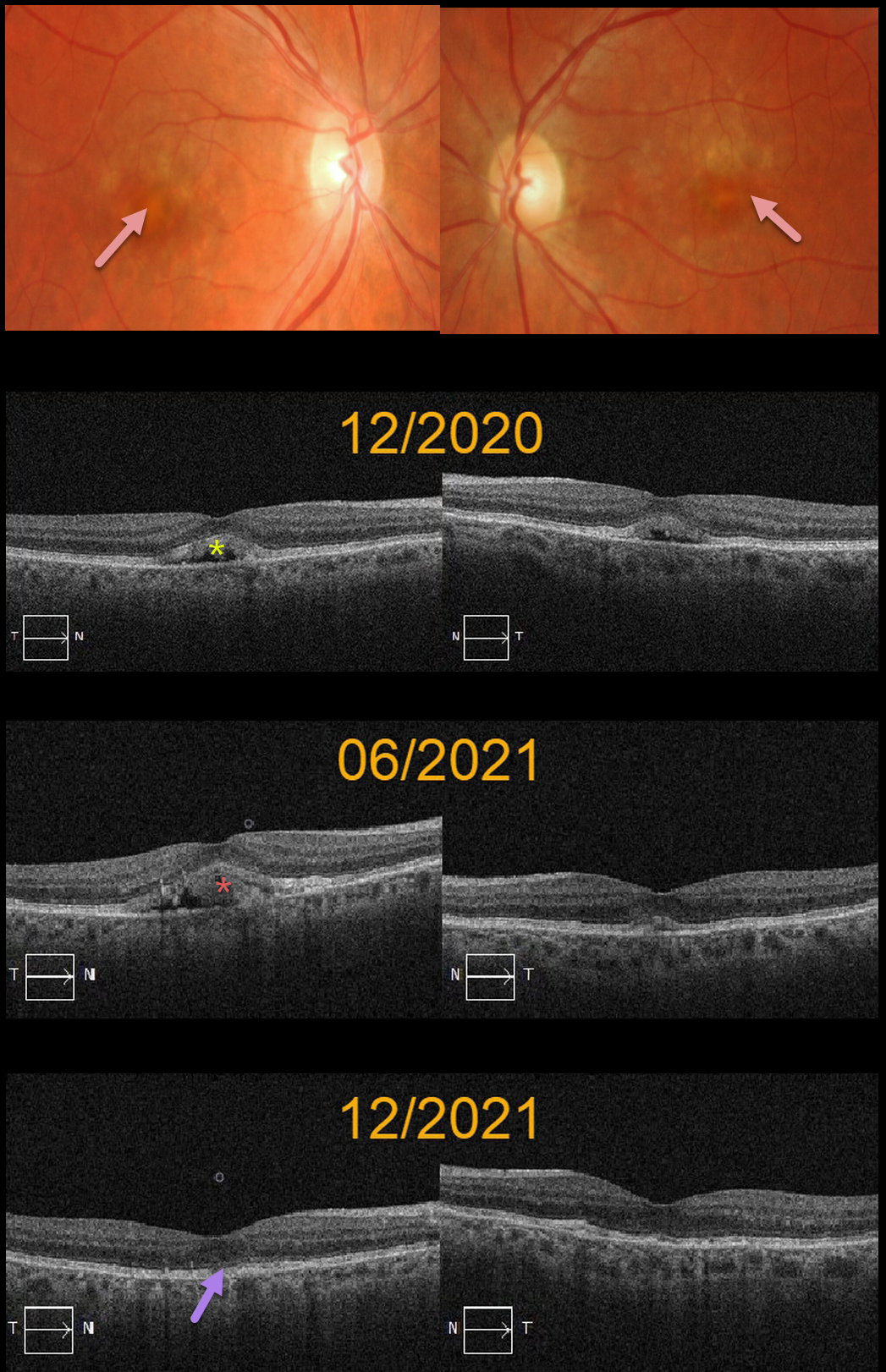
ADULT-ONSET FOVEOMACULAR VITELLIFORM DYSTROPHY
Originally posted on @retina.rocks January 19, 2022
This 73YO female presented in 12/20 with bilateral central vitelliform lesions and more peripheral yellowish macular lesions, consistent with a probable macular dystrophy.
OCT shows bilateral subretinal hyperreflective lesions with underlying hyporeflectivity. She was asymptomatic with 20/25 vision OU. There was no family history of macular dystrophy. Genetic testing was negative, including for BEST1.
Six months later, the vitelliform lesion increased OD and decreased OS. Another six months later, the vitelliform lesions collapsed OU, and vision remained 20/25.
Learning Points:
Vitelliform lesions are commonly found in association with other disorders, including retinal dystrophies such as Best disease, macular dystrophies, cuticular drusen, AMD, and vitreomacular traction.
The yellow subretinal material is felt to be debris from shed photoreceptor outer segments. These lesions markedly autofluoresce on fundus autofluorescence imaging, which unfortunately, is unavailable for this patient.
A great reference for learning more about AVLs is Freund et al, Retina 2011;31:13-25.
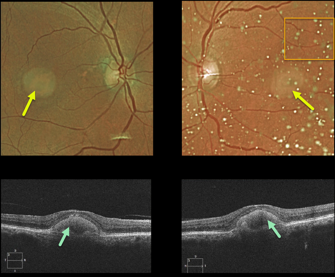
ADULT-ONSET FOVEOMACULAR VITELLIFORM DYSTROPHY
Originally posted on @retina.rocks January 18, 2022
This 87YO female shows classic findings for adult-onset foveomacular vitelliform dystrophy (Adult Best). Vision was 20/60 OD and 20/80 OS Optos imaging shows small, round, symmetrical subfoveal yellow lesions. Moderate asteroid hyalosis is noted as an incidental finding on the OS.
Triton swept-source OCT shows subretinal hyperreflective material above the RPE that extends into and indents the overlying retina.
Learning Points:
Adult Best is associated with a mutation in the BEST1 gene, which encodes the bestrophin-1 protein. Bestrophin-1, a calcium-activated chloride channel, is primarily found in the basolateral plasma membrane of the RPE.
BEST1 mutations cause a variety of phenotypes, including autosomal recessive bestrophinopathy, best vitelliform macular dystrophy, and autosomal dominant vitreoretinochoroidopathy.
These patients tend to maintain fairly good vision. Outer retinal and RPE atrophy can cause central vision loss, as can the rare development of macular neovascularization. Patients should therefore regularly monitor their vision with an Amsler grid.
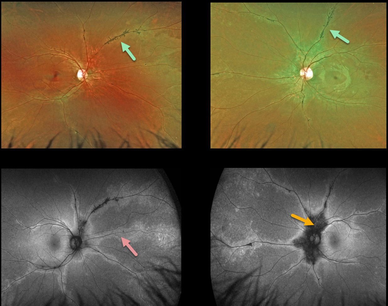
PIGMENTED PARAVENOUS CHORIORETINAL ATROPHY
Originally posted on @retina.rocks January 14, 2022
This 24YO female presented with 20/20 vision bilaterally and no visual symptoms. Optos ultrawidefield imaging shows normal central maculas with intraretinal pigment migration and depigmented RPE along the retinal veins. These changes extend from the peripapillary retina into the retinal peripheries.
Fundus autofluorescence (FAF) best highlights these changes with peripapillary hypo-FAF and paravenous hyper-FAF.
Our patient underwent Spark genetic testing. Results showed a pathogenic TNEM216 variant, which is associated with autosomal recessive Joubert Syndrome. However, she had absolutely no signs or symptoms of this syndrome.
Learning Points:
Pigmented paravenous chorioretinal atrophy (PPCRA) is a rare condition characterized by paravenous pigment clumps with peripapillary and radial zones of RPE atrophy.
It is bilateral, often asymmetric, with various causes, including genetic and pseudo-PPCRA (inflammatory, infectious, or unknown).
Patients tend to have some symptoms, usually nyctalopia, but most retain good vision, especially those without macular involvement.
The underlying pathophysiology is likely a primary choroidopathy that causes overlying RPE and retinal changes. The chorioretinal changes are best visualized with FAF. For a great clinical review, see Lee et al, AJO 2021;224:120-132.
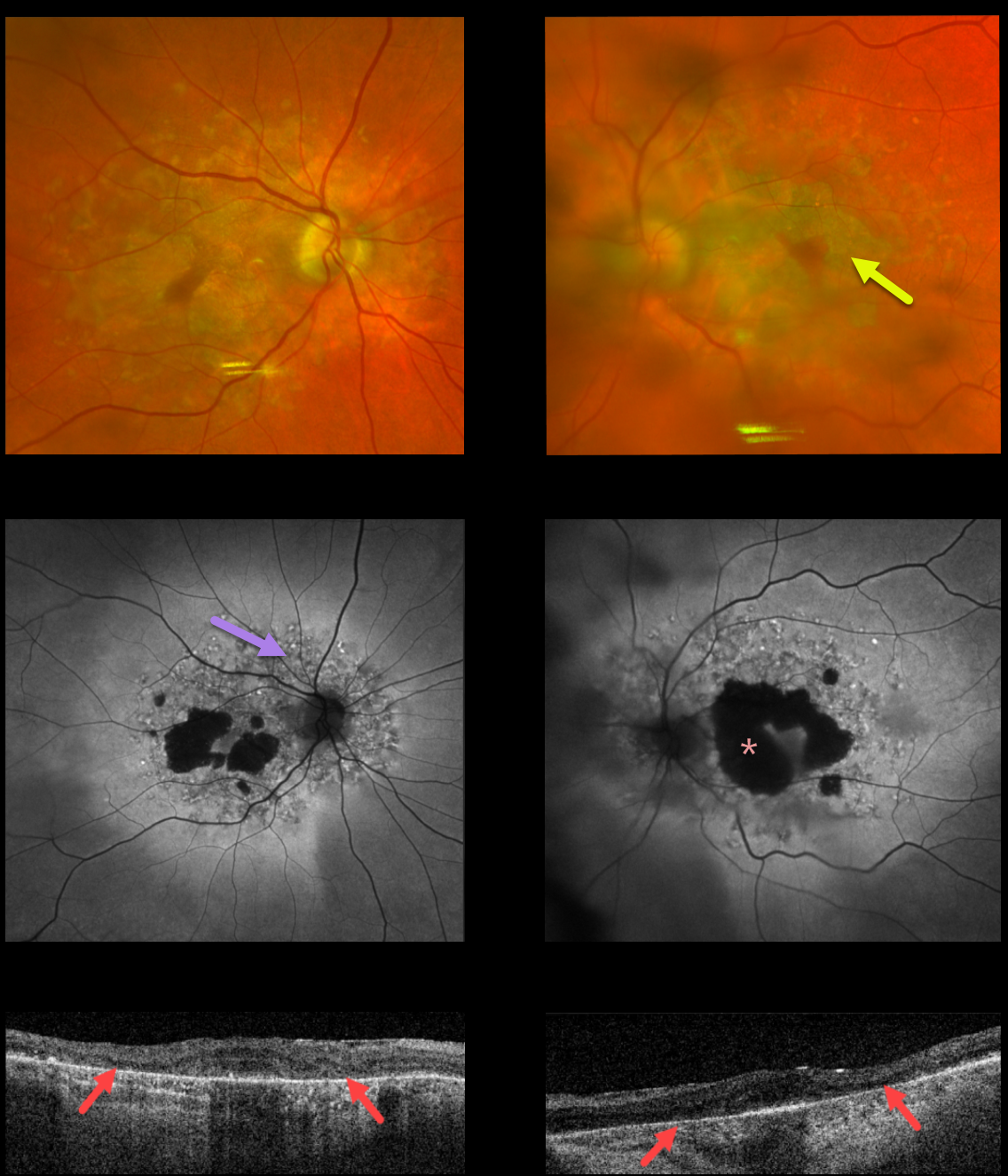
MATERNALLY INHERITED DIABETES AND DEAFNESS (MIDD)
Originally posted on @retina.rocks January 11, 2022
This 75YO male presented with 20/80 vision OU. Optos color imaging shows bilateral, relatively confluent patchy areas of macular atrophy.
Fundus autofluorescence (FAF) shows hypo-FAF within the areas of atrophy, with more extensive peripapillary and macular variably hyper-FAF spots.
Triton swept-source OCT shows abnormal macular architecture throughout, with the most pronounced outer retinal atrophy nasally and temporally within the areas of clinical atrophy.
Our patient has non-insulin-dependent type 2 diabetes, but there are no hearing abnormalities. MIDD patients need to be screened for other potential co-existing medical conditions, including cardiac arrhythmias, myopathy, and renal disease.
Learning Points:
Maternally inherited diabetes and deafness (MIDD) accounts for up to 3% of all cases of diabetes and results from a mutation in mitochondrial DNA at position A3243G.
MIDD often masquerades as a pattern macular dystrophy. In our experience, fundus autofluorescence (FAF) is the best way to visualize these changes. The FAF appearance somewhat resembles that seen with Elmiron toxicity.
Peripapillary hypoautofluorescence, more densely packed macular autofluorescent changes, and earlier central macular involvement suggest Elmiron toxicity over other causes (see Barnes et al., Ophthalmology Retina 2020;4:1196-1201). Note that our case spares the central macula and peripapillary retina, which is more consistent with MIDD.
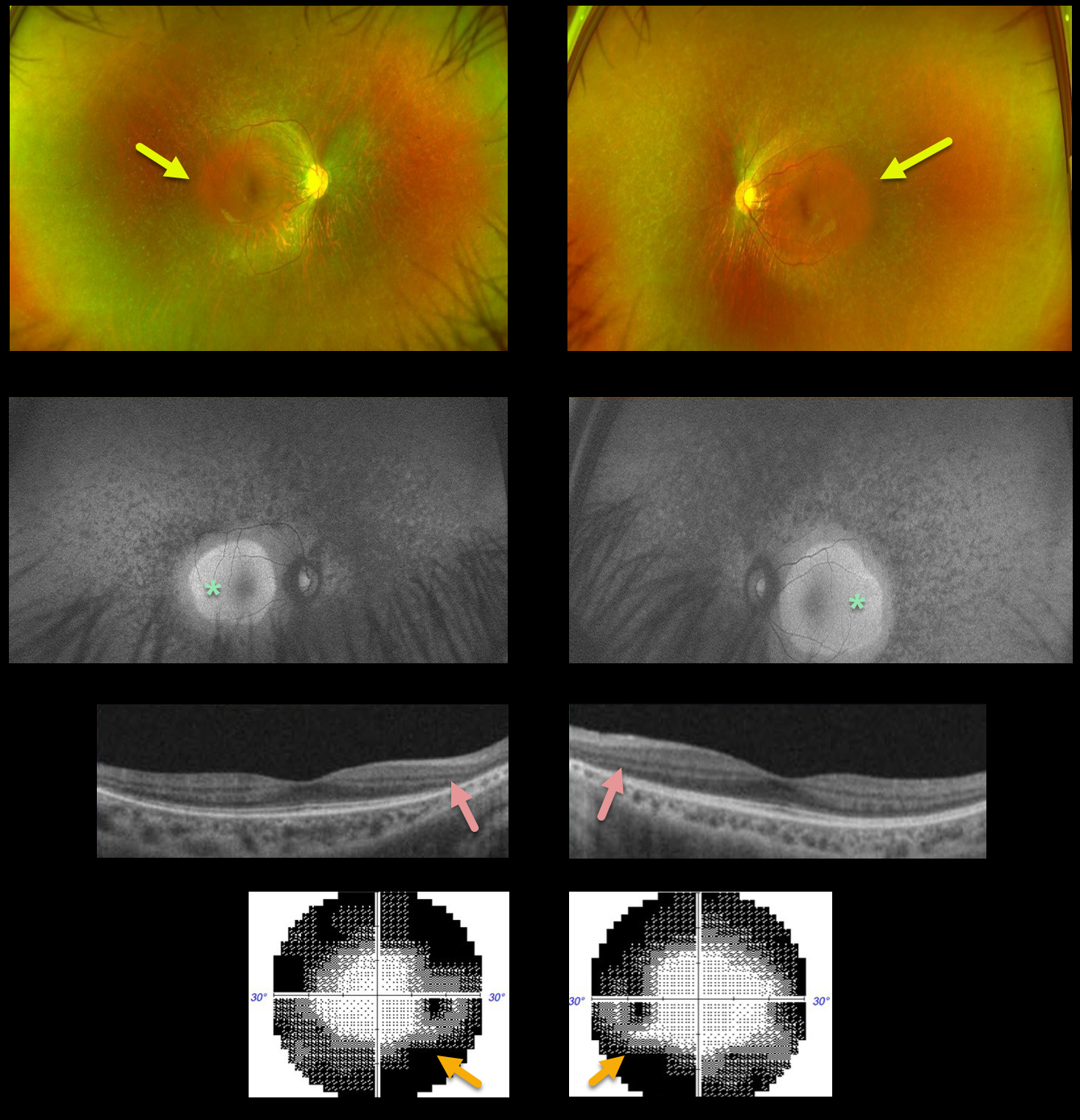
RETINITIS PIGMENTOSA
Alex Hynes
Originally posted on @retina.rocks December 14, 2021
This 25YO male presented with complaints of nyctalopia and peripheral vision loss. Vision was 20/25 OU.
Optos color imaging shows a diffuse moth-eaten appearance to the RPE, sparing each macula. Minimal bone spicules are noted. Fundus autofluorescence (FAF) shows macular hyperautofluorescence.
OCT scanning shows peripheral outer retinal thinning. Visual field testing shows classing bilateral constriction. Genetic testing showed no pathogenic mutations.
Learning Points:
Patients with retinitis pigmentosa (RP) classically have pale optic nerves, narrowed retinal blood vessels, variable outer retinal and RPE loss, and intraretinal pigment migration (bone spicules) due to loss of the inhibitory effect of the degenerated outer segment,s allowing the RPE to migrate along the retinal capillaries. In real life, patients often lack all these features.
As in this case, intraretinal pigment migration may be minimal or absent but often appears over time (see Takahashi et al, AJO 2018;195:176-180).
A truly normal fundus in a patient with RP is extremely unusual, as there are usually very subtle clinical and FAF findings.
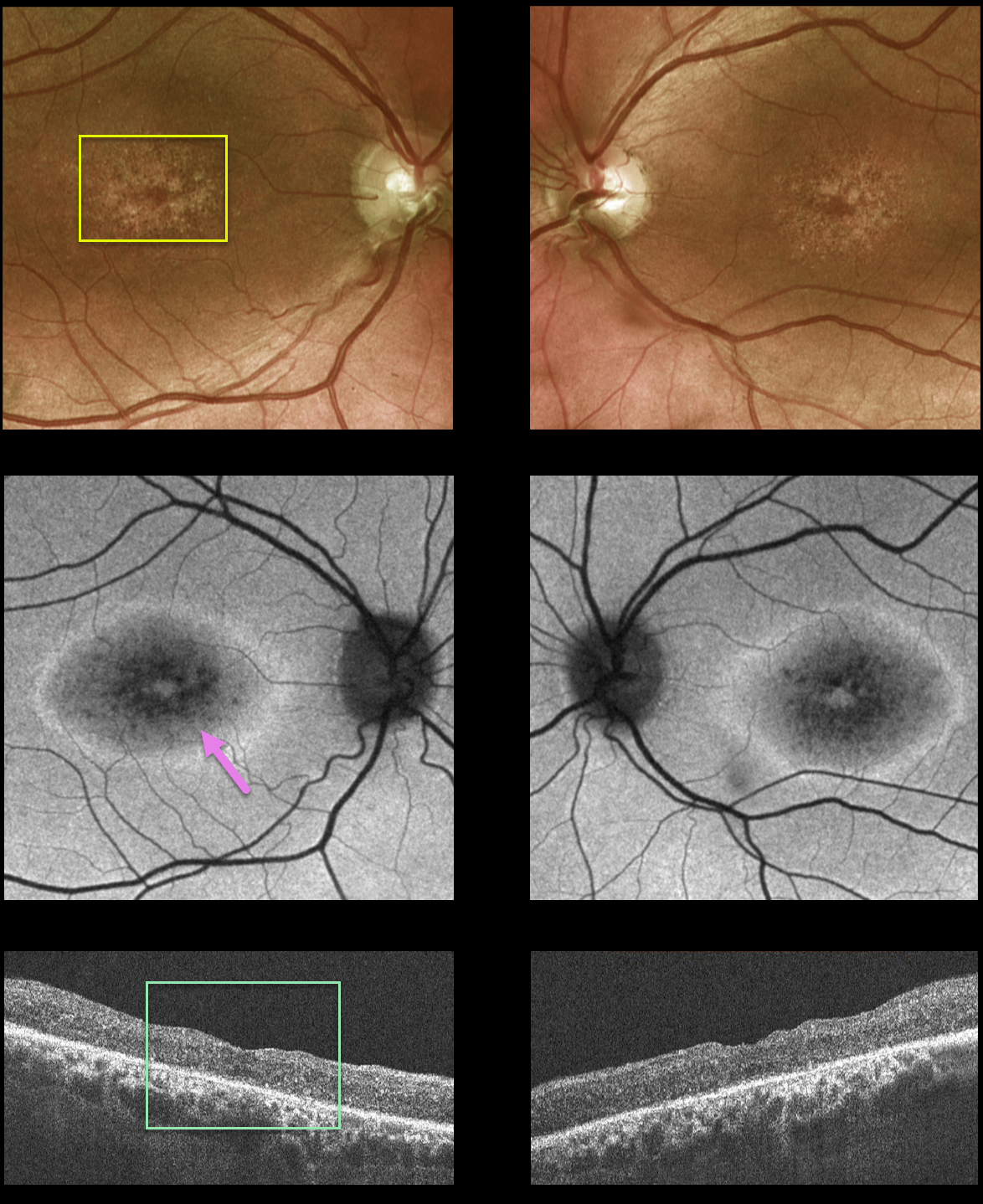
X-LINKED RETINOSCHISIS
Originally posted on @retina.rocks December 10, 2021
This 42YO male presented to our clinic with a history of X-linked retinoschisis diagnosed elsewhere. Vision was 20/70 OD and 20/200 OS.
Fundus photos show atrophic foveal pigmentary changes. Fundus autofluorescence (FAF) shows mottled central hypo-FAF with a ring of surrounding hyper-FAF. OCT scanning shows bilateral macular thinning with disorganization of the normal macular hyper- and hyporeflective bands.
Genetic testing confirmed the presence of a pathogenic RS1 mutation, which is associated with sex-linked retinoschisis.
Learning Points:
Although historically taught to involve the nerve fiber layer, sex-linked retinoschisis in the macula causes schisis throughout the macula, including the inner nuclear, outer nuclear, and outer plexiform layers (see Yu et al, AJO 2010;149:973-978).
Over time, the schisis often collapses with secondary atrophy, as occurred in our patient (see Menke et al, Retina 2011;31:1186-1192).
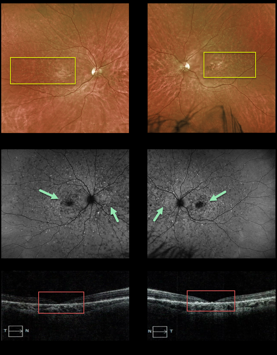
FUNDUS FLAVIMACULATUS
Originally posted on @retina.rocks November 18, 2021
This 28YO female presented with 20/400 vision in both eyes, with classic findings of fundus flavimaculatus, including bilateral subretinal flecks extending from the macula to the midperiphery, variable outer retinal and RPE atrophy, and fundus autofluorescence changes. OCT scanning showed marked outer retinal atrophy, especially centrally. Her brother has similar findings.
Learning Points:
Fundus flavimaculatus is an autosomal recessive disorder caused by a mutation in the ABCA4 gene. The ABCA4 protein is located in the photoreceptor outer segments and is involved in the recycling of 11-cis-retinal.
Mutation of this gene causes the accumulation of lipofuscin in the RPE, which eventually leads to photoreceptor and RPE degeneration.
Other ABCA4 disorders include Stargardt’s disease, cone-rod dystrophy, and retinitis pigmentosa.
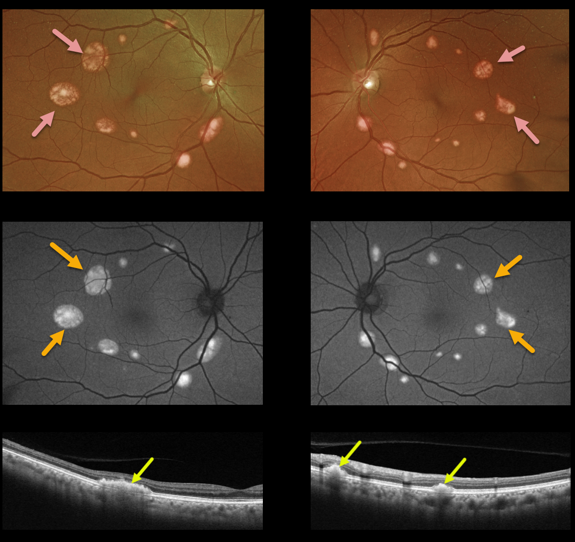
BEST DISEASE
Originally posted on @retina.rocks November 5, 2021
This 68YO female presented with these asymptomatic multifocal subretinal yellow posterior pole lesions. Vision is 20/20 OU.
The sharply demarcated yellowish lesions are markedly hyper-autofluorescent and are hyperreflective on Triton swept-source OCT. There was no known family history, but genetic testing revealed a heterozygous pathogenic BEST1 mutation.
Learning Points:
Best disease is associated with a mutation in the BEST1 gene, which encodes the bestrophin-1 protein. Bestrophin-1, a calcium-activated chloride channel, is primarily found in the basolateral plasma membrane of the RPE.
BEST1 mutations cause a variety of phenotypes, including autosomal recessive bestrophinopathy, best vitelliform macular dystrophy, and autosomal dominant vitreoretinochoroidopathy. Multifocal lesions are a rare phenotype and are not usually associated with subfoveal lesions.
Multifocal Best needs to be differentiated from other causes of multifocal serous detachments, including autosomal recessive bestrophinopathy (see Boon et al, Ophthalmology 2013;120:809-820) and idiopathic or paraneoplastic acute exudative polymorphous vitelliform maculopathy (see Barbazetto et al, Ophthalmology 2018;125:75-88).
HALLOWEEN RETINOPATHY
Originally posted on @retina.rocks October 29, 2021
This is a patient with retinitis pigmentosa, but do you see Jack O’Lantern or Jack Skellington? Maybe something else spooky?
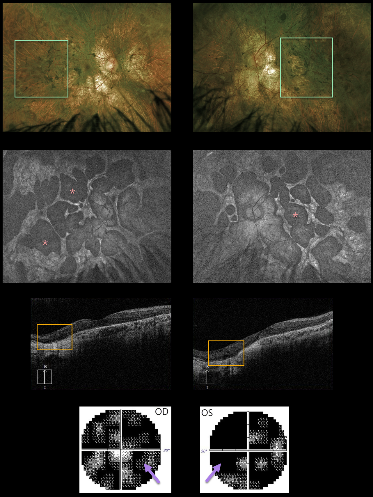
CHOROIDEREMIA IN A FEMALE CARRIER
Originally posted on @retina.rocks October 22, 2021
This 40YO female reported a 7-year history of reading difficulties and glare while driving at night.
Optos imaging shows severe diffuse chorioretinal scarring with intraretinal pigment migration extending from the optic nerves to the retinal peripheries.
The lobular nature of the chorioretinal changes is best seen with fundus autofluorescence. There was variable bilateral choroidal and outer retinal atrophy on OCT. Visual fields showed bilateral severe generalized depression.
Genetic testing showed that she was heterozygous for the pathogenic variant of the CHM gene. Vision remarkably was 20/20 OD and 20/25 OS due to sparing of the macular centers.
Learning Points:
Choroideremia is an X-linked recessive chorioretinal dystrophy caused by a mutation in the CHM gene located in the long arm of the X chromosome. Sons of female carriers have a 50% chance of developing choroideremia, and daughters have a 50% chance of becoming carriers. Some female carriers can still develop choroideremia due to irregular inactivation of the X chromosome (see Jauregui et al, AJO 2019;207:77-86).
Night blindness usually begins between the ages of 10 and 30, followed by peripheral visual field loss. Zones of patchy RPE and chorioretinal atrophy initially appear in the mid-periphery, gradually spreading anteriorly and posteriorly. Eventually, the patient is left with only a small island of vision, narrowed retinal vessels, and optic atrophy.
For a great choroideremia review article, see Pennesi et al, Retina 2019;39:2019-2069.
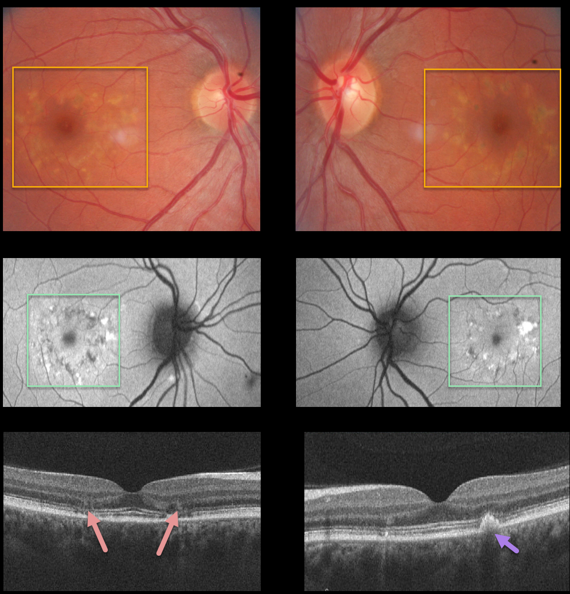
MACULAR DYSTROPHY
Originally posted on @retina.rocks October 20, 2021
This 34YO female, a local optometrist’s sister, was referred for these asymptomatic bilateral macular changes consistent with a macular dystrophy.
Fundus images show symmetrical yellowish subretinal pigmented drusen-like changes. Fundus autofluorescence reveals that these lesions are variably hyper- and hypoautofluorescent.
OCT scans show some variable ellipsoid zone loss and outer retinal atrophy, as well as subretinal hyperreflective material. Her brother had similar findings. Unfortunately, we have no further clinical information.
Learning Points:
Pattern macular dystrophy is a group of disorders characterized by pigment deposition of the macula and caused by mutations in the RDS/peripherin gene. The following are considered types of pattern dystrophies: adult-onset foveomacular vitelliform dystrophy, butterfly-shaped pigment dystrophy, reticular dystrophy, multifocal pattern dystrophy simulating Stargardt disease, and fundus pulverulentus.
Pattern macular dystrophy has also been found in systemic disorders such as pseudoxanthoma elasticum, myotonic dystrophy, and McArdle Disease.
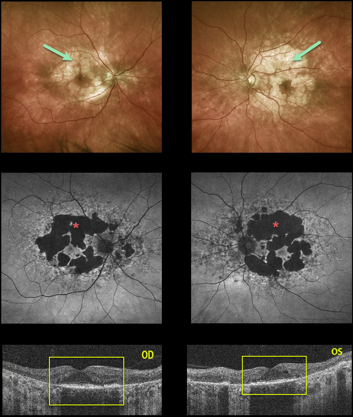
MATERNALLY INHERITED DIABETES AND DEAFNESS (MIDD)
Originally posted on @retina.rocks October 19, 2021
This 55YO male presented with 20/50 vision OD and 20/100 vision OS. Optos color imaging shows bilateral areas of relatively confluent patchy macular atrophy.
The RPE changes are much more pronounced on fundus autofluorescence. OCT scanning shows outer retinal and RPE loss within the areas of clinical atrophy.
Learning Points:
Maternally inherited diabetes and deafness (MIDD) accounts for up to 3% of all cases of diabetes and results from a mutation in mitochondrial DNA at position A3243G. MIDD often masquerades as a pattern macular dystrophy.
In our experience, fundus autofluorescence (FAF) is the best way to visualize these changes. The FAF appearance somewhat resembles that seen with Elmiron toxicity. Peripapillary hypoautofluorescence, more densely packed macular autofluorescent changes, and earlier central macular involvement suggest PPS toxicity over other causes (see Barnes et al Ophthalmology Retina 2020;4:1196-1201).
Note that our case spares the central macula and peripapillary retina, which is more consistent with MIDD.
MIDD patients need to be screened for other potential co-existing medical conditions, including cardiac arrhythmias, myopathy, and renal disease.
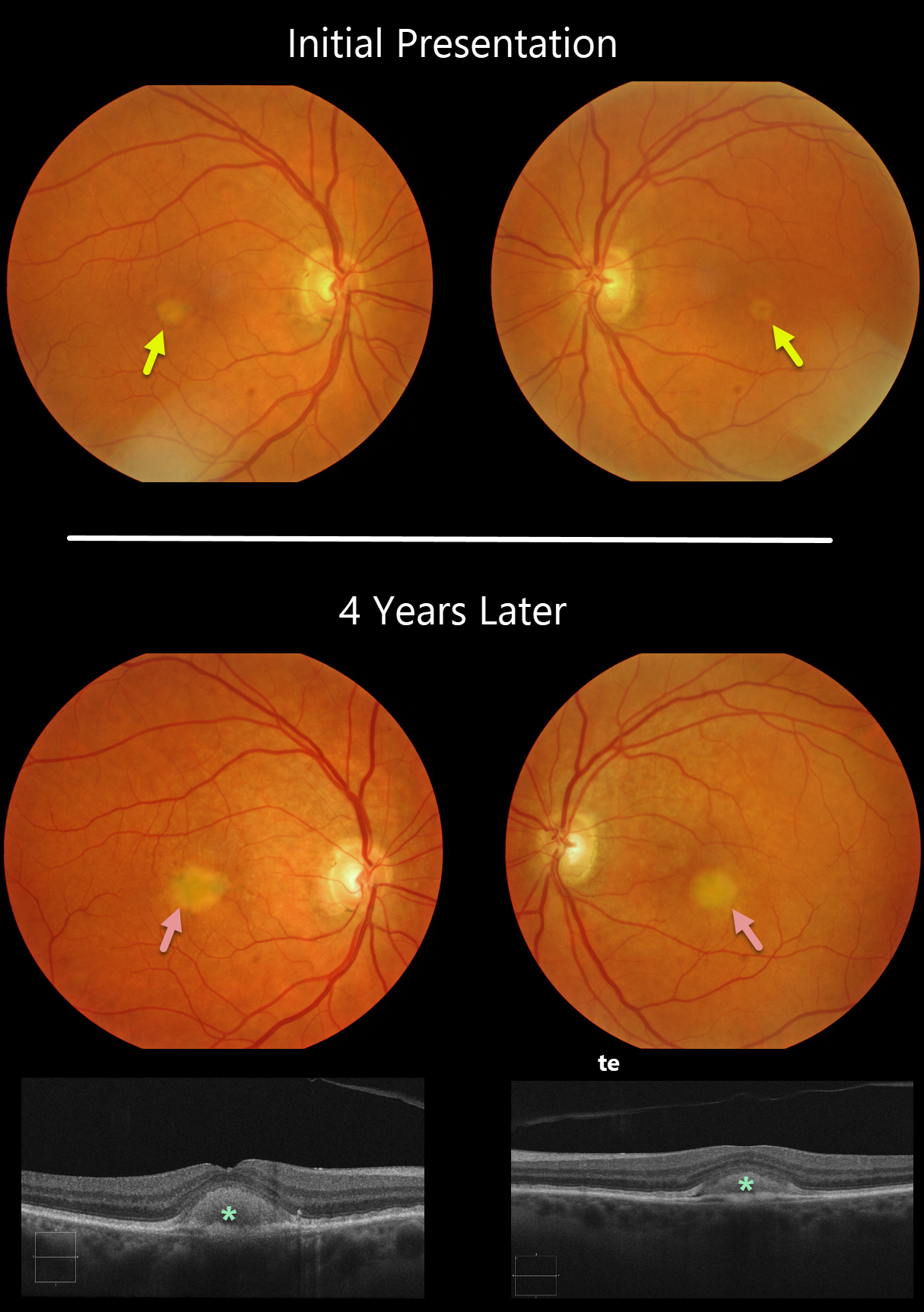
ADULT-ONSET FOVEOMACULAR VITELLIFORM DYSTROPHY
Originally posted on @retina.rocks September 13, 2021
Our patient shows classic findings for adult-onset foveomacular vitelliform dystrophy (Adult Best’s), including small, round, symmetrical subfoveal yellow spots. The vitelliform lesions enlarged over four years, which is seen on OCT as subretinal hyperreflective material between the RPE and outer retina. This material often indents the overlying retina.
Learning Points:
Adult Best’s is associated with a mutation in the BEST1 gene, which encodes the bestrophin-1 protein. Bestrophin-1, a calcium-activated chloride channel, is primarily found in the basolateral plasma membrane of the RPE.
BEST1 mutations cause a variety of phenotypes, including autosomal recessive bestrophinopathy, best vitelliform macular dystrophy, and autosomal dominant vitreoretinochoroidopathy.
These patients tend to maintain fairly good vision. Outer retinal and RPE atrophy can cause central vision loss, as can the rare development of macular neovascularization. Patients should therefore regularly monitor their vision with an Amsler grid.
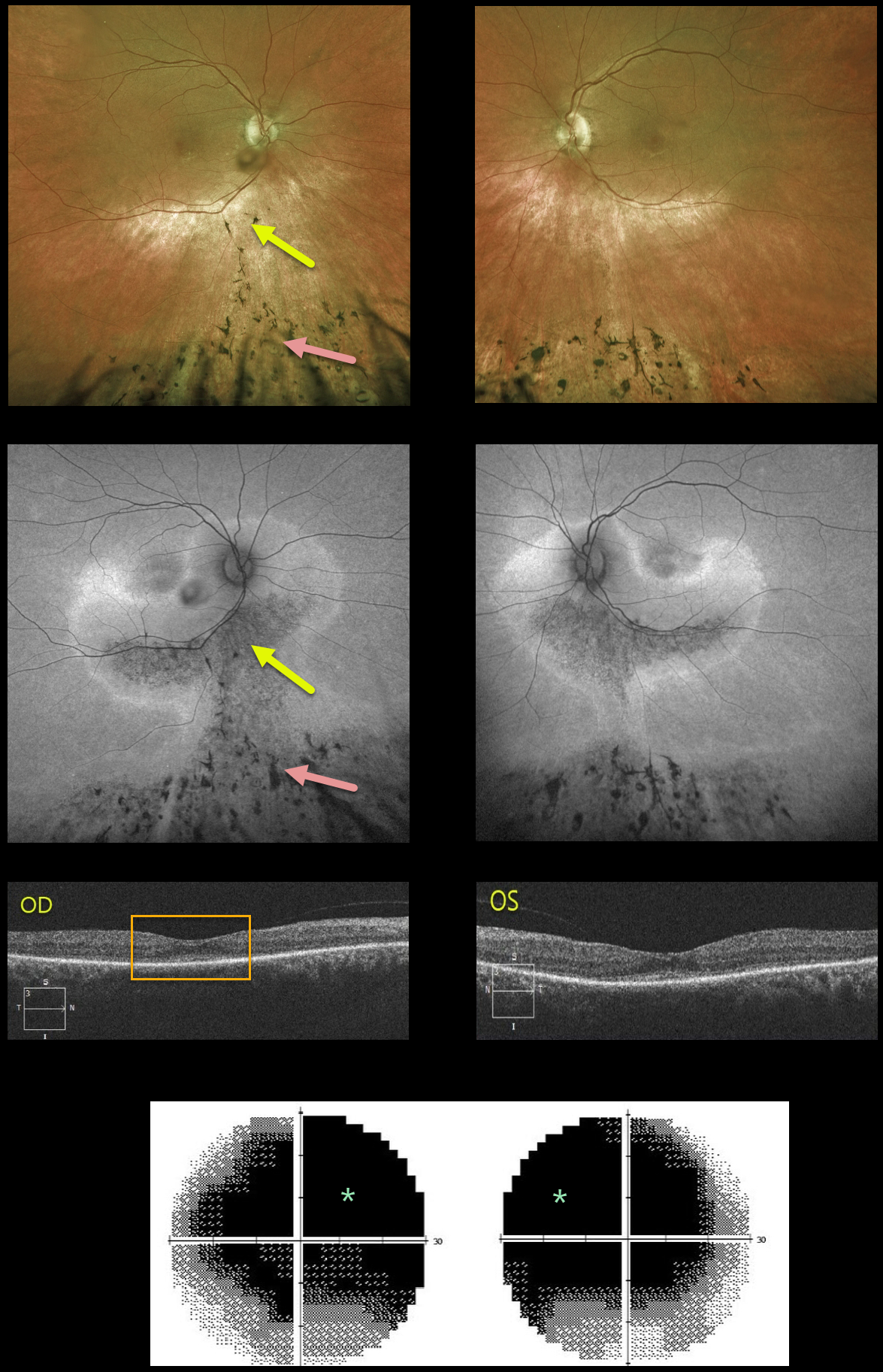
SENIOR-LOKEN SYNDROME
Originally posted on @retina.rocks August 13, 2021
This 51YO female presented with 2 years of progressive night vision loss. There was a history of a prior traumatic brain injury, along with a renal transplant in 2011. VA was 20/60 OD and 20/50 OS.
Optos color and fundus autofluorescence (FAF) imaging show extremely symmetrical findings. A gutter of pigment loss extends along the inferotemporal arcades and inferior to the optic nerve into the inferior peripheries, where there is more extensive chorioretinal scarring with intraretinal pigment migration.
The gutters are classic findings in subretinal exudation, most commonly central serous chorioretinopathy (Yannuzzi et al, Ophthalmology 1984;91:1554-1572). The intraretinal pigment migration can also be seen following blunt trauma (pseudo-RP).
OCT B-scanning shows outer retinal thinning and a probable thickened choroid bilaterally, possibly supporting the central serous diagnosis. However, the marked symmetry of all these findings made us feel that trauma or central serous was unlikely. VF testing shows marked diffuse depression.
Subsequent workup included full field electrophysiology and genetic testing. Dark-adapted ERG showed normal amplitudes, but the light-adapted and cone flicker responses were severely reduced. Her genetic testing revealed pathologic mutations in the NPHP1, ABHD12, LRP5, and PDE6A genes.
The NPHP1 mutation, coupled with her retinal findings and kidney history, confirmed a rare genetic autosomal recessive renal-retinal syndrome known as Senior-Løken Syndrome.
Learning Points:
Senior-Løken Syndrome was first described independently in 1961 by Senior et al as juvenile familial nephropathy and tapetoretinal degeneration (AJO 1961;52:625-633) and Løken et al as hereditary renal dysplasia and blindness (Acta Paediatrica 1961;50:177-184).
Cystic kidney disorders known as nephronophthisis are associated with retinal findings that fall within the broader category of retinal ciliopathies (Adams et al, Ophthalmic Genetics 2007;28:113-125), including Leber congenital amaurosis and some variants of retinitis pigmentosa. Less than 200 cases of Senior-Løken have been reported.
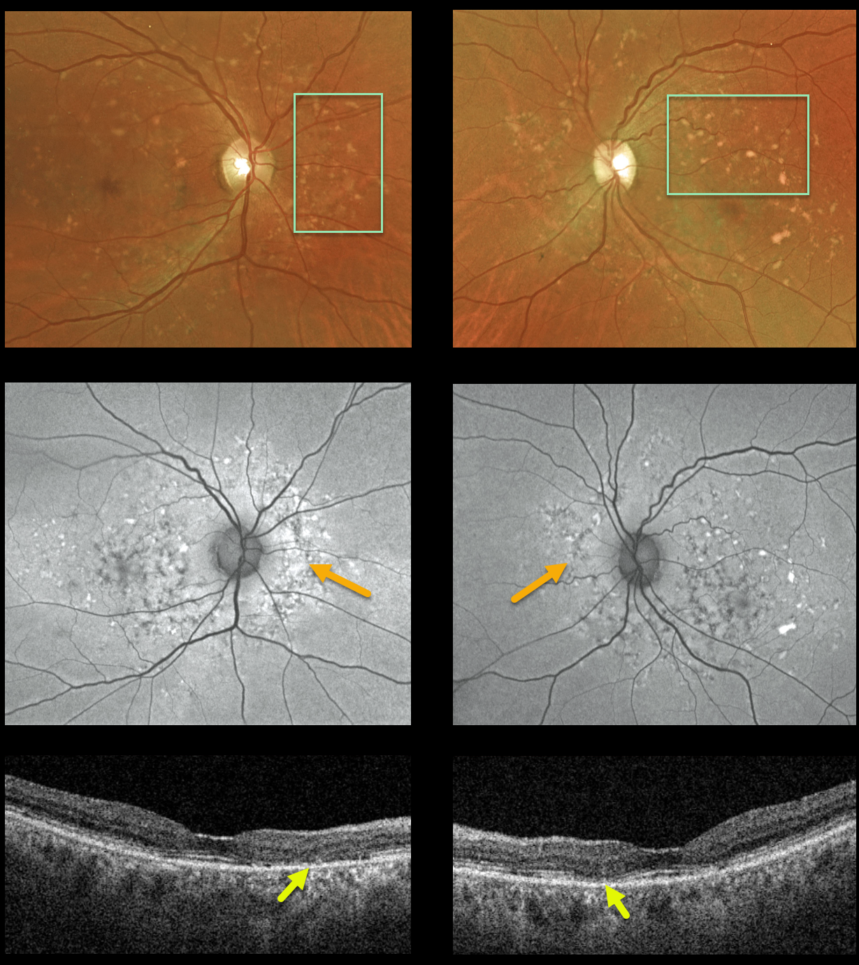
MACULAR DYSTROPHY
Originally posted on @retina.rocks July 29, 2021
This 74YO female came in to see us for a second opinion for age-related macular degeneration. She was visually asymptomatic and denied prior Elmiron (pentosan polysulfate sodium, PPS) use. Vision was 20/30 OU.
Bilateral drusenoid/fleck-like yellow-orange subretinal lesions are scattered throughout the macula and peripapillary regions. These are variably hyper- or hypofluorescent. OCT scanning shows variable outer retinal atrophy without drusen.
Learning Points:
We believe these findings likely represent a type of macular dystrophy. However, she declined genetic testing.
PPS, a medication used to treat interstitial cystitis, can also mimic the findings in age-related macular degeneration and macular dystrophies. Peripapillary hypoautofluorescence, more densely packed macular autofluorescent changes, and earlier central macular involvement suggest PPS toxicity over other causes (see Barnes et al, Ophthalmology Retina 2020;4:1196-1201). Thus, it is important to perform thorough history-taking and document all medications the patient is taking.
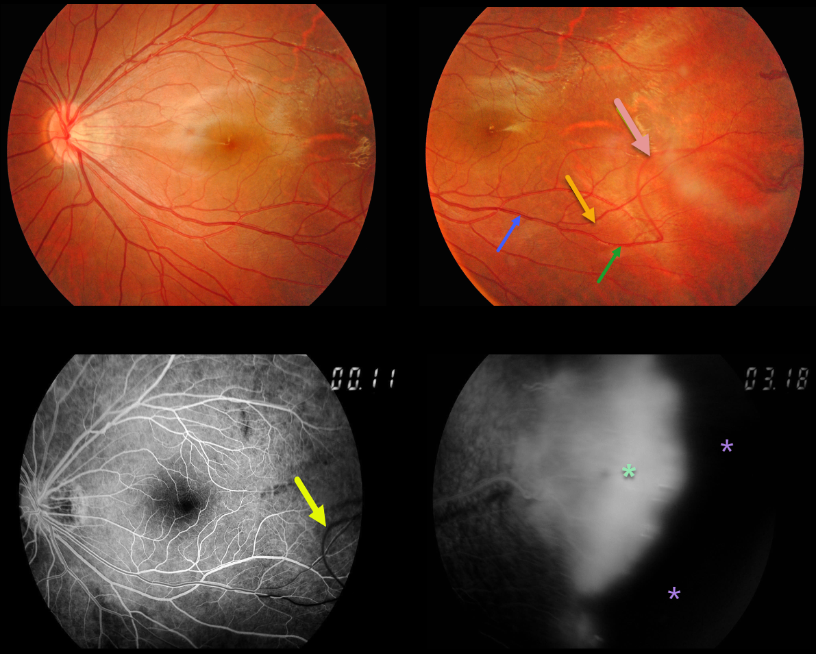
FAMILIAL EXUDATIVE VITREORETINOPATHY (FEVR)
Originally posted on @retina.rocks June 23, 2021
Our patient’s macular retinal vessels are dragged temporally. His brother has similar retinal findings.
The major inferotemporal vein branches into smaller superior and inferior venules. The inferior venule makes an abrupt turn more distally, arching superiorly before extending into the temporal periphery.
Arteries cross veins, veins cross arteries, but arteries never cross arteries, and veins never cross veins.
So why does this inferior venule eventually cross the superior venule? Although we don’t have any stereoscopic images, we suspect that vitreous traction pulled the inferior venule anteriorly and superiorly.
This is also evident on fluorescein angiography (FA), where the superior vessel, which has still not completely filled, is clearly anterior to the other venules, which have laminar filling. FA also shows leaking neovascularization and more peripheral retinal ischemia.
Learning Points:
Originally described by Criswick and Schepens (AJO 1969;58;578-594), familial exudative vitreoretinopathy (FEVR) can be inherited as an autosomal-dominant, recessive, or X-linked trait with high penetrance and variable expressivity.
FEVR is characterized by peripheral temporal retinal avascularity, lipid exudation, neovascularization, tractional retinal detachment, and temporal dragging of the macula and retinal vessels. These findings are somewhat similar to those found with retinopathy of prematurity (ROP).
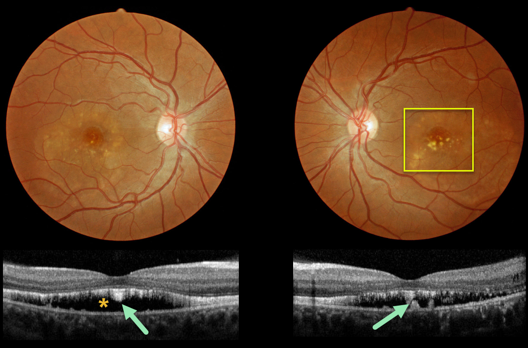
BEST DISEASE
Originally posted on @retina.rocks June 3, 2021
This patient has progressed to the more atrophic ‘scrambled egg’ appearance of Best’s disease.
OCT scanning shows the common hyporeflective subretinal space left behind after the yellow subretinal material absorbs, which often flattens over time.
Shaggy hyperreflective material, likely representing shed photoreceptors and residual vitelliform material, is noted along the posterior retinal surface and on top of the RPE.
Learning Points:
Best vitelliform macular dystrophy, or Best’s disease, is associated with a mutation in the BEST1 gene. This inherited disorder usually presents in childhood or early adulthood with a bilateral foveal “egg-yolk” appearance.
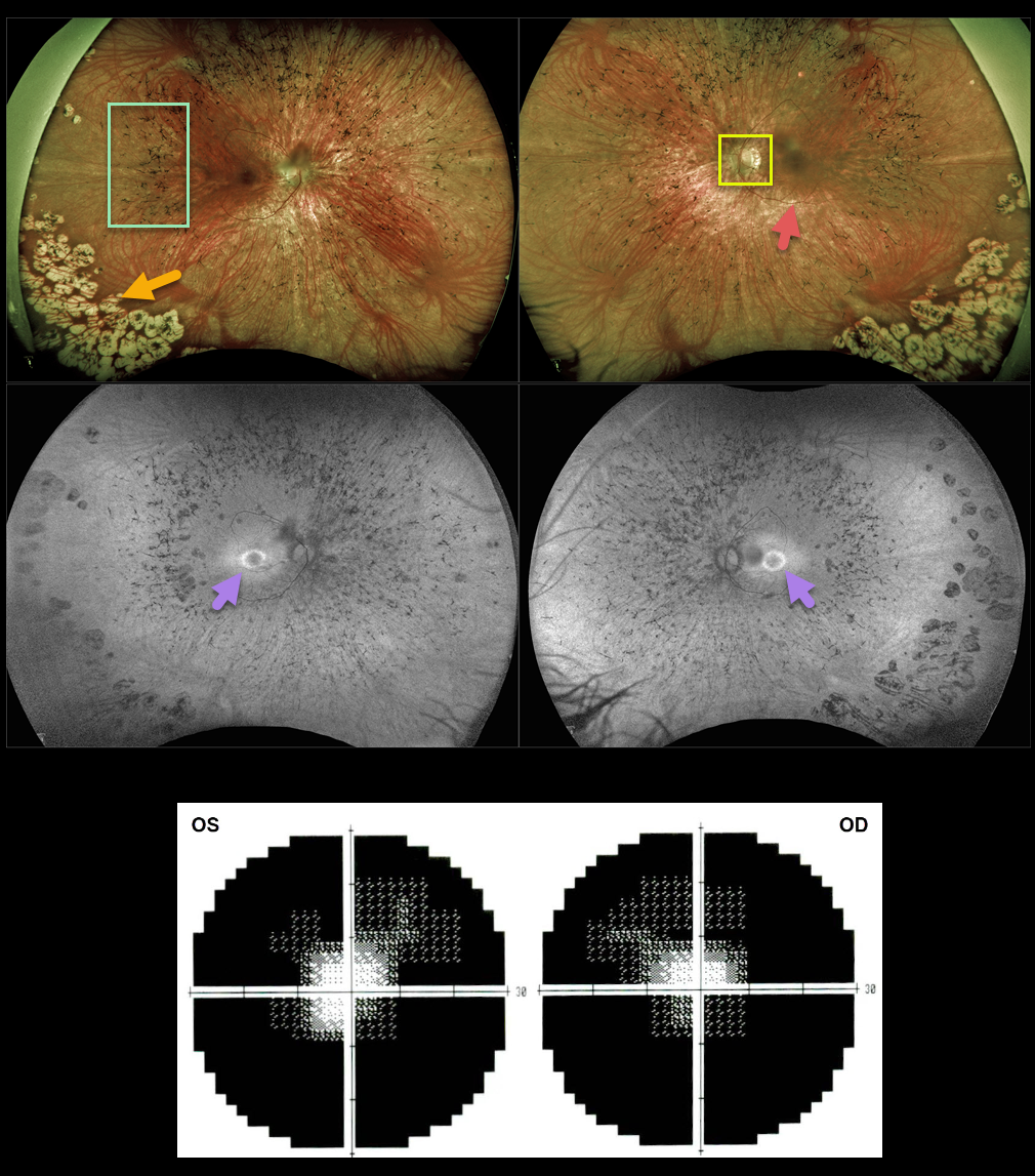
RETINITIS PIGMENTOSA
Originally posted on @retina.rocks June 2, 2021
This 35YO female has classic findings for retinitis pigmentosa. Optos imaging shows somewhat pale nerves, narrowed retinal vessels, and peripheral intraretinal pigment migration (bone spicules). There are also confluent cobblestone-like changes in the inferotemporal peripheries.
Fundus autofluorescence (FAF) shows a hyper-FAF ring around the central macula, indicating preserved photoreceptors that may be at-risk for future degeneration.
Visual fields are markedly constricted bilaterally.
Learning Points:
Photoreceptors normally inhibit RPE cells, so any process involving photoreceptor loss can cause intraretinal RPE migration that mimics retinitis pigmentosa, including chronic retinal detachment and blunt trauma.
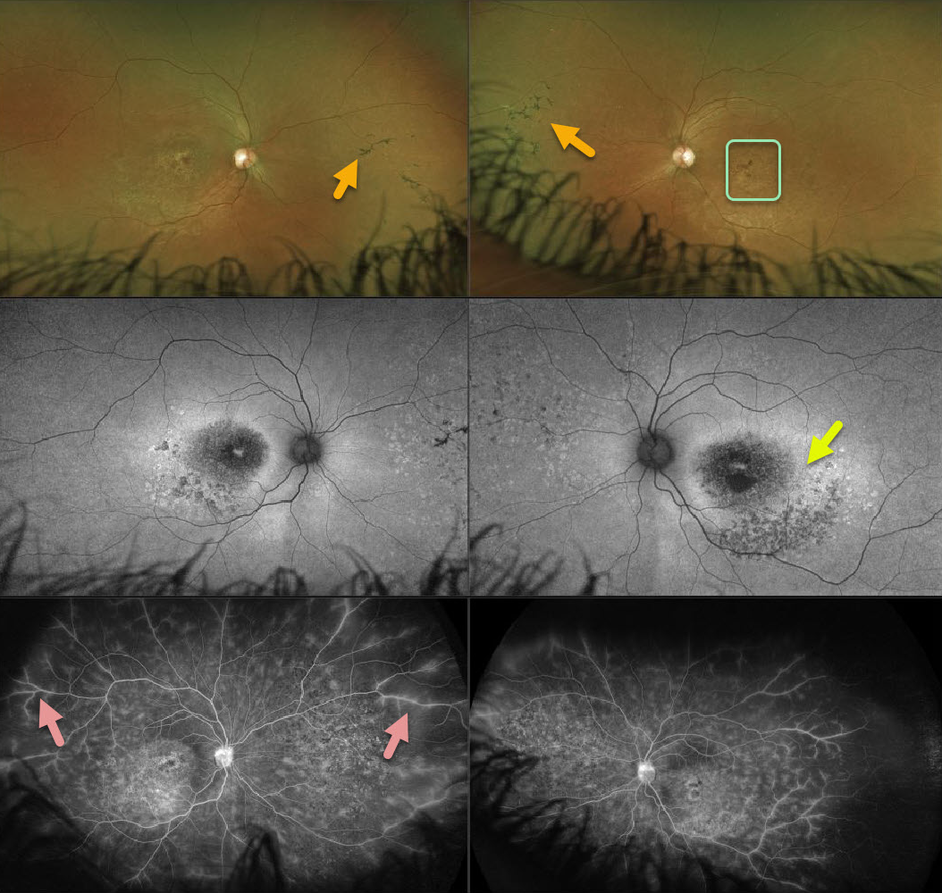
USHER SYNDROME
Originally posted on @retina.rocks April 23, 2021
This 26YO female presented with progressive bilateral vision loss and photophobia. Vision was 20/60 OD and 20/200 OS. She had moderate hearing loss since birth, and denied any family history of eye disease.
Ultra-wide-field imaging reveals areas of nasal mid-peripheral bone spicules (intraretinal pigment migration) and ‘beaten-bronze’ macular atrophy.
There is a ring of hyper-autofluorescence surrounding each macula, which identifies at-risk retina for future degeneration. Fluorescence angiography (FA) shows variable window defects due to pigmentary changes, with diffuse peripheral vascular staining and leakage.
Learning Points:
The association with neurosensory deafness/hearing loss and retinitis pigmentosa (RP) is called Usher Syndrome. About 10% of children who are deaf/hearing impaired have RP.
Vascular leakage is not uncommon with inherited retinal diseases. Photoreceptors normally inhibit RPE cells, and photoreceptor damage allows for RPE intraretinal migration along retinal capillaries. Vascular endothelial cells adjacent to these translocated RPE cells are thin and fenestrated, similar to the choriocapillaris, and may be the cause of this vascular leakage (see Li et al, Ophthalmology 1995;102;8805-816).
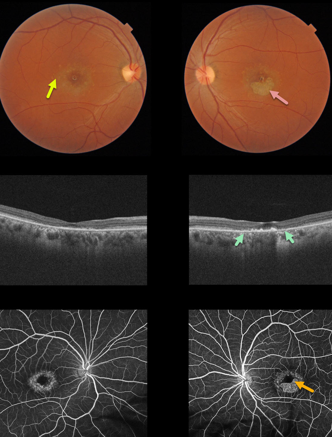
STARGARDT DISEASE
Originally posted on @retina.rocks April 12, 2021
This 40YO female presented with vision of 20/400 in her right eye and counting fingers (CF) in her left eye due to classic findings from Stargardt’s disease, including perifoveal yellow subretinal flecks and variable outer retinal/RPE atrophy.
The OCT of her left eye best visualizes the bull’s-eye pattern of outer retinal and RPE atrophy. Loss of photoreceptors is evident as the outer plexiform layer (OPL) descends towards the RPE, eventually resulting in fairly complete atrophy of the outer retina and RPE.
Fluorescein angiography (FA) characteristically shows a ‘dark choroid’ from the lipofuscin-engorged RPE blocking the choroid (not present in this case).
Learning Points:
Stargardt’s disease is an autosomal recessive disorder caused by a mutation in the ABCA4 gene. The ABCA4 protein is located in the photoreceptor outer segments and is involved in the recycling of 11-cis-retinal.
Mutation of this gene causes accumulation of lipofuscin in the RPE, eventually leading to photoreceptor and RPE degeneration. Other ABCA4 disorders include fundus flavimaculatus and cone-rod dystrophy.
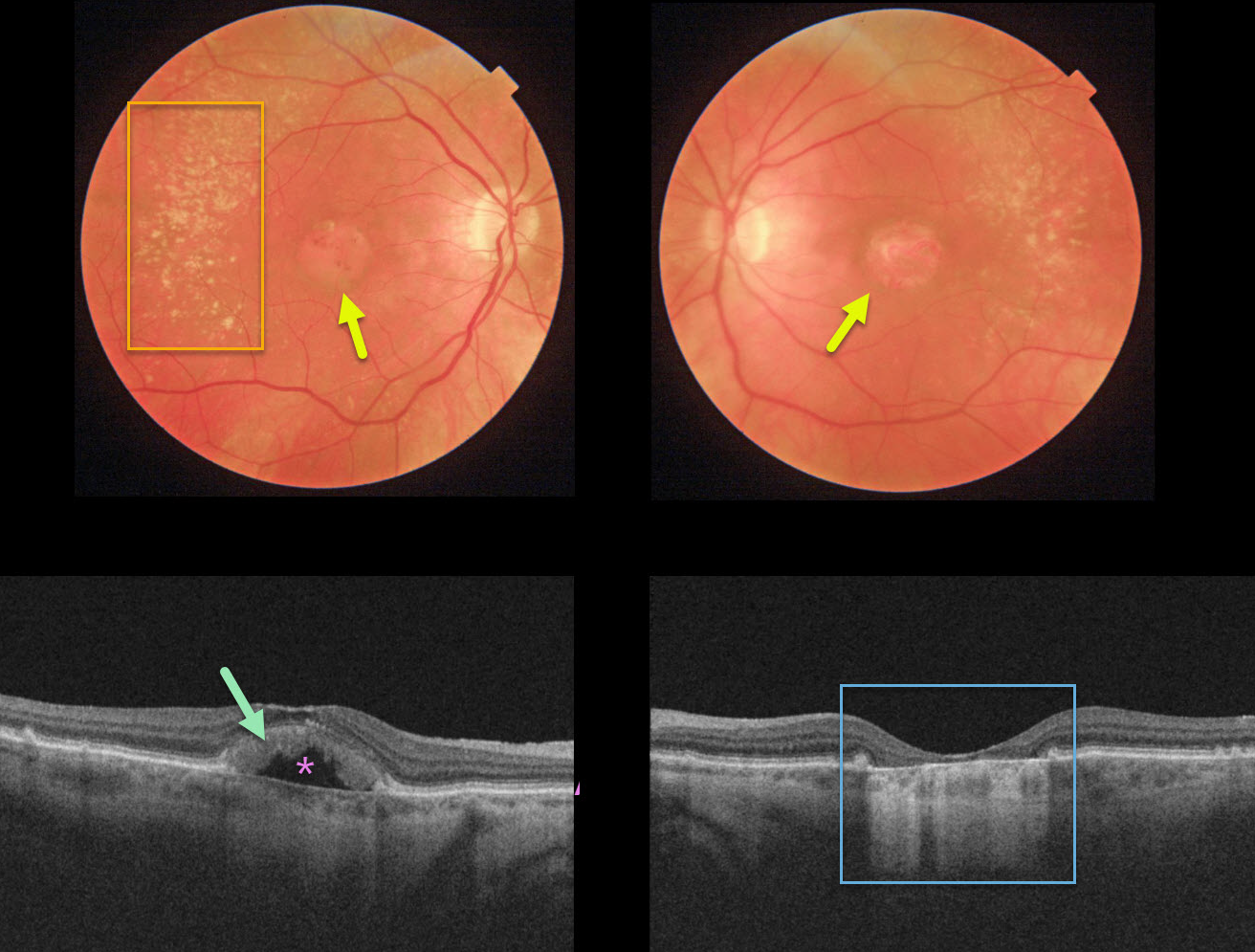
ADULT-ONSET FOVEOMACULAR VITELLIFORM DYSTROPHY
Originally posted on @retina.rocks April 5, 2021
This 80YO female was 20/400 in her right eye and counting fingers in her left eye due to probable adult-onset foveomacular vitelliform dystrophy (Adult Best’s). Funduscopically, there are bilateral round atrophic foveal lesions with peripheral drusen.
OCT of the right eye shows hyperreflective outer retinal material overlying a pocket of subretinal fluid. The underlying RPE is atrophic with bare Bruch’s membrane. OCT of the left eye shows complete outer retinal and RPE atrophy.
Learning Points:
Adult Best’s is associated with a mutation in the BEST1 gene, which encodes the bestrophin-1 protein.
Bestrophin-1, a calcium-activated chloride channel, is primarily found in the basolateral plasma membrane of the RPE. BEST1 mutations cause a variety of phenotypes, including autosomal recessive bestrophinopathy, best vitelliform macular dystrophy, and autosomal dominant vitreoretinochoroidopathy.
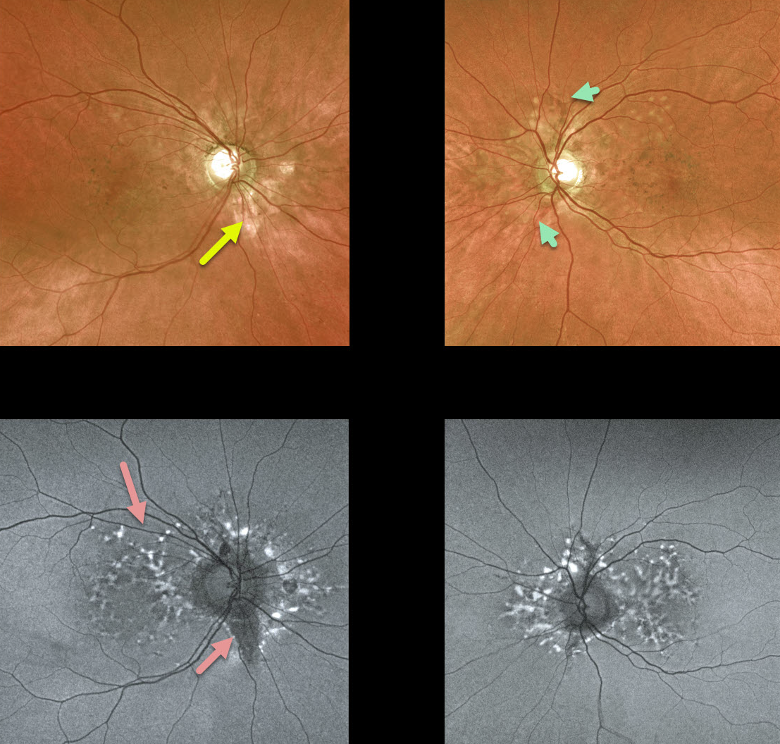
ANGIOID STREAKS
Originally posted on @retina.rocks April 1, 2021
We have been following this 54YO female with pseudoxanthoma elasticum (PXE) for many years. Vision is 20/20 bilaterally, and fortunately, she remains asymptomatic.
She has bilateral macular peripapillary pigmentary changes. Angioid streaks, always tough to find unless the clinician actively looks for them, are not prominent, although a few are visible in her left eye.
Fundus autofluorescence (FAF) shows more dramatic changes, including scattered hyper- and hypo-FAF peripapillary and macular spots.
Her sister became legally blind many years ago from bilateral macular neovascularization (MNV).
Learning Points:
Patients with angioid streaks and pseudoxanthoma elasticum (PXE) can present with retinal findings similar to those of pattern dystrophies.
These patients are at significant risk for macular neovascularization and need to be followed and treated as needed with anti-VEGF agents, as in patients with AMD.
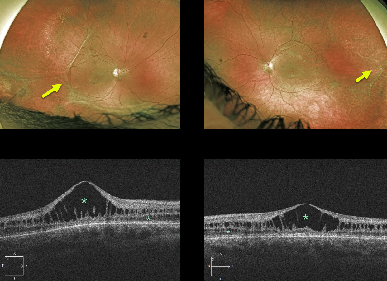
X-LINKED RETINOSCHISIS
Originally posted on @retina.rocks March 19, 2021
This 18YO male has classic findings for X-linked retinoschisis, including vitreous veils and bilateral foveal schisis. The macular schisis involves mostly the inner and outer nuclear layers, while peripherally it involves the nerve fiber layer (not pictured). Vision is 20/400 OD and 20/100 OS.
Learning Points:
X-linked retinoschisis is caused by a mutation in the RS1 gene, located on the X chromosome and encoding retinoschisin, a protein likely involved in retinal cell adhesion. Although it is usually transmitted as an X-linked recessive disorder in males, it can also affect females.
Over time, the macular schisis flattens with secondary atrophy. Vision usually levels off at about 20/100. Patients can develop spontaneous vitreous hemorrhage and rhegmatogenous retinal detachment from peripheral schisis.
Although there is no specific treatment, topical carbonic anhydrase inhibitors may reduce foveal thickness and possibly minimize foveal atrophy.
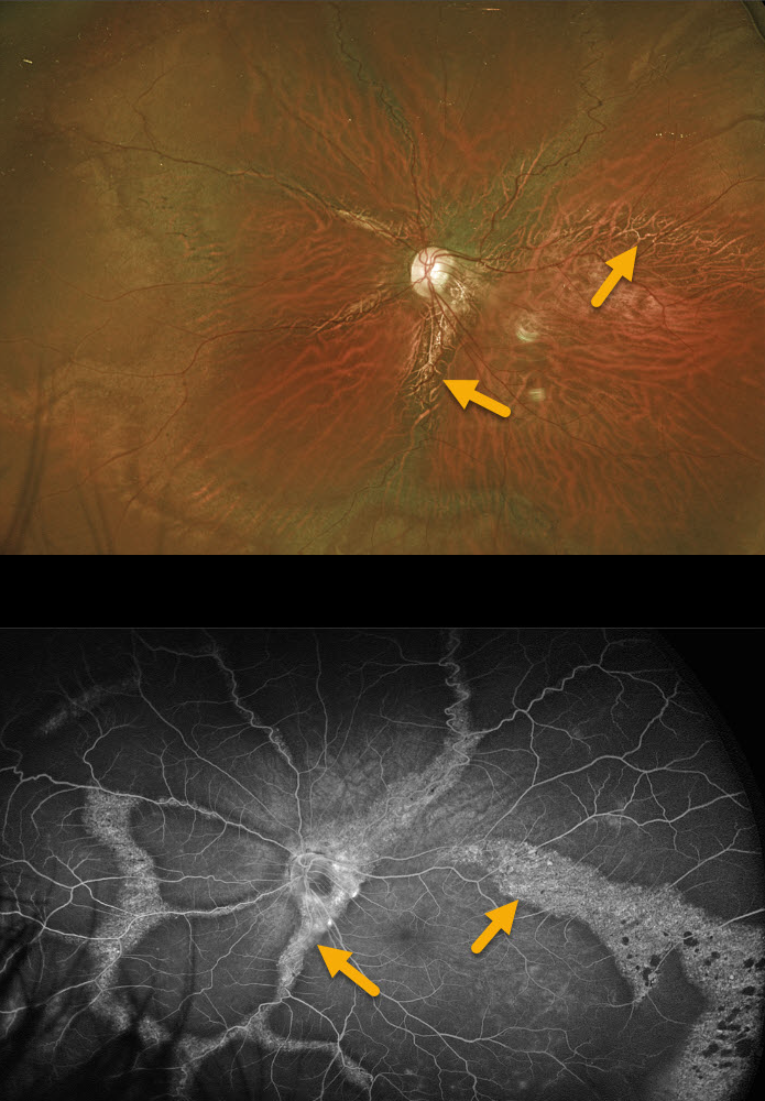
UNKNOWN DIAGNOSIS
Originally posted on @retina.rocks March 3, 2021
This unknown shows extensive and unusual, radial and circumferential scarring of the posterior pole in this 37 YO male’s left eye. The right eye (not pictured) had a failed retinal detachment repair at an outside practice many years earlier, resulting in no light perception vision and an opaque, white cornea.
Since the radial changes seem to mostly follow the arcades, pigmented paravenous retinochoroidal atrophy is considered in the differential, but there are no areas of intraretinal pigment migration (bone spicules), and the more peripheral connecting circumferential scarring would be unusual. No electrophysiology has been performed.
This patient presented like this years ago without symptoms, all findings have been stable, and we have been following him since with 20/30 vision.
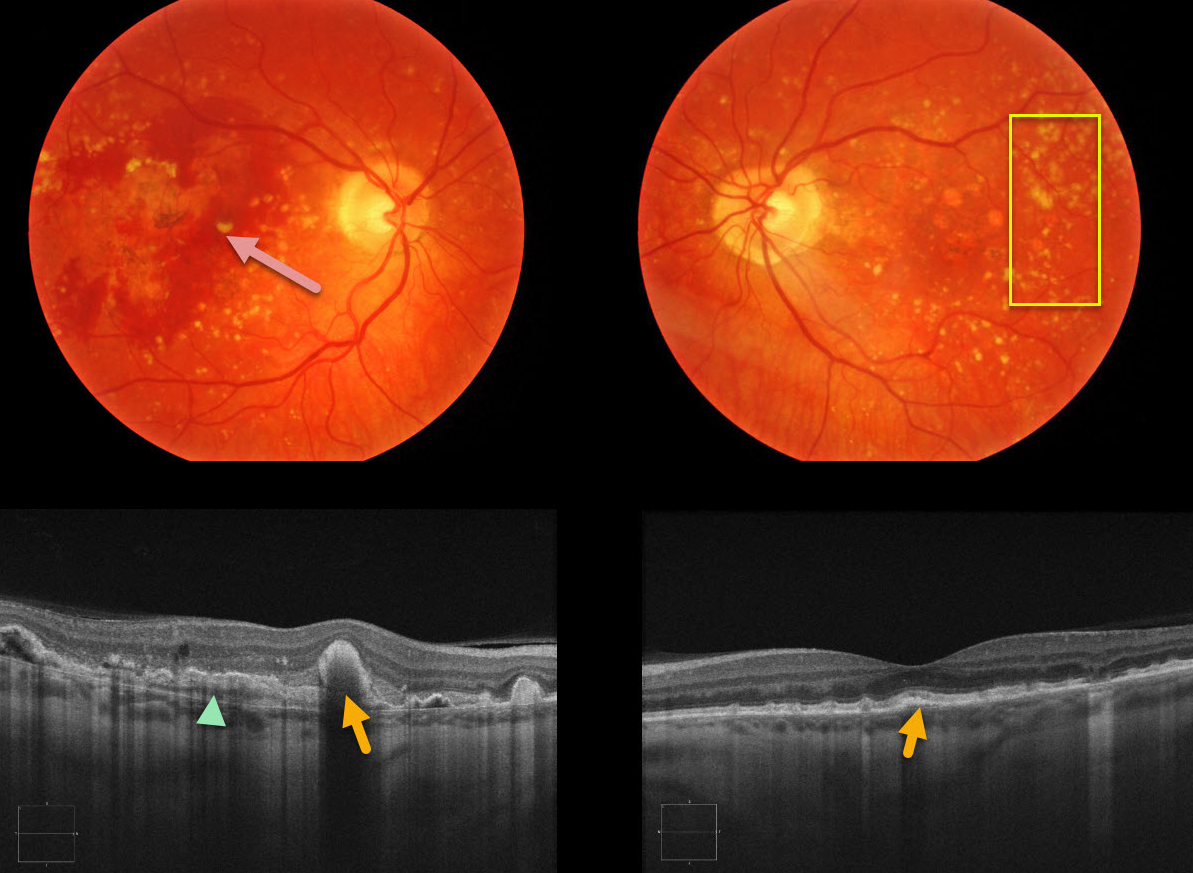
DOMINANTLY INHERITED RADIAL BASAL LAMINAR DRUSEN
Originally posted on @retina.rocks January 29, 2021
This 66YO female presented with acute vision loss of her right eye. There was no family history of eye disease. Extensive variably sized drusen were noted in both maculae, with subretinal blood from macular neovascularization (MNV) in her right eye.
OCT scanning shows variably sized and peaked drusen and subretinal hyperreflective material (SHRM) reminiscent of Doyne’s honeycomb macular dystrophy.
Learning Points:
Doyne’s honeycomb macular dystrophy, also known as dominantly inherited radial basal laminar drusen or Malattia Levantinese, is a rare macular disorder caused by a mutation in the EFEMP1 gene on chromosome 2p16. The EFEMP1 protein is a member of the fibulin family of extracellular matrix glycoproteins. The defective protein creates an abnormally thickened RPE basement membrane.
Centrally large, nodular, and confluent drusen are noted, along with a temporal radiating pattern of smaller cuticular drusen. Later, there may be variable amounts of RPE hyperplasia and fibrous metaplasia. MNV, as in this case, may also develop.
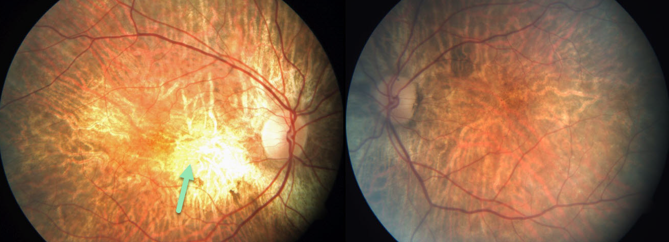
CHOROIDEREMIA
Originally posted on @retina.rocks January 28, 2021
This patient has choroideremia and, luckily, has preserved central 20/30 vision bilaterally. Fundus photography shows patchy areas of RPE and chorioretinal atrophy.
Learning Points:
Choroideremia is an X-linked recessive chorioretinal dystrophy caused by a mutation in the CHM gene. Some female carriers can still develop choroideremia due to irregular inactivation of the X chromosome (lyonization).
Night blindness usually begins between the ages of 10 and 30, followed by peripheral visual field loss. Zones of patchy RPE and chorioretinal atrophy initially appear in the mid-periphery, gradually spreading anteriorly and posteriorly.
Eventually, the patient is left with only a small island of vision, narrowed retinal vessels, and optic atrophy.
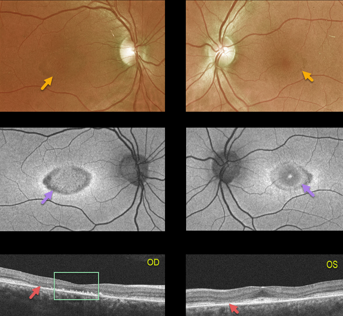
CONE ROD DYSTROPHY
Originally posted on @retina.rocks January 27, 2021
This 42YO female presented with counting fingers vision OD and 20-25 vision OS. There was no family history of vision loss.
A bull’s-eye pattern of perifoveal atrophy was noted on Optos color and fundus autofluorescence imaging, with loss of photoreceptors on OCT. The outer retinal atrophy extended through the macular center in her right eye, accounting for the counting-fingers vision.
Learning Points:
Cone-rod dystrophy is a group of inherited retinal disorders characterized by loss of cone photoreceptors followed by rod photoreceptors. Symptoms include decreased visual acuity, central vision loss, color vision abnormalities, and photophobia.
Several genes have been implicated, including ABCA4, CRX, GUCY2D, and RPGR. ABCA4 mutations are found in autosomal recessive cone-rod dystrophy as well as Stargardt Disease.
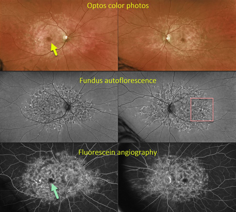
FUNDUS FLAVIMACULATUS
Originally posted on @retina.rocks January 7, 2021
This 44YO male presented with classic findings for fundus flavimaculatus, including perifoveal atrophy and variable hyper-autofluorescent subretinal yellow flecks extending from the macula to the midperiphery. Fluorescein angiography (FA) reveals a central dark choroid due to RPE cells becoming engorged with lipofuscin. Surprisingly, vision was 20/20 OU with a negative family history.
Learning Points:
Fundus flavimaculatus is an autosomal recessive disorder that is caused by mutations involving the ABCA4 gene.
This gene encodes one of a family of ATP-binding cassette (ABC) transmembrane proteins, which mediate the active transport of various substrates across cellular membranes. The ABCA4 protein is present in the photoreceptor outer segment disc membranes, where it participates in the recycling of 11-cis-retinal.
Stargardt’s disease is felt to be fundus flavimaculatus without peripheral findings. Vision in fundus flavimaculatus is often better than Stargardt’s due to later disease onset and less macular involvement. Both likely represent an RPE lipofuscin storage disease.
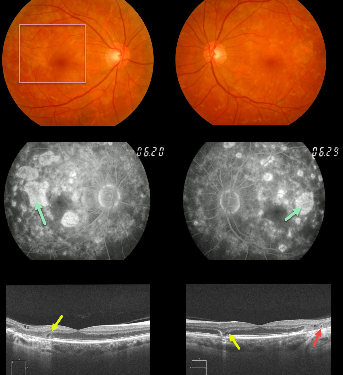
MATERNALLY INHERITED DIABETES AND DEAFNESS (MIDD)
Originally posted on @retina.rocks December 4, 2020
This 62yo female with a history of type 2 diabetes and mild hearing loss initially presented in 2012 with asymptomatic 20/30 vision OU and bilateral areas of focal macular atrophy and subretinal yellow fleck-like lesions.
The retinal pigment epithelial changes were much more pronounced on fluorescein angiography. OCT scanning shows classic wedge defects along the border of the macular atrophy as well as an area of outer retinal tubulation in the left eye.
Learning Points:
Maternally inherited diabetes and deafness (MIDD) accounts for up to 3% of all cases of diabetes and results from a mutation in mitochondrial DNA at position A3243G. MIDD often masquerades as a pattern macular dystrophy.
Fundus autofluorescence (FAF), in our experience, is the best way to visualize these changes, but it was not available at the office when we initially saw her. MIDD patients need to be screened for other potential co-existing medical conditions, including cardiac arrhythmias, myopathy, and renal disease.
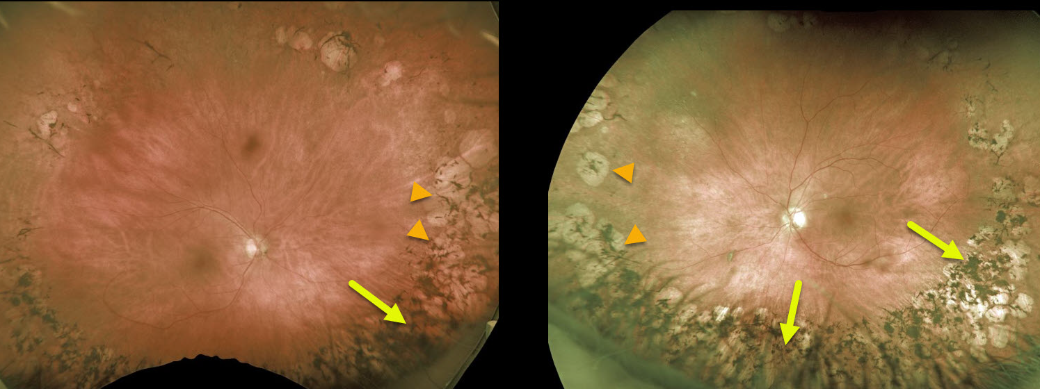
AUTOSOMAL DOMINANT VITREORETINO-CHOROIDOPATHY (ADVIRC)
Originally posted on @retina.rocks November 25, 2020
This patient has an extremely rare retinal dystrophy called autosomal dominant vitreoretinochoroidopathy (ADVIRC). Its most distinguishing feature is a band of circumscribed chorioretinal atrophy with intraretinal pigment migration extending circumferentially from the equator to the ora serrata.
Learning Points:
Autosomal dominant vitreoretinochoroidopathy was first described by Kaufman et al (Arch Ophthalmology 1982;100:272-278). It is caused by a mutation in the BEST1 gene, which encodes for bestrophin, a transmembrane protein in the basolateral plasma membrane of the RPE responsible for intracellular calcium homeostasis.
Aside from the pigmentary retinal changes, findings include punctate whitish pre-retinal opacities, vitreous cells and fibrillary condensation, and retinal neovascularization.
Anterior segment findings are common and include hyperopia and angle-closure glaucoma. There is wide phenotypic variability, and advanced cases can mimic retinitis pigmentosa (see Boulanger-Scemama et al Retina 2019;39:867-878).
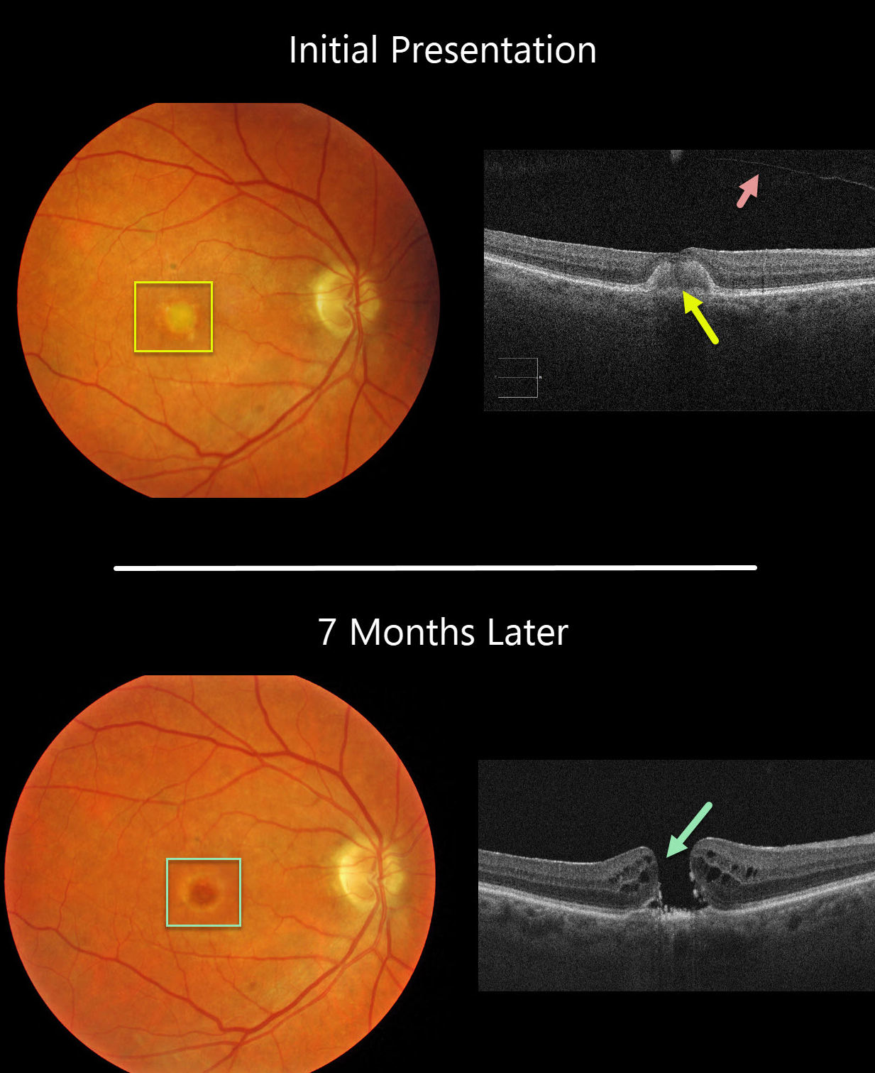
ADULT-ONSET FOVEOMACULAR VITELLIFORM DYSTROPHY
Originally posted on @retina.rocks November 18, 2020
This 77YO female was initially examined with 20/60 vision OD, 20/30 vision OS, and bilateral vitelliform lesions (only right eye pictured) consistent with Adult Best (adult-onset foveomacular vitelliform dystrophy). A complete posterior vitreous detachment (PVD) was noted in her right eye.
Seven months later, vision decreased to counting fingers OD due to a new full-thickness macular hole. This patient was asymptomatic due to good vision in her left eye and has opted against surgery for now.
Learning Points:
Macular holes are most commonly caused by a partial PVD with active vitreomacular traction. However, they can occasionally develop without traction following a prior PVD or vitrectomy surgery. Macular holes have also been reported as a rare complication of adult Best’s disease.
Adult-onset foveomacular vitelliform dystrophy is associated with a mutation in the BEST1 gene, which encodes the bestrophin-1 protein.
Bestrophin-1, a calcium-activated chloride channel, is primarily found in the basolateral plasma membrane of the RPE. BEST1 mutations cause a variety of phenotypes, including autosomal recessive bestrophinopathy, best vitelliform macular dystrophy, and autosomal dominant vitreoretinochoroidopathy.
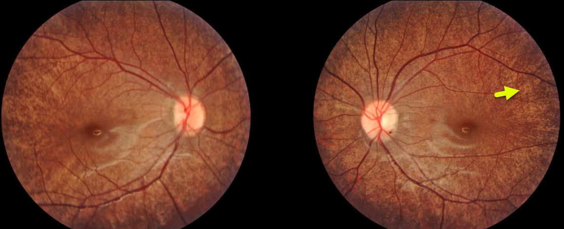
FUNDUS ALBIPUNCTATUS
Originally posted on @retina.rocks November 5, 2020
This patient has fundus albipunctatus, a rare autosomal recessive form of congenital stationary night blindness. The characteristic numerous small white-yellow specks spare the fovea.
Learning Points:
Fundus albipunctatus is caused by a mutation in the RDH5 gene, which codes for retinol dehydrogenase, which converts 11-cis retinol to 11-cis retinal within the RPE.
The optic disc, retinal vessels, visual acuity, visual fields, and color vision are usually normal. Electroretinograms (ERG) are typically depressed but normalize after prolonged dark adaptation, in contrast with retinitis punctata albescens.
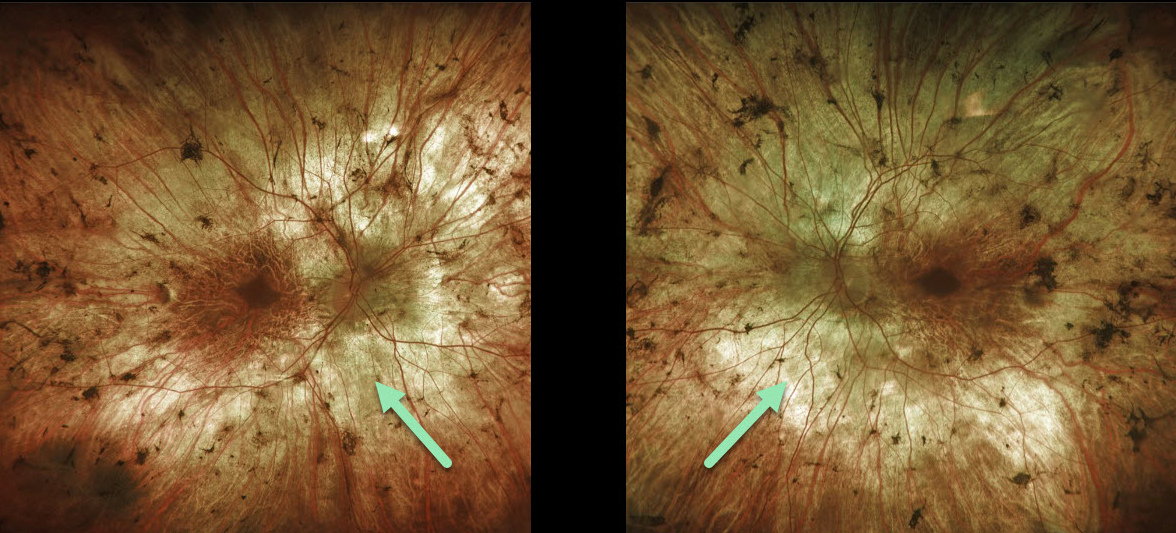
CHOROIDEREMIA
Originally posted on @retina.rocks October 28, 2020
This is a 68YO male with a likely diagnosis of choroideremia. Vision is remarkably 20/60 OD and 20/50 OS. Note the areas of patchy RPE and chorioretinal atrophy in both eyes.
Learning Points:
Choroideremia is an X-linked recessive chorioretinal dystrophy caused by a mutation in the CHM gene. Some female carriers can still develop choroideremia due to irregular inactivation of the X chromosome (lyonization) later in life.
Night blindness usually begins between the ages of 10 and 30, followed by peripheral visual field loss. Patchy RPE and chorioretinal atrophy initially appear in the mid-periphery, gradually spreading anteriorly and posteriorly. Eventually, the patient is left with only a small island of vision, narrowed retinal vessels, and optic atrophy.
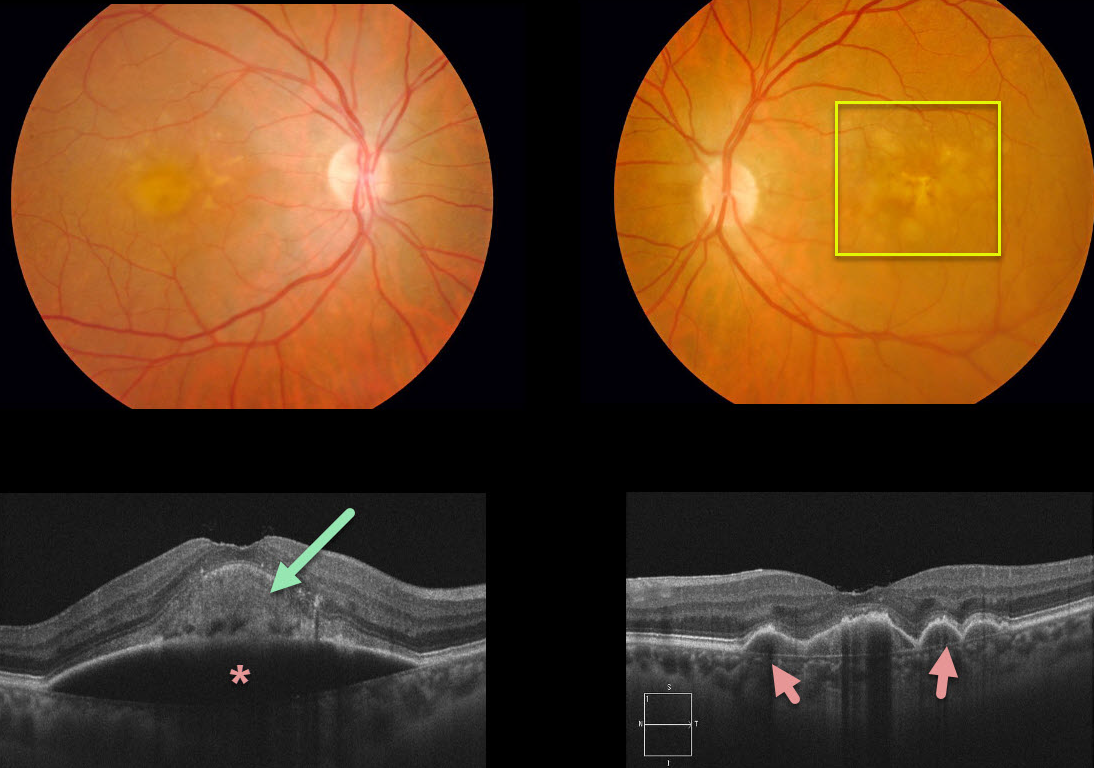
PIGMENT EPITHELIAL DETACHMENT (PED)
Originally posted on @retina.rocks September 30, 2020
This patient had been followed for age-related macular degeneration (AMD) with bilateral confluent soft drusen. She then developed a large acquired vitelliform lesion overlying a retinal pigment epithelial detachment (PED) in her right eye. There are several PEDs also shown in the left eye.
Learning Points:
Vitelliform lesions are commonly found with other disorders, including retinal dystrophies (Best’s disease and macular dystrophies), cuticular drusen, AMD, and vitreomacular traction.
The yellow subretinal material is felt to be debris from shed photoreceptor outer segments. These lesions markedly autofluoresce on fundus autofluorescence.
A great reference to learn more about AVLs is Freund et al Retina 2011;31:13-25.
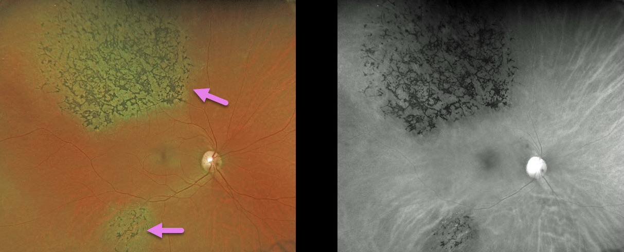
TRAUMA
Originally posted on @retina.rocks September 29, 2020
A 49YO female was involved in a motor vehicle accident 30 years ago.
Her retina showed signs of pigmentary retinopathy from the ocular injury, which could be mistaken for retinitis pigmentosa (RP). Unlike typical RP, our patient had multifocal, sectoral, unilateral findings.
Learning Point:
Intact photoreceptors inhibit RPE migration. Photoreceptor loss from numerous causes, including blunt trauma, retinal detachment, macular telangiectasia, and retinitis pigmentosa, can allow intraretinal RPE migration with this shared phenotype.
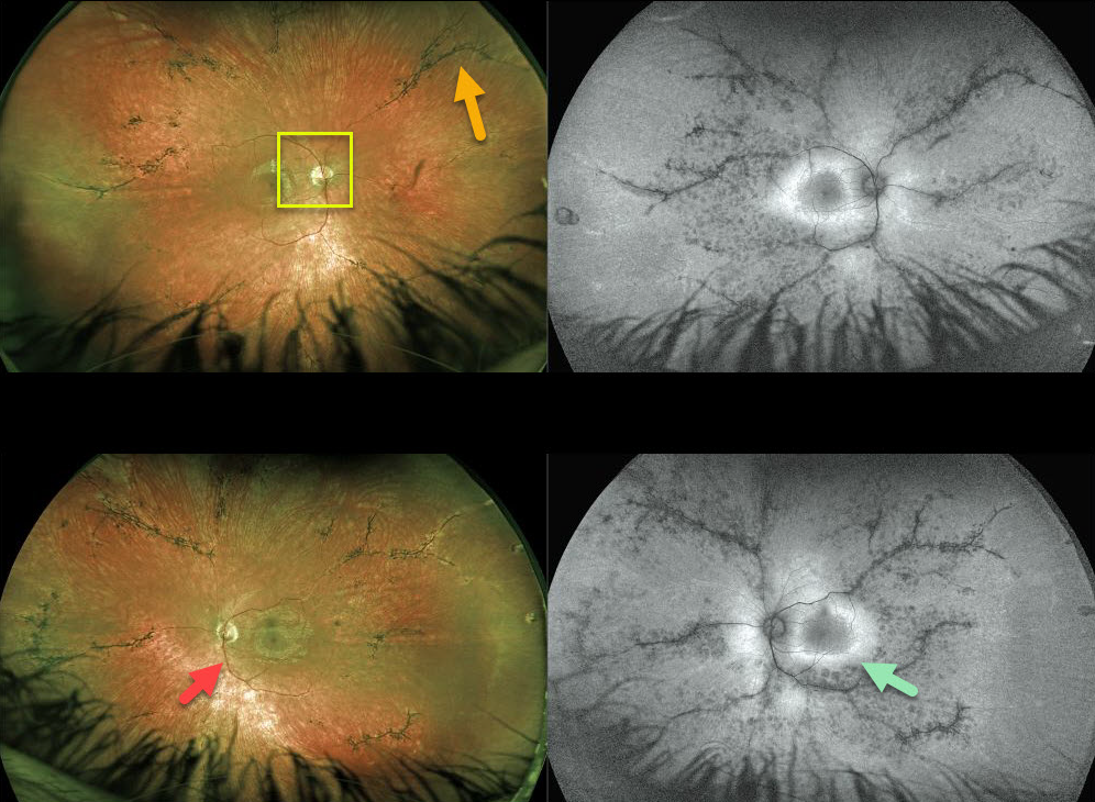
PIGMENTED PARAVENOUS CHORIORETINAL ATROPHY
Originally posted on @retina.rocks September 15, 2020
This 16YO patient with pigmented paravenous chorioretinal atrophy has excellent central vision of 20/20 OU but with significant nyctalopia.
Posterior segment findings include attenuated retinal vessels, bone spicules (intraretinal migration of the RPE along retinal capillaries), and optic disc pallor.
The bone spicules in this patient are primary perivascular, indicating the likely diagnosis of pigmented paravenous chorioretinal atrophy (PPCRA).
Fundus autofluorescence for this patient shows a ring of hyperautofluorescence around each central macula. This ring identifies at-risk retina for future degeneration and can help monitor the progression of this disease.
Learning Points:
Pigmented paravenous chorioretinal atrophy (PPCRA) is a rare condition characterized by paravenous pigment clumps with peripapillary and radial zones of RPE atrophy. It is bilateral, often asymmetric, with various causes, including genetic and pseudo-PPCRA (inflammatory, infectious, or unknown). Patients tend to have some symptoms, usually nyctalopia, but most retain good vision, especially those without macular involvement. The underlying pathophysiology is likely a primary choroidopathy that causes overlying RPE and retinal changes. The chorioretinal changes are best visualized with fundus autofluorescence.
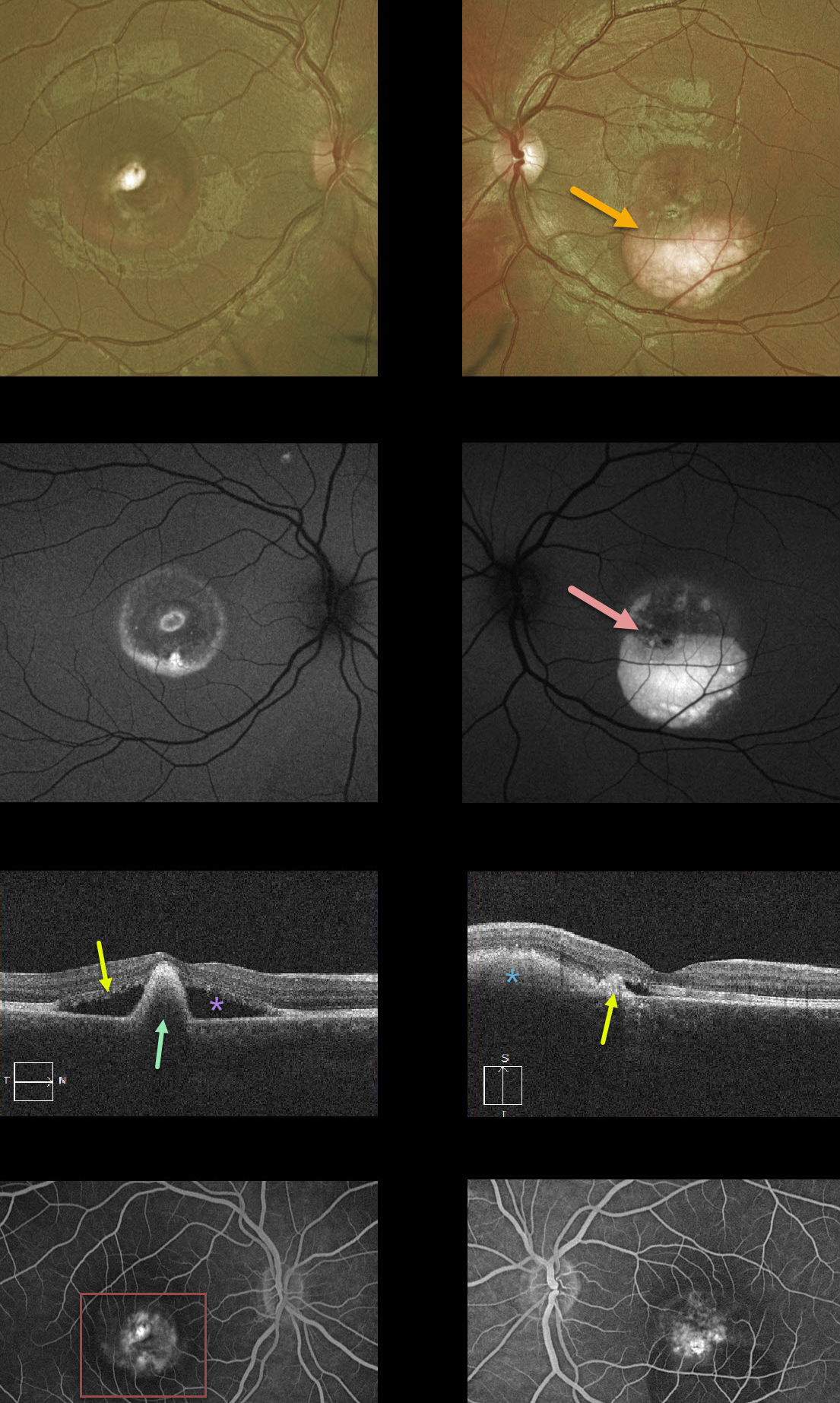
BEST DISEASE
Originally posted on @retina.rocks September 14, 2020
This patient has Best’s disease and has progressed to the more atrophic, ‘scrambled egg’ appearance. The fundus photos show asymmetric layering of the residual yellow-white subretinal vitelliform material.
This material is highly autofluorescent and can also appear as hyperreflective on OCT scans. The right eye has a large subfoveal pigment clump with surrounding non-exudative subretinal fluid. The left eye has a smaller subfoveal scar with a flattened vitelliform lesion without subretinal fluid. Fluorescein angiography (FA) shows staining without active leakage.
Learning Points:
Best vitelliform macular dystrophy (Best’s disease) is associated with a mutation in the BEST1 gene. This inherited disorder usually presents in childhood or early adulthood with a bilateral foveal “egg-yolk” appearance.
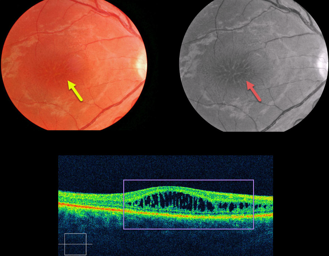
X-LINKED RETINOSCHISIS
Originally posted on @retina.rocks August 26, 2020
This patient presented with mild bilateral central vision loss (only the right eye is pictured).
Typical findings in sex-linked retinoschisis include radiating foveal cystoid macular edema-type changes, which are much more pronounced on red-free imaging.
Although classically described as nerve fiber layer schisis (compared to outer plexiform splitting in degenerative schisis), macular OCT here shows splitting in the inner and outer nuclear layers.
Fluoroscein angiography (not pictured) would show no leakage, as the findings are due to a structural defect rather than exudation.
Learning Points:
X-linked retinoschisis is caused by a mutation in the RS1 gene, which is located on the X chromosome and encodes retinoschisin, a protein likely involved in retinal cell adhesion. Although usually transmitted as an X-linked recessive disorder found in males, it can also sometimes affect females.
Over time, the macular schisis flattens with secondary atrophy. Vision usually levels off at about 20/100. Patients can develop spontaneous vitreous hemorrhage and rhegmatogenous retinal detachment from peripheral schisis.
Although there is no specific treatment, topical carbonic anhydrase inhibitors may reduce foveal thickness and possibly minimize foveal atrophy.
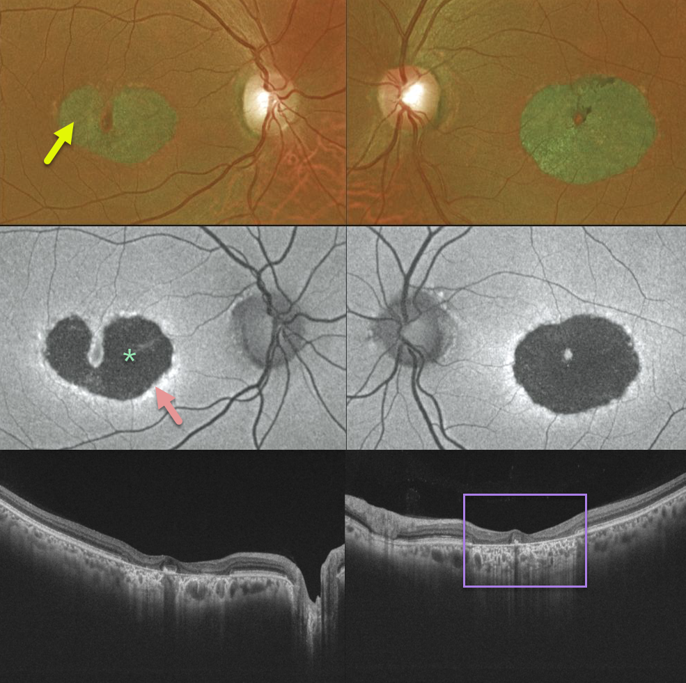
MACULAR DYSTROPHY
Originally posted on @retina.rocks August 19, 2020
Our patient has a bilateral bull’s-eye maculopathy with central foveal sparing. Vision was 20/30 OD and 20/60 OS.
Fundus autofluorescence shows central hypo-FAF from loss of the central RPE. The thin surrounding hyper-FAF ring likely represents diseased, at-risk RPE cells. The OCT scans show pericentral outer retinal and RPE loss.
Learning Points:
Our patient no doubt has some type of inherited macular disorder. Some possibilities include central areolar chorioretinal dystrophy or a cone-rod dystrophy.
Central areolar chorioretinal dystrophy is a genetic condition associated with mutations in the RDS/peripherin gene, which encodes for a cell surface glycoprotein found in the rod and cone outer segments. The RPE and choriocapillaris atrophy over time, eventually forming a round, atrophic lesion in the macula. Vision can vary from 20/20 to 20/200.
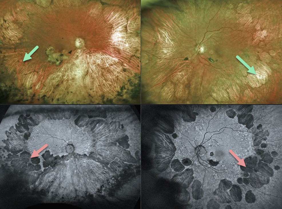
GYRATE ATROPHY
Originally posted on @retina.rocks August 14, 2020
This 88YO female presented with 20/200 vision OD and 20/50 vision OS. Although she complained of nyctalopia since childhood, her vision was good throughout her life up until about a year ago. There was no family history of eye disease.
She had bilateral large islands of scalloped chorioretinal atrophy throughout the mid-peripheral fundi, with extension more posteriorly and inferiorly in the right eye. The areas of chorioretinal atrophy are hypoautofluorescent.
Learning Points:
The history of night blindness with scalloped areas of atrophy is consistent with gyrate atrophy, although patients usually progress to much more severe macular atrophy and vision loss.
Our patient may also have late-onset retinal macular degeneration (LORMD or LORD), which is a rare autosomal-dominant mutation in the tumor necrosis factor-related protein 5 (C1QTNF5) gene, which is expressed in the RPE. LORMD/LORD presents in the fifth to sixth decades with bilateral night blindness and drusen-like deposits throughout the fundi. Islands of atrophy, resembling gyrate atrophy, develop over time.
We will likely order plasma ornithine levels, which should be elevated if she has gyrate atrophy. Regardless of the etiology, however, no treatment is indicated.
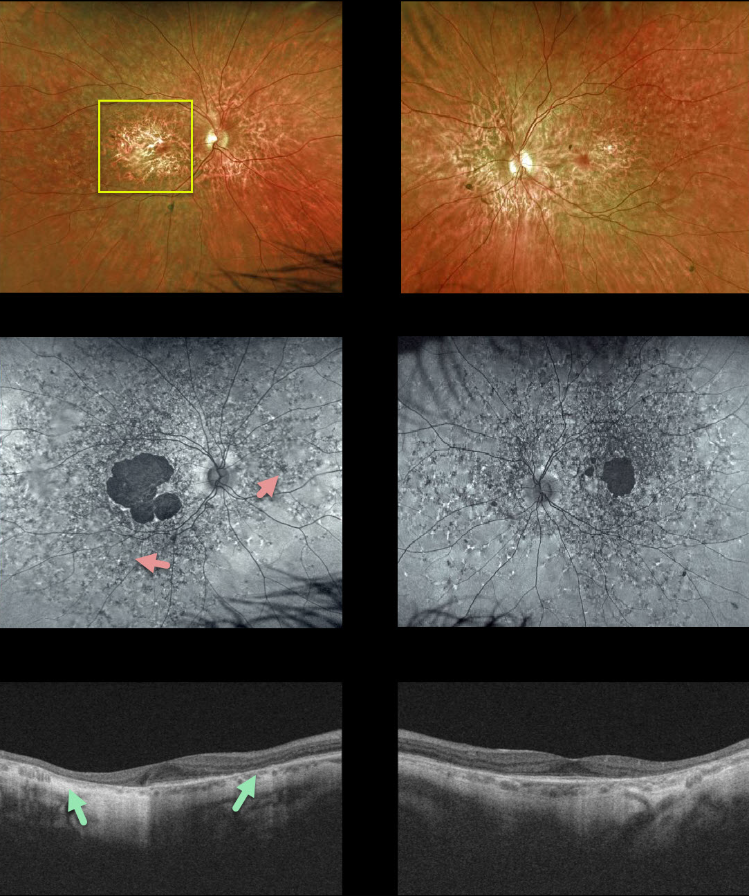
FUNDUS FLAVIMACULATUS
Originally posted on @retina.rocks July 17, 2020
Our patient shows fairly classic findings of fundus flavimaculatus. Central vision remains good at 20/40 OD and 20/30 OS, with preserved foveas.
Findings include bilateral macular atrophy and scattered yellow, subretinal, multifocal, and autofluorescent flecks extending to the mid-peripheral retina. OCT shows RPE and outer retinal atrophy.
Learning Points:
Fundus flavimaculatus is a genetic disorder, usually transmitted as an autosomal recessive trait, that is caused by mutations involving the ABCA4 gene.
ABCA4 encodes one of a family of ATP-binding cassette (ABC) transmembrane proteins that mediate the active transport of various substrates across cellular membranes. The ABCA4 protein is present in the photoreceptor outer segment disc membranes, where it participates in the recycling of 11-cis-retinal.
Stargardt’s disease is felt to be fundus flavimaculatus without peripheral findings. Vision in fundus flavimaculatus is often better than Stargardt’s due to later disease onset and less macular involvement.
Both likely represent an RPE lipofuscin storage disease. The RPE cells become engorged with lipofuscin, causing a dark choroid on fluorescein angiography.
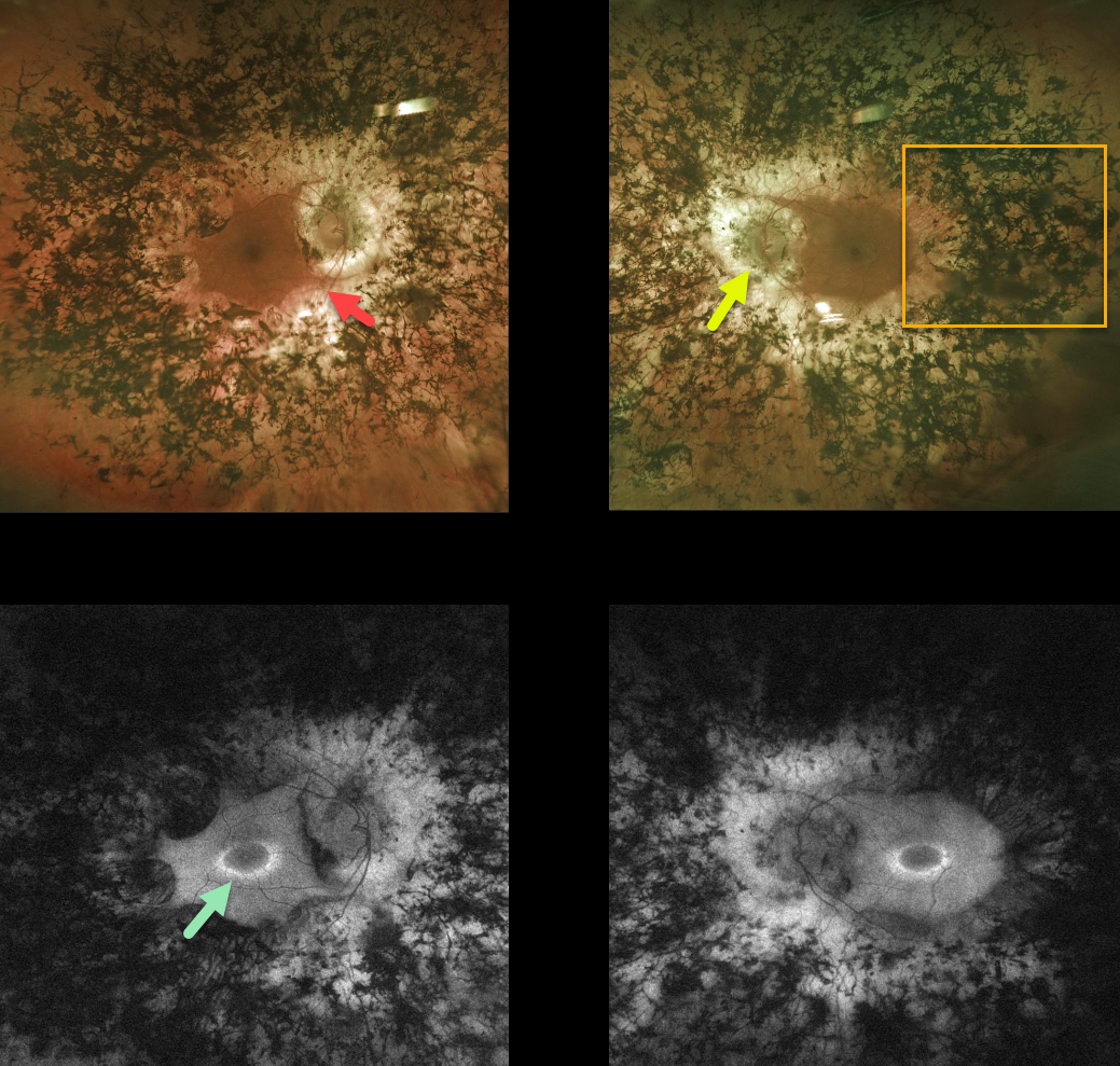
RETINITIS PIGMENTOSA
Originally posted on @retina.rocks June 11, 2020
This patient with retinitis pigmentosa (RP) surprisingly has good central vision 20/30 OU.
The fundus photos show all the classic findings for RP, including attenuated retinal vessels, extensive bone spicules (intraretinal migration of the RPE along retinal capillaries), and optic disc pallor.
Fundus autofluorescence for this patient shows a ring of hyperautofluorescence around each central macula. This ring identifies at-risk retina for future degeneration and can help monitor disease progression.
Learning Points:
RP is a retinal degenerative disorder that causes nyctalopia and progressive vision loss.
Photoreceptors normally inhibit RPE cells, so any process with photoreceptor loss can cause similar localized RPE migration.
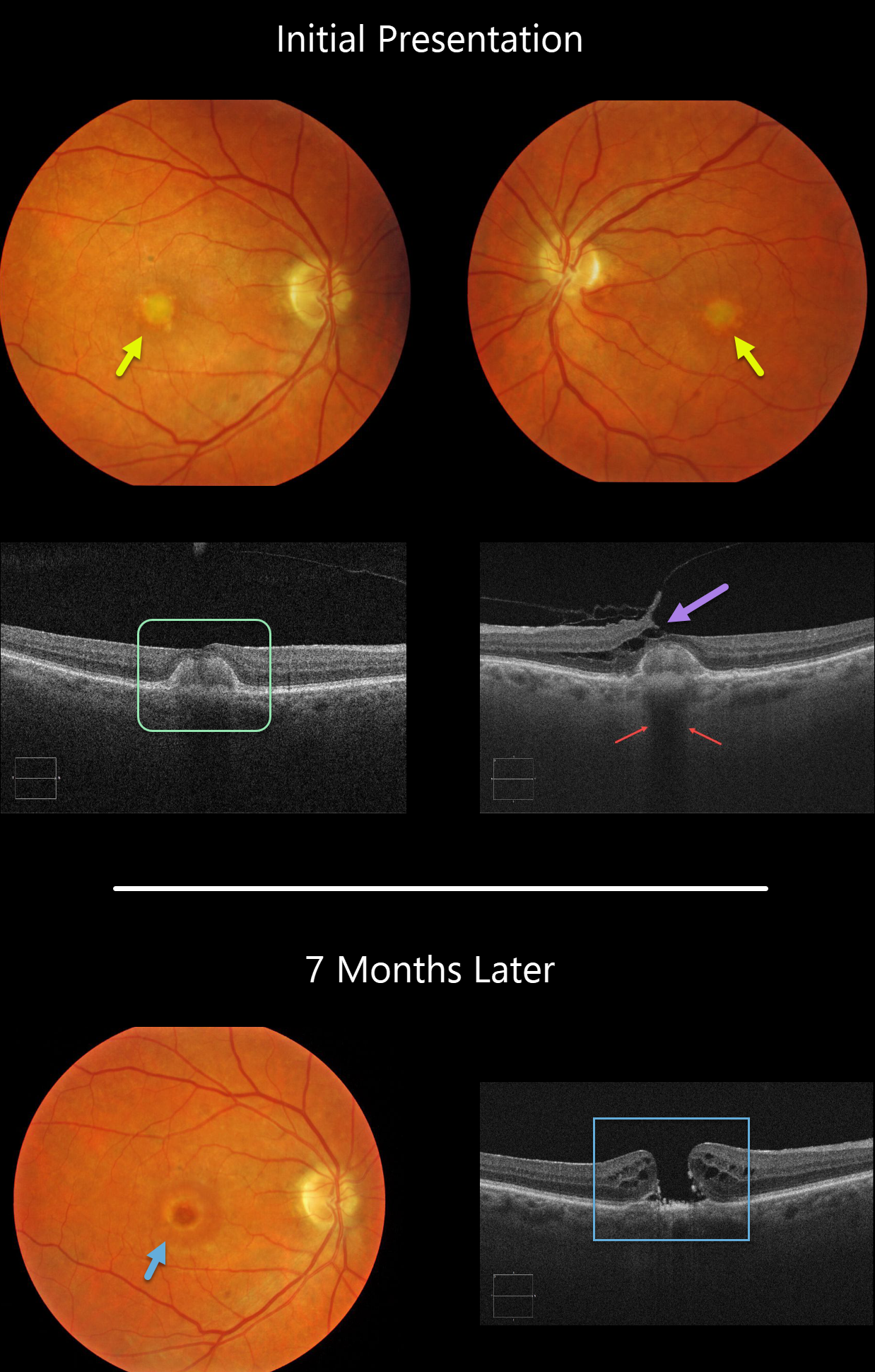
ADULT-ONSET FOVEOMACULAR VITELLIFORM DYSTROPHY WITH SECONDARY MACULAR HOLE
Originally posted on @retina.rocks May 28, 2020
Our patient shows classic findings for adult-onset foveomacular vitelliform dystrophy (Adult Best’s) including small, round, symmetrical subfoveal yellow spots, which is seen on OCT as subretinal hyperreflective material between the RPE and outer retina.
Additionally, the vitelliform lesions provide a great example of shadowing of the underlying choroid. Shadowing occurs when a hyperreflective structure prevents the OCT laser from penetrating to illuminate deeper structures.
A bonus finding in the left eye is focal nasal vitreofoveal traction lifting the retina, splitting the outer plexiform layer and forming a partial tractional lamellar macular hole.
Learning Points:
There are numerous causes for the subfoveal accumulation of yellow material (acquired vitelliform lesions. Adult-onset foveomacular vitelliform dystrophy (Adult Best’s) is associated with a mutation in the BEST1 gene, which encodes for the bestrophin-1 protein.
Bestrophin-1, a calcium-activated chloride channel, is primarily found in the basolateral plasma membrane of the RPE. BEST1 mutations cause a variety of varied phenotypes, also including autosomal recessive bestrophinopathy, best vitelliform macular dystrophy, and autosomal dominant vitreoretinochoroidopathy.
These patients tend to maintain fairly good vision. Outer retinal and RPE atrophy can cause central vision loss, as can the rare development of macular neovascularization. Patients should therefore regularly monitor their vision with an Amsler grid.
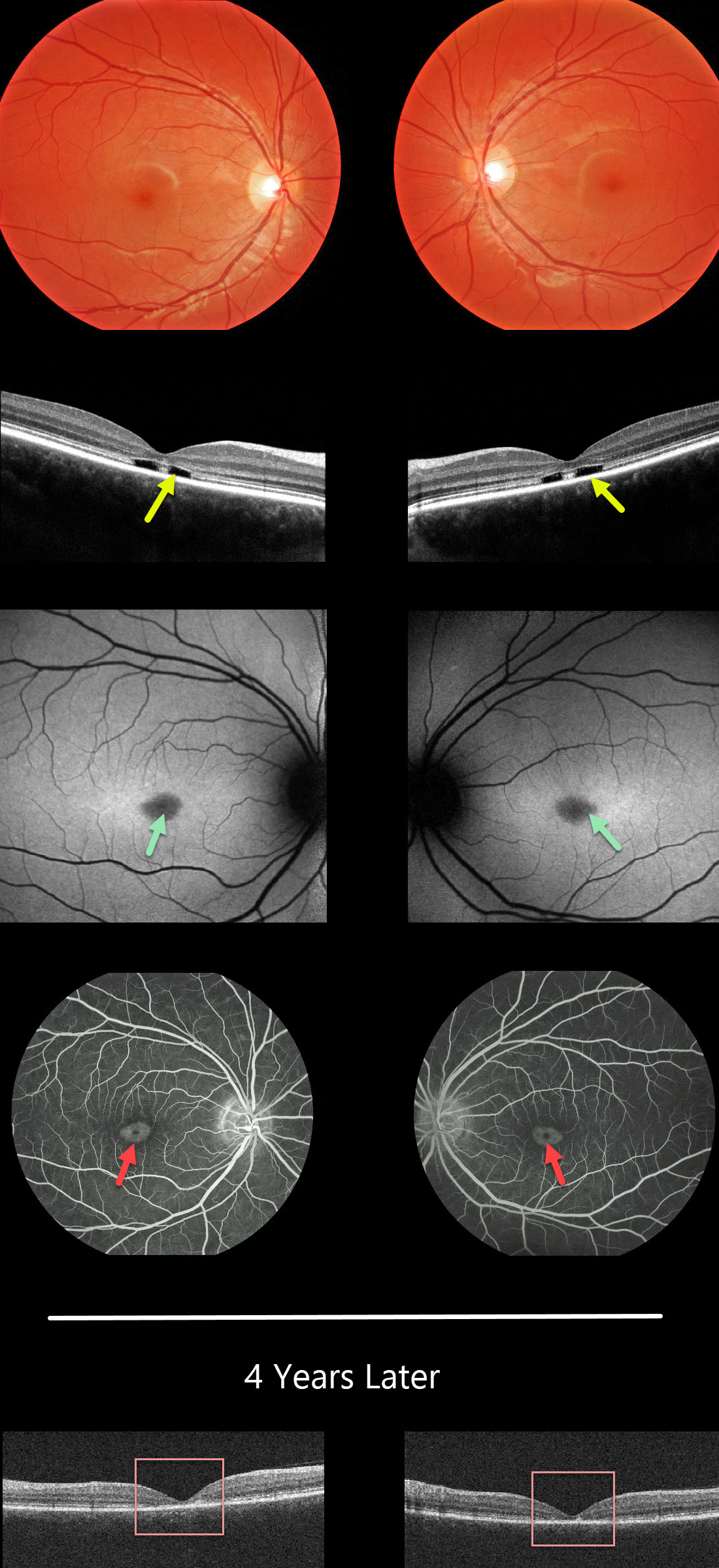
STARGARDT DISEASE
Originally posted on @retina.rocks May 26, 2020
This 14YO female presented with difficulty distinguishing colors and seeing the yellow softball when batting. Vision was 20/30 OD and 20/25 OS.
Her fundus examinations were normal. However, OCT scanning shows a pathognomonic loss of the photoreceptor layers outer to the external limiting membrane.
Fundus autofluorescence shows foveal hypofluorescence corresponding to the angiographic bull’s-eye lesions.
This patient tested positive for the ABCA4 gene, which is associated with both cone-rod dystrophy and Stargardt’s disease.
Four years later, vision and the clinical findings remain stable, but the OCT outer retinal defects have collapsed with increased outer retinal atrophy.
Learning Points:
Stargardt disease is an autosomal recessive disorder caused by a mutation in the ABCA4 gene. The ABCA4 protein is located in the photoreceptor outer segments and is involved in the recycling of 11-cis-retinal.
Mutation of this gene causes the accumulation of lipofuscin in the RPE, which eventually leads to photoreceptor and RPE degeneration. Other ABCA4 disorders include fundus flavimaculatus and cone-rod dystrophy.
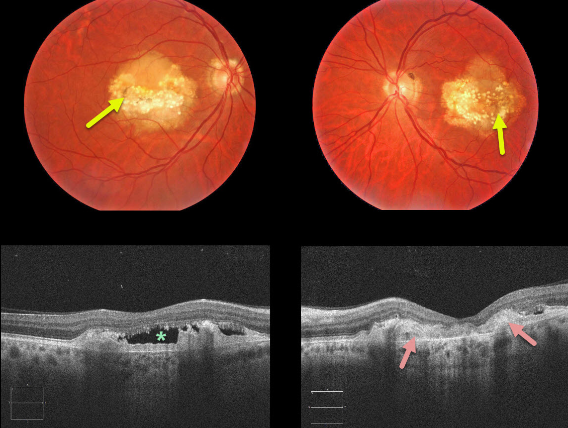
DOMINANTLY INHERITED RADIAL BASAL LAMINAR DRUSEN
Originally posted on @retina.rocks May 18, 2020
Our 54YO patient had a strong family history for macular problems. The fundus photos demonstrate radial cuticular drusen.
The OCT scans are even more dramatic with a saw-tooth pattern of drusen. Subretinal hyperreflective material (SHRM) is noted between Bruch’s membrane and the RPE and subretinal fluid is present in the right eye.
Learning Points:
Dominantly inherited radial basal laminar drusen (Doyne’s honeycomb macular dystrophy, Malattia Levantinese) is a rare genetic macular disorder. A defect in the EFEMP1 gene on chromosome 2p16 is thought to cause an abnormally thickened RPE basement membrane.
Centrally large, nodular and confluent drusen are noted, along with a temporal radiating pattern of smaller cuticular drusen. Later there may be variable amounts of RPE hyperplasia and fibrous metaplasia. Macular neovascularization may also develop.
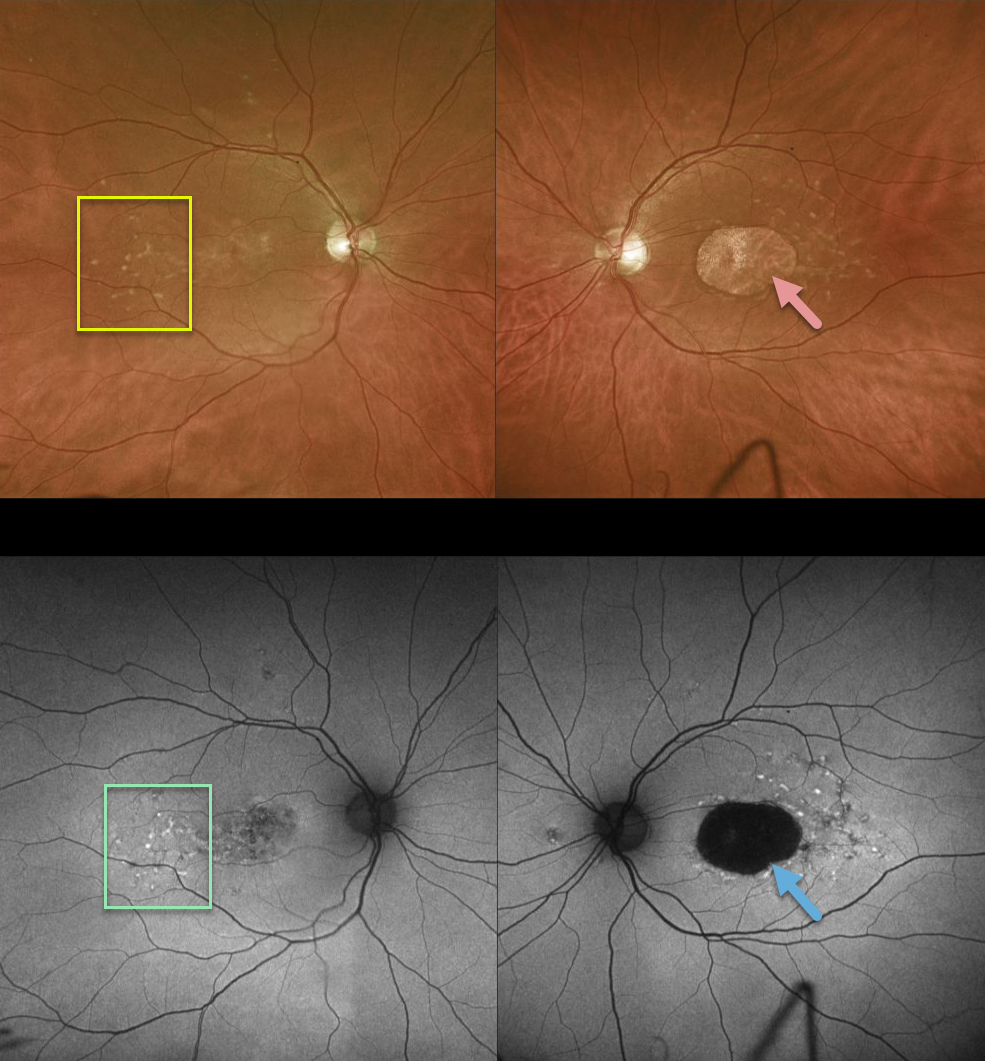
STARGARDT DISEASE
Originally posted on @retina.rocks May 5, 2020
This patient has bilateral 20/400 vision due to classic findings from Stargardt disease, including yellow subretinal flecks and outer retinal/RPE atrophy.
FAF primarily displays hyperfluorescence from RPE lipofuscin. Since Stargardt is essentially a lipofuscin storage disease, the subretinal flecks are hyperautofluorescent and the areas of RPE loss are hypoautofluorescent.
Learning Points:
Stargardt disease is an autosomal recessive disorder caused by a mutation in the ABCA4 gene. The ABCA4 protein is located in the photoreceptor outer segments and is involved in the recycling of 11-cis-retinal. The mutation of this gene causes accumulation of lipofuscin in the RPE, which eventually leads to photoreceptor and RPE degeneration.
Fluorescein angiography (not pictured) characterically shows a ‘dark choroid’ from the lipofuscin-engorged RPE blocking visualization of the underlying choroid.
Stargardt is one of the macular diseases where the vision loss is often much worse than expected from the fundus appearance.
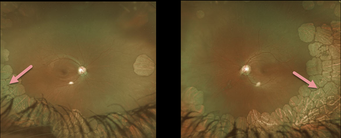
GYRATE ATROPHY
Originally posted on @retina.rocks March 23, 2020
This patient has gyrate atrophy, a rare autosomal recessive retinal dystrophy characterized by extensive areas of lobular peripheral chorioretinal degeneration that gradually spread posteriorly.
Learning Points:
Gyrate atrophy is caused by a lack of a vitamin B6-dependent ornithine aminotransferase mitochondrial enzyme, which leads to elevated ornithine levels.
Vitamin B6 supplementation and an arginine-restricted diet may slow the disease progression in some patients.
End-stage gyrate atrophy can be indistinguishable from choroideremia. Other findings include myopia and night blindness.
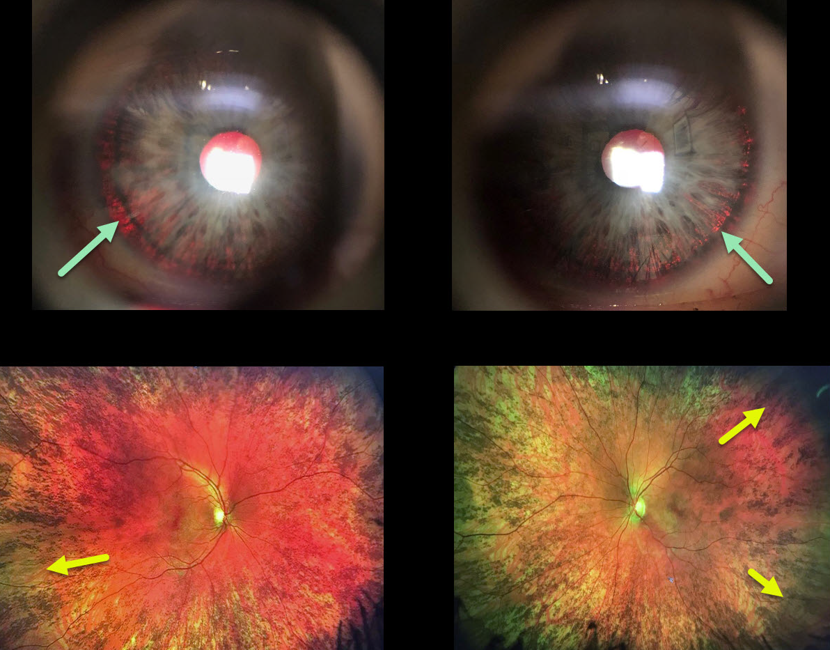
OCULAR ALBINISM
Originally posted on @retina.rocks March 9, 2020
This woman has a history for both a congenital rubella infection and a positive family history for albinism in both an uncle and cousin.
We think that she is likely a carrier for OA1 (ocular albinism type 1) given the presence of iris transillumination defects and pigmentary changes of the peripheral retina, although the fundus changes may also be due to congenital rubella.
Learning Points:
Ocular albinism is a X-linked recessive disorder that affects only the eyes and is caused by a mutation in the OA1 gene. Findings seen in ocular albinism include strabismus, nystagmus iris transillumination defects, blonde fundus, and absent or blunted foveal reflex (fovea plana). There is often a higher number of crossed nerve fibers at the optic chiasm.
OA1 carriers will often have partial iris transillumination defects and up to 90% of OAT1 female carriers exhibit a salt and pepper (or mud-splattered) fundus due to the patches of amelanotic RPE.
This salt and pepper appearance can also be seen in congenital rubella, Leber’s congenital amaurosis, congenital syphilis, and phenothiazine toxicity.
Oculocutanoeus albinism involves both eye and skin findings and is caused by mutations in the tyrosine gene. There is a complete tyrosinase-negative form in which no melanin is produced and a tyrosinase-positive form in which only partial amounts of melanin are produced,
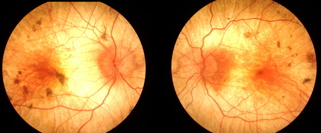
CHOROIDEREMIA
Originally posted on @retina.rocks March 6, 2020
This patient has choroideremia, an X-linked recessive chorioretinal dystrophy caused by mutation in the CHM gene.
It is characterized by a pale appearing fundus with RPE and choroidal atrophy initially in the mid-periphery that gradually spread anteriorly and posteriorly.
Learning Points:
Patients with choroideremia often develop severe central and peripheral vision loss with night blindness. During later stages only a small island of preserved foveal tissue remains.
Female carriers can present with a wide range of phenotypes, ranging from asymptomatic rubella-like pigmentary changes to severe disease.
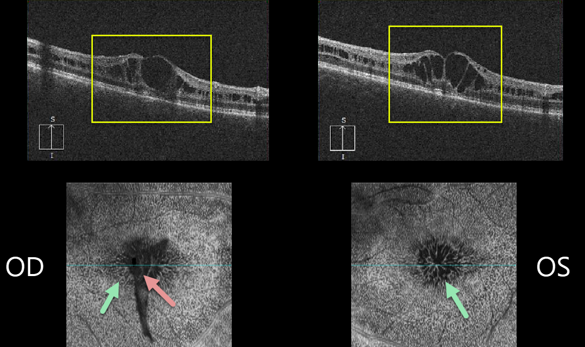
X-LINKED RETINOSCHISIS
Originally posted on @retina.rocks February 26, 2020
This 24YO male has bilateral 20-80 vision and foveal schisis.
B-scan OCTs show schisis mostly in the inner nuclear layer and en face OCTs show a cool perspective for this mid-retinal schisis. There is also an unusual funnel-shaped vitreous veil floating over the right fovea.
Learning Points:
Sex-linked retinoschisis is caused by a mutation of the RS1 gene which is located on the X chromosome and encodes for retinoschisin, a protein likely involved in retinal cellular adhesion.
Although usually transmitted as an X-linked recessive disorder found in males, it can also sometimes affect females.
Over time, the macular schisis flattens with secondary atrophy. Vision usually levels off at about 20/100. Patients can develop spontaneous vitreous hemorrhage and rhegmatogenous retinal detachment from peripheral schisis.
Although there is no specific treatment, topical carbonic anhydrase inhibitors may decrease the foveal thickness and possibly help minimize foveal atrophy (see Andreuzzi et al Retina 2017;37:1555-1561).
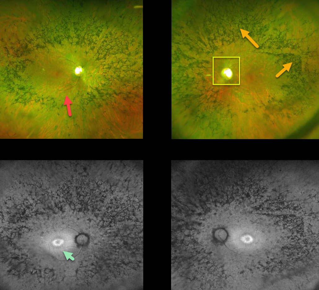
RETINITIS PIGMENTOSA
Originally posted on @retina.rocks January 24, 2020
This patient with retinitis pigmentosa (RP)surprisingly has good central vision 20/30 OU.
The fundus photos show all the classic findings for RP, including attenuated retinal vessels, bone spicules (intraretinal migration of the RPE along retinal capillaries), and optic disc pallor.
Fundus autofluorescence for this patient shows a ring of hyperautofluoresence around each central macula. This ring identifies at-risk retina for future degeneration and can help monitor the progression of this disease.
Learning Points:
Retinitis pigmentosa is a retinal degenerative disorder that causes nyctalopia and progressive vision loss.
Photoreceptors normally inhibit RPE cells, so any process with photoreceptor loss can cause similar localized RPE migration.
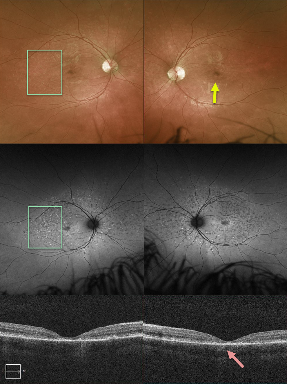
STARGARDT DISEASE
Originally posted on @retina.rocks January 3, 2020
This patient has Stargardt’s disease with beaten-bronze macular atrophy and scattered pisciform flecks, which are more obvious on fundus autofluorescence. OCT scanning shows central outer retinal atrophy.
Learning Points:
Stargardt’s disease is caused by a mutation in the ABCA4 gene, which codes for a protein in the outer segments responsible for facilitating movement of retinoids across the disc membrane.
Lipofuscin accumulates in the RPE, eventually causing degeneration of the photoreceptors and RPE.


