

Intrachoroidal Cavitation
<< Back to Cases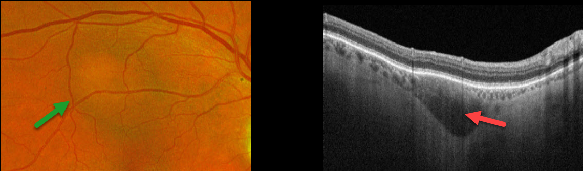
INTRACHOROIDAL CAVITATION
Originally posted on @retina.rocks April 8, 2024
This 84YO male was referred for an asymptomatic macular lesion in his right eye. He was a +6 diopter hyperope. Vision was 20/30 in his right eye and 20/30 in his left eye.
Optos color RG imaging shows a subretinal, mostly depigmented lesion in the superior macula. Triton swept-source OCT shows a hyporeflective outer choroidal lesion that bows posteriorly. The choroid surrounding this lesion is relatively hyperreflective and disorganized. Observation was recommended.
Learning Points:
Initially described by Freund et al (Arch Ophthalmol 2003;121:197-204), intrachoroidal cavitations are most commonly found in high myopes as a peripapillary yellow-orange subretinal lesion. Our case is quite unique, given the location and occurrence in a hyperope.
HIGH MYOPIA
Asma Samsudeen and Ashish Sharma
Originally posted on @retina.rocks September 14, 2023
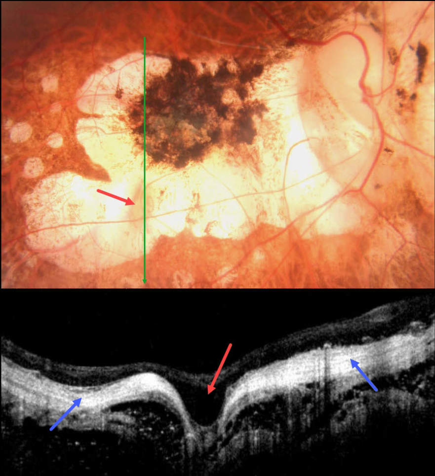
This 58YO male with a history of degenerative myopia presented with 6 months of vision loss in his right eye. He was blind in his left eye following vitrectomy surgery 7 years earlier. Vision was counting fingers OD.
Color photography shows severe variably pigmented atrophic myopic degeneration. An area of orange coloration is seen within the larger area of atrophy inferiorly.
On OCT, this area appears as a focal staphyloma. The overlying retina is draped across the staphyloma. The choroid is so thin as to be invisible on OCT, with a hyperreflective underlying sclera. The orbital tissue is well seen posterior to the sclera.
Learning Points:
Degenerative myopia is a major cause of worldwide visual impairment. Due to the stretching of the posterior retinal structures, there is an increased risk for numerous findings, including atrophic retinal and choroidal thinning, posterior staphyloma, macular schisis/traction, and macular neovascularization (see Ohno-Matsui et al for a recent review, Invest Ophthalmol Vis Sci 2021;62(5);5)..
Initially described by Freund et al (Arch Ophthalmol 2003;121:197-204), intrachoroidal cavitations are most commonly found in high myopes as a peripapillary yellow-orange subretinal lesion. It is difficult to determine whether our patient has a cavitation above the staphyloma.
RETINITIS PIGMENTOSA WITH CHOROIDAL CAVITATIONS
Omar Mulki and Faisal Fayyad
Originally posted on @retina.rocks August 16, 2023
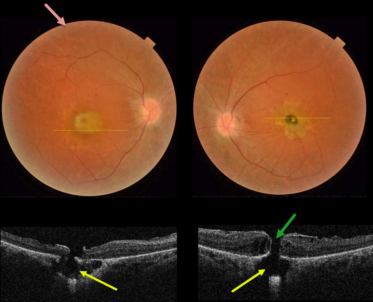
This 32YO female presented with a known history of severe vision loss from retinitis pigmentosa (RP). Her parents are first-degree cousins, and there was no family history of RP. Vision was hand motion OD and counting fingers OS.
Fundus photography shows bilateral irregular yellow central macular pigmentary changes, with a central hyperpigmented spot OS.
OCT scanning through each macula shows a full-thickness macular hole with an underlying defect in the RPE. Each macular hole is suspended over a choroidal cavitation.
Surgical repair was offered with appropriate counseling regarding the poor visual prognosis, and the patient elected to be observed.
Learning Points:
Initially described by Freund et al (Arch Ophthalmol 2003;121:197-204), intrachoroidal cavitations are most commonly found in high myopes as a peripapillary yellow-orange subretinal lesion.
Ornek and Ornek reported a patient with pathologic myopia and a unilateral macular hole communicating with a choroidal cavitation (Retinal Cases 2020;14:328-330).
Although macular holes can rarely be associated with RP (Jin et al, Retina 2008;28:610-614), to the best of our knowledge, ours is the first occurrence of RP with bilateral macular holes and underlying choroidal cavitations.
TOXOPLASMOSIS
Originally posted on @retina.rocks November 16, 2020
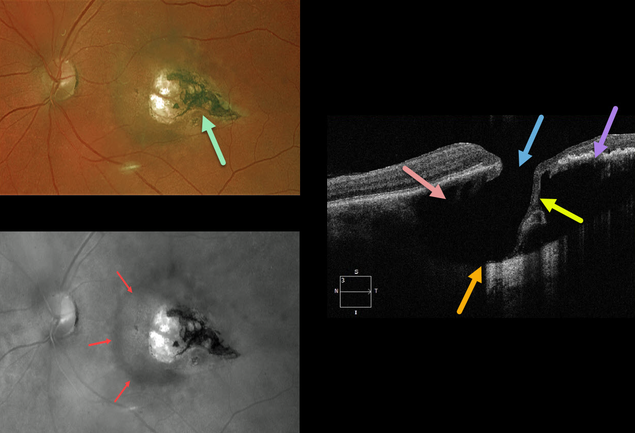
This 67YO female was referred with 20/30 vision and asymptomatic retinal findings. There was a tear-drop-shaped, variably pigmented chorioretinal scar in the temporal macula. The associated increased choroidal pigmentation is best seen in the red-channel image.
The OCT shows some unexpected findings. There is either a coloboma or posterior staphyloma causing marked posterior displacement of the sclero-choroidal junction. Temporally thinned retina dives posteriorly with an adjacent full-thickness retinal defect.
Temporally, the increased pigmentation of the scarring likely makes it difficult to see more posterior choroidal detail due to shadowing. Nasally, the faint, increased choroidal pigmentation also causes shadowing.
What do you think this is? Our best guess is a choroidal cavitation or an atypical colobomatous toxoplasmosis scar.
Learning Points:
For a great discussion on how to differentiate these infectious macular colobomatous lesions (congenital toxoplasmosis, Zika, and cytomegalovirus) from inherited dystrophies like North Carolina Macular Dystrophy (NCMD), see Kumar and Mahalingam AJO 2019;200:47-56. Also see the first OCT description of congenital toxoplasmosis macular lesions (Garg et al, Retina 2009;29;631-637).
HIGH MYOPIA & INTRACHOROIDAL CAVITATION
Originally posted on @retina.rocks July 9, 2020
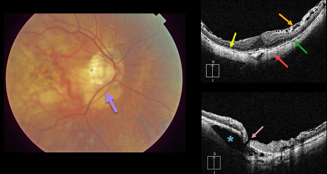
Peripapillary intrachoroidal cavitation is a relatively rare abnormality most commonly found in high myopia. Clinically, these subretinal lesions have an orange coloration and are usually located inferior to the optic nerve.
OCT scanning of the central macula shows a variety of typical myopic findings, including posterior bowing of the sclera, variable attenuation of the ellipsoid zone, markedly thinned choroid, and a variable epiretinal membrane with inner retinal schisis most prominent superiorly.
OCT scanning centered over the optic nerve reveals the intrachoroidal cavitation. There also seems to be a tiny full-thickness retinal defect that communicates directly with the cavitation.
Learning Points:
To learn more about intrachoroidal cavitations and high myopia, see Spaide et al Retina 2012;32;1037-1044.
See Ikuno Retina 2017;37;2347-2351 for a nice overview of macular findings in high myopia.


