

Intraocular Foreign Body (IOFB) + Siderosis
<< Back to Cases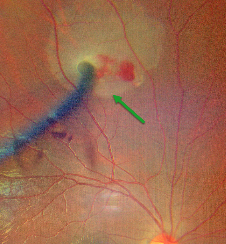
INTRAOCULAR FOREIGN BODY
Kanwaljeet Harjot Madan
Originally posted on @retina.rocks April 2, 2026
This 20YO male developed sudden, painful vision loss in his right eye while welding. Vision was counting fingers. Examination revealed a corneal laceration with a metal wire piercing the iris temporally and stuck in the superior retina with a localized retinal detachment.
Emergent surgery included corneal wound repair, lensectomy, and vitrectomy with removal of the intraocular foreign body (IOFB), fluid-air exchange, and endolaser photocoagulation. Four weeks postoperatively, vision was 20/80 with an aphakic correction, and the retina remained completely attached.
Learning Points:
Penetrating ocular injuries from IOFB are a true ocular emergency. These IOFBs are usually metallic and need to be removed due to the risk of siderosis, endophthalmitis, cataract, glaucoma, iris defects, and retinal degeneration with depressed ERG.
THORN INTRAOCULAR FOREIGN BODY
Ayushi Gupta and Vishal Agrawal
Originally posted on @retina.rocks October 13, 2025
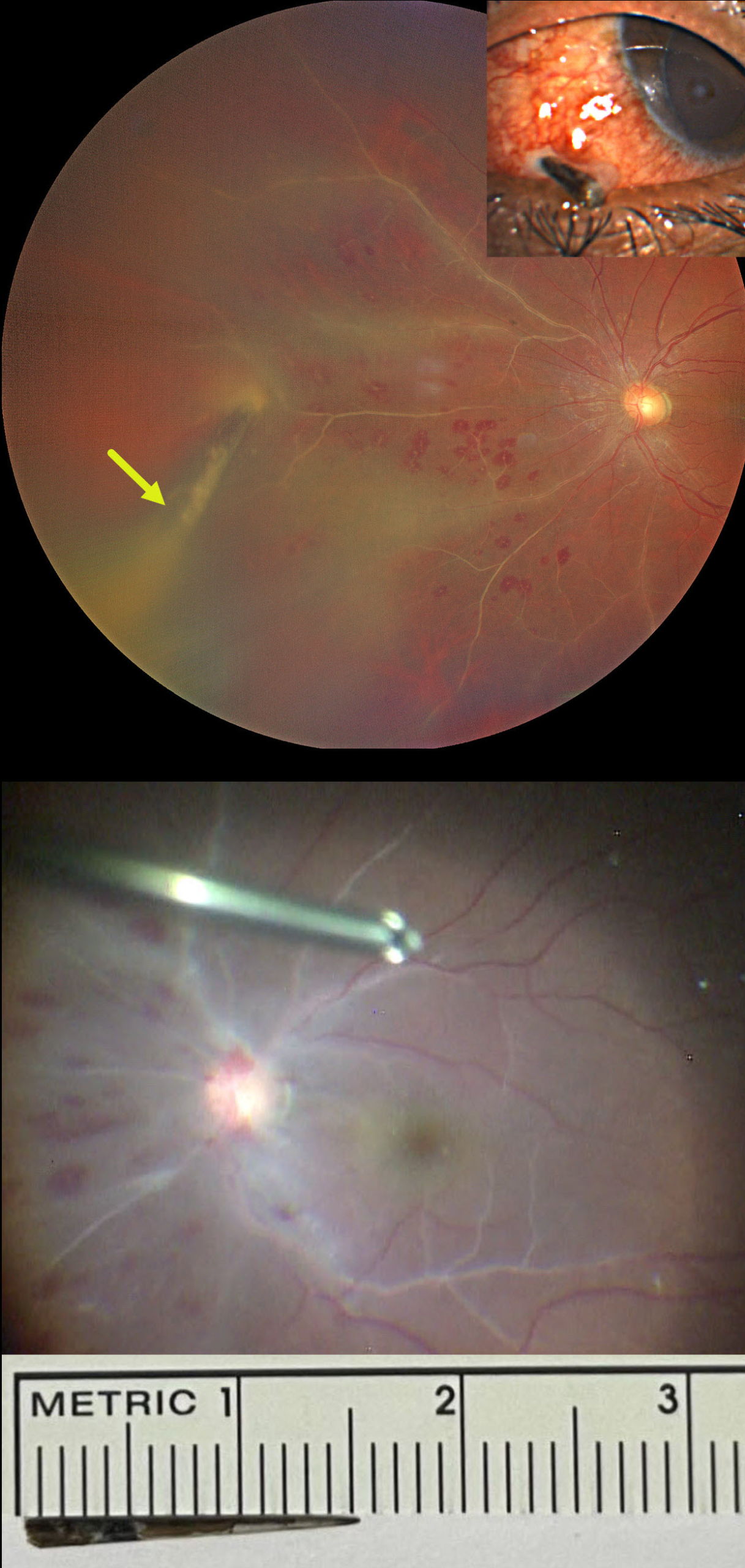
This 28YO male presented 5-6 hours following a perforating thorn injury to his left eye. He was standing in an open-moving loading vehicle when an Acacia tree branch suddenly hit his eye. Vision was 20/20 in his normal OD and 20/40 OS.
External examination showed a thorn lodged in the inferonasal sclera. There were moderate anterior chamber cells on slit lamp examination with a fibrinous pupillary membrane.
Fundus examination revealed vascular sheathing predominantly in the nasal quadrant, multiple, white-centered retinal hemorrhages, and vitreous exudates. The thorn penetrated the retina, extending into the vitreous cavity.
Despite being taken to surgery approximately two hours later, intraoperatively, there was a dramatic increase in vascular sheathing extending into the macula. Pars plana vitrectomy was performed with removal of the thorn foreign body, repair of the scleral laceration, cryotherapy, endolaser photocoagulation around the retinal break, silicone oil tamponade, and intravitreal antibiotic injection. The thorn measured approximately 15mm.
On the first postoperative day, the posterior segment appeared healthy with resolved vascular sheathing. One week postop, vision was 20/50 with a +5-diopter sphere, the retina remained completely attached, and good laser scarring was noted around the penetration site (not shown).
INTRAOCULAR FOREIGN BODY?
Originally posted on @retina.rocks April 1, 2025
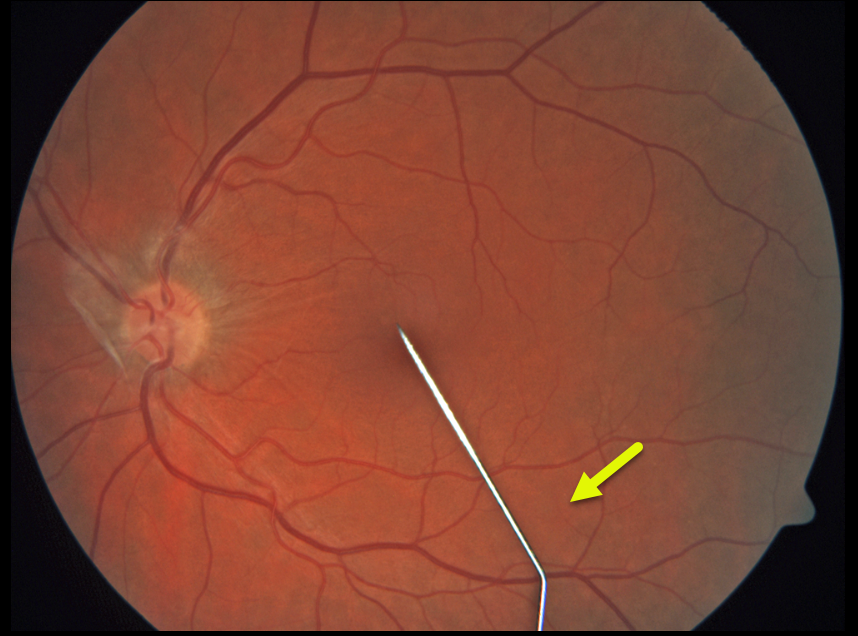
This healthy 24YO goth female presented with severe pain and a floater in her left eye immediately following a failed intraocular tattoo procedure earlier that day. Vision was 20/40.
Color photography shows a posteriorly dislocated intraocular tattoo needle lying on the inferior macular surface. Adherent vitreous is noted on the optic nerve. She was immediately taken to surgery for the removal of the metallic intraocular foreign body (IOFB).
Learning Points:
HAPPY APRIL FOOLS!!
Although this case is a fake-out from an old fundus camera utilizing a fixation stick that appears as a pseudo-IOFB, cosmetic tattooing is unfortunately a very real thing, including corneal, subconjunctival, and episcleral tattoos (https://eyewiki.org/Eye_Tattooing).
The procedure is performed by either the individual performing it on themselves or non-ophthalmic trained individuals and is obviously associated with numerous complications, including globe penetration, traumatic cataract, retinal detachment, endophthalmitis, and orbital cellulitis. Ink-related complications include dye migration into the anterior chamber, where it can coat the lens and posterior corneal surface and cause secondary
INTRAOCULAR FOREIGN BODY WITH SECONDARY BRAO
Anjana Mirajkar and Manish Nagpal
Originally posted on @retina.rocks August 5, 2024
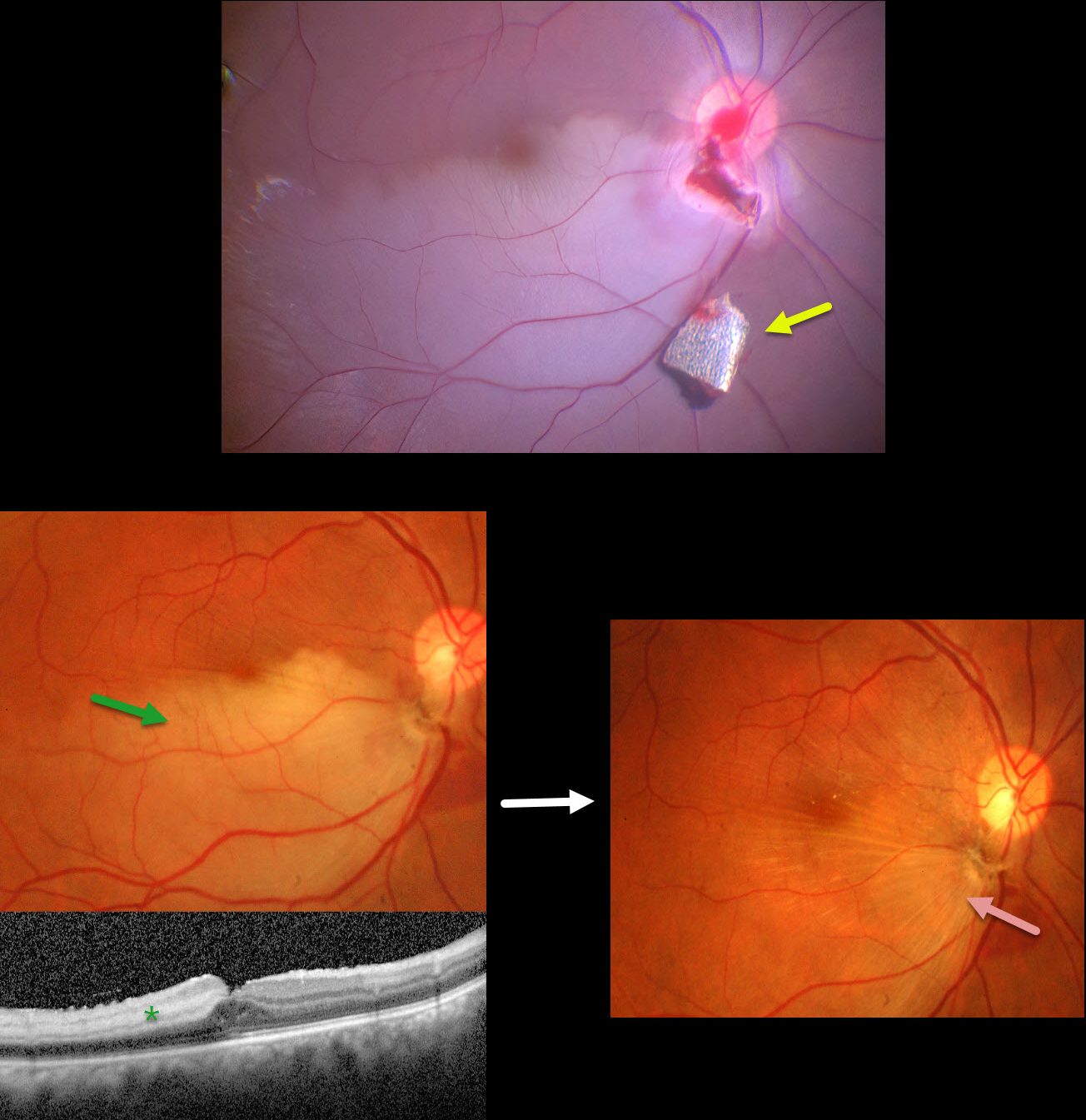
This 34YO male presented with pain and vision loss immediately following a metal-on-metal injury at work. Vision was light perception. Slit lamp examination revealed a corneal laceration and traumatic cataract. B-scan ultrasonography and CT scanning revealed an intraocular foreign body (IOFB). The patient was immediately taken to surgery for corneal wound repair, lensectomy, and vitrectomy.
Intraoperative photography shows a metallic intraocular foreign body (IOFB) resting just inferior to the optic nerve. The impact site is just inferior to the optic nerve, with an inferotemporal branch retinal artery occlusion (BRAO) distally. The IOFB was removed with a rare-earth magnet.
Two weeks following surgery, vision was 20/120 with an aphakic correction. The inferior retina remains opaque, and the OCT shows inner retinal hyperreflectivity from the BRAO. A secondary intraocular lens implantation was performed 3 months postoperatively. Eleven months following the initial surgery, the BRAO is fully resolved, and secondary retinal striae extend outwards from the impact site. Vision remarkably improved to 20/30.
This case was published in the Indian Journal of Ophthalmology (Nagpal M et al, Ind J Ophthalmol 2018;66;146-148).
INTRAOCULAR THUMB
Originally posted on @retina.rocks April 1, 2024
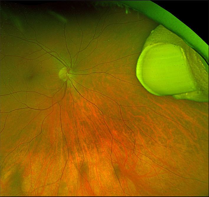
This healthy 7YO boy was playing with his older brother while watching their favorite Three Stooges bit about Niagara Falls.
Horseplay literally turned into tragedy when they presented to the emergency room with the older brother’s hand impaled into his younger brother’s right eye.
Both were immediately taken to surgery, where the attacking brother’s thumb was amputated at the distal phalanx, then removed internally via an open sky pars plana vitrectomy approach. Six months later, following removal of the silicone oil, vision was 20/30.
And Happy April Fool’s Day!
INTRAOCULAR FOREIGN BODY
Barbara Parolini, Veronika Matello, Giulia Freschi, and Roberta Penzani
Originally posted on @retina.rocks March 12, 2024
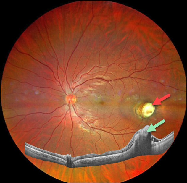
This 23YO male was referred for a second opinion regarding a traumatic intraocular foreign body that occurred about 5 months earlier.
The patient said he was pumping a large truck tire when the tube detached from the valve on the wheel. Metal powder then shot up from the ground and hit the eye. The initial surgical repair involved solely suturing the anterior wound. The foreign body was likely metallic but of an unknown type. The patient was asking why the foreign body had been left. Vision was 20/20.
Fundus photography shows a nodular fibrotic scar in the temporal midperiphery. Widefield OCT shows inner retinal hyperreflectivity with posterior shadowing from this lesion.
Learning Points:
Siderosis can develop from metallic intraocular foreign bodies (IOFBs, specifically iron) and cause cataract, glaucoma, iris defects, and retinal degeneration with depressed ERG. In this case, we felt no need to remove the IOFB given the encasement by scar tissue, lack of inflammation, and excellent vision. We plan on initially examining him monthly.
INTRAOCULAR FOREIGN BODY
Neelam Khatwani, Priyanka Gupta and Charu Gupta
Originally posted on @retina.rocks November 22, 2023
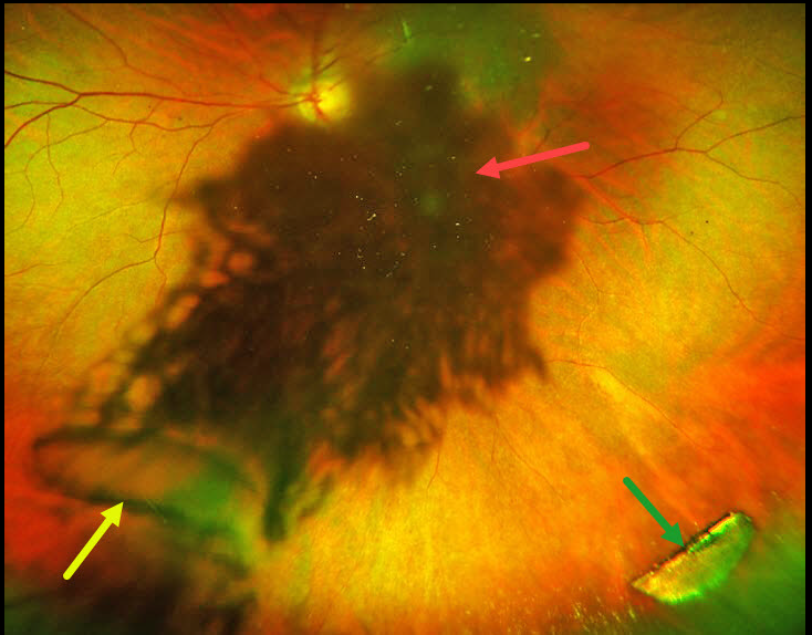
This 45YO male presented with a 4-day history of sudden vision loss in the OS immediately following penetrating trauma while working with a hammer and chisel. Vision was 20/20 in his normal OD and 20/200 OS.
Slit lamp examination OS showed a small peripheral self-sealing corneal laceration, an iris defect, and a ‘rosette cataract.’ On extreme left gaze, a semilunar defect in the posterior lens capsule and a linear defect in the anterior lens capsule were also noted.
Optos color RG imaging shows loss of central fundus detail from the cataract, the capsular defect, and a metallic intraocular foreign body (IOFB).
Fundus examination and localization of an IOFB are often difficult in the presence of secondary cataract. Ultrawide-field fundus imaging is an invaluable diagnostic tool in such cases, as it captures 200° (82% of the retina) in a single image, compared with standard fundus photography.
The rosette cataract in our patient is caused by seepage of aqueous humor through the broken lens capsule and between the lens fibers, resulting in opacification.
Urgent pars plana lensectomy, vitrectomy, removal of the IOFB, and secondary intraocular lens were performed. Vision was 20/40 six weeks following surgery.
INTRAOCULAR FOREIGN BODY
Originally posted on @retina.rocks September 21, 2021
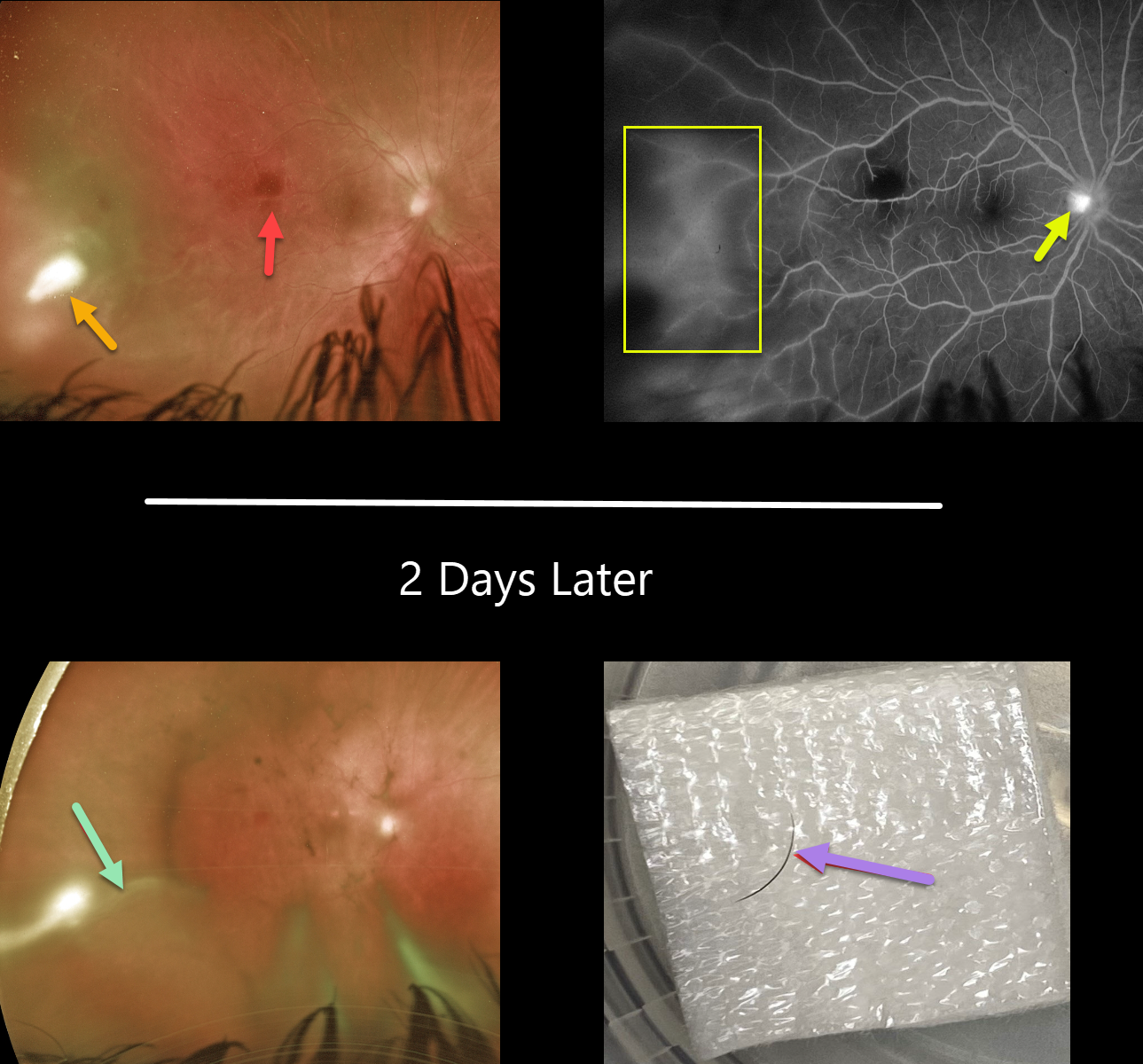
This 18YO female presented with a 10-day history of pain, redness, and blurred vision in her right eye. Her referring doctor put her on topical prednisolone acetate 1% and ciprofloxacin several days earlier.
Vision was 20/60 OD and 20/20 in her normal left eye. There was significant temporal conjunctival injection overlying a deeper scleral nodule with moderate chemosis. There were fine KP on the corneal endothelium, severe cell and moderate flare, and a 0.8mm hypopyon with scattered fibrin clumps (not shown).
Fundus examination shows an outer retinal hemorrhage in the distal macula, along with a patch of white retinal/vitreous inflammation in the temporal periphery. Fluorescein angiography shows diffuse leakage temporally, along with some optic nerve leakage.
She was placed empirically on oral fluconazole. Laboratory workup for various infectious (toxoplasmosis, bartonella, syphilis, tuberculosis, toxoplasma) and inflammatory (Behcet’s, HLA-B27, ANA, ESR) causes was ordered.
One week later, vision was stable, and the scleral and anterior chamber inflammation were only minimally better. Given the lack of significant improvement, oral Bactrim was started.
She returned 2 days later with increasing vitreous inflammation and elongation of the temporal white vitreous opacity. Laboratory results were completely negative. She was taken to surgery the following day to both clear the visual axis and to obtain vitreous for cultures and sensitivities.
During core vitrectomy, the vitreous was so thick that the cutter had to be pulsed on and off due to the tip being occluded. As the elongated vitreous opacity was excised, an embedded underlying eyelash cilia was discovered. The lash was ‘plucked’ with intraocular forceps and removed from the eye, and the site was prophylactically surrounded with laser.
One week later, vision improved to 20/30. The external and intraocular inflammation were dramatically improved. However, a new superotemporal dialysis with peripheral retinal detachment was found, which was repaired with vitrectomy.
Learning Points:
This is an unbelievable case, and it is difficult to fathom how the eyelash entered the vitreous cavity. There was no history of trauma or ocular surgery.
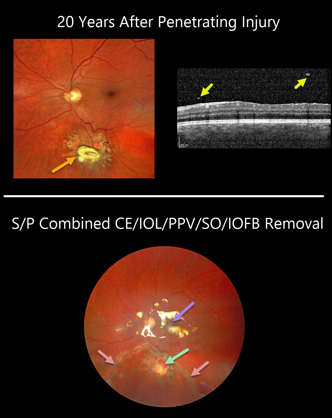
INTRAOCULAR FOREIGN BODY
Originally posted on @retina.rocks September 9, 2021
This 36YO male presented with recent blurred vision of 20/32 in his left eye.
Clarus imaging shows a whitish elevated lesion with surrounding chorioretinal scarring just below the inferotemporal arcade. OCT scanning shows overlying vitreous cells.
He gave a history of a retained retinal plastic foreign body 20 years earlier, following a penetrating injury from an exploding detonator. He had no prior visual complaints, and given the presumed inert nature of the intraocular foreign body (IOFB), surgery was not recommended at that time.
Combined phacoemulsification and vitrectomy with foreign body removal was performed due to increasingly symptomatic vitritis. The retina around the IOFB was necrotic and excised, and the area was surrounded with laser retinopexy. The retina was completely attached under silicone oil one week later.
After removal, the central portion of the IOFB was found to be metallic but coated in plastic. Presumably, as the plastic degraded over time, the metal’s toxicity caused secondary inflammation and visual symptoms.
Learning Point:
In retrospect, a CT scan should have been performed following the initial injury to ensure that the IOFB was fully non-metallic.
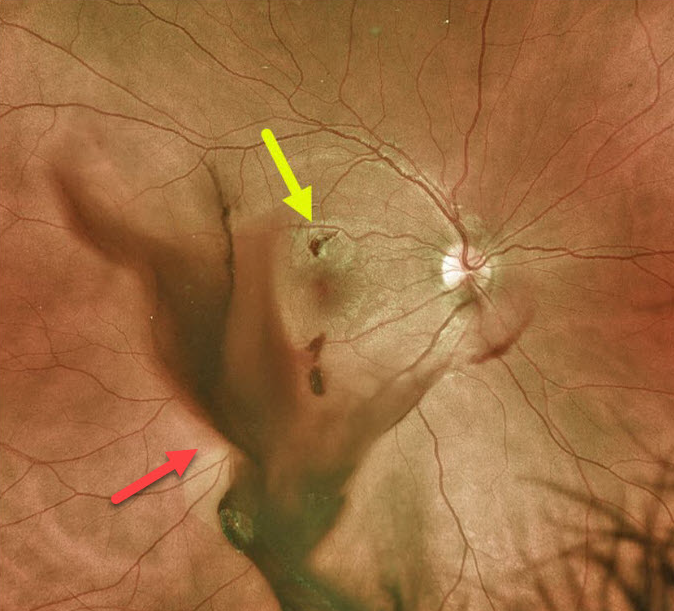
INTRAOCULAR FOREIGN BODY
Originally posted on @retina.rocks March 25, 2021
This 25YO male was utilizing a pneumatic hammer without appropriate eye protection when a metal projectile entered his eye and lodged in the superior macula with associated vitreous hemorrhage.
He underwent immediate vitrectomy with removal of the foreign body.
Learning Points:
Siderosis can develop from metallic intraocular foreign bodies (specifically iron) and lead to cataract, glaucoma, iris defects, and retinal degeneration with depressed ERG potentials.
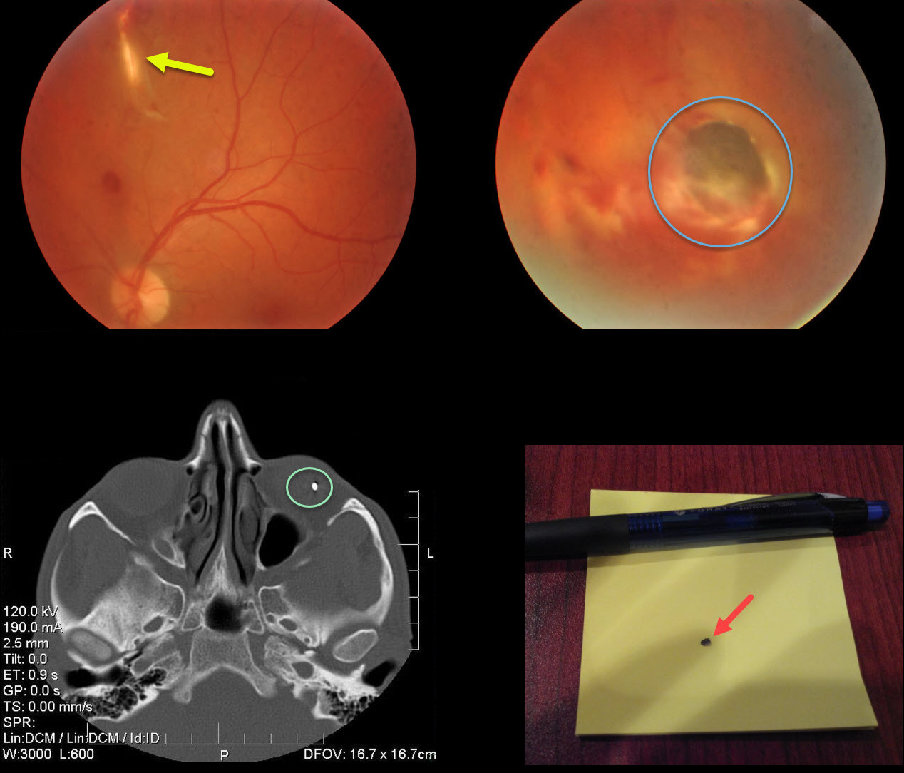
INTRAOCULAR FOREIGN BODY
Originally posted on @retina.rocks September 3, 2020
This patient was hammering so hard that a piece of metal was sent flying through his upper lid, into his orbit, bounced off the orbital roof, penetrated his globe from behind in the superior midperiphery, and lodged in the inferior retina.
The eye looked completely normal externally since the penetrating injury occurred from behind the eye. The entrance wound through the upper lid was also self-sealing, and the lid looked fairly normal when he presented a few days after the injury.
Take a look at the metal object on the CT scan and the removed foreign body.
Learning Points:
Siderosis can develop from metallic intraocular foreign bodies (specifically iron) and lead to cataract, glaucoma, iris defects, and retinal degeneration with depressed ERG potentials.
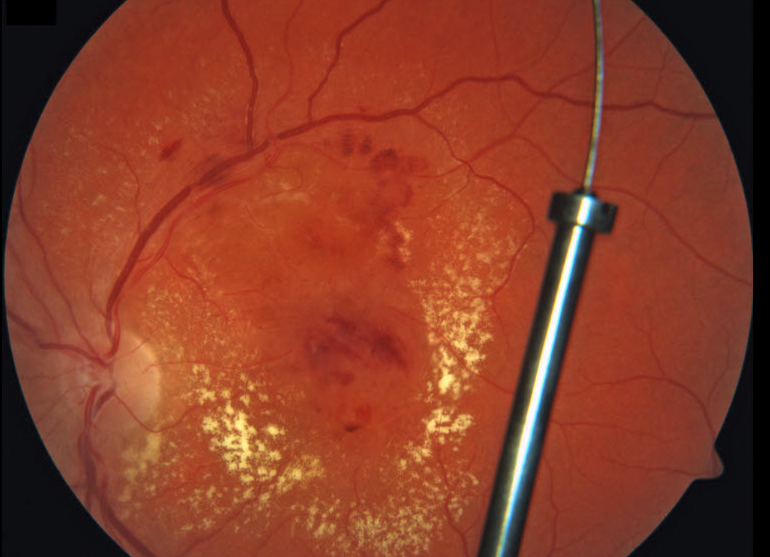
INTRAOCULAR FOREIGN BODY
Originally posted on @retina.rocks April 1, 2020
An automobile manufacturing worker from our local Ford plant presented with an acute injury to his left eye while installing a catalytic converter. A large metallic intraocular foreign body (IOFB) shot through the pars plana in a self-sealing wound and lodged in the posterior vitreous without any direct retinal injury. Since the material was made primarily of inert platinum, the IOFB was left in place and the patient has remained remarkably stable for 3 years.
We hypothesize that the platinum exhibited powerful anti-fungal and anti-bacterial effects helping to prevent infection. The diffuse diabetic macular edema with surrounding lipid also spontaneously resolved, sparking interest in intravitreal platinum as a more durable treatment for macular leakage. The results of the NIH-sponsored Phase 3 MORON Study, Might IntraOcular Platinum ResOlve Edema Better ThaN Really Expensive Anti-VEGF Agents, are eagerly anticipated in late 2020..
Yes, of course it’s APRIL FOOLS!
This patient has diabetic retinopathy with macular edema. The “IOFB” is really a broken fundus camera fixation stick projected onto the posterior pole image.


