

Large retinal capillary aneurysm (TelCap Telangiectatic capillary, PEVAC, Retinal venous macroaneurysm)
<< Back to Cases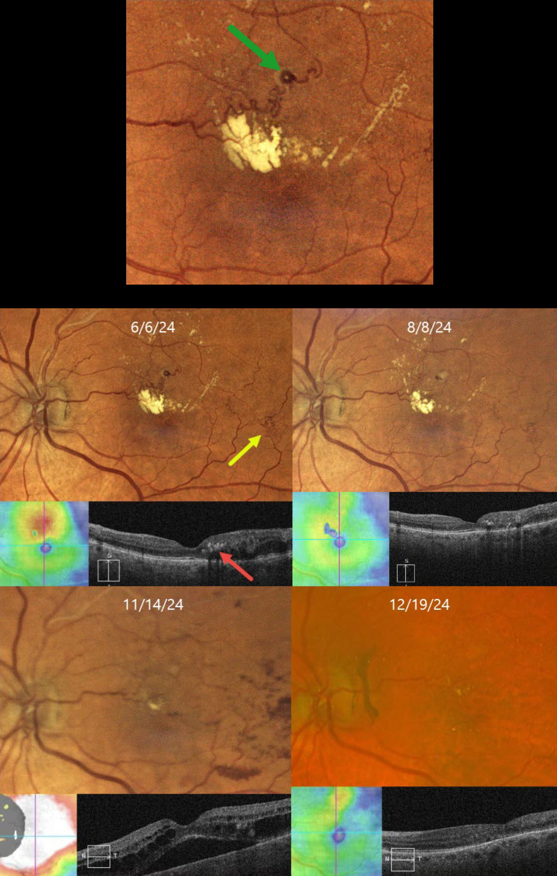
BRVO WITH RETINAL VENOUS MACROANEURYSM
Originally posted on @retina.rocks April 14, 2025
This 72YO female has been followed since 2017 for a branch retinal vein occlusion in her left eye. She has required ongoing anti-VEGF therapy for macular edema and failed macular laser for treatment burden several times. Although her eye was stable for years, on 6/6/24, there was new macular lipid with increased superior macular edema. Vision was stable at 20/25.
Color imaging shows a major superotemporal branch retinal vein occlusion with extensive collateral vessels and superior macular edema. Confluent lipid exudates threaten the macular center and appear as hyperreflective deposits in the mid-retina on OCT. A retinal venous macroaneurysm (RVM) is noted in the superior macula.
An intravitreal Eylea injection was administered, followed by focal thermal laser treatment of the RVM. Two months later, on 8/8/24, vision was 20/30. The lipid was decreasing, and the edema resolved. On 11/14/24, vision decreased to 20/60. Although the lipid was almost resolved, recurrent macular fluid persisted. Following a single Avastin injection, vision improved to 20/30, and the edema resolved. Macular grid laser was applied in hopes of reducing the treatment burden. We will continue to follow her closely and treat her as needed.
Learning Points:
In 1990, Cousins et al reported that RVMs can be an additional finding in occlusive venous disease, always in association with collateral vessels or capillary or arterial macroaneurysms (AJO 1990;109:567-570). These vascular abnormalities, also known as retinal capillary macroaneurysms or telangiectatic capillaries (TelCaps), tend to have a higher rate of intraretinal lipid (40%) but are less likely to affect central vision since they are usually outside the macula. They tend to have a higher association with retinal ischemia, thus being at a higher rate for developing retinal neovascularization (32%). Although these lesions will usually involute spontaneously, we treated our patient out of concern that the lipid might extend into the foveal center.
BRVO WITH RETINAL VENOUS MACROANEURYSM
Seif Anwar
Originally posted on @retina.rocks August 22, 2024
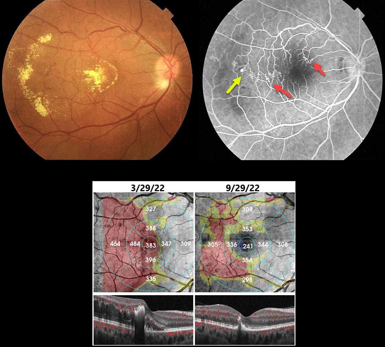
This 40YO hypertensive male presented with gradual vision loss in his right eye. Vision was 20/200 OD and 20/20 in his normal OS.
Color imaging shows central and temporal macular lipid and telangiectasia. Fundus fluorescein angiography shows a superotemporal branch retinal vein occlusion (BRVO) with superonasal and temporal collateral vessels. A retinal venous macroaneurysm (RVM) is noted distally.
Our patient was initially treated unsuccessfully with several monthly Lucentis injections. The macular edema and lipid improved following macular grid laser with focal treatment of the RVM.
Learning Points:
In 1990 Cousins et al reported that RVMs can be an additional finding in occlusive venous disease, always in association with collateral vessels or capillary or arterial macroaneurysms (AJO 1990;109:567-570). These vascular abnormalities tend to have a higher rate of intraretinal lipid (40%) but are less likely to affect central vision since they are usually outside the macula. They tend to be associated with retinal ischemia, thus increasing the risk of developing retinal neovascularization (32%).
RETINAL VENOUS MACROANEURYSM
Originally posted on @retina.rocks December 15, 2022
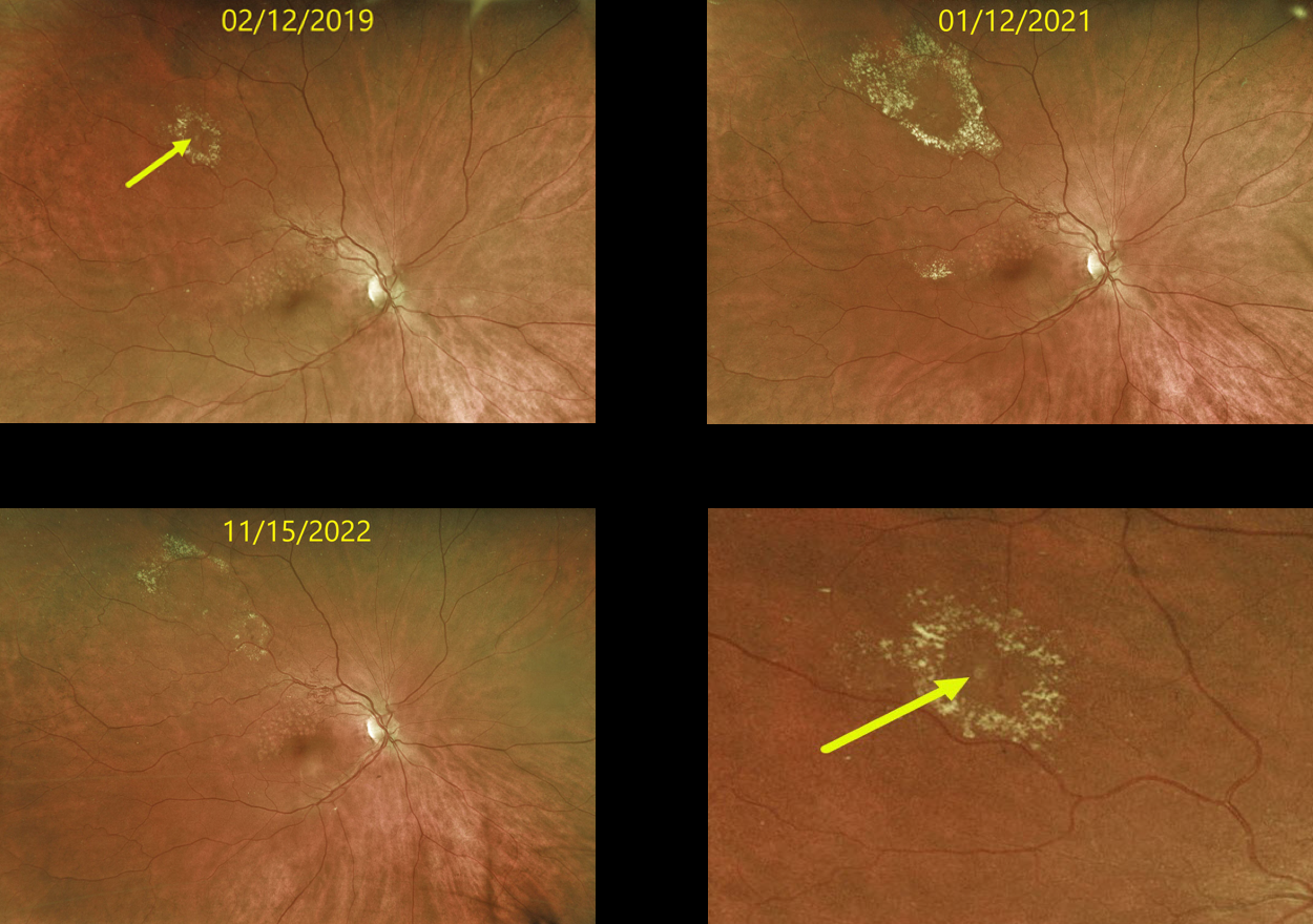
This 63 YO female originally presented in 2019 with a history of a major superotemporal branch retinal vein occlusion (BRVO). Successful macular laser was performed for macular edema.
When initially examined, a midperipheral retinal venous macroaneurysm (RVM) with some surrounding lipid was noted. This RVM has been observed due to its distant location from the fovea. The series of photos over several years shows the exudation to wax and wane. At the last examination on 11/15/22, there was almost complete lipid resolution. Vision remains at 20/25.
Learning Points:
In 1990 Cousins et al reported that RVMs can be an additional finding in occlusive venous disease, always in association with collateral vessels or capillary or arterial macroaneurysms (AJO 1990;109;567-1700).
These vascular abnormalities tend to have a higher rate of intraretinal lipid (40%) but are less likely to affect central vision since they are usually outside the macula.
They tend to have a higher association with retinal ischemia, thus being at a higher rate of developing retinal neovascularization (32%). The lesions will usually involute, as was the case in our patient.


